Ambulatory circadian monitoring in sleep disordered breathing patients and CPAP treatment - Nature
←
→
Page content transcription
If your browser does not render page correctly, please read the page content below
www.nature.com/scientificreports
OPEN Ambulatory circadian monitoring
in sleep disordered breathing
patients and CPAP treatment
Antonio Martinez‑Nicolas1,2, Marc Guaita3,6, Joan Santamaría3,4,6, Josep M. Montserrat3,5,7,
Juan Antonio Madrid1,2 & María Angeles Rol1,2*
Our aim was to evaluate the circadian rhythm of motor activity, body position and integrated
variable TAP (composed by wrist Temperature, motor Activity and body Position) in Sleep Disordered
Breathing (SDB), its relation to SDB severity and the effect of continuous positive airway pressure
(CPAP) on these circadian rhythms. To do this, we monitored motor activity and body position rhythms
of 78 SDB patients (53.3 ± 1.2 years old, 26.9% women) and 32 healthy subjects (51.4 ± 3.2 years old,
43.8% women) for 1 week. On the last day of that week, SDB patients underwent a polysomnography
followed by a Maintenance of Wakefulness Test, Multiple Sleep Latency Test and Sustained Attention
to Response Task protocol. A subgroup of 18 moderate to severe SDB patients was treated with CPAP
and monitored again after 3 months under treatment. A non-parametrical analysis was performed
to characterize the circadian patterns to assess differences between groups and associations
between sleep and circadian parameters. Circadian variables were altered in SDB, exhibiting a direct
relationship to SDB severity. The motor activity pattern showed a clear improvement with CPAP
treatment. Thus, circadian ambulatory monitoring, including the integrated variable TAP, could be
used to evaluate the circadian alterations caused by SDB and activity pattern to monitor the effect of
CPAP treatment.
The circadian system is the main responsible for the temporal regulation of most physiological processes, includ-
ing thermoregulation, metabolism or the sleep–wake c ycle1. Among these processes, the sleep–wake cycle is
regulated by an homeostatic sleepiness accumulation, a circadian process and sleep i nertia2,3.
The increasing prevalence of SDB through the world has become a major health concern, due to its multiple
consequences including hypertension, diabetes, cardiovascular and cerebrovascular disease, cognitive impair-
ment and even c ancer4. The chronodisruptive effect of SDB is supported by both the circadian system alteration
at several levels, such as thermoregulation or the sleep–wake cycle i tself5–8, and the circadian restoration of sleep,
blood pressure, thermoregulation, immune response or haemostatic system by Continuous Positive Airway
Pressure (CPAP)5,6,9–13. Also, this relationship seems bidirectional since the circadian system modulates the
incidence and length of apnoea e vents14.
The rest-activity marker rhythm is inherently associated to the sleep–wake c ycle15, used as choice method
for circadian sleep disorders assessment16 and has been validated against polysomnography in SDB patients17.
However, isolated marker rhythms are submitted to masking, thus multivariable r ecordings15,18,19 and vari-
able integration have been d eveloped15. In this sense, the integrated variable TAP (composed by wrist Tem-
perature, motor Activity and body Position) developed for sleep detection and successfully validated against
polysomnography15,20, can improve the accuracy provided by a single marker rhythm15,21.
Thus, the main purpose of this study was to evaluate the circadian rhythm of motor activity, body position
and the integrated TAP variable in order to determine how these rhythms are affected by sleep disordered breath-
ing severity and continuous positive airway pressure treatment, and in consequence Ambulatory Circadian
Monitoring (ACM) could constitute a screening tool for this disorder as well as CPAP efficacy under free-living
conditions.
1
Chronobiology Lab, Department of Physiology, College of Biology, University of Murcia, Mare Nostrum
Campus. IUIE. IMIB ‑ Arrixaca, 30100 Espinardo, Murcia, Spain. 2Ciber Fragilidad y Envejecimiento Saludable
(CIBERFES), Madrid, Spain. 3Multidisciplinary Sleep Disorders Unit, Hospital Clinic of Barcelona, Barcelona,
Spain. 4Neurology Department, Hospital Clinic of Barcelona, Barcelona, Spain. 5Pneumology Department,
Hospital Clinic of Barcelona, Barcelona, Spain. 6Institut d’Investigacions Biomèdiques August Pi i Sunyer (IDIBAPS),
Barcelona, Spain. 7Ciber Enfermedades Respiratorias (CIBERES), Madrid, Spain. *email: angerol@um.es
Scientific Reports | (2021) 11:14711 | https://doi.org/10.1038/s41598-021-94315-0 1
Vol.:(0123456789)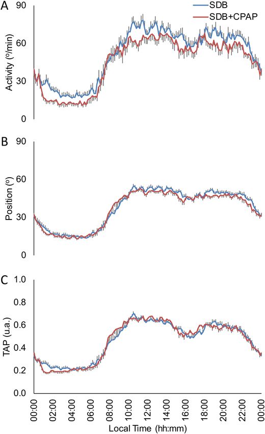
www.nature.com/scientificreports/
Results
Clinical and polysomnographic characteristics. From the consecutive SDB patients originally studied
(n = 98), twenty were excluded due to the following reasons: irregular sleep–wake rhythms (n: 3), acute sleep
deprivation prior to the sleep study (n: 1), REM sleep without atonia (n: 1), severe depressive symptoms (n: 1),
migraines during the nap protocol (n: 1) and removing the sensors (i.e. insufficient data for analysis) either at
home or during the PSG procedure (n: 13). Then, the group finally comprised 78 adults with a wide spectrum
of disease (see our previous w ork5 for clinical and polysomnographic characteristics). With regard to the 30
patients requiring CPAP, one refused to complete the protocol and 11 presented non valid recordings (remov-
ing the sensors or insufficient monitoring time); therefore, 18 moderate-to-severe SDB patients receiving CPAP
were revaluated at least 6 weeks after the baseline study. PSG revealed a complete resolution of SDB in all patients
and improved excessive daytime sleepiness measured by ESS, BSI sleepiness index, MSLT-SL, MWT-SL and
MWT-E. However, CPAP treatment did not improve SART errors (see Martinez-Nicolas et al.5).
Circadian characteristics of sleep disordered breathing. Daily mean patterns and circadian param-
eters of motor activity, body position and TAP variable for SDB patients (n: 78) and healthy subjects (n: 32) are
shown in Fig. 1 and Table 1. When comparing rhythms for motor activity of SDB patients and healthy subjects,
SDB patients showed lower circadian stability as indicated by decreased IS (General Linear Model controlled by
age, sex and BMI; p < 0.05), as well as higher RA and daytime values (M10) and a phase advance in TL5 for body
position than healthy subjects. Finally, TAP pattern (and index of general activation) of SDB patients showed
lower IS, CFI and daytime values (M10) compared with healthy subjects (General Linear Model controlled by
age, sex and BMI; p < 0.05).
The decision tree to discern between healthy subjects (n: 32) and SDB patients (n: 78) selected WT stability
(IS) with a maximum agreement rate (89.2%) at 0.53 (sensitivity = 88.5%; specificity = 91.7%; positive predic-
tive value = 97.5%; negative predictive value = 68.8%). The ROC curve shows an area under the curve of 0.89
(p < 0.001) as shown in Fig. 2. The second most discriminant variable chosen by the decision tree was the TAP
robustness (CFI) with a cut-off point of 0.79 (Fig. 2), reached an agreement rate of 80.2% (sensitivity = 79.4%;
specificity = 85.7%; a positive predictive value = 97.5%; negative predictive value = 37.5%) and the ROC area
under the curve was 0.794 (p < 0.001).
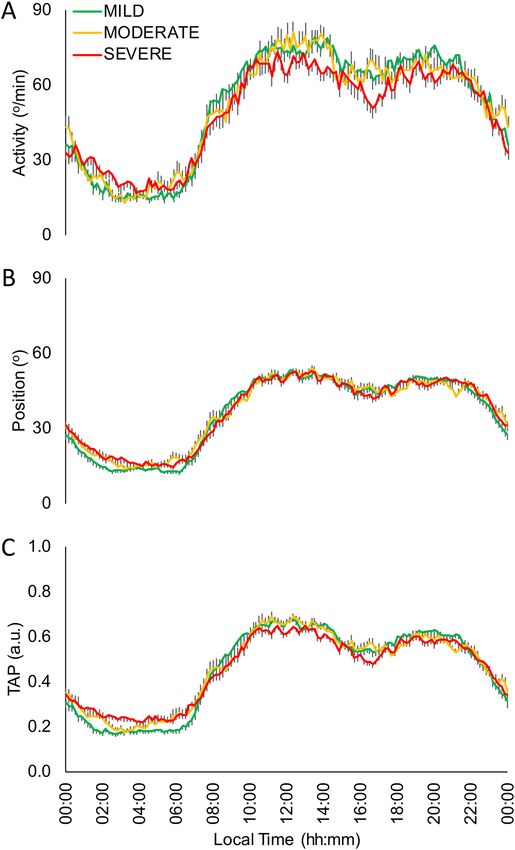
Circadian impairment by sleep disordered breathing severity. The SDB severity effect on the cir-
cadian rhythmic parameters (Fig. 3) was addressed by comparing snorers/mild (n: 31), moderate (n: 19) and
severe SDB (n: 28) by a General Linear Model controlled for age, sex and BMI, and followed by a Bonferroni’s
post hoc analysis (Table 2). The motor activity pattern of snorers/mild apnoea group compared to severe group
(Fig. 3A and Table 2) showed a more stable pattern and lower values at night (L5), this latter also occurred for
body position rhythm together with a phase advance (TL5) and higher day/night contrast (RA) (Fig. 3B and
Table 2), presenting moderate group intermediate values. Finally, the integrated variable TAP showed a more
stable (IS) and robust pattern (CFI) in the snorers/mild apnoea group compared to severe group (Fig. 3C and
Table 2), being the moderate group in an intermediate status. In addition, the mild/snorers group showed lower
N1, AI, AHI, CT90, ODI3, BSI and higher N3, REM, REM Episodes, MSAT, NSAT and MWT-E than severe SDB
group, with moderate group again with intermediate values as it was previously p ublished5.
The best criteria to discriminate, by means of a decision tree, from mild (n: 31) to severe (n: 28) SDB patients,
was TAP robustness (CFI) with a cut-off point of 0.62 yielding an agreement rate of 83.3% (sensitivity = 96.9%;
specificity = 67.9%; positive predictive value = 77.5%; negative predictive value = 95.0%). The ROC for TAP
robustness (Fig. 4) reached an area under the curve of 0.87 (p < 0.001). Since previously, WT pattern impair-
ment has been reported as changing according to SDB s everity5, the best WT parameter for SBD classification
was also assessed. Thus, WT robustness, as measured by CFI, with a cut-off point of 0.44, yielded an agreement
rate of 76.7% (sensitivity = 81.3%; specificity = 71.4%; a positive predictive value = 76.5%; negative predictive
value = 76.9%) and the ROC area under the curve was 0.76 (p < 0.01) as it is shown in Fig. 4, that it is, ROC
curve for TAP robustness tended to reach better results than for WT robustness (p = 0.067) for discriminating
SDB severity.
Regression analysis for sleep and circadian parameters. Regression analysis was performed to
determine the association between sleep and circadian parameters (n: 78). It was controlled for age, sex and BMI.
For motor activity (Supplementary Table 1), higher time in N1 stage and CT90 were associated with increased
night activity values (L5) and the subsequent decrease of RA. Higher percentage of N3 was correlated with
higher RA and lower CT90 was associated to higher CFI. In the case of body position (Supplementary Table 2),
MWT-E was positively associated with high IS, RA and CFI whereas a high ODI3 and low NSAT were related
with a delay in the nocturnal phase marker (TL5). Finally, the most relevant parameters of TAP variable were sig-
nificantly associated to decreased nocturnal sleep quality parameters and poorer outcomes of diurnal MWT-E
and MSLT-E tests (Supplementary Table 3).
CPAP treatment. The influence of CPAP treatment on circadian parameters was addressed by a Mixed
Effects Model (n: 18). For the motor activity pattern, CPAP reduced fragmentation (IV), nocturnal activity (L5)
and increased day-night contrast (RA) as it is shown in Fig. 5A and Table 3. In addition, diurnal body position
(M10) was decreased with the CPAP treatment (Fig. 5B and Table 3). CPAP tended to decrease TAP variable
nocturnal values (L5) while increasing day/night contrast (Fig. 5C and Table 3).
Finally, the decision tree to discern between pre-treatment and CPAP treated severe patients (n: 15)
selected a value of 10.4°/min for nocturnal activity (L5) with an agreement rate of 86.7% (sensitivity = 100.0%;
Scientific Reports | (2021) 11:14711 | https://doi.org/10.1038/s41598-021-94315-0 2
Vol:.(1234567890)
www.nature.com/scientificreports/
Figure 1. Mean waveforms for SDB patients (red line, n: 78) and control subjects (blue line, n: 32) for: (A)
Activity (º/min), (B) Body Position (º) and (C) integrated variable TAP (a. u.). All data are expressed as
mean ± SEM.
specificity = 73.3%; positive predictive value = 78.9%; negative predictive value = 100.0%). The Fig. 6 shows its
corresponding ROC curve reaching an area under the curve of 0.87 (p < 0.01). Again, when comparing with the
best WT parameter (CFI), agreement rate was 68.6% with a cut-off point of 0.44 (sensitivity = 66.7%; specific-
ity = 64.7%; positive predictive value = 66.7%; negative predictive value = 64.7%) and the ROC area under the
curve was 0.68 (p = 0.11). Then, ROC curve for nocturnal activity (L5) tended to reach better results than for
WT robustness (p = 0.063).
Discussion
Our results highlight that SDB impairs rest-activity, body position and TAP variable circadian rhythms in accord-
ance to SDB severity, whereas CPAP treatment improves motor activity pattern. Besides circadian parameters
allow quantitative classification of populations providing a useful clinical screening tool for SDB and CPAP
efficacy.
SDB patients showed lower stability and robustness for motor activity and the integrated variable TAP than
healthy subjects, conditions related to other sleep d isorders20, ageing22 and cancer chemotherapy circadian
impairment23. In the case of body position, SDB patients experienced a nocturnal phase advance, a characteristic
Scientific Reports | (2021) 11:14711 | https://doi.org/10.1038/s41598-021-94315-0 3
Vol.:(0123456789)www.nature.com/scientificreports/
TM10
IS IV RA CFI TL5 (hh:mm) L5 (o/min) M10 (o/min)
(hh:mm)
Activity Control 0.51 ± 0.02 0.63 ± 0.02 0.67 ± 0.02 0.62 ± 0.01 03:57 ± 00:14 15:34 ± 00:27 14.86 ± 0.92 76.74 ± 2.16
SDB 0.41 ± 0.01 0.73 ± 0.02 0.62 ± 0.01 0.55 ± 0.01 04:04 ± 00:06 15:12 ± 00:13 16.84 ± 0.76 70.50 ± 1.40
p 0.000 0.117 0.765 0.084 0.582 0.150 0.795 0.541
TM10 o
IS IV RA CFI TL5 (hh:mm) L5 ( ) M10 (o)
(hh:mm)
Position Control 0.58 ± 0.03 0.30 ± 0.02 0.53 ± 0.03 0.65 ± 0.02 04:53 ± 00:37 15:06 ± 00:25 14.90 ± 0.83 48.93 ± 1.70
SDB 0.55 ± 0.01 0.31 ± 0.01 0.55 ± 0.01 0.65 ± 0.01 04:16 ± 00:06 15:27 ± 00:11 14.59 ± 0.47 49.53 ± 0.54
p 0.237 0.865 0.021 0.103 0.013 0.191 0.204 0.026
TM10
IS IV RA CFI TL5 (hh:mm) L5 (a. u.) M10 (a. u.)
(hh:mm)
TAP Control 0.73 ± 0.01 0.26 ± 0.02 0.60 ± 0.02 0.74 ± 0.01 03:54 ± 00:12 14:48 ± 00:15 0.16 ± 0.01 0.64 ± 0.01
SDB 0.56 ± 0.01 0.31 ± 0.01 0.52 ± 0.01 0.64 ± 0.01 03:46 ± 00:07 14:48 ± 00:04 0.20 ± 0.01 0.61 ± 0.00
p 0.000 0.845 0.590 0.018 0.599 0.866 0.514 0.017
Table 1. Non-parametrical analysis of the circadian rhythms for activity, position and TAP in healthy subjects
(n: 32) and SDB patients (n: 78). Interdaily stability (IS); intradaily variability (IV); relative amplitude (RA);
circadian function index (CFI); mean of the 10 consecutive hours with the highest values (M10) and its
timing (TM10); mean of the 5 consecutive hours with the lowest values (L5) and its timing (TL5). Values are
expressed as the mean ± SEM. Significant differences between healthy and SDB subjects are highlighted in bold
(p < 0.05, General Linear Model controlled for gender, age and BMI).
Figure 2. ROC Curves for Diagnostic ability to detect SDB patients (n: 78) against controls (n: 32) according to
wrist temperature stability (WTIS, continuous line) versus TAP robustness (TAPCFI, discontinuous line).
associated with an aged circadian s ystem22,24,25, and a more standing position during daytime, probably due to
the less pronounced nap period in our sample.
The above-mentioned differences between healthy and SDB subjects´ circadian rhythms can be used as a
screening tool for SBD prone population. In a previous paper we showed that SDB patients exhibited lower wrist
temperature stability values (IS) with respect to healthy subjects with an agreement rate of 89%, probably due
to apnoea-dependent sympathetic activation at n ight26 and the subsequent excessive daytime s leepiness5 which
can contribute to frequent and unexpected or arrhythmic WT fluctuations.
The severity of SDB, as measured by the Apnea–Hypopnea I ndex27, seems to impair circadian indexes (sta-
bility, fragmentation, amplitude and robustness), reinforcing the hypothesis of circadian disruption caused by
sleep apnoea5, as also occurs with other pathological conditions such as obesity, metabolic syndrome, diabetes,
cardiovascular disease or, even, mortality r isk28–31. Nocturnal values were increased for activity, body position
and TAP variable as SDB severity progressed, which indicates a less deep sleep5,20,32, confirmed by the increase
Scientific Reports | (2021) 11:14711 | https://doi.org/10.1038/s41598-021-94315-0 4
Vol:.(1234567890)www.nature.com/scientificreports/
Figure 3. Mean waveforms for mild, moderate and severe SDB subjects according to their Apnoea/Hypopnoea
Index for: (A) Activity (º/min), (B) Body Position (º) and (C) integrated variable TAP (a. u.). Snorer/Mild group:
subjects with less than 15 events/hour (green line, n: 31); Moderate: between 15 and 30 events/hour (yellow line,
n: 19); Severe: more than 30 events/hour (red line, n: 28). All data are expressed as mean ± SEM.
in N1 duration and the decrease of N3 and REM duration. Besides, and according to regression analyses, TAP
stability, fragmentation, amplitude, robustness and nocturnal values impairment were related to higher daytime
sleepiness as measured by MWT and MSLT as it was previously suggested for WT5, and could be a reflect of the
sleep structure impairment15. Thus, these circadian alterations can be used as a screening procedure to detect
individuals prone to suffer SDB before their definitive diagnosis in a sleep unit. Moreover, low TAP robustness,
assessed by CFI, allowed distinction in SBD severity reaching an agreement rate of 83%. Given that the CFI
variable can be used as an indicator of circadian system status15, SDB worsening could implicate a progressive
deterioration of the circadian system.
The restorative effect of CPAP treatment has been thoroughly demonstrated for sleepiness5 and sleep
itself33,34, but also for other alterations such as core body temperature rhythm6, cognitive impairment4, metabolic
syndrome35, fibrinolytic10 and inflammatory m arkers13, coagulation f actors11, blood p
ressure12 or autonomic
36
function . Although the main limitation of this study is the absence of the individual adherence and compliance
CPAP data, all monitored patients accomplished at least five hours per night during six consecutive w eeks37 of
CPAP treatment, an threshold value for an objective CPAP use that has demonstrated its efficacy for improving
Scientific Reports | (2021) 11:14711 | https://doi.org/10.1038/s41598-021-94315-0 5
Vol.:(0123456789)www.nature.com/scientificreports/
TM10 M10 (o/
IS IV RA CFI TL5 (hh:mm) L5 (o/min)
(hh:mm) min)
Mild 0.45 ± 0.01 0.69 ± 0.02 0.65 ± 0.02 0.58 ± 0.01 03:55 ± 00:09 15:15 ± 00:24 14.18 ± 1.08 70.22 ± 2.28
Activity
Moderate 0.41 ± 0.02 0.72 ± 0.03 0.61 ± 0.02 0.55 ± 0.02 04:04 ± 00:13 15:04 ± 00:24 17.63 ± 1.40 72.34 ± 2.68
Severe 0.37 ± 0.02* 0.77 ± 0.03 0.59 ± 0.02 0.53 ± 0.02 04:15 ± 00:11 15:12 ± 00:20 19.30 ± 1.14* 69.56 ± 2.44
p 0.003 0.080 0.163 0.187 0.386 0.977 0.006 0.723
TM10
IS IV RA CFI TL5 (hh:mm) L5 (o) M10 (o)
(hh:mm)
Mild 0.57 ± 0.02 0.29 ± 0.01 0.58 ± 0.02 0.67 ± 0.01 04:04 ± 00:09 15:38 ± 00:19 12.70 ± 0.68 48.41 ± 0.85
Position
Moderate 0.53 ± 0.03 0.34 ± 0.02 0.51 ± 0.02* 0.62 ± 0.02 03:59 ± 00:14 15:14 ± 00:25 15.83 ± 0.88* 49.30 ± 1.01
Severe 0.56 ± 0.02 0.31 ± 0.02 0.53 ± 0.02 0.64 ± 0.02 04:42 ± 00:09* 15:23 ± 00:17 15.92 ± 0.72* 50.98 ± 0.91
p 0.415 0. 301 0.041 0.078 0.006 0.946 0.002 0.162
TM10
IS IV RA CFI TL5 (hh:mm) L5 (a. u.) M10 (a. u.)
(hh:mm)
Mild 0.60 ± 0.02 0.28 ± 0.02 0.55 ± 0.02 0.67 ± 0.01 03:37 ± 00:12 14:44 ± 00:17 0.18 ± 0.01 0.62 ± 0.01
TAP
Moderate 0.54 ± 0.02* 0.31 ± 0.02 0.51 ± 0.02 0.63 ± 0.02 03:39 ± 00:13 14:38 ± 00:20 0.20 ± 0.01 0.62 ± 0.01
Severe 0.53 ± 0.02* 0.34 ± 0.02 0.48 ± 0.02 0.62 ± 0.01* 04:00 ± 00:13 14:58 ± 00:19 0.22 ± 0.01 0.61 ± 0.01
p 0.014 0.169 0.069 0.026 0.363 0.725 0.057 0.590
Table 2. Non-parametrical analysis of the circadian patterns for activity, position and TAP according to
SDB severity. Snorer/Mild: subjects with AHI lower than 15 (n: 31); Moderate: AHI higher than 15 and lower
than 30 (n: 19); Severe: AHI higher than 30 (n: 28). Interdaily stability (IS); intradaily variability (IV); relative
amplitude (RA); circadian function index (CFI); mean of the 10 consecutive hours with the highest values
(M10) and its timing (TM10); mean of the 5 consecutive hours with the lowest values (L5) and its timing
(TL5). Values are expressed as the mean ± SEM. p denotes global significance level according to General Linear
Model (in bold probability values lower than 0.05). * Indicates statistical differences when compared to mild
SDB patients (General Linear Model controlled for gender, age and BMI followed by a Bonferroni’s post hoc,
p < 0.05).
Figure 4. ROC Curves for SDB classification for severe (n: 28) and mild (n: 31) SDB patients according to TAP
robustness (TAPCFI, discontinuous line) versus to wrist temperature robustness (WTCFI, continuous line).
nocturnal sleep34. In this sense, the activity pattern a great improvement occurred, probably because CPAP treat-
ment reduces nocturnal awakenings38 and, hence, would reduce nocturnal activity. Previous studies reported no
effect of CPAP treatment39,40, probably due to differences in treatment compliance or monitoring device sensi-
tivity. Activity nocturnal values diminution, and the subsequent increase in amplitude, were compatible with a
less fragmented41 and deeper sleep due to the reduced number of a rousals5, as also supported by lesser time in
Scientific Reports | (2021) 11:14711 | https://doi.org/10.1038/s41598-021-94315-0 6
Vol:.(1234567890)www.nature.com/scientificreports/
Figure 5. Circadian patterns for 18 SDB subjects at baseline (SDB, red line) and after 6 months of CPAP
treatment (SDB + CPAP, in blue). (A) Activity (º/min), (B) Body Position (º) and (C) integrated variable TAP (a.
u.). All data are expressed as mean ± SEM.
N1 and the AI and SDB indexes improvement. In accordance with the nocturnal decrease in activity level, L5
values can be used to assess CPAP efficacy with an agreement rate of 86%.
In summary, although WT robustness allow us to discern between SDB patients and healthy subjects, when
motor activity, body position and TAP robustness are considered reliability for discriminating between mild
and severe SDB as well as CPAP usage increases with respect to wrist temperature alone (83% vs 77%, and 87%
vs 65%, respectively).
Considering jointly motor activity, body position and WT in the integrated variable TAP reveals that Sleep
Disordered Breathing patients present circadian disruption, that increases with the severity of the disease and
improves with Continuous Positive Airway Pressure treatment. Thus, TAP variable could constitute, a useful
screening tool for SBD prevalence and severity as well as treatment efficacy adherence.
Scientific Reports | (2021) 11:14711 | https://doi.org/10.1038/s41598-021-94315-0 7
Vol.:(0123456789)www.nature.com/scientificreports/
IS IV RA CFI TL5 (hh:mm) TM10 (hh:mm) L5 (o/min) M10 (o/min)
SDB 0.40 ± 0.02 0.78 ± 0.04 0.59 ± 0.03 0.54 ± 0.02 03:58 ± 00:12 15:10 ± 00:27 18.40 ± 1.72 68.92 ± 2.60
Activity
CPAP 0.43 ± 0.02 0.72 ± 0.03 0.68 ± 0.02 0.58 ± 0.02 04:02 ± 00:10 14:45 ± 00:21 12.10 ± 1.30 62.26 ± 3.78
p 0.139 0.016 0.011 0.061 0.794 0.455 0.003 0.112
IS IV RA CFI TL5 (hh:mm) TM10 (hh:mm) L5 (o) M10 (o)
SDB 0.57 ± 0.03 0.31 ± 0.02 0.55 ± 0.02 0.66 ± 0.02 04:19 ± 00:13 15:02 ± 00:16 14.86 ± 0.98 50.43 ± 1.00
Position
CPAP 0.54 ± 0.02 0.31 ± 0.01 0.54 ± 0.02 0.64 ± 0.01 04:13 ± 00:12 14:50 ± 00:19 14.51 ± 0.88 48.34 ± 0.97
p 0.251 0.745 0.745 0.433 0.662 0.569 0.656 0.041
IS IV RA CFI TL5 (hh:mm) TM10 (hh:mm) L5 (a. u.) M10 (a. u.)
SDB 0.55 ± 0.03 0.35 ± 0.02 0.46 ± 0.02 0.61 ± 0.02 03:44 ± 00:11 14:44 ± 00:24 0.23 ± 0.01 0.62 ± 0.01
TAP
CPAP 0.56 ± 0.03 0.33 ± 0.01 0.51 ± 0.02 0.63 ± 0.02 03:16 ± 00:07 14:08 ± 00:16 0.20 ± 0.01 0.62 ± 0.01
p 0.772 0.430 0.076 0.280 0.105 1.000 0.051 0.802
Table 3. Non-parametrical analysis of CPAP treatment effect on circadian parameters in SDB patients.
Interdaily stability (IS); intradaily variability (IV); relative amplitude (RA); circadian function index (CFI);
mean of the 10 consecutive hours with the highest values (M10) and its timing (TM10); mean of the 5
consecutive hours with the lowest values (L5) and its timing (TL5). Values are expressed as the mean ± SEM. p
denotes significance level (Mixed Model Analysis). In bold are the probability values lower than 0.05.
Figure 6. ROC Curves for SDB classification of pre- and after CPAP therapy in severe patients (n: 15)
according to their night activity level (AL5, discontinuous line) versus wrist temperature robustness (WTCFI,
continuous line).
Materials and methods
In a previous s tudy5, we analysed the wrist temperature (WT) circadian pattern in the same group of subjects,
using the information from the ACM devices. In the current study we will focus on the information provided
by the circadian pattern of motor activity, body position and integrated variable TAP as well as data provided by
the Maintenance of Wakefulness Test (MWT), and the Multiple Sleep Latency Test (MSLT).
Ninety-eight consecutive patients with suspected SDB (Table 4) were evaluated at the Hospital Clinic of Barce-
lona in the Multidisciplinary Sleep Disorders Unit, as previously described in Martinez-Nicolas et al., 2 0175. The
patients were informed about the objectives of the study, the willingness of their participation and that whatever
their decision, it would not affect their treatment. Participants who accepted to participate in the study signed an
informed consent. Exclusion criteria were being under 18 years of age, use of medications affecting wakefulness
or sleep, shift work or irregular sleep–wake schedules during the four weeks before the sleep study, and major
medical or psychiatric disorders. Patients with any concomitant sleep disorder other than SDB were excluded
by nocturnal polysomnography (PSG).
Scientific Reports | (2021) 11:14711 | https://doi.org/10.1038/s41598-021-94315-0 8
Vol:.(1234567890)www.nature.com/scientificreports/
Healthy vs. SDB SDB severity CPAP Effect
Healthy SDB Mild Moderate Severe PRE-CPAP CPAP
Age (y) 51.4 ± 3.2 53.3 ± 1.2 51.2 ± 2.2 56.4 ± 2.1 53.7 ± 1.5 55.3 ± 2.3 55.9 ± 2.3#
Sex (M/W) 18/14 57/21 18/13 16/3 23/5 16/2 16/2
BMI (kg/m2) 24.7 ± 0.7 30.0 ± 0.6* 27.3 ± 0.6 29.7 ± 1.2 33.4 ± 1.1ab 31.3 ± 1.2 31.6 ± 1.1
Table 4. Subjects characteristics. Healthy: control group, n: 32 SDB: Sleep Disordered Breathing, n: 78; Snorer/
Mild: subjects with less than 15 events/hour (n: 31); Moderate: between 15 and 30 events/hour (n: 19); Severe:
more than 30 events/hour (n: 28). Pre-CPAP and CPAP: SDB subgroup before and after CPAP treatment (n:
18). BMI: Body Mass Index. All data are expressed as mean ± SEM, except for the men/women ratio. *Indicates
statistical differences between healthy and SDB patients (General Linear Model, p < 0.05). For mild, moderate
and severe SDB groups, “a” indicates statistical differences when compared to mild SDB patients, “b” indicates
statistical differences with moderate SDB patients (General Linear Model, p < 0.05, Bonferroni’s post hoc). #
Denotes statistical differences between pre-CPAP and post-CPAP treatment (Mixed Model Analysis, p < 0.05).
A total of 32 healthy subjects were recruited (age and gender balanced with the SDB patients) at the Chrono-
biology Lab of the University of Murcia (Table 4). They are included in this study in order to illustrate healthy
circadian patterns for activity, body position and TAP. Exclusion criteria were the same as those applied to the
SDB patients, but also included snoring (as reported during a personal interview) and/or excessive daytime
sleepiness (Epworth Sleepiness Scale score higher than 12)42.
The study follows the bioethical principles set out by the Declaration of Helsinki. Data from the volunteers
were protected according to Spanish Law 15/1999 from 13 September. The study was approved by the Hospital
Clinic of Barcelona and the University of Murcia ethics committees for the SDB patients and healthy subjects,
respectively.
Design. Healthy volunteers were monitored for an entire week whereas SDB patients were monitored 6 days
under free-living conditions and an additional day, the last one, under controlled conditions in hospital. During
this week, the subjects were encouraged to maintain their habitual life style. Since PSG and nap-protocols could
interfere with the normal sleep pattern of the p atients43, both test were performed at the end of the monitoring
period, allowing at least 6 days of continuous recordings (the minimum for a reliable non-parametrical analysis,
according to literature44).
For ambulatory circadian monitoring, all subjects wore a Thermochron iButton DS1921H (Maxim Integrated
Products, Sunnyvale, CA) programmed to sample every 10 min over the whole week to measure W T32, placed
on the wrist of the non-dominant hand over the radial artery as already d escribed5,45–47, and isolated from the
environment by a double-sided cotton sport wrist band32. In addition, every subject was monitored with a
HOBO Pendant G Acceleration Data Logger UA- 004–64 actimeter (Onset Computer, Bourne, MA) programmed
every 30 s, positioned on the non-dominant arm by means of a sport band for motor activity and body position
monitoring as already described15,20,45. Briefly, motor activity was measured as the rate of change in degrees per
minute and position represents the angle between the X-axis of the accelerometer (parallel to the humerus bone
of the arm) and the horizontal plane. Data from activity and body position were averaged for 10-min intervals
to facilitate WT comparisons. Wrist Temperature, motor Activity and body Position were combined in order
to obtain the integrated variable TAP, according to the algorithms previously d escribed15. In summary, and as
5,15,20–22
previously described , each variable was normalized between 0 and 1 and averaged to obtain the integrated
variable (TAP). Thus, a TAP value of 1 indicates the lowest values of WT, the highest values of activity and a
standing position (compatible with wake periods), whereas a score of 0 corresponds to the highest WT values,
the lowest activity values and a horizontal position (compatible with sleep periods)15.
On the last day of the week, patients with suspected SDB underwent a 24-h sleep study, including question-
naires and neurophysiological tests as previously described5. After admission, the Epworth Sleepiness Scale (ESS)
and Barcelona Sleepiness Index (BSI) were used to assess subjective daytime sleepiness42,48. Nocturnal PSG was
performed according to standard practice parameters and diagnostic c riteria49,50 with sleep stages independently
and manually double scored according to the American Academy of Sleep Medicine (AASM) criteria, using 30 s
epochs51. The nap protocol was initiated the morning after PSG in order to objectively measure daytime sleepi-
ness throughout the day (review Guaita et al., 2015 for further d etails48). In brief, we used the research version
protocol comprising 5 sets of the Maintenance of Wakefulness Test (MWT), the Multiple Sleep Latency Test
(MSLT) that started at 08:30 and every 2 h o nwards52. Each nap set was preceded by a measurement of vigilance
with Sustained Attention to Response Task (SART), with a duration of 4 m in53 and using commission errors,
54
missed errors and total e rrors . Due to their skewed distribution, MWT and MSLT were log transformed previ-
ously to statistical analysis.
Continuous positive airway pressure treatment. From the cohort of 78 patients, eighteen moderate
or severe SDB patients with resistant hypertension or excessive daytime sleepiness were treated with CPAP and
evaluated again following the same protocol (nocturnal PSG followed by SART-MSLT-MWT protocol). CPAP
titration was performed following the recommendations of the Spanish Sleep Society55. CPAP compliance was
Scientific Reports | (2021) 11:14711 | https://doi.org/10.1038/s41598-021-94315-0 9
Vol.:(0123456789)www.nature.com/scientificreports/
measured objectively using a built-in CPAP meter. For the analysis, five hours per night, measured by the CPAP,
during six consecutive weeks were considered the strict minimum use of C PAP37.
Data analysis. The WT data were obtained, filtered and processed from data previously reported5. The
mean daily patterns for motor activity, body position and TAP variable were calculated per individual, and then
averaged per group.
In order to characterize the aforementioned circadian rhythms a nonparametric analysis was performed as
previously described5. This analysis determines the following parameters: interdaily stability (the constancy of the
24-h rhythmic pattern over days, IS), intradaily variability (rhythm fragmentation, IV), relative amplitude (RA)
and circadian function index (CFI) calculated by the integration of IS, IV and RA oscillating between 0 (absence
of circadian rhythmicity) and 1 (robust circadian rhythm)15,56. RA was calculated as the difference between M10
(average of 10-min intervals for the 10 consecutive hours with maximum values) and L5 (average of 10-min
intervals for the 5 consecutive hours of minimum values) divided by the sum of M10 and L5 for variables with
acrophase during daytime (activity, body position and TAP), as previously published41,56. The timing for L5 and
M10 were used as nocturnal and diurnal phase markers (TL5 and TM10, respectively).
Circadian variables parameters in healthy subjects and SDB patients were compared using a General Linear
Model controlled by age, gender and body mass index (BMI). Differences between snorers/mild SDB (AHI < 15
events/hours), moderate SDB (AHI between 15 and 30 events/hour) and severe SDB (AHI > 30 events/hour)
were assessed by a General Linear Model controlled by age, gender and BMI (followed by post hoc pairwise
comparisons and a Bonferroni test). In addition, a regression analysis was performed between sleep and circadian
parameters controlling again for age, gender and BMI (with Bonferroni correction for multiple comparisons). A
Mixed Effects Model was performed to determine the effect of CPAP treatment on SDB patients.
Circadian parameters for WT (showed in a previous work5), activity, body position and TAP were entered
in WEKA version 3.8.3 (University of Waikato, Hamilton, New Zealand)57. An independent classification was
then performed to discern between i) healthy subjects and SDB patients, ii) mild and severe SDB patients and
iii) pre-treatment and CPAP therapy groups by means of a J.4.8 decision tree that uses the C 4.5 algorithm for
decision making58, which selects the decision that maximizes information gain at each step. In order to simplify
the algorithm, all decision trees were required to use only one decision. Each individual decision tree corre-
sponds to the best of 100 iterations performed with 66% of the data randomly selected and checked against the
other 33%. Then, specificity (test’s ability to detect correctly (i) SDB patients, (ii) severe SDB patients and (iii)
pre-treatment group), sensitivity (test’s ability to detect correctly (i) healthy subjects, (ii) mild SDB patients and
(iii) CPAP therapy group), positive predictive value (proportion of positive results that are true positive; the
positive results were (i) SDB, (ii) severe SDB patients and (iii) pre-treatment patients), negative predictive value
(proportion of negative results that are true negative; the negative results were (i) healthy subjects, (ii) mild SDB
patients and (iii) CPAP therapy group), agreement rate and ROC curve for every decision rule were calculated,
which were compared by a Wilcoxon test. Data were processed using Microsoft Office Excel 2016, and all statisti-
cal analyses were performed using SPSS version 23.0 software (SPSS, Chicago, Illinois, USA). Values of p < 0.05
were considered to be statistically significant for General Linear Model and Wilcoxon test and p < 0.00625 for
Regression analysis.
Data availability statement
The raw data supporting the conclusions of this manuscript are available on request to the corresponding author.
Received: 4 March 2021; Accepted: 17 June 2021
References
1. Buijs, R. M. et al. The suprachiasmatic nucleus balances sympathetic and parasympathetic output to peripheral organs through
separate preautonomic neurons. J. Comp. Neurol. 464, 36–48 (2003).
2. Akerstedt, T. & Folkard, S. Predicting duration of sleep from the three process model of regulation of alertness. Occup. Environ.
Med. 53, 136–141 (1996).
3. Kräuchi, K., Cajochen, C. & Wirz-Justice, A. Thermophysiologic aspects of the three-process-model of sleepiness regulation. Clin.
Sports Med. 24, 287–300 (2005).
4. Lim, D. C. & Pack, A. I. Obstructive sleep apnea: Update and future. Annu. Rev. Med. 68, 99–112 (2017).
5. Martinez-Nicolas, A. et al. Circadian impairment of distal skin temperature rhythm in patients with sleep-disordered breathing:
The effect of CPAP. Sleep 40, 2 (2017).
6. Moog, R., Fietze, I., Penzel, T., Peter, J. H. & Vogel, M. Circadian reactions to nCPAP treatment. Chronobiol. Int. 15, 265–273
(1998).
7. Lemmer, B., Scholtze, J. & Schmitt, J. Circadian rhythms in blood pressure, heart rate, hormones, and on polysomnographic
parameters in severe obstructive sleep apnea syndrome patients. Blood Press. Monit. 21, 136–143 (2016).
8. Fleetham, J. et al. Canadian Thoracic Society guidelines: Diagnosis and treatment of sleep disordered breathing in adults. Can.
Respir. J. 13, 387–392 (2006).
9. Silva, G. E. et al. Longitudinal evaluation of sleep-disordered breathing and sleep symptoms with change in quality of life: The
Sleep Heart Health Study (SHHS). Sleep 32, 1049–1057 (2009).
10. Bagai, K. et al. Circadian variability of fibrinolytic markers and endothelial function in patients with obstructive sleep apnea. Sleep
37, 359–367 (2014).
11. Phillips, C. et al. Effects of continuous positive airway pressure on coagulability in obstructive sleep apnoea: A randomised,
placebo-controlled crossover study. Thorax 67, 639–644 (2012).
12. Tamisier, R., Tan, C. O., Pepin, J.-L., Levy, P. & Taylor, J. A. Blood pressure increases in OSA due to maintained neurovascular
sympathetic transduction: Impact of CPAP. Sleep 2, 2 (2015).
Scientific Reports | (2021) 11:14711 | https://doi.org/10.1038/s41598-021-94315-0 10
Vol:.(1234567890)www.nature.com/scientificreports/
13. Jelic, S. et al. Inflammation, oxidative stress, and repair capacity of the vascular endothelium in obstructive sleep apnea. Circulation
117, 2270–2278 (2008).
14. Butler, M. P. et al. The circadian system contributes to apnea lengthening across the night in obstructive sleep apnea. Sleep 2, 2
(2015).
15. Ortiz-Tudela, E., Martinez-Nicolas, A., Campos, M., Rol, M. Á. & Madrid, J. A. A new integrated variable based on thermometry,
actimetry and body position (TAP) to evaluate circadian system status in humans. PLoS Comput. Biol. 6, e1000996 (2010).
16. Morgenthaler, T. I. et al. Practice parameters for the clinical evaluation and treatment of circadian rhythm sleep disorders. An
American Academy of Sleep Medicine report. Sleep 30, 1445–1459 (2007).
17. Dick, R. et al. AASM standards of practice compliant validation of actigraphic sleep analysis from SOMNOwatch TM versus
polysomnographic sleep diagnostics shows high conformity also among subjects with sleep disordered breathing. Physiol. Meas.
31, 1623–1633 (2010).
18. Kolodyazhniy, V. et al. Estimation of human circadian phase via a multi-channel ambulatory monitoring system and a multiple
regression model. J. Biol. Rhythms 26, 55–67 (2011).
19. Kolodyazhniy, V. et al. An improved method for estimating human circadian phase derived from multichannel ambulatory moni-
toring and artificial neural networks. Chronobiol. Int. 29, 1078–1097 (2012).
20. Ortiz-Tudela, E. et al. Ambulatory circadian monitoring (ACM) based on thermometry, motor activity and body position (TAP):
A comparison with polysomnography. Physiol. Behav. 126, 30–38 (2014).
21. Martinez-Nicolas, A. et al. Assessing chronotypes by ambulatory circadian monitoring. Front. Physiol. 10, 1396 (2019).
22. Martinez-Nicolas, A. et al. Circadian monitoring as an aging predictor. Sci. Rep. 8, 15027 (2018).
23. Ortiz-Tudela, E., Innominato, P. F., Rol, M. A., Lévi, F. & Madrid, J. A. Relevance of internal time and circadian robustness for
cancer patients. BMC Cancer 16, 2 (2016).
24. Batinga, H. et al. Ontogeny and aging of the distal skin temperature rhythm in humans. Age 37, 29 (2015).
25. Roenneberg, T. et al. Epidemiology of the human circadian clock. Sleep Med. Rev. 11, 429–438 (2007).
26. Idiaquez, J., Santos, I., Santin, J., Del Rio, R. & Iturriaga, R. Neurobehavioral and autonomic alterations in adults with obstructive
sleep apnea. Sleep Med. 15, 1319–1323 (2014).
27. Rapoport, D. Point: Is the AHI the best way to quantify the severity of sleep disordered breathing? - YES. Chest https://doi.org/10.
1378/chest.15-1319 (2015).
28. Zuurbier, L. A. et al. Fragmentation and stability of circadian activity rhythms predict mortality—The Rotterdam study. Am. J.
Epidemiol. 181, 54–63 (2015).
29. Sohail, S., Yu, L., Bennett, D. A., Buchman, A. S. & Lim, A. S. Irregular 24-hour activity rhythms and the metabolic syndrome in
older adults. Chronobiol. Int. 32, 802–813 (2015).
30. Luik, A. I., Zuurbier, L. A., Hofman, A., Van Someren, E. J. W. & Tiemeier, H. Stability and fragmentation of the activity rhythm
across the sleep-wake cycle: The importance of age, lifestyle, and mental health. Chronobiol. Int. 30, 1223–1230 (2013).
31. Garaulet, M. et al. Fragmentation of daily rhythms associates with obesity and cardiorespiratory fitness in adolescents: The HELENA
study. Clin. Nutr. 36, 1558–1566 (2017).
32. Sarabia, J. A., Rol, M. Á., Mendiola, P. & Madrid, J. A. Circadian rhythm of wrist temperature in normal-living subjects. A candidate
of new index of the circadian system. Physiol. Behav. 95, 570–580 (2008).
33. Dongol, E. M. & Williams, A. J. Residual excessive sleepiness in patients with obstructive sleep apnea on treatment with continuous
positive airway pressure. Curr. Opin. Pulm. Med. 22, 589–594 (2016).
34. Neikrug, A. B. et al. Continuous positive airway pressure improves sleep and daytime sleepiness in patients with parkinson disease
and sleep apnea. Sleep 37, 177–185 (2014).
35. Kanimozhi, S., Balaji, C., Saravanan, A. & Ravi, K. Effect of short term CPAP therapy in obstructive sleep apnea patients with
metabolic syndrome. J. Clin. Diagnostic Res. 9, CC07-CC10 (2015).
36. Maser, R. E., Lenhard, M. J., Rizzo, A. A. & Vasile, A. A. Continuous positive airway pressure therapy improves cardiovascular
autonomic function for persons with sleep-disordered breathing. Chest 133, 86–91 (2008).
37. Weaver, T. E. & Grunstein, R. R. Adherence to continuous positive airway pressure therapy: The challenge to effective treatment.
Proc. Am. Thorac. Soc. 5, 173–178 (2008).
38. Cooke, J. R. et al. Continuous positive airway pressure deepens sleep in patients with Alzheimer’s disease and obstructive sleep
apnea. Sleep Med. 10, 1101–1106 (2009).
39. West, S. D., Kohler, M., Nicoll, D. J. & Stradling, J. R. The effect of continuous positive airway pressure treatment on physical activity
in patients with obstructive sleep apnoea: A randomised controlled trial. Sleep Med. 10, 1056–1058 (2009).
40. Bamberga, M. et al. Relationship between energy expenditure, physical activity and weight loss during CPAP treatment in obese
OSA subjects. Respir. Med. 109, 540–545 (2015).
41. Van Someren, E. J. et al. Circadian rest-activity rhythm disturbances in Alzheimer’s disease. Biol. Psychiatry 40, 259–270 (1996).
42. Johns, M. A new method for measuring daytime sleepiness: The Epworth sleepiness scale. Sleep 14, 540–545 (1991).
43. Le Bon, O. et al. The first-night effect may last more than one night. J. Psychiatr. Res. 35, 165–172 (2001).
44. Van Someren, E. J. W. Improving actigraphic sleep estimates in insomnia and dementia: How many nights?. J. Sleep Res. 16, 269–275
(2007).
45. Martinez-Nicolas, A., Ortiz-Tudela, E., Rol, M. Á. & Madrid, J. A. Uncovering different masking factors on wrist skin temperature
rhythm in free-living subjects. PLoS ONE 8, e61142 (2013).
46. Kräuchi, K., Cajochen, C. & Wirz-Justice, A. Waking up properly: Is there a role of thermoregulation in sleep inertia?. J. Sleep Res.
13, 121–127 (2004).
47. Kräuchi, K. et al. Diurnal and menstrual cycles in body temperature are regulated differently: A 28-day ambulatory study in healthy
women with thermal discomfort of cold extremities and controls. Chronobiol. Int. 31, 102–113 (2014).
48. Guaita, M. et al. The Barcelona sleepiness index: A new instrument to assess excessive daytime sleepiness in sleep disordered
breathing. J. Clin. Sleep Med. 11, 1289–1298 (2015).
49. American Academy of Sleep of Medicine. The international classification of sleep disorders: diagnostic and coding manual. (American
Academy of Sleep Medicine, 2005). doi:https://doi.org/10.1212/WNL.41.1.160
50. Kushida, C. A. et al. Practice parameters for the indications for polysomnography and related procedures: An update for 2005.
Sleep 28, 499–521 (2005).
51. Iber, C., Ancoli-Israel, S., Chesson, A. L. & Quan, S. F. The AASM manual for the scoring of sleep and associated events: Rules,
terminology and technical specifications. (2007).
52. Littner, M. R. et al. Practice parameters for clinical use of the multiple sleep latency test and the maintenance of wakefulness test.
Sleep 28, 113–121 (2005).
53. Robertson, I. H., Manly, T., Andrade, J., Baddeley, B. T. & Yiend, J. ‘Oops!’: Performance correlates of everyday attentional failures
in traumatic brain injured and normal subjects. Neuropsychologia 35, 747–758 (1997).
54. Fronczek, R., Middelkoop, H. A., Van Dijk, J. G. & Lammers, G. J. Focusing on vigilance instead of sleepiness in the assessment
of narcolepsy: High sensitivity of the sustained attention to response task (SART). Sleep 29, 187–191 (2006).
55. Lloberes, P. et al. Diagnosis and treatment of sleep apnea-hypopnea syndrome. Arch. Bronconeumol. 47, 143–156 (2011).
56. Witting, W., Kwa, I. H., Eikelenboom, P., Mirmiran, M. & Swaab, D. F. Alterations in the circadian rest-activity rhythm in aging
and Alzheimer’s disease. 563–572 (1990).
Scientific Reports | (2021) 11:14711 | https://doi.org/10.1038/s41598-021-94315-0 11
Vol.:(0123456789)www.nature.com/scientificreports/
57. Hall, M. et al. The WEKA data mining software: An update mark. SIGKDD Explor. 11, 10–18 (2009).
58. Kotsiantis, S. Supervised machine learning: A review of classification techniques. Informatica 31, 249–268 (2007).
Acknowledgements
The authors wish to express their appreciation to the late Dr. Joaquín Terán Santos for his interest in integrating
different circadian variables for the detection of apnea.
Author contributions
A.M.N., M.G., J.S., J.A.M. and M.A.R. had the initial conception of the study, A.M.N. and M.G. performed
the data analysis, M.G. and J.S. recruited and monitored subjects, A.M.N., J.A.M. and M.A.R. wrote the main
manuscript text, which was then discussed and refined with the other authors.
Funding
This work was supported by the Ministry of Economy and Competitiveness, the Instituto de Salud Carlos III
through CIBERFES (CB16/10/00239 and CB06/06/0025, awarded to JM and JMM, respectively); the Séneca
Foundation through grant 19899/GERM/15 awarded to JM, the Ministry of Science Innovation and Univer-
sities and the Agencia Estatal de Investigación (AEI) through RTI2018-093528-B-I00 and Call H2020-sc1-
BHC-2018–2020 (Grant agreement 825546, Diabfrail-Latam) to MAR (all of them co-financed by FEDER).
Competing interests
The authors declare no competing interests.
Additional information
Supplementary Information The online version contains supplementary material available at https://doi.org/
10.1038/s41598-021-94315-0.
Correspondence and requests for materials should be addressed to M.A.R.
Reprints and permissions information is available at www.nature.com/reprints.
Publisher’s note Springer Nature remains neutral with regard to jurisdictional claims in published maps and
institutional affiliations.
Open Access This article is licensed under a Creative Commons Attribution 4.0 International
License, which permits use, sharing, adaptation, distribution and reproduction in any medium or
format, as long as you give appropriate credit to the original author(s) and the source, provide a link to the
Creative Commons licence, and indicate if changes were made. The images or other third party material in this
article are included in the article’s Creative Commons licence, unless indicated otherwise in a credit line to the
material. If material is not included in the article’s Creative Commons licence and your intended use is not
permitted by statutory regulation or exceeds the permitted use, you will need to obtain permission directly from
the copyright holder. To view a copy of this licence, visit http://creativecommons.org/licenses/by/4.0/.
© The Author(s) 2021
Scientific Reports | (2021) 11:14711 | https://doi.org/10.1038/s41598-021-94315-0 12
Vol:.(1234567890)You can also read


