Amazonian Guarana- and Açai-Conjugated Extracts Improve Scratched Fibroblast Healing and Eisenia fetida Surgical Tail Amputation by Modulating ...
←
→
Page content transcription
If your browser does not render page correctly, please read the page content below
Hindawi
Oxidative Medicine and Cellular Longevity
Volume 2022, Article ID 3094362, 16 pages
https://doi.org/10.1155/2022/3094362

Research Article
Amazonian Guarana- and Açai-Conjugated Extracts Improve
Scratched Fibroblast Healing and Eisenia fetida Surgical Tail
Amputation by Modulating Oxidative Metabolism

 Fellipe D. Felin,1 Ednea A. Maia-Ribeiro,2 Carollina D. Felin,3 Nathália A. C. Bonotto,4
 Bárbara O. Turra,5 Isabel Roggia,4 Verônica F. Azzolin,4 Cibele F. Teixeira,5
 Moisés H. Mastella,5 Carolina Rodrigues de Freitas,6 Jaqueline Greijanim,6 Daniel Santos,7
 Erico M. M. Flores,6 Fernanda Barbisan,4,5,8 Ivana B. M. Cruz ,4,5 and Tiango A. Ribeiro 1
 1
 Mestrado Profissional em Ciências da Saúde, Universidade Federal de Santa Maria (UFSM), Santa Maria, RS, Brazil 97105-9000
 2
 Universidade do Estado do Amazonas-Fundação Universidade Aberta da Terceira Idade (FUNATI), Manaus, AM, Brazil 69029-000
 3
 Pontíficia Universidade Católica do Rio Grande do Sul, Porto Alegre, RS, Brazil 90619-900
 4
 Programa de Pós-Graduação em Gerontologia, Universidade Federal de Santa Maria (UFSM), Santa Maria, RS, Brazil 97105-9000
 5
 Programa de Pós-Graduação em Farmacologia, Universidade Federal de Santa Maria (UFSM), Santa Maria, RS, Brazil 97105-9000
 6
 Laboratório de Biogenômica, Universidade Federal de Santa Maria (UFSM), Santa Maria, RS, Brazil 97105-9000
 7
 Departamento de Química, Universidade Federal de Santa Maria (UFSM), Santa Maria, RS, Brazil 97105-9000
 8
 Departamento de Patologia, Universidade Federal de Santa Maria (UFSM), Santa Maria, RS, Brazil 97105-9000

 Correspondence should be addressed to Ivana B. M. Cruz; ivana.ufsm@gmail.com

 Received 25 January 2022; Accepted 9 June 2022; Published 26 June 2022

 Academic Editor: Eisa Tahmasbpour

 Copyright © 2022 Fellipe D. Felin et al. This is an open access article distributed under the Creative Commons Attribution
 License, which permits unrestricted use, distribution, and reproduction in any medium, provided the original work is
 properly cited.

 Background. Previous studies have suggested that guarana (Paullinia cupana) and açai (Euterpe oleracea) have antioxidant, anti-
 inflammatory, and proliferative properties, indicating their potential therapeutic action in wound healing. We produced a
 conjugated guarana-açai (GA) extract and tested its healing action on earthworms (Eisenia fetida) subjected to tail amputation
 by surgical incision. Methods. Extract from roasted guarana seeds and fresh açai seed berries was produced. The antioxidant
 and genoprotective capacity of GA extract was tested. The concentration with the most remarkable healing potential was used
 in subsequent tests. The last three posterior segments of the clitellate earthworm tail reared under standardized conditions
 were surgically amputated. Next, topical PBS or GA extract was applied to the surgical wound. The rate of cell migration and
 tissue regeneration at the local wound site was histologically evaluated after the procedure. Expression of the SOX4 gene that
 acts in epithelial-to-mesenchymal transition was determined by RT-qPCR. Results. Sixteen bioactive molecules, including some
 previously described substances, were identified. All tested concentrations exhibited antioxidant and genoprotective effects. The
 GA extract accelerated the healing processes as observed through macroscopic and histological analyses and increased
 expression of SOX4. Conclusion. The GA extract has a potential role in the healing of surgically induced wounds.

1. Introduction personal autonomy, and health. Although aesthetic surgical
 procedures are typically elective and usually performed on
There is a clear correlation involving demographic popula- healthy patients, some undesirable postsurgical complica-
tion aging and an increase in the frequency of plastic sur- tions can occur including infections, local anaesthetic sys-
gery, since people are living longer and are more temic toxicity, electrolyte and hematologic alterations,
concerned with maintaining a more youthful appearance, intravascular fluid shifts, and wound healing abnormalities
2 Oxidative Medicine and Cellular Longevity

[1]. Therefore, optimal management of postoperative entific), thermal cycler, MaxyGene II, Axygen® (Corning
wounds is important to prevent potential complications Incorporated, Nova York, USA), Rotor-Gene Q 5plex
such as the appearance of thick, deformed scars and keloids HRM System (Qiagen). The HFF-1 and HaCaT cell lines
and development of nodules under the skin, which is caused were obtained from the American Type Culture Collection
by the formation of hard tissue [2]. (ATCC, USA). Protocols involving spectrophotometric and
 One factor that seems to mitigate the occurrence of com- fluorimetric analyses were performed using a 96-well micro-
plications in surgical healing is patient nutritional status. plate reader (SpectraMax M2/M2e Multimode Plate Reader;
Evidence has shown that malnutrition or deficiency in cer- Molecular Devices, Sunnyvale, CA, USA), Leica DMI 4000 B
tain nutrients and bioactive molecules present in some foods microscope (Leica Microsystems GmbH, Wetzlar, Ger-
can cause impairment of the complex biological and molec- many), and FACSCanto Flow Cytometer™ II (BD Biosci-
ular events of the wound healing process. For example, defi- ences, San Jose, California, USA). Use Annexin V-FITC
ciency can alter postsurgical events such as coagulation, Apoptosis Detection Kit (BD Biosciences, San Jose, Califor-
inflammation, migration, proliferation, and skin remodeling nia, USA). Data acquisition and cell content analysis were
[3]. In addition to oral ingestion, current studies have sug- performed using FlowJo vX.0.7 software (Tree Star, Inc.,
gested that the topical application of certain extracts or Ashland, OR, USA).
drugs based on bioactive molecules from fruits and other
herbal plants could have beneficial effects on wound healing 2.2. Plant Materials. Toasted guarana powder and fresh açai
processes [4–8]. berries were obtained from agricultural producers in Maués
 Populations living in the Amazon rainforest use herbal city, Amazonas, and transferred to the laboratory where the
plants and products obtained from certain fruits to relieve extract was produced. The açai berries were pulped, and only
various clinical symptoms such as inflammation, pain, and the seeds were used to produce the GA-conjugated extract.
fever and for wound healing. This is the case for guarana The use of açai seed is justified because the industrialization
(Paullinia cupana) seed powder and açai (Euterpe oleracea), of açai pulp generates a large amount of residues that are
which have important antioxidant and anti-inflammatory considered environmental pollutants and which may still
properties [9–15]. Studies have shown that these fruits have have biological properties of interest to human health [21].
an effect on the reversal of mitochondrial dysfunctional The extract was produced only with hot water at a low pH
states and antiaging properties [16–19]. An investigation via the addition of citric acid. The GA-conjugated extract
performed by [20] also reported that interactions between was then lyophilized for further analysis of its chemical com-
low-level laser therapy and guaraná extract present antioxi- position using high-resolution electrospray ionization time-
dant, anti-inflammatory, and antiapoptotic effects and pro- of-flight mass spectrometry (ESI-ToF-MS), as well as
mote fibroblast proliferation. in vitro, noncellular, and in vivo assays.
 These previous results support the hypothesis that a con-
jugated extract based on roasted guarana seeds and fresh 2.3. General Experimental Design. The following experimen-
açai berry seeds (GA) could have an effect on the healing tal protocols were conducted: (1) safety indicators of the
processes resulting after acute trauma, such as surgical pro- GA-conjugated extract at concentrations of 0, 1, 3, 5, 10,
cedures. To test this hypothesis, we produced an aqueous and 30 μg/mL to evaluate the effect on the viability and pro-
GA-conjugated extract in which the main bioactive mole- liferation rate of two human commercial cell lines: keratino-
cules were identified. After safety tests using keratinocyte cytes (HaCaT) and fibroblasts (HFF-1). These two cell lines
and fibroblast cell lines, in vitro and in vivo assays were con- are used once keratinocytes and fibroblasts participate
ducted using human fibroblasts and surgical amputation of actively in the healing process, and our intention was to
the tails of the Californian red earthworm Eisenia fetida. mimic the wound healing process in the surface scars and
The role of oxidative metabolism modulation in healing pro- usual acute scars. Additionally, both cell lines have been pre-
cesses in these experimental models was also determined. viously used in scratch assay by other authors [22, 23]. The
 antioxidant and genoprotective capacities of the extracts
2. Materials and Methods were determined by 2,2-diphenyl-1-(2,4,6-trinitrophenyl)-
 hydrazyl (DPPH) and GEMO noncellular tests. Overall,
2.1. Chemicals and Equipment. All chemicals used in this these results allowed us to choose the minimal concentration
study were purchased from the following companies: Gibco® of GA-conjugated extract with adequate safety and efficacy
Life Technologies Inc. (Grand Island, NY, USA), Vitrocell- properties. The following protocols were performed with
Embriolife (Campinas, São Paulo, Brazil), Qiagen (Hilden, the chosen GA-conjugated extract. (2) The GA-conjugated
North Rhine-Westphalia, Germany), Ludwig Biotechnology extract was evaluated in vitro on fibroblast cultures through
(Alvorada, Rio Grande do Sul, Brazil), Bio-Rad Laboratories scratched wound healing assays analyzing cultures 3, 6, 24,
(Hercules, CA, USA), and Sigma-Aldrich Co. (St. Louis, and 72 h after monolayer physical injury [24]. The modula-
MO, USA). Molecular biology reagents were as follows: TRI- tory effect of GA extract on oxidative markers, apoptosis, cell
zol reagent (Thermo Fisher Scientific, Grand Island, NY, cycle, and gene expression associated with fibroblast func-
USA), iScript cDNA synthesis kit (Bio-Rad Laboratories), tion was analyzed in 24 h and 72 h cell cultures. (3) In vivo
DNase (Invitrogen Life Technologies; Carlsbad, CA, USA), assays were performed using E. fetida as an experimental
QuantiFast SYBR Green PCR Kit (Qiagen), and Equipment model in which the terminal 5 posterior segments of the tail
NanoDrop™ 1000 Spectrophotometer (Thermo Fisher Sci- were surgically removed using a scalpel. The wounds were
Oxidative Medicine and Cellular Longevity 3

washed with 1 mL of physiological salt solution immediately The plates were then incubated for 3 h at 37°C in the dark.
after cutting and once a day for 3 days. The earthworm tail The cells were then washed with PBS pH 7.4, and 100 μL
wound was then exposed to 2 μL of buffer solution (control) of the desorption solution (50% EtOH, 49% H2O, 1% glacial
or GA-conjugated extract. Cellular migration and scarring acetic acid solution) was added. Optical absorbance was
events were histologically evaluated 1, 3, 6, 12, and 24 h after measured at a wavelength of 540 nm [27].
the procedure. As SOX4 transcription factor gene is consid-
ered an important gene in epithelial-to-mesenchymal transi- 2.6.3. ATP Assay. According to the manufacturer, Promega
tion (EMT) [25], its modulation was compared in injured Corporation, the CellTiter-Glo® cell viability assay detects
tissues of earthworms with and without exposure to GA- live cells by quantifying the amount of ATP present in living
conjugated extract 3 and 24 h after the surgical tail incision. cells. The reagent quickly lyses the cells, stabilizes the ATP
 present, and generates a luminescent signal proportional to
2.4. Cell Culture and Treatments. In vitro investigations used the amount of ATP present. The signal is directly propor-
two commercial cell lines, keratinocytes (HaCaT) and a tional to the number of living cells in the sample. The test
fibroblast cell line (HFF-1), which were cultured in Dulbec- was performed according to the manufacturer’s instructions
co’s Modified Eagle’s Medium (DMEM) with 10% fetal and those contained in the package insert of the kit.
bovine serum (FBS), supplemented with 1% penicillin/strep-
tomycin and amphotericin B. Cells were maintained at 37°C 2.7. Noncellular Antioxidant and Genoprotective Assays
in a 5% CO2 and 95% humidified atmosphere and were
 2.7.1. DPPH Assay. The antioxidant capacity of the GA-
expanded by obtaining the optimal amount for experiments.
 conjugated extract was evaluated using 1,1-diphenyl-2-
After cell attachment, cells were treated with GA-conjugated
 picryl-hydrazyl (DPPH) assays to compare samples with
extract at concentrations of 1, 3, 5, 10, and 30 μg/mL. After
 rutin, a pure antioxidant molecule. All tests were performed
determining the concentration of GA-conjugated extracts,
 in triplicate. The antioxidant capacities were described in
a time curve was plotted over 1, 3, 6, and 24 h to evaluate
 terms of IC50 (concentration of sample required to scavenge
reactive oxygen species (ROS) production and superoxide
 50% of DPPH free radicals).
anion levels, nitric oxide, and lipoperoxidation. Other
parameters were evaluated after 24 and 72 h of incubation. 2.7.2. Genoprotective–GEMO Assay. Double-stranded DNA
 (dsDNA) from calf thymus, a reference prooxidant, H2O2,
2.5. Scratch Assay In Vitro. At 80% confluence, the fibroblast
 and a dye specific for dsDNA, PicoGreen, which is part of
monolayer cell was streaked in a straight line using a 200 μL
 the Quant-IT™ kit (Thermo Fisher Scientific Waltham, Mas-
sterile pipette, thus simulating an in vitro wound. We then
 sachusetts, USA), were used to verify the genoprotective
used sterile PBS to wash the cells to remove cellular debris.
 potential of the extracts. PicoGreen DNA. The test assumes
Then, we added the appropriate culture medium and treat-
 that if the investigated compound has a protective capacity,
ment with the conjugated GA extract. Wound photographs
 DNA fragmentation will be attenuated and fluorescence of
were taken at 0, 6, 24, and 72 h to investigate and analyze
 a test group will decrease compared to a positive control
the scratched wound. The scratched area was measured
 group (obtained by exposing calf DNA to H2O2). The 96-
using ImageJ software. Digital photographs were obtained
 well plate was filled with 10 μL of calf thymus DNA (1 μg/
using a Leica DMI 4000 B microscope.
 mL plus 70 μL of TE buffer) containing varying concentra-
2.6. Cell Viability Assays. Three complementary protocols tions of the GA-conjugated extract and 70 μL of H2O2
were used for viability analysis after 24 h of exposure to (3 μM). The reaction mixture was then incubated for
GA-conjugated extracts: (1) MTT, (2) neutral red, and (3) 30 min. After 30 min, PicoGreen® DNA dye was added,
ATP assays. and the fluorescence was read (excitation at 480 nm/emis-
 sion at 520 nm). The genoprotective effect was considered
2.6.1. MTT. MTT (3-(4,5-dimethyl-2-thiazolyl)-2,5-diphe- to be present when the absorbance was lower than that of
nyl-2H-tetrazolium bromide) assays were performed the positive control group [28].
according to the instructions provided by [26]. Briefly, the
supernatants of the treatments were removed, and the cells 2.8. Apoptosis Detection by Flow Cytometry. Complementary
were resuspended in PBS (0.01 M; pH 7.4). MTT was dis- analysis via flow cytometry confirmed that treatment with
solved to 5 mg/mL in PBS, and 10 μL was added to a 96- the GA-conjugated extract was safe. Cytotoxicity was
well plate containing sample treatments and was subse- assessed using the Annexin V-FITC Apoptosis Detection
quently incubated for 1 h at 37°C. The supernatant was Kit, allowing the assessment of early apoptotic cells (annexin
removed from the wells, and the cells were resuspended in V positive, propidium iodide (PI) negative), necrotic cells
200 μL of dimethyl sulfoxide (DMSO). Optical absorbance (annexin V positive, PI positive), and viable cells (annexin
was measured at 560 nm. V negative, PI negative). The protocol was performed
 according to BD Biosciences® instructions. Briefly, cells were
2.6.2. Neutral Red. The neutral red uptake assay provides a seeded in 6-well plates at density 1 × 10⁶ cells/mL; after 24
quantitative estimate of the number of viable cells in a cul- hours, wound “scratching” was carried out. Plates were
ture, based on the ability of viable cells to bind and incorpo- immediately treated with GA-conjugated extract (5 μg/mL)
rate neutral red dye in lysosomes. Briefly, 100 μL (50 μg/mL) and incubated for 24 hours. After incubation, cells were
of neutral red dye was added to 96-well cell culture plates. trypsinized, washed twice with PBS pH 7.0, and resuspended
4 Oxidative Medicine and Cellular Longevity

in 1× binding buffer. The resuspended cells were gently vor- 2.11. Histological Analysis. Earthworm samples subjected to
texed and stained with 5 μL of annexin-V-FITC and 5 μL of tail amputation with and without GA-conjugated extract
PI as described by kit manufacturer. To avoid induction of topical exposure were euthanized with -70% cold alcohol
apoptosis by mechanical stimulation, the sample was vor- and then fixed in 4% paraformaldehyde in 0.1 M PBS, pH
texed for more than five seconds. After 15 min incubation 7.4 at 4°C. Paraplast (Bio-Optica Spa, Milan, Italy)-embed-
in the dark at 22-25°C, 400 μL of 1× binding buffer was ded cells were cut into 7 μm thick sections. Histological
added to each tube, and the fluorescence of each cell was observations were performed using Masson Goldner’s tri-
analyzed by flow cytometry according to the manufacturer’s chrome stain. Sections were examined using a Leica DMRB
specifications. light and epifluorescence microscope, and images were
 acquired by optical microscopy (×100 and ×200 magnifica-
2.9. Oxidative Marker Assays. Modulation of oxidative stress tion) using a Leica DMI 4000 B microscope (Leica Microsys-
was evaluated in cultured cells of the HFF-1 lineage 3, 6, 24, tems GmbH, Wetzlar, Germany).
and 72 h after scratching and treatment with GA-conjugated
extract (5 μg/mL) by analyzing the levels of superoxide, ROS, 2.12. Gene Expression Protocol. Earthworms were rinsed
nitric oxide, and lipoperoxidation. with distilled water, and the last three rings were excised
 with a scalpel and immediately placed in TRIzol for mRNA
2.9.1. Superoxide. Superoxide levels were quantified using a extraction. RNA was extracted using Quick-Zol (TRIzol,
colorimetric assay that produces a formazan salt via reaction Ludwig Biotech Co., Alvorada, Brazil), according to the
with nitroblue tetrazolium chloride (NBT), according to a manufacturer’s instructions, and was quantified using a
protocol established by [29]. NanoDrop™ 1000 spectrophotometer (Thermo Fisher Sci-
 entific). Further, RNA samples were treated with 0.2 μL of
2.9.2. ROS by 2,7-Dichlorodihydrofluorescein Diacetate. DNase (Invitrogen Life Technologies) at 37°C for 5 min to
DCFH-DA is a nonfluorescent chemical that is deacetylated digest any DNA contamination and at 65°C for 10 min. Gene
by mitochondrial esterase enzymes to DCFH, which reacts expression was evaluated by qPCR using a thermocycler
with ROS to form DCF, a fluorescent molecule. The assay (Axygen® 261 MaxyGene II Thermal Cycler, Corning-Life
was performed as described by [30]. Sciences, Tewksbury, MA, USA).
 RT-qPCR was performed using 19 μL of a mix con-
2.9.3. Nitric Oxide. Nitric oxide levels were indirectly quan- taining the iTaq Universal SYBR Green Supermix (Bio-
tified by analyzing nitrate abundance using the Griess Rad Laboratories) and 1 μL cDNA sample. The parame-
reagent, as described by [31]. ters used were an initial denaturation step of 3 min at
 95°C, followed by 40 cycles of 95°C for 10 s, 60°C for
2.9.4. Lipoperoxidation. Lipid peroxidation was spectropho- 30 s, and a melting step to generate a melting curve from
tometrically estimated through the formation of thiobarbitu- 65°C to 95°C with an increase of 0.5°C over 5 s. PCR
ric acid reactive substances (TBARS), as previously primers for evaluation of SOX4 gene expression were as
described by [32].
 follows: forward, 5- ′ CAGGGAGTACCCGGACTACA-3 ′
2.10. Earthworm Rearing Conditions and Ethical Issues. and reverse 5-CCACGAGTCACTTACCAGCA-3 ′ . The
Earthworms were commercially obtained and acclimated in expression level of β-actin was used as an internal control.
the laboratory of 144 Biogenomics, Federal University of Relative expression was calculated using the comparative
Santa Maria, Brazil, for 7 days at 21° C ± 1° C in a Bio- Ct method and was expressed as fold expression com-
Oxygen Demand (BOD) (Thoth, São Paulo, Brazil) incuba- pared to the control.
tor under a 12/12 h dark/light cycle. Before being used in
experiments, the earthworms were reared for 30 days in 2.13. Statistical Analysis. Comparisons among treatments
small plastic boxes (18:5 × 18:5 × 6:5 cm) protected from were performed using GraphPad Prism software (v.8.02,
light and containing sterilized soil and cattle manure (10 : 1 2019). Data were compared among treatments using analysis
proportion) at 80%–85% humidity. The soil used in this of variance (ANOVA), followed by Tukey’s post hoc test or
treatment was sterilized in an oven at 200°C for 2 h to kill by Kruskal-Wallis nonparametric analysis of variance,
nematodes and other potential worms, parasites, and patho- followed by the Wilcoxon-Mann–Whitney post hoc test.
gens. Approximately 500 g of wet soil was placed in each Data in the graphs are presented as means plus 95% confi-
container. The experiments were conducted using juvenile dence intervals (CIs), which were the most representative
earthworms selected based on the absence of well- statistical parameters for the comparison of treatments. Cat-
developed clitella [33], to avoid possible reproduction- egorical variables were compared using chi-squared or Fish-
related bias. The experiments were performed independently er’s exact tests and are presented as relative frequencies (%).
in triplicate. In many countries, including Brazil, prior The area of brown bodies (BB; in μm) was compared using
approval by an Animal Ethics Committee for studies involv- the Digimizer image analysis software package (v.5.4.1,
ing earthworms is not required. However, all experiments MedCalc Software, Belgium), which allowed manual mea-
were performed in accordance with the ethical principles surements, as well as automatic object detection, for the
of animal experimentation that aim to avoid discomfort measurement of object characteristics. All tests were consid-
and unnecessary suffering. ered statistically significant at p ≤ 0:05.
Table 1: Identification of the chemical compounds from acai-guarana extract by ESI-ToF-MS.
 Observed, MS/MS fragmentation
No. Possible molecular structure Error, ppm Elucidation References
 m/z (collision energy)
Positive ionization mode, [M-H]+
 C14H9N10O4 -1.0 doi:10.1002/rcm.4662
1 381.0804 C13H13N6O8 +2.4 219.0287; 201.0166 (20) 4-CDOA (derivate from caffeic acid) doi:10.1016/j.pharmthera.2018.05
 -4.7 .006
 C17H17O10
 C10H17N6O9
 -0.8
 C10H17N6O9 The m/z 365.1067 is a possible precursor ion of
 +2.7
 aconitate D or aconitate F (m/z 203.0552), doi:10.3390/md15120374
2 365.1067 C27H13N2 -3.3 203.0552; 185.0441 (20)
 and the m/z 185.0441 is doi:10.1016/j.foodchem.2014.06.011
 -4.7
 C12H9N14O associated to methyl gallate
 -4.7
 C14H21O11
 The m/z 295.1854 is a possible
 C9H19N12 -0.7 precursor ion of diosmetin (m/z 256.0335), doi:10.3389/fphar.2020.00824
 Oxidative Medicine and Cellular Longevity

3 295.1854 256.0335; 148.0975 (9)
 C8H23N8O4 +4.1 and the m/z 148.0975 is doi:10.1007/s00216-011-5639-2
 associated to d-fagomine
 The m/z 215.0175 and 166.0320 are possible
4 215.0175 C4H3N6O5 +4.7 166.0320 (13) doi:10.1039/c7cc05913b
 sequential fragments from oscillacyclamide A
 172.0115; 160.0625;
5 203.0527 C4H7N6O4 -1.0 Unknown Unknown
 136.0645 (10)
6 195.0883 C8H11N4O2 +0.5 138.0671; 110.0730 (16) Caffeine doi:10.1021/es047985v
 C7H6O5 -2.9
7 170.0785 149.0327; 128.5182 (3) Gallic acid doi:10.1016/j.chmed.2020.06.002
 C3H12N3O5 +4.7
Negative ionization mode, [M-H]-
 C22H18O11 +3.7
8 458.9561 190.9990 (11) Epigallocatechin gallate doi:10.1002/rcm.1135
 C17H3N2O14 -5.0

 C14H3N10O5 +2.0 216.9986; 191.0019; The m/z 391.0296 is a possible precursor
9 391.0296 doi:10.1016/j.foodchem.2017.03.050
 C18H7N4O7 -4.9 172.9928; 110.9950 (8) ion of citric acid (m/z 191.0019)

 C12H21O11
 +0.3
 C10H9N14O +0.3
 179.0456; 161.0357; Cistanoside F (caffeic acid
10 341.1085 C25H13N2 +1.8 doi:10.3390/foods8100432
 89.0168 (18) hexosidedeoxyhexoside)
 -3.5
 C13H17N4O7
 +4.4
 C9H2N10O5
 Reduced signal for
11 333.0244 C15H9O9 -0.9 Unknown Unknown
 MS/MS analysis
12 289.0689 C15H14O6 -1.8 245.0700 (13) Catechin doi:10.1002/bmc.4807
 Reduced signal for
13 245.0804 C14H13O4 -4.1 Torachrysone doi:10.1039/C4AY02506G
 MS/MS analysis
 Reduced signal for
14 228.9726 Unknown — Unknown Unknown
 MS/MS analysis
15 191.0192 C6H7O7 0.0 111.0062 (16) Citric acid doi:10.1016/j.foodchem.2017.03.050
 Reduced signal for The m/z 173.0069 is a possible
16 173.0069 Unknown — doi:10.1016/j.bjp.2016.03.009
 MS/MS analysis sequential fragment from quercetin
 5
6 Oxidative Medicine and Cellular Longevity

 150 p < 0.001 p < 0.001 p < 0.001

 C C C

 Fibroblast HFF-1 viability (% control)
 A
 B B B B B
 A A
 A A A A
 A A
 100
 B

 50

 0
 0 1 3 5 10 30 0 1 3 5 10 30 0 1 3 5 10 30
 GA‑conjugated extract ( g/mL)
 Assays
 MTT
 Neutral red
 ATP
 (a)
 200 p < 0.001 p < 0.001 p < 0.001
 B
 B B
 Keratinocytes hacat viability (% control)

 C
 150

 A B B B B
 B B
 A A A
 A A A
 100 B

 50

 0
 0 1 3 5 10 30 0 1 3 5 10 30 0 1 3 5 10 30
 GA‑conjugated extract ( g/mL)

 Assays
 MTT
 Neutral red
 ATP
 (b)

 Figure 1: Continued.
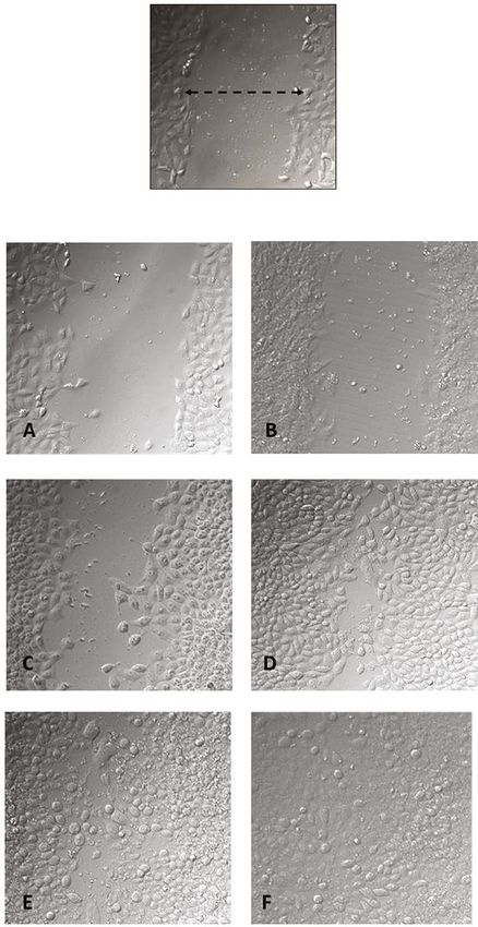
Oxidative Medicine and Cellular Longevity 7

 150 p < 0.001

 Fibroblast HFF‑1 cell proiiferation (% control)
 p < 0.001
 p < 0.001 B B B B
 B
 C
 A AB AB
 A A A
 A A A
 AB A
 100
 B

 50

 0
 0 1 3 5 10 30 0 1 3 5 10 30 0 1 3 5 10 30
 GA‑conjugated extract ( g/mL)
 Assays
 MTT
 Neutral red
 ATP
 (c)
 150 p < 0.001 150
Keratinocytes hacat cell proiiferation (% control)

 p < 0.001

 C C p < 0.001
 Antioxidant capacity (% of DPPH)

 B
 B A A
 A A A A
 A A
 100 A A 100 B
 C C
 B B
 B B B D
 E
 B

 50 50
 F

 C D D
 E

 0
 0 DPPH 1 3 5
 10 30 DPPH 1 3 5 10 30
 0 1 3 5 10 30 0 1 3 5 10 30 0 1 3 5 10 30
 Concentration ( g/mL)
 GA‑conjugated extract ( g/mL)
 GA-Conjugated extract
 Assays Rutin
 MTT
 Neutral red
 ATP
 (d) (e)

 Figure 1: Continued.
8 Oxidative Medicine and Cellular Longevity

 200
 p < 0.001

 Gemo assay (% H2O2 dsDNA degradation)
 D
 150
 B
 C C C

 A
 100

 50

 0
 H2O2 1 3 5 10 30
 GA‑conjugated extract ( g/mL)
 (f)

Figure 1: The in vitro effects of GA-conjugated extract at different concentrations on HFF-1 human fibroblasts and HaCaT keratinocytes
cultures: (a, b) cellular viability in 24 h cell cultures; (c, d) cellular proliferation in 72 h cell cultures. Data were generated from three different
protocols: MTT assay (3-[4,5-dimethylthiazol-2-yl]-2,5 diphenyl tetrazolium bromide), neutral red assay, and ATP assay. The (e)
antioxidant and (f) genoprotective capacity of GA-conjugated extract at different concentrations was quantified by (e) DPPH and (f)
GEMO assays, respectively. Statistical comparisons were performed by one-way analysis of variance followed by post hoc Tukey test.
Different letters indicated significant differences at p < 0:05.

3. Results these cells, when considering the ATP assay. Whereas fibro-
 blasts presented higher ATP levels than controls
3.1. Identification of Chemical Compounds in GA- (Figure 1(c)), keratinocytes exhibited lower ATP concentra-
Conjugated Extracts. An overview of all compounds identi- tions than controls (Figure 1(d)). The GA-conjugated
fied in the GA-conjugated extracts analyzed by ESI-ToF- extracts showed high antioxidant capacity and genoprotec-
MS is shown in Table 1. In addition to caffeine, which is tive capacity in the analysis performed for DPPH and
an alkaloid, polyphenols present mainly in guarana extracts GEMO assays (Figures 1(e) and 1(d)). Overall, the results
were identified, such as catechin, gallic acid, quercetin, and indicated that GA-conjugated extract at 5 μg/mL concentra-
epigallocatechin gallate (EGGC). However, other polyphe- tion could result in biological activity in the wound healing
nols were also observed, such as diosmetin, which is a fla- process.
vone derived from luteolin, methyl gallate, and
torachrysone, which belong to the class of naphthalenes. A 3.3. In Vitro Wound Scratch Assay. The GA-conjugated
macrocyclic peptide molecule (oscillacyclamide A) was also extract effects in in vitro scratched assays were employed
detected in the extract. Three molecules detected in the anal- to evaluate dermal fibroblast migration, which is a very
ysis could not be identified. The presence of citric acid was important event for wound contraction and further cellular
expected because this molecule was used to lower the pH proliferation. Cell migration was observed at 6, 12, and
in the extraction process, which gave rise to the GA- 24 h after scratching (Figure 2). Cell cultures supplemented
conjugated extract. with 5 μg/mL GA-conjugated extract exhibited a greater
 extent of migration than controls (Figures 2(a)–2(d)). More-
3.2. Indicators of Safety and Biological Capacity of GA- over, cellular proliferation observed in confluent monolayers
Conjugated Extracts. The cytotoxic effects of GA- after 72 h of cell culture was higher in fibroblast GA extract-
conjugated extract on keratinocytes and fibroblasts were exposed cells than in controls (Figures 2(e) and 2(f)).
analyzed using three complementary assays. Data from
MTT and neutral red assays showed that all extract concen- 3.4. Oxidative Metabolism Modulation in Scratched
trations significantly increased cell viability compared to Fibroblasts. The potential role of oxidative metabolism mod-
controls in 24 h cell culture. However, the ATP assay showed ulation in the migration and regeneration of scratched fibro-
an increase in the viability of fibroblasts cultured at 10 and blast monolayers was analyzed after 3, 6, 24, and 72 h of cell
30 μg/mL (Figures 1(a) and 1(b)). In 72 h cultures, GA con- culture. Superoxide and NO levels decreased significantly in
jugates triggered slightly increased cellular proliferation in 3 h fibroblasts cultures supplemented with GA extract com-
MTT and neutral red assays for both fibroblasts and kerati- pared to controls. However, the concentrations of these oxi-
nocytes. However, an antagonistic action was observed in dant molecules were similar between the treatment groups
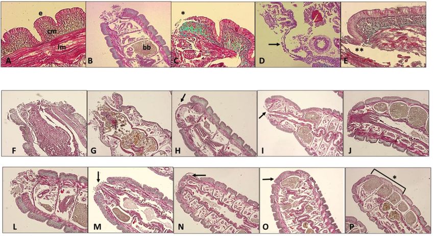
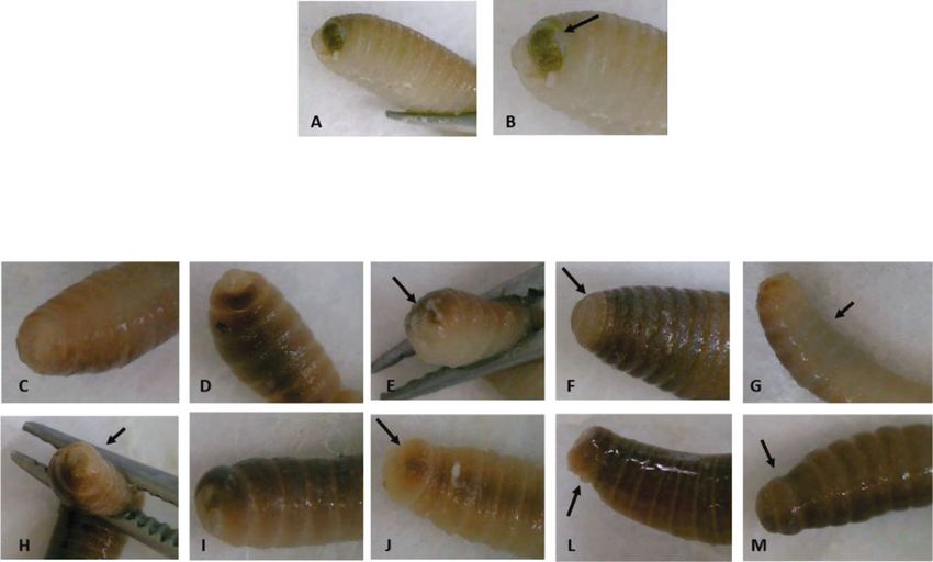
Oxidative Medicine and Cellular Longevity 9

(Figures 3(a) and 3(b)). In contrast, ROS levels decreased in
a time-dependent manner in fibroblasts exposed to GA com-
pared to those in controls (Figure 3(c)). The levels of lipo-
peroxidation measured by TBARS quantification also Scratch cells (0 h)
decreased significantly in 6, 24, and 72 h cell cultures when
compared to controls.
 To assess the impact of GA extracts on the survival and
modulation of apoptosis and necrosis of scratched cells, flow
cytometric analysis in 24 h cultures was conducted. In this CONTROL GA-EXTRACT
analysis, the frequency of live cells was significantly lower
in cultures exposed to GA extract than in controls. An
inverse situation was observed in relation to the frequency
of cells in early and late apoptosis, which was higher in the
control group than in cells exposed to GA extract. However, 6
the frequency of necrotic cells was similar between the two
groups (Figures 4(a)–4(c)).
 Cells exposed to GA extract showed higher collagen con-
centration in 24 h cultures, which was consistent with the
scratched closure pattern observed in Figure 2. However,
in 48 h and 72 h cultures, collagen levels were similar
between cells exposed or not exposed to GA extract
(Figure 4(d)). Considering the more rapid pattern of cell
confluence in cultures exposed to GA extract, analysis of 24
its effects on the modulation of genes related to healing pro-
cesses and dermal fibroblast function was also conducted.
The transcriptional levels of FGF2 were similar between
the two groups. However, FGF7 and COL1 genes were over-
expressed in cultures exposed to GA extract compared to the
controls. On the other hand, downregulation was observed
for the MMP1 and NLRP3 genes in the group exposed to
the GA extract compared to the controls (Figure 4(e)).
 The second group of experiments involved in vivo anal- 72
yses using E. fetida as an experimental model in which the
last three segments of the tail were surgically removed and
treated topically with GA extract. We concomitantly ana-
lyzed the results of macroscopic and microscopic observa-
tions, as summarized in Figures 5 and 6. Figure 2: Evaluation of GA-conjugated extract at 5 μg/mL
 In comparisons between the regeneration processes in concentration on HFF-1 human dermal fibroblasts submitted to
controls and earthworms topically treated with GA extract, scratch assay experiment. Fibroblast migration was evaluated by
the following events were considered. Macroscopically, microphotographs (200x magnification) 6, 24, and 72 h after the
 cell cultures are torn with the aid of a pipette tip. To determine
immediately after tail amputation, it was possible to visualize
 the cell numbers migrating during scratch closure, the perimeter
the intestines, which had a brownish color (Figures 5(a) and of each scratch was traced and the cells inside the area of closure
5(b)). Soon after, there was a large production of mucus at were counted using Digimizer software. Six wells were counted
the wound site (Figure 5(e)), followed by wound retraction for each condition, and three independent experiments were
and cell proliferation initially in a longitudinal direction, performed, and data are presented as mean ± SD of cells that
forming a more elongated structure covered by whitish tis- migrated inside the scratch of a single representative experiment.
sue (Figure 5(f)). Afterwards, this tissue darkened, and the The migration of cultures supplemented with GA-conjugated
three cut segments could be visualized again (Figure 5(m)). extract in relation to nonsupplemented cultures was compared by
 In histophysiological terms, it is important to highlight the Student t-test and was significantly higher (p < 0:05) in those
that in the posterior terminal region, the structure of the tis- receiving the extracts than in controls at the three time points (6,
sues of the earthworm body wall is very similar to that of 24, and 72 h) in which they were evaluated.
other sites. The body wall tissue is superficially composed
of a pseudostratified epithelial layer interspersed with tion, they do not have red cells, and hemoglobin is directly
mucous cells, which secrete a layer of chitin containing col- dissolved in the hemolymph.
lagen and a basal lamina that is held together with the adja- Immediately after cutting, the terminal part of the intes-
cent circular muscular layer. Below the circular muscle layer tines was exposed, followed by rapid tissue contraction and
is a thick layer of longitudinal muscle cells with a cytohisto- mucus production (Figures 5(b) and 6(c)). This process
logical pattern resembling that of vertebrate smooth muscle was visualized 1 h after tail amputation and was more rapid
(Figure 6(a)). Although earthworms have complete circula- in GA extract-treated earthworms than in controls
10 Oxidative Medicine and Cellular Longevity

 150 150

 Superoxide anion (% control)

 Nitric oxide (% control)
 100 100 ⁎
 ⁎⁎

 50 50

 0 0
 3 6 24 72 3 6 24 72
 Time (hours) Time (hours)
 Control Control
 GA-Conjugated extract GA-Conjugated extract
 (a) (b)
 150 150

 Lipoperoxidation (% control)
 ROS (% control)

 100 ⁎⁎ ⁎⁎ 100 ⁎⁎⁎ ⁎⁎⁎
 ⁎⁎⁎ ⁎⁎⁎
 ⁎⁎⁎

 50 50

 0 0
 3 6 24 72 3 6 24 72
 Time (hours) Time (hours)
 Control Control
 GA-Conjugated extract GA-Conjugated extract
 (c) (d)

Figure 3: The in vitro modulatory effects in four oxidative metabolism markers (superoxide, nitric oxide, reactive oxygen species (ROS), and
lipoperoxidation) of GA-conjugated extract at 5 μg/mL concentration on 3, 6, 24, and 72 h cultures of HFF-1 human dermal fibroblasts.
Data are presented as relative percentage of control. Statistical comparisons were performed by one-way analysis of variance followed by
post hoc Tukey test. Different letters indicated significant differences at p < 0:05.

(Figures 5(c), 5(h), 6(f), and 6(l)). The earthworm does not cess of migration and wound closure with these cells
have well-established connective tissue, but circular muscle occurred up to 6 h after cutting (Figures 6(g) and 6(h)).
cells secrete a network of collagen that supports this tissue However, in earthworms topically treated with GA extract,
(Figure 6(a)). When an injury occurs, hemoleucocytes pres- 3 h after the cut, the wound was almost completely closed,
ent in the fluid coelom and circular muscle cells migrate rap- and at 6 h, it was already possible to observe the formation
idly to the injury site. Therefore, these latter cells operate as of multiple layers at the incision site (Figures 6(m) and
fibroblast-like cells, migrating to the local incision 6(n)). This process can also be visually observed, as shown
(Figures 6(a) and 6(d)). At this point, reepithelialization in Figures 5(e) and 5(j).
and production of the collagen network in the circular mus- Twenty-four hours after the tail incision in earthworms
cle layer begin. Concomitantly, the longitudinal muscle layer treated with GA extract, complete and darkened segmenta-
also proliferated (Figure 6(e)). The final stage of regenera- tion was observed in the ventral and dorsal regions, whereas
tion was observed when the three segments were darkened in control earthworms, this process was more advanced only
in both the dorsal and ventral regions and could be visually in the dorsal region (Figures 5(g) and 5(m)). Histological
identified. As tail clipping involves only three segments, a analysis also confirmed that regeneration of the three seg-
large part of this regenerative process takes place within ments was in the process of completion in earthworms
the first 24 h after incision. treated with GA extract (Figures 6(j) and 6(p)). These results
 Comparison of this process in the two groups of earth- indicated that, on average, the regeneration rate was 6-12 h
worms showed an acceleration of the relevant differences faster in earthworms treated with GA extract than in control
between the two treatments. In control earthworms, the pro- animals.
Oxidative Medicine and Cellular Longevity 11

 Control GA extract
 104 Q1 Q2 10 4
 Q1 Q2
 1,08 48,2 0,39 20,3
 103 103

 102

 PI
 102

 PI
 101 101
 Q4 Q3 Q4 Q3
 49,7 1,08 77,6 1,78
 100 10 0

 100 101 102 103 104 100 101 102 103 104
 Annexin Annexin
 (a) (b)
 150
 ⁎⁎⁎
 100
 p < 0.001

 Collagen (% control)
 Cells in flow cytometry (%)

 80 100
 p < 0.001
 60 ⁎⁎⁎

 40 50
 ⁎⁎⁎
 20

 0 0
 Control GA‑extract Control GA‑extract 24 48 72
 Live cells Time (hours)
 Apoptotic cells Control
 GA-Conjugated extract
 (c) (d)
 2.5
 Gene expression (x times to control)

 2.0

 1.5

 1.0

 0.5

 0.0
 FGF‑2 FGF‑7 COL‑1 MMP‑1 NLRP‑3
 (e)

Figure 4: The in vitro modulatory effects of GA-conjugated extract at 5 μg/mL concentration on HFF-1 human dermal fibroblasts by
evaluation of apoptosis rate, collagen quantification, and expression of genes related to wound healing processes. (a, b) Representative
flow cytometry of cell cultures in the apoptosis analysis with and without extract supplementation. (c) Comparison of dead and live cell
frequency between culture extract supplemented and control by the Student t-test. Data are presented as relative percentage of control
calculated from three replications. (d) Collagen concentration in fibroblasts 24, 48, and 72 h cell cultures compared by one-way analysis
of variance followed by post hoc Tukey test. Data are presented as relative percentage of control. Different letters indicated significant
differences at p < 0:05. (e) Gene expression: values are normalized by beta-actin 1 gene and represent their value expressed in relation to
the control with reference value 1 (dashed line in the graph).

4. Discussion results showed that GA extract did not present cytotoxic or
 extensive proliferative effects on human keratinocytes and
Based on previous evidence, the effects of a GA-conjugated fibroblast cells in culture, showing potential antioxidant
extract prepared from roasted guarana seed and fresh seeds and genoprotective capacities that are relevant properties
of açai berries on wound healing models were tested. The in healing or in regenerative substances of the integument.
12 Oxidative Medicine and Cellular Longevity

 After tail amputation

 Hours

 1 3 6 12 24

 CONTROL

 GA EXTRACT

Figure 5: Representative photographs of the surgical incision in the anterior segments of Eisenia fetida earthworms topically treated with
GA-conjugated extract at 5 μg/mL concentration. Soon after the incision, the wound received a single topical dose of 2 μL of phosphate
buffer (control) or extract solution. The earthworms were then kept in Petri dishes, and the regeneration process was evaluated in
different periods of time. (a, b) Right after the incision, it is possible to observe the worm’s intestinal tube (in yellowish-brown color
indicated by the arrow). (c, h) One hour after cutting, the entire region was covered with mucus and seems to be more contracted in
earthworms that received the extract treatment than in controls. (d, i) Three hours after cutting, the contraction of the wound is already
well established and it is possible to see new transparent tissue starting to form. At some point, the wound is still open. Six hours after
cutting, the wound still has open parts in the control worms (e, arrow) and is completely closed with new tissue in the worm topically
treated with the extract (j, arrow); 12 hours after the incision, the wound of the control worms is completely regenerated with new
whitish tissue (f, arrow). In earthworms treated with the extract, the regenerated area is already pigmenting and differentiating into the
segments that have been cut (l, arrow). 24 h after the incision, the entire area was regenerated. However, pigmentation in the ventral
region, which is slower than the dorsal region, has not yet occurred in the control earthworms (g, arrow), while in those treated topically
with the extract, pigmentation is practically complete both in the dorsal region and in the ventral region of the animal’s body (m, arrow).

In both fibroblasts and earthworms, GA extract increased inflammatory activities, as described in the literature
healing speed, with no cytohistological abnormalities [37–41]. Some of these molecules appear to have healing
observed during the process. Analysis of possible causal properties, including EGCG [42] and gallic acid [43]. In
mechanisms associated with the effects of GA extract on addition, gallic acid, EGCG, and caffeine seem to act against
human fibroblasts revealed antioxidant and genomodulatory complications related to healing processes, such as keloid
activities related to the healing process. formation [44–46].
 These results will be discussed in greater detail below, Three other less-investigated polyphenols were also
starting with the chemical matrix of the GA-conjugated detected in the extract: the flavone diomestine, found in
extract identified by ESI-ToF-MS. The GA-conjugated legumes such as Acacia farnesiana, and in the leaves of Olea
extract was produced by mixing roasted guarana powder europaea [47], torachrysone, a molecule belonging to the
seeds with fresh pulped açai seeds. The presence of caffeine group of naphthoquinones, molecules widely distributed in
is expected in guarana extracts, as this plant has high con- plants, and cystonoside F. Previous studies have described
centrations of this alkaloid [15]. The identification of poly- the antioxidant, anti-inflammatory, and antimicrobial prop-
phenols such as gallic acid, catechin, EGCG, and quercetin erties of these polyphenols [48–52]. In addition to these
could also be anticipated, as many studies have described polyphenols, a macrocyclic peptide was identified, oscillacy-
the presence of these molecules in guarana extracts, as well clamide A, for which there are practically no studies regard-
as in açai [18, 34, 35]. In a study conducted by [36], the ing its biological properties. Even so, as highlighted by [53],
authors described the presence of four flavones, luteolin these peptides show great promise as therapeutics because
and velutin, in açai. they have increased target binding affinity and selectivity,
 Diosmetin, whose precursor was detected in the chemi- are more stable against proteolytic enzymes, and often have
cal matrix of the GA-conjugated extract, is also a flavone higher membrane permeability than their linear counter-
chemically close to luteolin. Therefore, it is possible that this parts. Therefore, this information indicates that the chemical
molecule derives from the extraction of fresh açai seeds. All matrix of the GA-conjugated extract contains bioactive ele-
of these known molecules have antioxidant and anti- ments that contribute to their antioxidant and anti-
Oxidative Medicine and Cellular Longevity 13

 1 3 6 12 24 hours

 Control

 GA‑conjugated extract

Figure 6: Representative histological analysis of Eisenia fetida earthworms submitted to posterior surgical incision of the three last segments
with and without topical treatment using GA-conjugated extract at 5 μg/mL concentration. The slides were stained with Masson-Goldner
trichrome kit that is used for visualization of muscles, collagen fibers, connective tissues, and keratin. (a) Body wall histology showing
the (e) epidermic layer that is directly connected with circular muscle layer (cm) immersed in an extracellular matrix richest in collagen.
Immediately afterwards, there is the longitudinal muscular layer (lm) that is covered with the peritoneum that delimits the earthworm’s
coelomic cavity. (b) Posterior segments (metameres) showing the region of the surgical incision. In this place, there are spaces full of
brown bodies (bb), which are structures that contain immune cells (cellomyocytes) that trapped and destroyed pathogens or linked to
impurities and underwent melanization. When these cavities are completely filled, the worm performs autotomy to free them and then
regenerates the posterior region. (c, d) Detail of the incision site showing migration (∗) of circular muscle cells migrating to initially
close the wound. These cells behave similarly to human fibroblasts when skin damage occurs. (e) Only after migration and onset of
proliferation of the smooth muscle layer does epidermis proliferation occur. (f–j) Sequence of healing processes in control earthworms at
1.3, 6, 12, and 24 hours. After this period, the region is in the final stage of regeneration of the incised segments. (l–p) Sequence of
healing processes in earthworms topically treated with GA-conjugated extract showing acceleration in regenerative processes in relation
to control. After 24 hours, the segments are fully regenerated (p, ∗).

inflammatory properties and to modulation of the expres- oprotective capacity. In the GEMO test, described by [28],
sion of relevant genes in healing processes. isolated dsDNA is exposed to H2O2, which causes extensive
 Plant extracts often have relevant therapeutic properties, strand breaks in the DNA backbone. Since the PicoGreen
but their use is restricted because of the toxicity associated dye has high affinity for dsDNA but does not bind to nucle-
with them. For this reason, we initially evaluated the poten- otides and single-stranded DNA, these breaks reduce the
tial cytotoxic and cell proliferative effects of GA-conjugated fluorescence of the reaction. However, the presence of GA
extract using keratinocytes and fibroblasts that are in direct extract decreased DNA breakage rates, indicating its geno-
contact when topically applied. Although there is some level protective activity. These results allowed the choice of the
of variation between the results obtained from the three dif- GA extract concentration to be 5 μg/mL, which was later
ferent assays employed here (MTT, neutral red, and ATP), used in complementary tests in vitro with fibroblasts and
the set of results did not reveal any relevant toxic effects. in vivo with earthworms.
These results are in accord with those of previous studies The results showed that, in both fibroblasts and earth-
that did not indicate toxicity associated with the supplemen- worms, GA extract induced a greater speed in the wound
tation of cell cultures with guarana or açai extract [20, 34]. healing process. Macroscopic and histological analyses of
 Two tests were conducted to determine the biological surgical incisions of earthworm tails revealed accelerated
capacity of the extracts. The first evaluation of the scaveng- healing processes, corroborating the results observed in
ing power of the DPHH radical by the GA-conjugated human fibroblasts. We highlight here that the use of earth-
extract showed that it has a relevant antioxidant capacity. worms as an experimental model for regeneration studies
The second trial evaluated the extent to which the extract has been gaining popularity for a number of reasons. First,
was able to decrease the rate of degradation of dsDNA mol- earthworms have long been used in ecotoxicity studies to
ecules exposed to high concentrations of H2O2. The results assess soil quality in relation to environmental pollutants.
also suggested that the GA-conjugated extract exerted a gen- This more primitive organism also has great regenerative
14 Oxidative Medicine and Cellular Longevity

capacity while having some cytofunctional elements that cellular matrix, facilitating fibroblast migration, and
have certain similarities to the human immune system remodulation of granulation tissue [57]. Again, it is possible
[10]. Furthermore, earthworm husbandry involves low that the low expression of MMP1 observed in 24 h cell cul-
maintenance cost and incurs fewer ethical problems than tures may be related to the fact that the GA extract acceler-
the use of vertebrates in experiments involving surgical inci- ated the wound healing process compared to control
sion. Although the data using earthworms provided evi- cultures.
dence of the role of GA extract in wound healing in E. Since in vitro assays have some limitations in assessing
fetida, analyses of potential causal mechanisms were per- the properties of certain extracts or drugs, the results
formed only in fibroblasts. This is because E. fetida is still a described here from fibroblast cell culture were corroborated
relatively new model in studies of regeneration involving by in vivo assays involving E. fetida. In summary, despite
environmental variables, such as plant extracts. Conse- limitations related to in vitro and in vivo studies, the results
quently, it will be necessary to establish future analyses of obtained from this translational investigation suggest that
biochemical and molecular markers related to the role of GA extract has a potential therapeutic effect on the healing
inflammation in healing and other metabolic pathways. of acute wounds, such as those that occur during surgical
 Therefore, the potential effect of GA extracts on oxida- interventions.
tive metabolism was investigated in fibroblasts. The results
showed a generalized antioxidant effect when the levels of Data Availability
total ROS were evaluated. These values remained lower than
in control cells at all time points analyzed. The lipoperoxida- The data will be made available if the reviewers and/or edi-
tion rate was also lower in 6 h fibroblast cultures subjected to tors of this journal so request.
the scratch assay. Two other oxidative markers that are
directly associated with inflammatory responses (superoxide Disclosure
and NO) were also analyzed. The results showed a decrease
in the levels of these two oxidative molecules in 3 h cultures. Some results of this article were presented in the form of an
Thereafter, superoxide and NO levels remained similar to abstract in the 53rd Brazilian Congress of Pharmacology and
those in control cells. In addition, scratched cultures supple- Experimental Therapeutics. A part of the data was presented
mented with GA extract exhibited lower apoptosis rates than as a poster at the 53rd Brazilian Congress of Pharmacology
controls. and Experimental Therapeutics in the following link
 Previous investigations such as the study performed by https://www.sbfte.org.br/wp-content/uploads/2021/12/
[54] showed that, in the presence of prosenescence mole- Livro-2021_fnv2.pdf/ [58].
cules such as advanced glycation end products (AGEs), der-
mal fibroblasts are induced to undergo apoptosis, presenting Conflicts of Interest
an increase in ROS and overexpression of the NRLP3 gene,
which is directly associated with inflammasome formation. The authors declare that they have no known competing
Although this study did not expose dermal fibroblasts to financial interests or personal relationships that could have
molecules such as AGEs, supplementation of cultures with influenced the work reported in this paper.
GA extract showed the opposite effect. That is, 24 h cultures
exposed to GA extract showed lower levels of ROS and Authors’ Contributions
downregulation of the proinflammatory gene NRLP3. It is
possible that the peak oxidative stress and proinflammatory Fellipe Danezi Felin and Ednea Aguiar Maia-Ribeiro have
responses caused by scratching decreased in cultures treated contributed equally to this work.
with GA extract. This is because there is evidence that the
triggering of initial oxy-inflammatory processes is relevant
 Acknowledgments
for wound healing to occur [55].
 Other relevant results concern the induction of greater This work was supported by Fundação de Apoio a Pesquisa
collagen formation, Col-1 gene overexpression, and faster do Amazonas (FAPEAM), by project protocol number
scratch closure in fibroblast cultures and those supple- 38133.UNI699.56949.07052018, Coordenação de Aperfei-
mented with GA extract. Moreover, differential modulation çoamento de Pessoal de Nível Superior (CAPES), and Con-
of other genes related to tissue regeneration and dermal selho Nacional de Desenvolvimento Científico e
function was observed in fibroblast cultures supplemented Tecnológico (CNPq). We appreciate the contributions of
with GA extracts. This is the case with FGF7 overexpression laboratory technicians Marina de Souza Vencato and Bruno
in fibroblast GA extract supplements, which is considered a Tomazele Rovani.
relevant gene for organogenesis and mediation of wound
healing in mammals [56]. In addition to downregulated
NRPL3 expression, this result was also observed for MMP1.
 References
Collagenases such as MMP-1 play a major role in the healing [1] T. Montrief, K. Bornstein, M. Ramzy, A. Koyfman, and B. J.
process. When a physical lesion occurs, generating a wound, Long, “Plastic surgery complications: a review for emergency
MMP-1 is activated assisting the healing process by elimina- clinicians,” The Western Journal of Emergency Medicine,
tion of damaged proteins, destroying the provisional extra- vol. 21, no. 6, pp. 179–189, 2020.
Oxidative Medicine and Cellular Longevity 15

 [2] C. L. Harris, J. Kuhnke, J. Haley, K. Cross, R. Somayaji, and [16] D. M. de Oliveira, G. Barreto, P. Galeano et al., “Paullinia
 J. Dubois, “Best practice recommendations for the prevention cupanaMart. var.Sorbilisprotects human dopaminergic neuro-
 and management of surgical wound complications,” Founda- blastoma SH-SY5Y cell line against rotenone-induced cytotox-
 tions of Best Practice for Skin and Wound Management. A Sup- icity,” Human & Experimental Toxicology, vol. 30, no. 9,
 plement of Wound Care Canada, vol. 668, 2017. pp. 1382–1391, 2011.
 [3] M. Barchitta, A. Maugeri, G. Favara et al., “Nutrition and [17] A. K. Machado, F. C. Cadoná, V. F. Azzolin, E. B. Dornelles,
 wound healing: an overview focusing on the beneficial effects F. Barbisan, and I. B. M. da Cruz, “Guarana (Paullinia cupana)
 of curcumin,” International Journal of Molecular Sciences, improves the proliferation and oxidative metabolism of senes-
 vol. 20, no. 5, p. 1119, 2019. cent adipocyte stem cells derived from human lipoaspirates,”
 [4] F. Alminderej, S. Bakari, T. I. Almundarij, M. Snoussi, Food Research International., vol. 67, pp. 426–433, 2015.
 K. Aouadi, and A. Kadri, “Antimicrobial and wound healing [18] A. K. Machado, A. C. Andreazza, T. M. da Silva et al., “Neuro-
 potential of a new chemotype from Piper cubeba L. essential protective effects of açaí (Euterpe oleracea Mart.) against rote-
 oil and in silico study on S. aureus tyrosyl-tRNA synthetase none in vitro exposure,” Oxidative Medicine and Cellular
 protein,” Plants., vol. 10, no. 2, p. 205, 2021. Longevity, vol. 2016, 14 pages, 2016.
 [5] S. B. Khedir, S. Bardaa, N. Chabchoub, D. Moalla, Z. Sahnoun, [19] J. R. Souza-Monteiro, G. P. F. Arrifano, A. I. D. G. Queiroz
 and T. Rebai, “The healing effect ofPistacia lentiscusfruit oil on et al., “Antidepressant and antiaging effects of açaí (Euterpe
 laser burn,” Pharmaceutical Biology, vol. 55, no. 1, pp. 1407– oleracea Mart.) in mice,” Oxidative Medicine and Cellular Lon-
 1414, 2017. gevity, vol. 2019, 16 pages, 2019.
 [6] N. Ojeh, O. Stojadinovic, I. Pastar, A. Sawaya, N. Yin, and [20] D. R. Maldaner, N. L. Pellenz, F. Barbisan et al., “Interaction
 M. Tomic-Canic, “The effects of caffeine on wound healing,” between low-level laser therapy and guarana (Paullinia
 International Wound Journal, vol. 13, no. 5, pp. 605–613, cupana) extract induces antioxidant, anti-inflammatory, and
 2016. anti-apoptotic effects and promotes proliferation in dermal
 [7] P. Orlowski, M. Zmigrodzka, E. Tomaszewska et al., “Polyphe- fibroblasts,” Journal of Cosmetic Dermatology, vol. 19, no. 3,
 nol-conjugated bimetallic Au@AgNPs for improved wound pp. 629–637, 2020.
 healing,” International Journal of Nanomedicine, vol. 15, [21] I. B. Santos, G. F. de Bem, C. A. da Costa et al., “Açai seed
 pp. 4969–4990, 2020. extract prevents the renin-angiotensin system activation, oxi-
 [8] S. Ud-Din, P. Foden, M. Mazhari et al., “A double-blind, ran- dative stress and inflammation in white adipose tissue of
 domized trial shows the role of zonal priming and direct topi- high-fat diet-fed mice,” Nutrition Research, vol. 79, pp. 35–
 cal application of epigallocatechin-3-gallate in the modulation 49, 2020.
 of cutaneous scarring in human skin,” The Journal of Investi- [22] B. Sadowska, J. Rywaniak, A. Cichocka et al., “Phenolic and
 gative Dermatology, vol. 139, no. 8, pp. 1680–1690, 2019. non-polar fractions of the extracts from fruits, leaves, and
 [9] T. D. Algarve, C. E. Assmann, F. C. Cadoná, A. K. Machado, twigs of Elaeagnus rhamnoides (L.) A. Nelson-the implications
 M. F. Manica-Cattani, and I. B. M. da Cruz, “Guarana for human barrier cells,” Molecules, vol. 25, no. 9, 2020.
 improves behavior and inflammatory alterations triggered by [23] C. C. Lu, J. S. Yang, Y. J. Chiu et al., “Dracorhodin perchlorate
 methylmercury exposure: an in vivo fruit fly and in vitro neu- enhances wound healing via β-catenin, ERK/p38, and AKT
 ral cells study,” Environmental Science and Pollution Research signaling in human HaCaT keratinocytes,” Experimental Med-
 International, vol. 26, no. 15, pp. 15069–15083, 2019. icine Journal, vol. 22, no. 2, p. 822, 2021.
[10] A. O. Alves, G. C. C. Weis, T. C. Unfer et al., “Caffeinated bev-
 [24] G. Á. Borges, S. T. Elias, S. M. M. da Silva et al., “In vitro eval-
 erages contribute to a more efficient inflammatory response:
 uation of wound healing and antimicrobial potential of ozone
 evidence from human and earthworm immune cells,” Food
 therapy,” Journal of Cranio-Maxillo-Facial Surgery, vol. 45,
 and Chemical Toxicology, vol. 134, p. 110809, 2019.
 no. 3, pp. 364–370, 2017.
[11] P. A. Benatrehina, L. Pan, C. B. Naman, J. Li, and A. D. King-
 [25] A. R. Lourenço and P. J. Coffer, “SOX4: joining the master reg-
 horn, “Usage, biological activity, and safety of selected botani-
 ulators of epithelial-to-mesenchymal transition?,” Trends in
 cal dietary supplements consumed in the United States,”
 Cancer, vol. 3, no. 8, pp. 571–582, 2017.
 Journal of Traditional and Complementary Medicine, vol. 8,
 no. 2, pp. 267–277, 2018. [26] V. F. Azzolin, F. C. Cadoná, A. K. Machado et al., “Superoxide-
[12] S. K. Chang, C. Alasalvar, and F. Shahidi, “Superfruits: phyto- hydrogen peroxide imbalance interferes with colorectal cancer
 chemicals, antioxidant efficacies, and health effects - a compre- cells viability, proliferation and oxaliplatin response,” Toxicol-
 hensive review,” Critical Reviews in Food Science and ogy In Vitro, vol. 32, pp. 8–15, 2016.
 Nutrition, vol. 59, no. 10, pp. 1580–1604, 2019. [27] G. Repetto, A. del Peso, and J. L. Zurita, “Neutral red uptake
[13] M. T. S. S. de Almeida, M. P. C. de Oliveira, A. Converti, and assay for the estimation of cell viability/cytotoxicity,” Nature
 L. Á. A. Neves, “The use of Euterpe oleracea Mart. as a new Protocols., vol. 3, no. 7, pp. 1125–1131, 2008.
 perspective for disease treatment and prevention,” Biomole- [28] F. C. Cadoná, M. F. M. R. Cattanni, A. K. Machado et al.,
 cules, vol. 10, no. 6, p. 813, 2020. “Genomodifier capacity assay: a non-cell test using dsDNA
[14] D. V. de Souza, L. Pappis, T. T. Bandeira et al., “Acai (Euterpe molecules to evaluate the genotoxic/genoprotective properties
 oleracea Mart.) presents anti-neuroinflammatory capacity in of chemical compounds,” Analytical Methods (point), vol. 6,
 LPS-activated microglia cells,” Nutritional Neuroscience, no. 21, pp. 8559–8568, 2014.
 vol. 25, no. 6, pp. 1188–1199, 2022. [29] G. Morabito, D. Trombetta, B. K. Singh et al., “Antioxidant
[15] F. C. Schimpl, J. F. da Silva, J. F. Gonçalves, and P. Mazzafera, properties of 4-methylcoumarins in in vitro cell-free systems,”
 “Guarana: revisiting a highly caffeinated plant from the Ama- Biochimie, vol. 92, no. 9, pp. 1101–1107, 2010.
 zon,” Journal of Ethnopharmacology, vol. 150, no. 1, pp. 14–31, [30] B. Halliwell and M. Whiteman, “Measuring reactive species
 2013. and oxidative damage in vivo and in cell culture: how should
You can also read