Chemical pancreatectomy treats chronic pancreatitis while preserving endocrine function in preclinical models
←
→
Page content transcription
If your browser does not render page correctly, please read the page content below
The Journal of Clinical Investigation                                                                                                                              RESEARCH ARTICLE

Chemical pancreatectomy treats chronic pancreatitis
while preserving endocrine function in preclinical models
Mohamed Saleh,1,2 Kartikeya Sharma,1 Ranjeet Kalsi,1 Joseph Fusco,1 Anuradha Sehrawat,1 Jami L. Saloman,3 Ping Guo,4
Ting Zhang,1 Nada Mohamed,1 Yan Wang,1 Krishna Prasadan,1 and George K. Gittes1
Division of Pediatric Surgery, 2Division of Pediatric Endocrinology, UPMC Children’s Hospital of Pittsburgh, Pittsburgh, Pennsylvania, USA. 3Department of Medicine, Division of Gastroenterology, Hepatology,
1

and Nutrition, Department of Neurobiology, Pittsburgh Center for Pain Research, University of Pittsburgh School of Medicine, Pittsburgh, Pennsylvania, USA. 4Department of Clinical Science, Colorado State
University, Fort Collins, Colorado, USA.

              Chronic pancreatitis affects over 250,000 people in the US and millions worldwide. It is associated with chronic debilitating
              pain, pancreatic exocrine failure, and high risk of pancreatic cancer and usually progresses to diabetes. Treatment options are
              limited and ineffective. We developed a new potential therapy, wherein a pancreatic ductal infusion of 1%–2% acetic acid in
              mice and nonhuman primates resulted in a nonregenerative, near-complete ablation of the exocrine pancreas, with complete
              preservation of the islets. Pancreatic ductal infusion of acetic acid in a mouse model of chronic pancreatitis led to resolution
              of chronic inflammation and pancreatitis-associated pain. Furthermore, acetic acid–treated animals showed improved
              glucose tolerance and insulin secretion. The loss of exocrine tissue in this procedure would not typically require further
              management in patients with chronic pancreatitis because they usually have pancreatic exocrine failure requiring dietary
              enzyme supplements. Thus, this procedure, which should be readily translatable to humans through an endoscopic retrograde
              cholangiopancreatography (ERCP), may offer a potential innovative nonsurgical therapy for chronic pancreatitis that relieves
              pain and prevents the progression of pancreatic diabetes.

Introduction                                                                                             life-threatening hypoglycemia (14, 15). Overall, CP patients con-
Chronic pancreatitis (CP) affects approximately 250,000 people in                                        sume copious resources and cause a substantial economic burden
the US and millions worldwide (1–4), and the incidence is increas-                                       (16). CF-related diabetes (CFRD) is also a distinct form of pan-
ing with the increase in alcohol abuse (5). Also, cystic fibrosis (CF),                                  creatic diabetes associated with increased morbidity and mortal-
the most common inherited disease in White people in the US, is                                          ity (6). The etiology of CFRD is multifactorial, with the primary
characterized by pancreatic inflammation leading to islet loss and                                       etiology being relative insulin insufficiency due to islet loss (6).
diabetes mellitus (6). Patients with CP suffer from chronic pain that                                    Approximately 40%–50% of adults with CF develop CFRD, and
compromises quality of life (7), have exocrine pancreatic insuf-                                         the incidence is rising (17).
ficiency, and develop a brittle form of diabetes (8). Additionally,                                           Surgical intervention in CP can entail major surgeries, includ-
patients with CP have a roughly 13-fold increased risk of pancreatic                                     ing partial and total pancreatectomy (8). Total pancreatectomy
ductal adenocarcinoma (9), a common and highly lethal form of                                            with islet autotransplantation (TPIAT) has evolved to treat CP-
cancer (10, 11). The treatment options for CP are limited, marginal-                                     associated pain while preserving some endocrine function, but it
ly effective, and usually cause endocrine tissue loss (12).                                              is a surgery with high morbidity with variable outcomes, and sub-
     Pancreatic diabetes occurs in approximately 80% of patients                                         stantial islet loss occurs during both islet isolation and reinfusion
with CP (13). Management of pancreatic diabetes is challenging                                           (18). Thus, new therapeutics for CP are needed.
because, besides loss of insulin-producing β cells, there is concom-                                          Herein, we demonstrate a potentially novel therapy for CP
itant loss of the glucagon-producing α cells (presumably because                                         through ablation of the exocrine pancreas by delivering a chemical
all islet cells are subjected to the inflammatory milieu). Glucagon                                      agent, acetic acid (AcA), through the pancreatic duct. Remarkably,
is a counterregulatory hormone that rescues hypoglycemia. With                                           the islets of Langerhans remain completely intact. In this study,
glucagon deficiency, a slight insulin overdose could cause severe                                        because of the small size of the animals, a surgical laparotomy to
                                                                                                         access the pancreatic duct was required. However, pancreatic duct
                                                                                                         access is routinely and readily achieved in humans via the nonsur-
       Related Commentary: https://doi.org/10.1172/JCI146210                                             gical endoscopic procedure endoscopic retrograde cholangiopan-
                                                                                                         creatography (ERCP). This “chemical pancreatectomy” technique
Authorship note: KS and RK contributed equally to this work.                                             appears to relieve CP-associated pain. Moreover, it may rescue ail-
Conflict of interest: The authors have declared that no conflict of interest exists.
                                                                                                         ing islets by removing the source of injury (inflamed exocrine tis-
Copyright: © 2021, American Society for Clinical Investigation.
Submitted: August 13, 2020; Accepted: December 1, 2020; Published: February 1, 2021.
                                                                                                         sue) and thus potentially prevent the progression to, or even foster
Reference information: J Clin Invest. 2021;131(3):e143301.                                               rescue from, pancreatic diabetes. This approach could also be bene-
https://doi.org/10.1172/JCI143301.                                                                       ficial for preventing CFRD if given early in the course of the disease

                                                                                                                                                                                                       1
RESEARCH ARTICLE                                                                                       The Journal of Clinical Investigation

before the destruction of islets. The incurred loss of exocrine tissue     no statistically significant difference in serum pancreatic enzymes
in this procedure would not typically require further management           among the 3 groups, indicating resolution of the infusion-induced
in CP and CF patients because they usually develop pancreatic              transient pancreatitis (Supplemental Figure 1, C and D).
exocrine failure requiring dietary enzyme supplements. Finally,                 Panoramic histology of the whole pancreas 1 year after AcA
near-total exocrine pancreas ablation may well minimize the risk of        infusion revealed persistent near-complete loss of the exocrine
future pancreatic ductal adenocarcinoma in these patients.                 pancreas with stable fatty replacement and normal-appearing
                                                                           islets (Figure 2E).
Results                                                                         Chemical pancreatectomy preserves the integrity of the islets. After
Chemical pancreatectomy ablates the exocrine pancreas, but spares          AcA infusion, despite near-complete ablation of the acinar cells,
islets. Eight-week-old outbred (CD1) mice from either sex were             the islets appeared normal (Figure 1, D and E). Immunostaining for
divided into 3 groups: AcA-treated, saline-treated, and unoperated         laminin and collagen 4 demonstrated an intact islet capsule (Sup-
(naive). Gross morphology of the pancreas 2 days after AcA infu-           plemental Figure 2A). Immunostaining for the endothelial marker
sion showed a markedly abnormal appearance, with tissue whit-              CD31 after AcA infusion showed a normal dense islet vasculature
ening, suggesting protein denaturation (Figure 1A). At 2 weeks,            (Figure 3A). Quantification of the CD31+/islet area ratio showed
the pancreas appeared translucent, suggesting acinar tissue loss,          no difference between AcA-treated mice and control groups at 6
with the islets readily visible (Figure 1A). At 8 weeks, the pancreas      months after AcA infusion (Figure 3B). Six months after AcA infu-
appeared fatty, with the islets remaining visible. The gross mor-          sion, the whole-mount IHC allowed detailed visualization of an
phology of the pancreas did not change at 6 months compared                intact islet vasculature (Figure 3C). Islet capillaries are highly spe-
with 8 weeks after AcA infusion. Fatty replacement is a well-              cialized, with dense fenestrations in the vicinity of endocrine cells
described common phenomenon in the human pancreas that can                 that facilitate rapid glucose sensing and release of hormones (23).
occur with aging, obesity, CF, pancreatitis, and diabetes (19–22).         Six months after AcA infusion, electron microscopy (EM) showed
     Pancreatic histopathology showed acinar cell injury and necro-        intact dense fenestrations (Figure 4, A and B). Immunostaining for
sis 2 days after AcA infusion (Figure 1, B and C). At 2 weeks, there       the neuron marker PGP9.5 revealed that the pancreas remained
was near-complete loss of the acinar cells, with inflammation and          innervated at serial time points after AcA infusion (Figure 4C).
accompanying fibrosis (Figure 1D and Supplemental Figure 1A;               Whole-mount IHC at 6 months after AcA infusion revealed intact
supplemental material available online with this article; https://doi.     nerve endings supplying the islets (Figure 4D). Additionally,
org/10.1172/JCI143301DS1). At 4 weeks, fat cells started to replace        although 1% AcA is sufficient to destroy acinar cells, the number of
the exocrine pancreas, and this process was complete by 8 weeks            neurons in the dorsal root ganglion (DRG) at the T12 level (which
(Figure 1D). Pancreatic histology remained unchanged at 6 months           normally contains the highest concentration of pancreas afferents)
(Figure 1D) compared with at 8 weeks after AcA infusion. Two days          was normal in AcA-treated mice, suggesting that AcA is not grossly
after AcA infusion, some areas in the pancreas showed apparently           cytotoxic to sensory neurons (Supplemental Figure 2, B and C).
preserved acinar cell architecture (Figure 1C); however, IHC showed             Mode of cell death and acinar cell susceptibility to cell injury after
a marked decrease in amylase staining, but with normal insulin             AcA treatment. Immunostaining for cleaved caspase-3 showed no
staining (Figure 1E). The loss of amylase immunostaining here may          evidence of apoptosis (Supplemental Figure 3A). The absence of
be due to degradation of zymogen enzymes in the injured acinar             caspase-3 in the setting of inflammation (Figure 1 and Supplemen-
cells. In line with the IHC findings, at 2 days, a Western blot showed     tal Figure 1) suggests necrosis as the mechanism of AcA-induced
decreased amylase in AcA-infused pancreata compared with that of           acinar cell death. EM of pancreas 2 days after AcA infusion con-
control groups (Figure 2, A and B), while insulin protein expression       firmed signs of cell injury and necrosis (Figure 5, A–D). To deter-
was not statistically different among the 3 groups (Figure 2, A and        mine whether AcA-induced cell death was explicitly due to acid-
C). Two weeks onwards after AcA infusion, immunostaining showed            ity, we infused citric acid (6 mM), which has a pH equivalent to
no amylase, with normal insulin staining (Figure 1E). Time course          1% AcA (174 mM, pH of approximately 2.75) into the pancreatic
of inflammatory changes in the exocrine pancreas following AcA             duct. The pancreatic histology was normal 2 weeks after citric acid
infusion showed the exocrine pancreas infiltrated with lymphocytes,        infusion, indicating that AcA’s toxicity is not merely due to low pH
macrophages, and neutrophils at 2 days, but there was no infiltration      (Supplemental Figure 3B).
into the islets (Supplemental Figure 1A). The inflammation peaked               We then examined the direct effect of AcA on acinar cell clus-
at 2 weeks, then regressed by 4 weeks, and eventually disappeared          ters versus islets in vitro. Acini and islets were isolated from naive
by 8 weeks (Supplemental Figure 1A). Also, cytokeratin (Figure 2D)         mice and cultured for 1 hour, then exposed to 1% AcA for 1 minute
and SOX9 (Supplemental Figure 1B) immunostaining demonstrat-               or 5 minutes, followed by immediate washing with PBS. Cell via-
ed increased ducts and duct-like structures at 2 weeks, followed by        bility was assessed by propidium iodide (PI) staining and FACS.
a decrease at 4 weeks, and a complete absence of pancreatic ducts          Acinar and islet viability were normalized to their respective con-
at 8 weeks after AcA infusion. Those early duct-like structures may        trol acini and islets not exposed to AcA. The islet cells had signifi-
represent an aborted attempt at exocrine pancreas regeneration.            cantly higher viability compared with acinar cells after exposure
     Two days after surgery, serum pancreatic enzymes (amylase and         to AcA at both 1 minute (P < 0.0001) and 5 minutes (P < 0.0001),
lipase) in AcA-treated mice and saline-treated controls were elevat-       suggesting that acinar cells are more vulnerable to AcA-induced
ed over those of naive mice, suggesting that the AcA and saline infu-      cell death (Figure 5E and Supplemental Figure 3, C and D).
sions similarly caused pressure-induced pancreatitis (Supplemental              To study the role of islet integrity in their relative resistance
Figure 1, C and D). One week and 8 weeks after surgery, there was          to AcA, islets were isolated from naive mice and cultured in cell

   2                                   J Clin Invest. 2021;131(3):e143301 https://doi.org/10.1172/JCI143301
The Journal of Clinical Investigation                                                                                          RESEARCH ARTICLE

Figure 1. Morphological and histological changes in mouse pancreas following chemical pancreatectomy. (A) After AcA infusion, gross morphology of the
pancreas was abnormally white and edematous at 2 days, translucent with visible islets (arrow) at 2 weeks, and replaced by fatty tissue with visible islets
(arrow) at 8 weeks. (B and C) Normal pancreas histology 2 days after saline infusion (B). Two days after AcA infusion, there was exocrine tissue necrosis
(asterisks) with intact islets (i). Magnification (inset) of the apparently preserved acinar cells revealed cell swelling and cytoplasmic vacuolization (arrows)
(C). (D) Histology of the pancreas after AcA infusion. Intact islets are denoted by i. Arrows denote fat cells at 4 weeks. (E) Immunostaining after AcA
infusion showed negative amylase staining (arrows denote amylase remnants at 2 days), with normal insulin and glucagon staining. Illustrative histology
results from 5 animals per time point are shown. Scale bars: 200 μm.

                                           J Clin Invest. 2021;131(3):e143301 https://doi.org/10.1172/JCI143301                                           3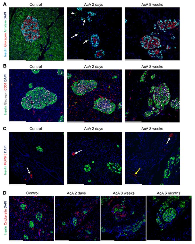
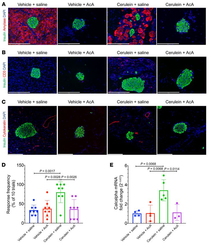
RESEARCH ARTICLE                                                                                              The Journal of Clinical Investigation

Figure 2. Chemical pancreatectomy causes nonregenerative ablation of exocrine pancreas and spares islets. (A–C) Protein isolated from pancreata, 2
days after surgery (3 mice/group), were loaded on the same gel (A). Western blot protein quantification by densitometry showed decreased amylase pro-
tein (B) in the AcA-infused pancreata compared with control groups (1-way ANOVA followed by Holm-Šidák test for multiple comparisons, F2,6=7.553;
P = 0.0230), with no difference in the insulin protein among the 3 groups (1-way ANOVA, F2,6=0.5592; P = 0.5988) (C). β-Actin was used as a protein loading
control for Western blot analysis. (D) Immunostaining for cytokeratin (duct cell marker) in control and AcA-infused pancreata showed the presence of
ducts and duct-like structures at 2 weeks and 4 weeks, while at 8 weeks, immunostaining was negative for cytokeratin, suggesting involution of the ducts.
(E) One year after AcA infusion, there was persistent loss of exocrine pancreas with intact islets (i). Illustrative histology results from 5 animals per time
point are shown. Scale bars: 200 μm unless otherwise specified. Data are presented as mean ± SD.

media for 24 hours. A subset of islets was dissociated with trypsin                   Chemical pancreatectomy resolves inflammation and amelio-
before exposure to 1% AcA. Another group of intact islets was first              rates pain-like behavior in cerulein-induced CP. Eight-week-old
exposed to AcA and then dissociated with trypsin for FACS after                  CD1 mice were divided into 2 groups. Group 1 received i.p. ceru-
treatment. Viability of islet cells was normalized to their respec-              lein injections as described in Methods for 8 weeks to induce
tive controls not exposed to AcA. Here, the cells from the intact                CP (Figure 6, A and B). Group 2 received i.p. vehicle injections
islets exposed to AcA had significantly higher viability than the                (normal saline). At 8 weeks, each group was split into 2 sub-
dissociated islet cells exposed to AcA at 1 minute (P < 0.0001), and             groups; one underwent AcA pancreatic ductal infusion, and the
5 minutes (P < 0.0001). Thus, islet integrity is likely an additional            other underwent a normal saline infusion. Thus, 4 experimen-
protective factor against AcA-induced cell death during chemical                 tal subgroups were created: vehicle+saline, vehicle+AcA, ceru-
pancreatectomy (Figure 5F and Supplemental Figure 3, E and F).                   lein+saline, and cerulein+AcA.

   4                                       J Clin Invest. 2021;131(3):e143301 https://doi.org/10.1172/JCI143301
The Journal of Clinical Investigation RESEARCH ARTICLE
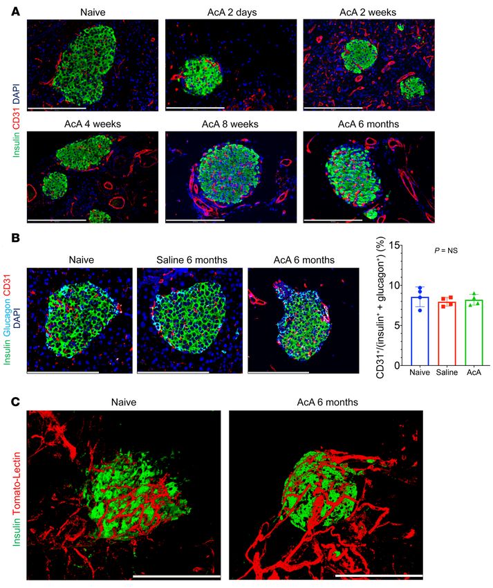
Figure 3. Chemical pancreatectomy preserves the integrity of islet vasculature. (A) Immunostaining for CD31 and insulin showed an intact microvascu-
lature of the islets in AcA-treated mice at all time points. (B) Representative images for CD31 immunostaining of the islets in naive, saline-treated, and
AcA-treated mice at 6 months after infusion (left panel). There was no significant difference in the quantification of islet microvasculature among the 3
groups (right panel). One-way ANOVA, F2,9=0.5013; P = 0.6217. n = 4 mice per group, 12 islets per mouse. (C) Whole-mount immunostaining for endothelial
cells (Tomato-lectin) and insulin showed intact microvasculature of islets at 6 months after AcA infusion compared with that of naive mice. Illustrative
histology results from 5 animals per time point are shown. Scale bars: 200 μm. Data are presented as mean ± SD.
One week after the mice recovered from surgery, we resumed cantly lower serum pancreatic enzymes than the cerulein+saline
i.p. injections of cerulein or vehicle for 8 more weeks to prevent group, and there was no difference in the serum pancreatic enzymes
the resolution of CP, which occurs after cerulein cessation (24). between the cerulein+AcA group and the 2 vehicle subgroups,
Following the two 8-week cycles of i.p. injections, histopathology indicating that AcA treatment normalized the elevated pancreatic
revealed persistence of the inflamed exocrine pancreas in the ceru- enzymes seen in the CP model (Supplemental Figure 4, A and B).
lein+saline group (Figure 6C and Figure 7A), while the cerulein+ In line with the data in the WT mice, AcA pancreatic ductal infusion
AcA group showed near-complete ablation of the acinar cells (Fig- ablated ducts in cerulein+AcA group, as evidenced by the cytokera-
ure 6D and Figure 7A). Immunostaining for CD3 showed absence tin (Figure 7C) and SOX9 staining (Supplemental Figure 4C).
of the characteristic CP lymphocytic infiltration in the cerulein+ For pain assessment, normal 8-week-old CD1 mice (without
AcA group (Figure 7B). Also, the cerulein+AcA group had signifi- CP) were divided into 3 groups (AcA treated, saline treated, and
J Clin Invest. 2021;131(3):e143301 https://doi.org/10.1172/JCI143301 5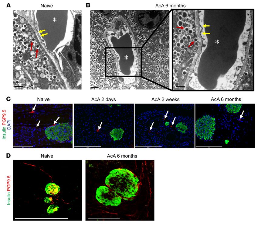
RESEARCH ARTICLE                                                                                               The Journal of Clinical Investigation

Figure 4. Chemical pancreatectomy preserves the ultrastructure of islet capillaries and innervation. (A and B) EM of the islet capillary-endocrine cell
interface in controls (A) and 6 months after AcA infusion (B). Red arrows denote hormone-containing granules of the islet cells. Yellow arrows denote the
intact endothelial fenestrations, characteristic of mature islet vessels. Asterisks denote red blood cells in the capillary lumen. Scale bars: 600 nm (A and B,
right panel); 1 μm (B, left panel). (C) Immunostaining for PGP9.5 (arrows) and insulin showed intact neurons in the parenchyma of the AcA-treated pancre-
ata at serial time points. (D) Whole-mount immunostaining for neurons (PGP9.5) and insulin in naive pancreas and 6 months after AcA infusion showed
apparently intact nerve endings supplying the islets. Illustrative histology results from 5 animals per time point are shown. For the EM, illustrative data
from 3 animals are shown. Scale bars: 200 μm (C and D).

naive). The abdominal von Frey monofilament assay was per-                        AcA; Figure 7D). Importantly, AcA infusion significantly abrogat-
formed 8 weeks after surgery by a blinded experimenter. This test                 ed the cerulein-induced mechanical allodynia (Figure 7D). Tho-
is typically abnormal in pancreatitis due to pain and measures                    racic DRGs were harvested from a subset of mice in each treat-
the neural response to mechanical stimulation. von Frey testing                   ment subgroup. DRGs from the cerulein+saline group exhibited
revealed no differences among the 3 groups, indicating that chem-                 a significant upregulation of CGRP mRNA, a neuropeptide asso-
ical pancreatectomy alone does not induce hypersensitivity (Sup-                  ciated with neurogenic inflammation and peripheral sensitization
plemental Figure 4D). Next, we determined whether chemical                        (Figure 7E). Similarly to what occurred with the behavioral chang-
pancreatectomy could reverse cerulein-induced pain (mechanical                    es, AcA treatment significantly reversed the cerulein-induced
allodynia). Mice received i.p. cerulein or vehicle for 8 weeks as                 upregulation in CGRP mRNA levels (Figure 7E).
described. At the 8-week time point, we performed an open-field                       Chemical pancreatectomy in nonhuman primates. For human
test. The cerulein-treated mice showed significantly less distance                translation, we developed a pancreatic ductal infusion technique
traveled (Supplemental Figure 4E) and rearing duration (Supple-                   in nonhuman primates (NHPs) (Supplemental Figure 5, A–D). The
mental Figure 4F), confirming the pain-like behavior of the ceru-                 NHPs (Cynomolgus macaques) recovered remarkably well from
lein-induced CP. Using the 4 previously described subgroups, and                  the procedure. There was no significant difference in the NHPs’
after the two 8-week cycles of i.p. cerulein, assessment of mechan-               comprehensive metabolic profile at baseline and 2 days after
ical sensitivity (von Frey assay) demonstrated, as expected, that                 surgery (Supplemental Table 1), including liver function testing.
the cerulein+saline group exhibited significant mechanical allo-                  NHPs were sacrificed 2 days, 8 weeks, and 6 months after AcA
dynia as compared with the vehicle-treated subgroups (saline and                  infusion. Grossly, at 8 weeks, the NHP pancreas was small with

   6                                       J Clin Invest. 2021;131(3):e143301 https://doi.org/10.1172/JCI143301
The Journal of Clinical Investigation                                                                               RESEARCH ARTICLE

                                                                                       Figure 5. Mode of cell death and acinar cell susceptibility
                                                                                       to cell injury after AcA treatment. (A–D) EM images. Naive
                                                                                       pancreas shows normal zymogen granules (white arrows),
                                                                                       normal nuclei (black arrows), and normal endoplasmic
                                                                                       reticulum (yellow arrows) (A). Two days after AcA infusion,
                                                                                       pancreata showed abnormal acinar cells with signs of cell
                                                                                       injury and necrosis (B–D). In B and D, pleomorphic nuclei
                                                                                       (black arrows), mitochondrial swelling (red arrow), disor-
                                                                                       ganized rough endoplasmic reticulum (yellow arrows), and
                                                                                       debris-filled vacuoles (green arrows). In C, abnormal appear-
                                                                                       ing zymogen granules (white arrow), pleomorphic nuclei
                                                                                       (black arrows), enlarged Golgi apparatus (blue arrow), and
                                                                                       disorganized rough endoplasmic reticulum (yellow arrows).
                                                                                       n = 3/time-point. Scale bars: 5 μm (A–C); 2 μm (D). (E) In
                                                                                       vitro cell viability assessed by PI staining using flow cytom-
                                                                                       etry comparing acinar cell clusters and isolated islets 1 hour
                                                                                       after harvesting. Acinar and islet viability were normalized
                                                                                       to their respective control acini and islets not exposed to 1%
                                                                                       AcA. Viability of the islet cells was significantly higher than
                                                                                       acinar cells after exposure to AcA at 1 and 5 minutes
                                                                                       (n = 3/time point in each group). Two-way repeated-mea-
                                                                                       sures ANOVA followed by Holm-Šidák test for multiple
                                                                                       comparisons. F2,8=832.9; P < 0.0001. ****P < 0.0001. (F) In
                                                                                       vitro cell viability assessed by PI staining using flow cytom-
                                                                                       etry comparing intact islets (dissociated with trypsin after
                                                                                       exposure to 1% AcA) and dissociated islets (dissociated
                                                                                       with trypsin before exposure to AcA) 24 hours after harvest-
                                                                                       ing. Islet cell viability was normalized to their respective
                                                                                       control not exposed to AcA. The viability of the intact islet
                                                                                       cells was significantly higher than the dissociated islet cells
                                                                                       after exposure to AcA at 1 and 5 minutes. n = 3/time point
                                                                                       in the intact islets group; n = 2/time point in the dissociated
                                                                                       islets group. Two-way repeated-measures ANOVA followed
                                                                                       by Holm-Šidák test for multiple comparisons. F2,8=462.2;
                                                                                       P < 0.0001. ****P < 0.0001.

loss of normal architecture (Supplemental Figure 5E). Similarly          AcA-treated mice had significantly lower body weight than naive
to what occurred in mice, histopathology at 2 days showed acinar         mice (Supplemental Figure 6A); all mice were maintained on an
cell injury and necrosis, while islets remained intact (Figure 8, A      elemental diet/normal chow combination (1:1 ratio). However, 8
and B). At 8 weeks (Figure 8, C and D) and 6 months (Figure 8, E         weeks after surgery, there were no differences in the body weight
and F), the exocrine pancreas was replaced by fibrous tissue and         among the 3 groups (Supplemental Figure 6B). i.p. glucose toler-
fat cells. In line with the mouse data, measurement of serum pan-        ance testing (i.p. GTT) at 2 weeks (Supplemental Figure 6, C and
creatic enzymes showed a peak at 2 days with return to baseline          D) and 8 weeks (Figure 10, A and B) after surgery revealed a sur-
by 7 days (Figure 8, G and H). Immunostaining revealed a marked          prising improved glucose tolerance in the AcA-treated mice when
decline in amylase staining at 2 days and negative amylase stain-        compared with control groups. Insulin tolerance testing (ITT)
ing at 8 weeks after AcA infusion (Figure 9A). After AcA infusion,       demonstrated no differences in insulin sensitivity among the 3
CD31 and PGP9.5 immunostaining revealed normal islet vascula-            groups at 2 weeks (Supplemental Figure 6, E and F) and 8 weeks
ture (Figure 9B) and intact nerve fibers (Figure 9C). As opposed         (Figure 10, C and D), indicating that the improved glucose toler-
to what occurred in mice, immunostaining for cytokeratin (Figure         ance was not due to increased insulin sensitivity.
9D) and SOX9 (Supplemental Figure 5F) in NHP showed the pres-                An in vivo i.v. glucose-stimulated insulin secretion (GSIS) test
ence of ducts and duct-like structures at 8 weeks after AcA infu-        was done to allow assessment of early phase insulin secretion. At
sion; however, ducts were absent at 6 months.                            8 weeks after surgery, i.v. GSIS (Figure 10, E–H) showed increased
     Chemical pancreatectomy improves glucose tolerance and insu-        insulin secretion in the AcA-treated group compared with that
lin secretion. Two weeks after surgery, both saline-treated and          in normal controls, suggesting that loss of the exocrine tissue in

                                     J Clin Invest. 2021;131(3):e143301 https://doi.org/10.1172/JCI143301                                       7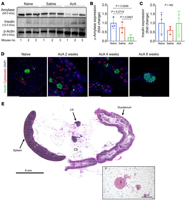
RESEARCH ARTICLE                                                                                     The Journal of Clinical Investigation

                                                                                                             Figure 6. Panoramic view of his-
                                                                                                             tology of the pancreas before and
                                                                                                             after chemical pancreatectomy
                                                                                                             in cerulein-induced CP. (A and B)
                                                                                                             Pancreas histology after 4 weeks
                                                                                                             and 8 weeks of i.p. cerulein showed
                                                                                                             abnormal acini (black arrows),
                                                                                                             acinar to ductal metaplasia (blue
                                                                                                             arrows), and fibrosis (asterisks).
                                                                                                             (C)Pancreas histology after two
                                                                                                             8-week cycles of i.p. injections in
                                                                                                             the cerulein+saline group showed
                                                                                                             fibrosis (inset arrows) and fatty
                                                                                                             replacement (inset asterisks).
                                                                                                             (D) Pancreas histology after two
                                                                                                             8-week cycles of i.p. injections in
                                                                                                             the cerulein+AcA group showed
                                                                                                             near-complete loss of pancreatic
                                                                                                             parenchyma with intact islets
                                                                                                             (I). Illustrative histology results
                                                                                                             from 4 animals per time point are
                                                                                                             shown. Scale bar: 200 μm unless
                                                                                                             otherwise specified.

normal mice improves insulin secretion. There was no significant         K–N). To confirm the finding that loss of the exocrine pancreas is
difference in β cell mass at 8 weeks among the 3 groups, indicating      the main cause of the improved glucose tolerance, we used anoth-
that chemical pancreatectomy does not cause irreversible loss of         er mouse model (ElaCreERT2-R26DTR) that uses the diphtheria toxin
β cells (Figure 10I), and suggesting that the improvement in glu-        receptor (DTR), where diphtheria toxin (DT) quickly ablates acinar
cose tolerance was not due to an increased β cell mass. Next, in         cells through apoptosis (25). Two days after the last DT injection,
vitro GSIS of isolated islets from AcA-treated, saline-treated, and      again, the i.p. GTT showed significantly improved glucose toler-
naive mice showed no difference in insulin secretion among the 3         ance in the ElaCreERT2-R26DTR mice compared with their littermate
groups (Figure 10J), suggesting that the difference seen in the in       controls (Supplemental Figure 6, G–I). Additionally, 3 weeks after
vivo GSIS is likely secondary to local in vivo anatomical/physio-        DT, to allow regeneration of exocrine pancreas (25), there was no
logical changes accompanying chemical pancreatectomy.                    difference in the repeat i.p. GTT between the 2 groups (Supple-
    In line with these data, using the same 4 subgroups described        mental Figure 6, J–L), supporting our finding that the acute loss of
in the CP model experiments, i.p. GTT 1 day after the last ceru-         acinar cells improves glucose tolerance.
lein injection showed improved glucose tolerance in AcA-treated               In line with the mouse data, 8 weeks after AcA infusion,
subgroups compared with saline-treated subgroups (Figure 10,             i.v. GTT in NHPs showed improved glucose tolerance in the

   8                                 J Clin Invest. 2021;131(3):e143301 https://doi.org/10.1172/JCI143301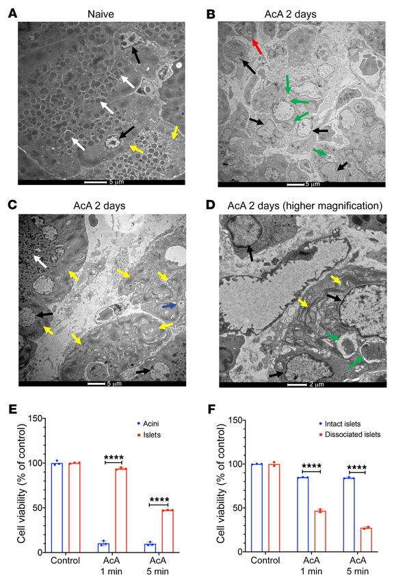
The Journal of Clinical Investigation                                                                                        RESEARCH ARTICLE

Figure 7. Chemical pancreatectomy ablates exocrine pancreas, resolves inflammation, and ameliorates pain in cerulein-induced CP. (A)Immunostaining
for amylase was positive in vehicle+saline and negative in AcA-treated groups while cerulein+saline group showed abnormal patchy amylase staining typ-
ical of CP. (B)Immunostaining for CD3 showed presence of lymphocytes only in the cerulein+saline group, indicating chronic inflammation. (C)Immunos-
taining for cytokeratin showed absence of ducts only in the AcA-treated groups, indicating involution of the ducts. (D)Abdominal von Frey monofilament
testing was performed following the two 8-week cycles of i.p. cerulein or vehicle and revealed that cerulein+saline group exhibited significant mechanical
allodynia pain as compared with cerulein+AcA group, as well as compared with both vehicle-treated groups, indicating that AcA treatment resolved the
pain in cerulein-induced CP. n = 8/group. One-way ANOVA followed by Holm-Šidák test for multiple comparisons. F3,28=7.884; P = 0.0006. (E)Levels of
CGRP (calcalpha) mRNA were quantified by RT-PCR from DRGs harvested from the 4 subgroups. In keeping with the in vivo pain assessment, there was a
significant upregulation in the cerulein+saline (n = 5) group compared with the other 3 groups, cerulein+AcA (n = 3), vehicle+saline (n = 4), and vehicle+AcA
(n = 4), indicating that AcA treatment reversed the cerulein-induced upregulation in CGRP mRNA levels. One-way ANOVA followed by Holm-Šidák test for
multiple comparisons. F3,12=9.015; P = 0.0021. Illustrative histology results from 4 animals per time point are shown. Scale bars: 200 μm. Data are present-
ed as mean ± SD. Only significant P values are depicted.

AcA-treated NHPs compared with weight- and age-matched con-                      brittle form of diabetes. Therapeutic options for CP are limited
trols (Figure 10, O and P, and Supplemental Figure 6M).                          and suboptimal and usually do not reduce the risks of diabetes
                                                                                 and pancreatic cancer (26). Here we present a paradigm-shifting
Discussion                                                                       nonsurgical “chemical pancreatectomy” as a potential thera-
CP- and CF-associated pancreatic inflammation are common                         py for CP. Remarkably, in mice and NHPs, this procedure led to
problems that can be debilitating and often cause a particularly                 near-total ablation of the exocrine pancreas, but with complete

                                           J Clin Invest. 2021;131(3):e143301 https://doi.org/10.1172/JCI143301                                         9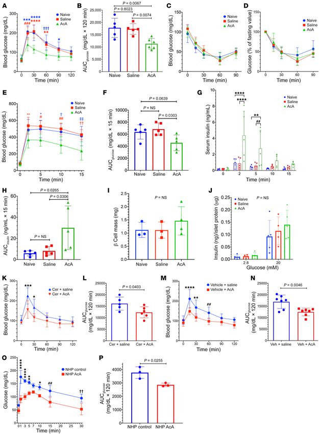
RESEARCH ARTICLE                                                                                      The Journal of Clinical Investigation

                                                                                                               Figure 8. Histological and
                                                                                                               biochemical changes following
                                                                                                               chemical pancreatectomy in
                                                                                                               NHPs. (A) Histology of normal
                                                                                                               NHP pancreas. i denotes islets. (B)
                                                                                                               Histology of NHP pancreas 2 days
                                                                                                               after AcA infusion showed exten-
                                                                                                               sive areas of exocrine tissue necro-
                                                                                                               sis (asterisks) with intact islets.
                                                                                                               Magnification of the exocrine
                                                                                                               tissue with apparently preserved
                                                                                                               architecture revealed abnormal
                                                                                                               acini with acinar cell swelling, indi-
                                                                                                               cating cell injury. (C–F) Histology
                                                                                                               of the pancreas 8 weeks (C and
                                                                                                               D), and 6 months (E and F) after
                                                                                                               AcA infusion showed disappear-
                                                                                                               ance of the exocrine tissue with
                                                                                                               intact islets. The acinar cells are
                                                                                                               replaced by fibrosis (arrows) and
                                                                                                               fat cells (asterisks). (G and H)
                                                                                                               Measurement of serum pancreatic
                                                                                                               enzymes in NHPs; serum amylase
                                                                                                               (G) and lipase (H) levels returned
                                                                                                               to preoperative baseline 7 days
                                                                                                               after surgery. Illustrative results
                                                                                                               from 2 animals per time point are
                                                                                                               shown (for 6-month time point, n
                                                                                                               = 1). Scale bars: 200 μm.

preservation of the endocrine cells, and relieved CP-associated           cell swelling and breakdown of the plasma membrane with the
pain. Since pancreatic diabetes in both CP and CF is thought to be        release of intracellular contents and local inflammation (27, 28).
due to an “innocent bystander” injury to islets from the inflam-               Histology at 2 days after AcA infusion revealed several areas of
mation, nonsurgical removal of the inflamed exocrine pancreas             advanced exocrine necrosis. Even though the remaining exocrine
should theoretically protect the islets indefinitely and possibly         areas showed apparently preserved tissue architecture, the acinar
ameliorate existing diabetes.                                             cells were swollen with vacuolated cytoplasm and distorted archi-
     The in vivo infusion of AcA into the pancreatic duct allows          tecture, indicating significant cell injury (Figure 1 and Figure 8).
direct contact of the AcA with ducts and acinar cells, resulting in       Also, amylase immunostaining at 2 days markedly decreased and
widespread cell injury and death. The mechanism of cell death is          was absent by 2 weeks. Other studies reported preservation of tis-
likely necrosis (Figure 1, Figure 2, and Figure 5), characterized by      sue architecture for 7 days after exposure to an injurious stimulus

   10                                 J Clin Invest. 2021;131(3):e143301 https://doi.org/10.1172/JCI143301
The Journal of Clinical Investigation RESEARCH ARTICLE
Figure 9. Chemical pancreatectomy ablates exocrine pancreas, and preserves pancreas innervation and islet vasculature in NHPs. (A) Immunostaining
for insulin, glucagon, and amylase in NHP’s pancreas 2 days after AcA infusion; there was marked decrease in amylase staining (arrows denote amylase
remnants) with normal insulin and glucagon staining. At 8 weeks, there was no amylase staining, with normal insulin and glucagon staining. (B)Immunos-
taining for CD31 showed intact microvasculature of islets at 2 days and 8 weeks after AcA infusion. (C)Immunostaining for PGP9.5 showed intact neurons
(white arrows) in the NHP pancreas at 2 days and 8 weeks after AcA infusion. Yellow arrow denotes intact nerve fibers 8 weeks after AcA infusion. (D) Immu-
nostaining for cytokeratin showed presence of ducts at 2 days and 8 weeks after AcA infusion. However, at 6 months, the cytokeratin immunostaining was
negative indicating absence of ducts. Illustrative results from 2 animals per time point are shown (for 6-month time point, n = 1). Scale bars: 200 μm.
(28). By EM, the acinar cells showed alterations in the plasma and 30), the complete absence of pancreatic ducts in this model (Fig-
organelle membranes, indicating cell injury and necrosis, except ure 2 and 9, and Supplemental Figures 1 and 5) may explain the
for the nuclear envelope, which can initially remain intact (ref. 29 inability of the exocrine pancreas to regenerate. Also, the absence
and Figure 5). One year after AcA infusion, the loss of exocrine of ducts to drain enzymes may further contribute to the death of
pancreas was persistent, indicating a lack of regeneration (Figure any acinar cells that might survive the initial AcA-induced inju-
2). As duct cells can contribute to regeneration of the pancreas (25, ry. Inadequate drainage of pancreatic secretions is well known
J Clin Invest. 2021;131(3):e143301 https://doi.org/10.1172/JCI143301 11RESEARCH ARTICLE The Journal of Clinical Investigation 12 J Clin Invest. 2021;131(3):e143301 https://doi.org/10.1172/JCI143301
The Journal of Clinical Investigation                                                                                    RESEARCH ARTICLE

Figure 10. Chemical pancreatectomy improves glucose tolerance and                es of serum amylase and lipase and/or small residual remnants
insulin secretion. (A and B) i.p. GTT 8 weeks after surgery (A) comparing        of pancreatic acinar tissue. It has been shown that removal of the
AcA group (n = 6) and controls (n = 5/group). Two-way repeated-measures
                                                                                 pancreas and salivary glands in rodents did not cause a significant
ANOVA, F10,65=7.231; P < 0.0001. For A, *P = 0.035; **P = 0.0026; ***P
= 0.0001; ****P < 0.0001; ###P = 0.0006; †††P = 0.0004. AUC (B), 1-way           decrease in blood amylase levels (33). Also, significant levels of
ANOVA, F2,13=9.234; P = 0.0032. (C and D) ITT at 8 weeks (C). (D) Percentage     amylase activity remain in the serum for an indefinite time after
change in glucose levels compared with fasting glucose. n = 4 naive;             pancreatectomy in dogs and humans (34, 35). Additionally, serum
5 saline, and 5 AcA. Two-way repeated-measures ANOVA, F8,44=1.130;               amylase levels were found to be associated with various physiolog-
P = 0.3626. (E and F) i.v. GTT (E) n = 5 per group. Two-way repeated-
                                                                                 ical changes, such as impaired hepatic function, and are not mere-
measures ANOVA, F8,48=3.241; P = 0.005. For E, *P = 0.0367; **P = 0.0082;
#
 P = 0.0108; ##P = 0.0023; †P = 0.0112; ††P = 0.0021; ‡‡P = 0.0035. AUC (F),     ly a marker of exocrine pancreatic function (36, 37).
1-way ANOVA, F2,12=5.167; P = 0.0241. (G and H) In vivo GSIS (G). n = 5/group.        We have frequently observed small remnants of acinar tis-
Two-way repeated-measures ANOVA, F8,48=5.267; P < 0.0001. For G, **P             sue in proximity to the duodenum, which we suspect results from
= 0.0024; ****P < 0.0001; ##P = 0.0034. AUC (H), 1-way ANOVA F2,12=5.92;         these areas having separate small ducts draining directly into the
P = 0.0163. (I) Eight weeks after surgery, β cell mass quantification. n = 4
                                                                                 duodenum and thus not in communication with the main ductal
AcA, 3 naive, and 3 saline. One-way ANOVA, F2,7=0.8376; P = 0.4719. (J) In
vitro GSIS. n = 5 naive, 4 saline, and 5 AcA. Two-way repeated-measures          system that receives the infusion. This type of drainage has been
ANOVA, F2,11=0.5903; P = 0.5708.(K–N) i.p. GTT comparing the cerulein+           described for heterotopic pancreas in humans elsewhere along the
AcA group (n = 6) and cerulein+saline group (n = 5) (K). Two-way repeat-         gastrointestinal tract (38).
ed-measures ANOVA, F5,45=4.141; P = 0.0035. For K, *P = 0.0150; ***P =                The resolution of the CP-associated pain is likely due to the
0.0006. AUC (L), unpaired t test, t9=2.394; P = 0.0403. i.p. GTT comparing
                                                                                 resolution of the inflammation, suggesting that the pain is second-
vehicle+AcA and vehicle+saline groups. n = 7/group (M). Two-way repeated-
measures ANOVA, F5,60=5.1; P = 0.0006. For M, **P = 0.0016; ****P <              ary to damaged perineurium as well as direct activation and sen-
0.0001; ##P = 0.0068. AUC (N), unpaired t test, t12 =3.475; P = 0.0046.          sitization of sensory nerve fibers by factors in the inflammatory
(O and P) i.v. GTT 8 weeks after surgery in NHPs. n = 3/group. Two-way           milieu (39, 40). CP-associated pain may evolve to central sensiti-
repeated-measures ANOVA, F7,28 =5; P = 0.0008. For O, *P = 0.0152; **P =         zation, where removal of the pancreatic source of the pain may not
0.0026; ##P = 0.0024; ††P = 0.0025, ****P < 0.0001). AUC (P), unpaired t
                                                                                 remove the pain, and such may be the case in a subset of patients
test, t4 =3.472; P = 0.0255. Data are presented as mean ± SD. Holm-Šidák
test was used for multiple comparisons.                                          (41, 42). However, surgical total pancreatectomy can typically
                                                                                 improve CP-associated pain (43). Thus, chemical pancreatectomy
                                                                                 will likely be beneficial for improving pain in CP.
                                                                                      In NHPs, the texture and anatomy of the pancreas are quite
to cause acinar cell death, as seen in the pancreatic duct ligation              different from those in the mouse (2% AcA was more effective in
model (31). In vivo, in order to reach the islets, the AcA must dif-             NHPs). We saw many similarities between the histological chang-
fuse across the gap junctions between acinar cells, the acinar base-             es in NHPs and in mice early on after the AcA treatment. However,
ment membrane, the interstitial space, and finally, the protective               at the 8-week and 6-month time points, instead of seeing a com-
islet capsule (32). Also, during this journey, the concentration of              plete fatty replacement of the acinar cells that occurs in mice, in
AcA likely becomes progressively more diluted, well below the                    NHPs, we saw fatty replacement plus fibrosis in the nonendocrine
starting concentration, by interstitial fluid and by lymphatic and               portion of the pancreas. This difference may be due to a faster
vascular clearance from the peri-islet region. Furthermore, in                   pace of pathological changes in mice compared with primates and
vitro, islets were substantially more resistant to AcA toxicity than             differences in the healing process between the 2 species. Also, the
acini. Collectively, these factors could explain why the islets were             islets of Langerhans, islet vasculature, and pancreatic innervation
spared. We also found that in vitro, intact islets were even more                were preserved. These findings make the translation of chem-
resistant than dissociated islet cells, perhaps due to the multiple              ical pancreatectomy to humans possible, a potential dramatic
layers of cells in the intact islet (Figure 5).                                  advancement in the management of patients with CP.
     Interestingly, the loss of the exocrine pancreas improved glu-                   Although chemical pancreatectomy has shown favorable out-
cose tolerance and enhanced insulin secretion in vivo (Figure 10,                comes in animal models, the effect of the chemical pancreatecto-
and Supplemental Figure 6). However, in vitro GSIS was unaffect-                 my procedure on a preexisting premalignant lesion, if present, in
ed. Islet histology, innervation, and vasculature (Figures 3, 4, and             patients with CP is not known at this point. This will require further
9) remained intact after AcA treatment; therefore, loss of the exo-              studies in order to translate this procedure to humans successfully.
crine pancreas is likely the main reason for improved β cell func-                    Another potential limitation of this study is that abnormal
tion. This conclusion is further supported by the ElaCreERT2-R26DTR              pancreatic duct architecture in patients with CP might lead to a
mouse model data (Supplemental Figure 6).                                        nonuniform diffusion of AcA throughout the pancreas, which may
     The rapid rise in serum pancreatic enzymes early after the AcA              cause exacerbation of the pancreatic inflammation. In humans
or saline infusion is likely related to infusion-induced pressure and            with CP, a pancreatic duct obstruction may result from a stric-
acute acinar cell damage. The normalization of serum enzymes                     ture or a stone, which may theoretically prevent the feasibility of
at 1 week indicates no ongoing pancreatic inflammation caused                    an effective chemical pancreatectomy. To account for this poten-
by the AcA. Similarly, we found that the animals did not appear                  tial technical limitation in humans, assessment of the pancreatic
to be in pain from the infusion after recovering from the surgery.               morphology and ductal anatomy will be crucial before proceed-
Interestingly, the serum pancreatic enzymes after AcA treatment                  ing with intervention. Currently, several imaging modalities are
did not fall below normal levels despite the near absence of acinar              used to visualize the pancreatic duct anatomy and potential sites
tissue. This observation may be attributable to other tissue sourc-              of obstruction including the following: ERCP, which has a high

                                            J Clin Invest. 2021;131(3):e143301 https://doi.org/10.1172/JCI143301                                 13
RESEARCH ARTICLE The Journal of Clinical Investigation
sensitivity to detect even mild cases of CP (44) and magnetic res- infusion into the liver and gall bladder. We then cannulated the com-
onance cholangiopancreatography (MRCP), which is sensitive in mon pancreatico-biliary channel using an umbilical catheter, and we
detecting moderate to severe pancreatitis (45). Additionally, when held the catheter in place by a bulldog clamp to prevent backflow into
MRCP is combined with secretin hormone injection, a more com- the duodenum. Next, we infused 2% AcA, with a volume of 2 mL/kg,
prehensive evaluation of the pancreatic duct can be obtained (46). at a flow rate of 1 mL/min. After the laparotomy closure, the jacket and
According to the 2014 Cambridge Classification System, CP is tether system were reapplied. NHPs received i.v. fluids, including dex-
classified into 4 stages based on imaging findings; only stage 4 is trose 5% and normal saline overnight before resuming oral intake.
marked by significant intraductal filling defects and obstructions
(47). Patients who have a severe duct obstruction can undergo Induction of CP in mice
endoscopic therapy to relieve the obstruction before proceeding CP was induced by the cholecystokinin analogue cerulein (Bachem,
with the chemical pancreatectomy. Specifically, pancreatic duct 4030451). Cerulein was dissolved in normal saline and administered
stones can be removed through a process of sphincterotomy, litho- i.p. (50 μg/kg) every hour for 8 doses, twice weekly (Mondays and
tripsy, stenting, and/or extraction (48–50). Also, pancreatic ductal Thursdays) for two 8-week cycles (40). Simultaneously, we had a con-
strictures can be dilated with subsequent stent placement (51, 52). trol group injected with vehicle (normal saline), and the i.p. injections
There is only a small number of patients in which ERCP interven- were given as per the regimen described above. Subgroups of the mice
tions are not successful, with failure to decompress the obstructed were sacrificed at 4 and 8 weeks, and pancreata were harvested for his-
pancreatic duct (53). Theoretically, these patients may not be can- tology to confirm the occurrence of CP.
didates for a chemical pancreatotomy. However, the larger pop-
ulation of patients suffering from the symptoms of CP will likely In vivo glucose homeostasis studies
benefit from this innovative therapy. All the in vivo glucose homeostasis studies were performed on age-
matched CD1 female mice.
Methods i.p. GTT. Overnight 16-hour–fasted mice were injected i.p. with 2
g/kg glucose (Sigma-Aldrich). Blood glucose was measured from the
Animals and animal manipulation tail vein at 0, 15, 30, 60, 90, and 120 minutes after injection using a
Mice. Eight-week-old CD1 mice from both sexes were purchased from glucometer (Contour NEXT EZ).
the Charles River Laboratories. The mice were maintained in an envi- i.v. GSIS. Overnight 16-hour–fasted mice were injected into the
ronment with a 12-hour light/ 12-hour dark cycle with the lights on tail vein with 2 g/kg glucose. During i.v. GTT, tail vein blood was col-
from 7 am until 7 pm. lected at 0, 2, 5, 10, and 15 minutes for measuring serum insulin con-
Pancreatic ductal infusion. The anesthetized mice underwent mid- centration using an ELISA kit (Alpco).
line laparotomy. The common bile duct was exposed, clamped close ITT. Six-hour–fasted mice were injected i.p. with 0.75 units/kg
to the liver, and cannulated by a blunt catheter across the duodenum. regular insulin. Blood glucose was measured from the tail vein at 0, 15,
The catheter was clamped near its insertion at the ampulla, and 150 30, 60, and 90 minutes after insulin injection using the glucometer.
μl of the AcA was infused at a rate of 50 μl/min (54). All mice were i.v. GTT in NHP. i.v. GTT in NHPs was done as described previ-
maintained on elemental diet (Peptamen, 1.0CAL/55620A) mixed ously (55). After a 16-hour overnight fast, the NHP was sedated using
with mushed normal chow 1:1. ketamine (10 mg/kg intramuscular) and kept sedated during the test.
ElaCreERT2-R26DTR mice. To allow DTR expression selectively in We then placed a peripheral i.v. catheter and obtained a fasting blood
acinar cells, we crossed R26-DTR mice (Jackson Laboratory) with glucose using a glucometer (Contour NEXT EZ). Next, we adminis-
ElaCreERT2 mice. Six-week-old mice were first injected i.p. with tamox- tered dextrose (0.5 mL/kg of 50% dextrose) i.v. and measured blood
ifen (Sigma-Aldrich) for 5 days (80 mg/kg/dose) to activate the Cre- glucose from the heel pad at the following time points: 1, 3, 5, 7, 10, 15,
recombinase enzyme. Seven days after the last tamoxifen dose, to and 30 minutes.
allow Cre-recombinase action, DT injections (50 μg/kg) were given
for 3 consecutive days to ablate acinar cells. Measurement of serum pancreatic enzymes and comprehensive
NHPs. Experiments were performed on male Cynomolgus macaques metabolic profile
who were 4 to 8 years old and purchased from Alpha Genesis Inc. NHPs Mice. We collected approximately 50 μL of blood from the tail vein
weighed 3.5 to 6.5 kg. Each NHP required a 30-day quarantine, fol- via the Microvette CB300 Capillary Blood Collection Tube. Serum
lowed by 1 to 2 weeks acclimatization. The NHPs were maintained in pancreatic enzyme concentrations were measured using a mouse
an environment with a 12-hour light/12-hour dark cycle with the lights amylase ELISA kit (LifeSpan BioSciences) and mouse lipase ELISA
on from 7 am to 7 pm. All animals had free access to water and received kit (Aviva Systems Biology).
a diet consisting of biscuits, fruits, vegetables, and forage mix. NHP. Blood was collected from the central line or peripherally
Pancreatic ductal infusion in NHP. Prior to surgery, we employed a from the saphenous vein. Pancreatic enzymes and comprehensive
jacket and tether system for 2 weeks to allow the NHP to acclimatize to metabolic profile were measured using the Dimension Vista 500
the system. Before the surgery, the NHP was sedated using ketamine chemistry analyzer (Siemens Medical Solutions USA Inc.).
(10 mg/kg), then intubated and administered inhalational anesthesia
with isoflurane. Following the placement of a tunneled central venous Pain assessment in mice
catheter into the right internal jugular vein, we performed a midline Open-field activity. Open field activity was monitored by a trained pro-
laparotomy and created a duodenotomy to identify the major papil- fessional. The total distance traveled and the duration of rearing were
la. We placed a temporary clamp on the common bile duct to prevent measured. Per previous studies, rodents thought to be in pain move
14 J Clin Invest. 2021;131(3):e143301 https://doi.org/10.1172/JCI143301The Journal of Clinical Investigation RESEARCH ARTICLE
shorter distances, and those with CP-associated abdominal pain have (2.5% glutaraldehyde, 2% PFA in 0.1 M sodium phosphate buffer), tis-
been shown to rear significantly less than control animals (40, 56). sues were rinsed twice in 0.1 M monosodium phosphate (NaH2PO4) for
Mechanical allodynia. An expert pain neurobiologist performed 30 minutes and then placed in 1% osmium tetroxide in water for 1 hour.
the abdominal von Frey assay on mice in order to directly test mechan- Tissues were rinsed twice in deionized water. The samples were then
ical sensitivity. Mice were placed in wire mesh chambers on a standard dehydrated in 50% EtOH, 75% EtOH, 95% EtOH (30 minutes each),
von Frey table and acclimatized. Following adaptation of previously and finally in 100% EtOH (20 minutes ×2). Tissues were placed in
described protocols, von Frey filament no. 3.61 (equivalent to 0.4 g of propylene oxide (15 minutes ×2) and were preinfiltrated in half resin/
force) was applied to the upper abdominal area overlying the pancre- half propylene oxide overnight. The next day, tissues were infiltrated in
as (upper left) for approximately 2 seconds and withdrawal respons- 100% resin for 5 hours. Then tissues were embedded with fresh resin
es recorded (56–58). The monofilament was applied 10 times with a and polymerized at 60°C overnight. The embedded tissues were sec-
minimum of 30 seconds between applications. The number of positive tioned with a Leica EM UC6 ultramicrotome at a thickness of 90 nm
withdrawal responses was converted into a percentage, with 10 posi- and collected on copper mesh grids. The sections were stained with 4%
tive responses corresponding to 100%. The experimenter was blinded aqueous uranyl acetate for 30 minutes and 2 minutes in 0.2% lead citrate
to mouse treatment groups for all behavioral tests. in 0.1 N sodium hydroxide (NaOH). TEM imaging was performed on an
FEI Talos L120C at 80 kV using Thermo Scientific Ceta 16 MP camera.
Tissue processing and histology
All pancreas samples were fixed with 4% paraformaldehyde (PFA) for 24 β Cell mass quantification
hours at 4°C. The tissues were then embedded into paraffin, and 5 μm Pancreata were harvested and fixed in 4% PFA for 24 hours at 4°C,
sections were cut. For IHC, antigen retrieval was performed (heat and/or after which they underwent standard paraffinization. Sections at 50
acid buffer). Slides were incubated with primary antibodies (Supplemen- μm intervals of the whole pancreas were stained with insulin and
tal Table 2) at 4°C overnight. The following day the slides were incubat- DAPI. The cross-sectional area of β cells and cross-sectional area of
ed with fluorescent-conjugated (FITC, CY3, CY5) secondary antibodies total tissue were measured by ImageJ software. The β cell mass of each
(Jackson ImmunoResearch Labs) for 1 hour at room temperature (RT). pancreas was calculated by multiplying the average value of relative
Nuclear staining and mounting were performed using Fluroshield with cross-sectional area of β cells and the weight of the pancreas.
DAPI (Sigma-Aldrich). Histological assessments were made on paraf-
fin-embedded 5 μm sections of pancreata stained with H&E. Islet vasculature quantification
Islet vasculature was calculated by dividing CD31-positive area by the
DRG harvesting and processing sum of the insulin- and glucagon-positive areas. Then the mean per-
DRG from the T12 level were harvested and fixed in 4% PFA. Follow- centage of 12 islets/mouse was calculated.
ing cryopreservation in 25% sucrose in 0.1 M phosphate buffer at 4°C,
DRG were embedded in Tissue-Tek OCT compound (Fisher Scientif- Islet isolation
ic), sectioned (14 μm), and mounted on Superfrost Plus slides (Fisher Islet isolation was performed following a previously published proto-
Scientific). Sections were stained for PGP9.5 and were photographed col (61). The pancreatic duct was infused and subsequently digested
using LAS software and a Leica DM 4000B microscope. DRG neurons with type V collagenase (1.95 mg/mL). In AcA-treated pancreata, col-
in every fifth section were counted using ImageJ software (NIH). lagenase was injected directly into the residual pancreas tissue due to
the absence of ducts. Islets were separated from the exocrine tissue
Whole-mount technique with a discontinuous Ficoll gradient and then washed with Hanks’ Bal-
The whole-mount immunostaining was done as described previously anced Salt Solution (Gibco, Thermo Fisher Scientific) containing 20
(59). Briefly, the pancreas was minced into small pieces, 0.5 mm × 0.5 mM HEPES buffer (Gibco, Thermo Fisher Scientific) and 0.2% BSA
mm, and then 100 pancreatic pieces were placed per well in a 96-well (Sigma-Aldrich). Islets were then handpicked to eliminate any con-
plate. Pancreatic pieces were washed on a rotator in PBS for 10 min- tamination from exocrine tissue.
utes at RT ×3 and then washed in methanol for 15 minutes at RT ×5.
Afterwards they were blocked with 10% normal donkey serum (NDS) Acinar tissue isolation
for 45 minutes at RT; incubated with primary antibody in 1% NDS at Acinar cell clumps were isolated as described previously (62). Mice
4°C on a rotator overnight; washed with 0.1% PBST on a rotator at RT were euthanized, and the harvested pancreas was rinsed in 6 mL
once an hour for 5 hours; incubated with secondary antibody in 1% of collagenase digestion buffer and was minced until the resulting
NDS at 4°C on rotator overnight; washed with PBST on rotator at RT solution appeared evenly dispersed. The minced product was trans-
for 10 minutes ×6, and mounted on chamber slides with whole-mount ferred to a 125 mL Erlenmeyer plastic flask, and an additional 5 mL
media, after which coverslip was applied. Primary antibodies used of collagenase digestion buffer was added. Afterwards, the Erlen-
were anti-insulin, and anti-PGP9.5 (Supplemental Table 2). DyLight meyer plastic flask was placed into a 37°C water bath with shaking at
594-labeled Lycopersicon esculentum (Tomato) Lectin (t-lectin; 90 rpm for 5 minutes. The suspension was then transferred to a con-
1:200; catalog DL-1177, Vector Laboratories; RRID: AB_2336416) was ical tube, cells were allowed to settle, and the collagenase digestion
used as a marker of blood vessels as described previously (60). buffer was exchanged. Next, the tube was shaken vigorously for 10
seconds to disperse the cells into smaller clusters, and tissues were
Transmission electron microscopy allowed to settle down. Then media was exchanged with fresh BSA
Ultrastructural changes of pancreatic acinar cells and islets were exam- incubation buffer, and the incubation was repeated with shaking at
ined using transmission electron microscopy (TEM). After fixation 90 rpm for 5 minutes. The digest was vigorously resuspended using
J Clin Invest. 2021;131(3):e143301 https://doi.org/10.1172/JCI143301 15You can also read