AFM study of changes in properties of horseradish peroxidase after incubation of its solution near a pyramidal structure
←
→
Page content transcription
If your browser does not render page correctly, please read the page content below
www.nature.com/scientificreports
OPEN AFM study of changes in properties
of horseradish peroxidase
after incubation of its solution
near a pyramidal structure
Yuri D. Ivanov1,2*, Tatyana O. Pleshakova1, Ivan D. Shumov1, Andrey F. Kozlov1,
Irina A. Ivanova1, Anastasia A. Valueva1, Maria O. Ershova1, Vadim Yu. Tatur3,
Igor N. Stepanov3, Victor V. Repnikov4 & Vadim S. Ziborov1,2
In our present paper, the influence of a pyramidal structure on physicochemical properties of a
protein in buffer solution has been studied. The pyramidal structure employed herein was similar to
those produced industrially for anechoic chambers. Pyramidal structures are also used as elements
of biosensors. Herein, horseradish peroxidase (HRP) enzyme was used as a model protein. HRP
macromolecules were adsorbed from their solution onto an atomically smooth mica substrate, and
then visualized by atomic force microscopy (AFM). In parallel, the enzymatic activity of HRP was
estimated by conventional spectrophotometry. Additionally, attenuated total reflection Fourier-
transform infrared spectroscopy (ATR-FTIR) has been employed in order to find out whether or not
the protein secondary structure changes after the incubation of its solution either near the apex of
a pyramid or in the center of its base. Using AFM, we have demonstrated that the incubation of the
protein solution either in the vicinity of the pyramid’s apex or in the center of its base influences
the physicochemical properties of the protein macromolecules. Namely, the incubation of the HRP
solution in the vicinity of the top of the pyramidal structure has been shown to lead to an increase in
the efficiency of the HRP adsorption onto mica. Moreover, after the incubation of the HRP solution
either near the top of the pyramid or in the center of its base, the HRP macromolecules adsorb onto
the mica surface predominantly in monomeric form. At that, the enzymatic activity of HRP does not
change. The results of our present study are useful to be taken into account in the development of
novel biosensor devices (including those for the diagnosis of cancer in humans), in which pyramidal
structures are employed as sensor, noise suppression or construction elements.
Studying pyramidal structures attracts growing interest owing to a concentration of electromagnetic radia-
tion near the apex and base of these structures—as was demonstrated by Balezin et al.1. Moreover, the use of
pyramidal structures in biosensor devices was demonstrated2. Pyramidal structures are also employed in atomic
force microscopy (AFM)-based highly sensitive sensors with probes of pyramidal s hape3. Shielded equipment
cabinets or even whole rooms, based on anechoic chambers, are widely employed in order to decrease external
electromagnetic impacts on both the electronic sensors and the biological systems under study. Such chambers
are fabricated on the basis of acanthoid structures, including pyramidal o nes4–6. As a rule, these structures are
organized in the form of an ordered set of pyramids, or in the form of a multilayer system of p yramids5. Some
computational studies, considering the electric noise reduction effects of various structures, were r eported7;
therein, it was shown that a spatial change in the structure of an electric field occurs owing to both the reflec-
tion from the pyramids’ surface and the damping in pyramids’ material. In biological experimental studies,
pyramidal elements can also be employed—for instance, as supporting or directional elements for positioning
measuring cells in the sensor devices, or as sensor elements. The use of pyramidal elements can influence the
spatial distribution of an electromagnetic field and, accordingly, have an effect on both the biological objects
under study and the electronic measuring equipment. Thus, the influence of external electromagnetic radiation,
1
Institute of Biomedical Chemistry, Pogodinskaya str., 10, Moscow 119121, Russia. 2Joint Institute for High
Temperatures of the Russian Academy of Sciences, Izhorskaya st. 13 Bd.2, Moscow 125412, Russia. 3Foundation of
Perspective Technologies and Novations, Moscow 115682, Russia. 4Bruker Ltd., Moscow 119017, Russia. *email:
yurii.ivanov.nata@gmail.com
Scientific Reports | (2021) 11:9907 | https://doi.org/10.1038/s41598-021-89377-z 1
Vol.:(0123456789)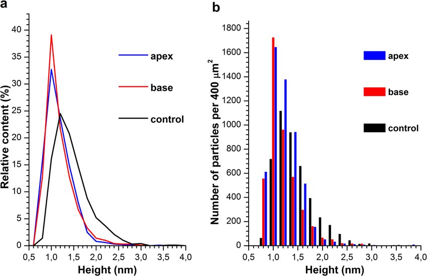
www.nature.com/scientificreports/
concentrated or scattered by such pyramidal elements, on biological objects in analytical systems represents an
important problem of modern biomedical research.
Herein, the influence of a single pyramidal structure on the physicochemical properties of a protein in
solution has been investigated. In our experiments, a structure similar to the industrially produced pyramidal
structures8,9, has been employed. Samples of a buffered solution of horseradish peroxidase protein were placed
and incubated in various positions relative to the pyramidal structure. After the incubation, the HRP macro-
molecules were adsorbed from the sample solutions onto an atomically smooth surface of bare mica substrates,
and visualized on this surface by AFM. The heights of the AFM images of the visualized macromolecules and
their amount on the substrate surface were estimated. The use of HRP enzyme in the study was motivated by the
fact that this protein is comprehensively characterized in the literature, thus being widely employed as a model
in biomedical research10.
As was demonstrated in our previous study, atomic force microscopy (AFM) allows one to reveal the effect
of electromagnetic fields on physicochemical properties of proteins—in particular, on their aggregation state
and adsorbability onto solid substrate s urfaces10. It should be emphasized that in the latter paper10, the electro-
magnetic field was generated intentionally. At the same time, electromagnetic fields of various intensities and
frequencies are now extensively used in both laboratory practice and everyday life. These fields can concentrate at
the expense of a resonance effect from various resonator structures, as was demonstrated by Balezin et al.1. These
structures form specific spatial topology of electromagnetic fi eld1. The effect of these external electromagnetic
fields on living systems—including enzymes—can depend on the surrounding environment.
Herein, AFM has been employed to find out whether the incubation of the solution of horseradish peroxidase
enzyme protein in certain locations in relation to a pyramidal structure (namely, in the vicinity of the apex of the
pyramid and in the center of its base) under conventional biochemical laboratory conditions affects the protein’s
properties. Alterations in the structural properties of biological macromolecules can induce changes in their
functionality (one of the examples is myeloperoxidase, which is only functional in dimeric f orm11). Generally,
aggregation of a protein often leads to loss in its f unctionality12. Accordingly, the results obtained are useful to be
taken into account in the development of highly sensitive biosensors, in which proteins are analyzed or employed
as surface-immobilized ligands. In highly sensitive biosensor systems, the measurements are performed at the
level of single molecules, providing ultra-high detection sensitivity. Application of such systems will allow one
to solve a number of important problems of biomedicine, including early diagnosis of diseases in humans. In
this respect, however, biomedical applications of single-molecule measurements are reported in pioneer studies.
Thus, all factors, including external electromagnetic fields, which potentially may influence the measurement
results, must be thoroughly studied.
Results
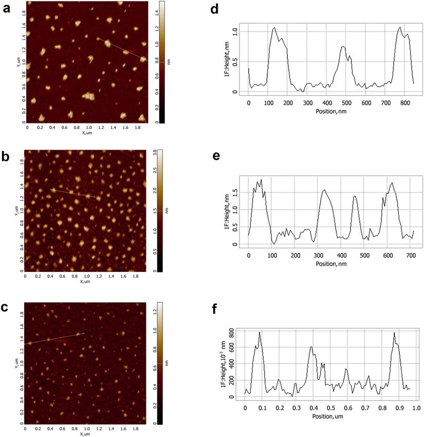
Atomic force microscopy analysis. Figure 1 displays typical AFM images of freshly cleaved mica surface
with HRP macromolecules, adsorbed from the test solutions in control (Fig. 1a) and working (Fig. 1b,c) experi-
ments, when the test solution was placed either in the vicinity of the pyramid’s apex (Fig. 1b) or in the center of
its base (Fig. 1c).
As seen from Fig. 1, in control and working experiments for the both cases—when the test solution was
incubated either near the apex of the pyramid or in the center of its base—compact objects with heights from
1 to 2 nm were visualized on the substrate surface. Since no such objects were observed on the surface of the
substrates incubated in protein-free buffer, these objects can be attributed to HRP macromolecules. After pro-
cessing the AFM data obtained, the distributions of the visualized objects with height (density functions ρ(h))
were plotted. The density function plots are shown in Fig. 2a.
These ρ(h) curves indicate that in working experiments the maximum height amounts to hmax = (1.0 ± 0.2)
nm, and in control experiments this value also makes up hmax = (1.2 ± 0.2) nm. That is, the density functions’
maxima are equal within the measurement error. The distribution width at half-height is, however, quite dif-
ferent, amounting to 0.4 nm in working experiments, while in control experiments its value makes up 0.8 nm.
The molecular weight of HRP is known to be 40–44 kDa13,14. According to the literature data, AFM images of
protein molecules of similar molecular weights have heights from 1 to 1.8 nm15–18, depending on experimental
conditions19,20. One can, hence, conclude that the objects of ~ 1 nm height correspond to the monomeric form
of HRP, and it is the form which predominantly adsorbs onto mica in both working and control experiments.
But in control experiments, broader height distribution was obtained, and the contribution of its right wing
(corresponding to higher objects visualized) was more significant—in comparison with that observed in work-
ing experiments. Thus, in control samples, an increased contribution of oligomeric forms of HRP to the height
distribution of the objects, visualized on mica surface, was observed.
Figure 2b displays a histogram indicating the number of objects, adsorbed onto and visualized on the mica
substrate surface. One can see that the number of objects, adsorbed onto the mica surface both in control experi-
ments and when the HRP solution was incubated in the center of the pyramid’s base, differs from that observed
when the HRP solution was incubated over the pyramid’s apex. Namely, both when the test tube containing the
HRP solution was placed in the center of the pyramid’s base, and in control experiments, the number of HRP
molecules, adsorbed onto mica in monomeric and in oligomeric forms, amounted to N400 ~ 4500 objects per
400 µm2 area. At the same time, in the case of placing the test tube over the pyramid’s apex, this number was
considerably higher, making up N400 ~ 5400 objects per 400 µm2.
Results of spectrophotometric estimation of enzymatic activity of HRP. In parallel with the
AFM measurements, the enzymatic activity of HRP was estimated by conventional spectrophotometry, employ-
ing the reaction with ABTS substrate in the presence of hydrogen peroxide as described in the “Methods” sec-
Scientific Reports | (2021) 11:9907 | https://doi.org/10.1038/s41598-021-89377-z 2
Vol:.(1234567890)
www.nature.com/scientificreports/
Figure 1. Typical AFM images (a–c) and cross-section profiles (d–f) of mica-adsorbed HRP, obtained in
control experiments (a,d) and after 40-min incubation of the HRP solution in the vicinity of the apex of the
pyramid (b,e) or in the center of its base (c,f). The cross-section profiles correspond to the lines shown in the
respective AFM images. The images were obtained using NOVA Px software (ver. Nova_3.4_20014; NT-MDT,
Moscow, Zelenograd, Russia).
tion. Samples treated in both working (when the test tube with the sample solution was incubated either near the
apex of the pyramid or in the center of its base) and control experiments similar to those used for the AFM meas-
urements, were studied by spectrophotometry. Figure 3 displays typical curves indicating the time dependencies
of the solution absorbance at 405 nm wavelength A405(t), recorded for working and for control HRP solutions.
The so-obtained A405(t) curves shown in Fig. 3 indicate that the difference in the functional activity, observed
for the HRP solutions tested, was comparable with the value of experimental error.
Results of attenuated total reflection Fourier‑transform infrared spectroscopy experi-
ments. Figure 4 displays typical ATR-FTIR spectra obtained upon studying 10−4 M HRP solution, which was
incubated in the center of the pyramid’s base, and for control HRP solution incubated at a > 10 m distance from
the pyramid. In the latter case, the pyramid was suspected to have no influence on the protein solution.
Within the wavelength range from 1700 to 1500 cm−1, the spectrum for the control solution, shown in Fig. 4
(black curve), exhibits two characteristic peaks at 1660 cm−1 and 1550 cm−1, corresponding to Amide I and
Amide II, r espectively21. Within the wavelength range studied, no change in the ATR-FTIR spectrum of the
solution incubated near the pyramid’s apex was observed (data not shown)—in contrast to the case with the
solution incubated in the center of the pyramid’s base. Namely, for the solution incubated in the center of the
base, the intensity of the 1550 cm−1 peak (Amide II band) is considerably lower in comparison with that obtained
for the control solution. That is, the ratio between the intensities of Amide I and Amide II peaks has changed.
This indicates that incubation of HRP solution in the center of the pyramid’s base leads to an alteration in the
protein secondary structure.
Scientific Reports | (2021) 11:9907 | https://doi.org/10.1038/s41598-021-89377-z 3
Vol.:(0123456789)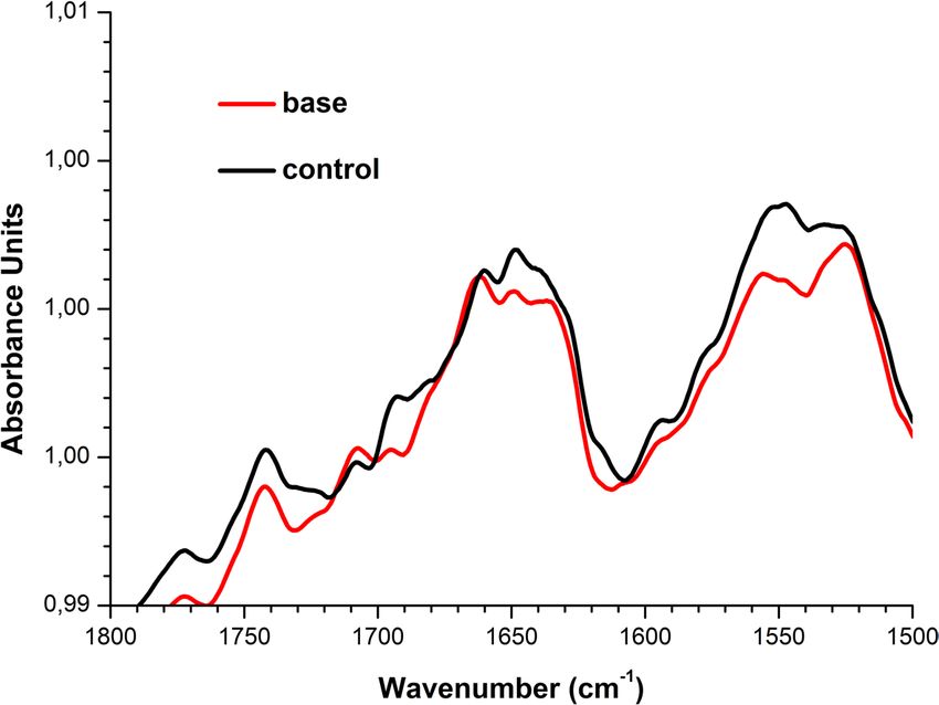
www.nature.com/scientificreports/
Figure 2. Density function ρ(h) plots (a) and the number of the AFM-visualized objects per 400-µm2 area
vs. height (b) obtained in control experiments (black line and bars) and after 40-min incubation of the HRP
solution in the vicinity of the apex of the pyramid (blue line and bars) or in the center of its base (red line and
bars).
Figure 3. Time dependencies of the absorption at 405 nm obtained for the HRP samples in control
experiments (black curve) and after the incubation of the HRP solution either near the apex of the pyramid
(blue curve) or in the center of its base (red curve). Experimental conditions: HRP:ABTS:H2O2 = 10−9 M:0.3
mM:2.5 mM, room temperature (22 °C).
Scientific Reports | (2021) 11:9907 | https://doi.org/10.1038/s41598-021-89377-z 4
Vol:.(1234567890)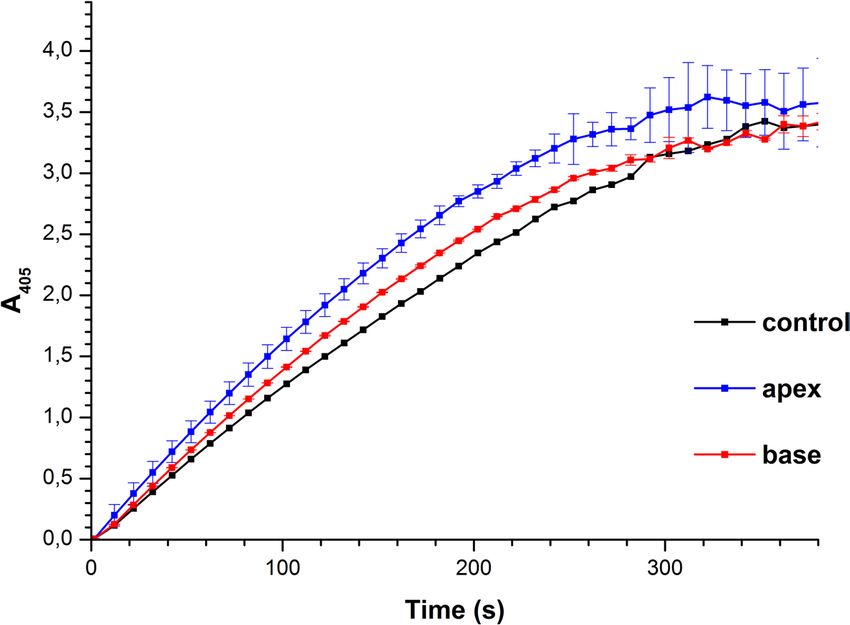
www.nature.com/scientificreports/
Figure 4. Results of attenuated total reflection Fourier-transform infrared spectroscopy (ATR-FTIR)
experiments. Typical ATR-FTIR spectra obtained for 1 0−4 M HRP solutions incubated in the center of the
pyramid’s base (red), or far away from the pyramid (control solution; black).
Discussion
As was reported by Ignatenko et al., in aqueous solutions, HRP is present in the form of a mixture of monomers
and aggregates22. Our results obtained herein indicate that the incubation of HRP solution in the vicinity of the
pyramid’s apex leads to an increase in the efficiency of adsorption of the protein molecules onto the mica sur-
face. An increased contribution of objects, whose heights are characteristic for monomeric HRP, to the height
distribution of AFM-visualized objects shown in Fig. 2a, also indicates an increase in the content of the HRP
macromolecules, adsorbed onto mica in monomeric form after the incubation of the HRP solution both in the
vicinity of the pyramid’s apex and in the center of its base. At that, the enzymatic activity of HRP does not change.
Thus, in the case of the HRP solution incubated over the pyramid’s apex, changes in both the number of the
adsorbed objects and the degree of the protein oligomerization were observed, while only the oligomerization
degree changed in the case of the HRP solution incubated in the center of the pyramid’s base —in comparison
with the parameters obtained for the control samples incubated far away from the experimental setup.
The changes in adsorption properties of the biological macromolecules indicate alterations in their struc-
ture. The adsorption of proteins onto solid surfaces is known to be primarily driven by electrostatic interactions
between the charged protein macromolecules and the charged surface23,24, though other types of interactions
(such as hydrophobic ones) can also have a certain (sometimes governing) effect on protein adsorbability25. In
our case, the adsorption of HRP onto mica represents a consequence of electrostatic interaction between the
negatively charged groups of the mica surface with the surface groups of the protein globules. The change in the
spatial structure of the HRP protein apparently leads to changes in the distribution of charge over the surface
of the protein globule. At that, these changes do not affect the active site of HRP, as otherwise a change in its
enzymatic activity would be observed. Accordingly, the strength of the interactions of the HRP molecules both
between each other and with the substrate surface has changed. This change in the protein structure is expected
to be connected with alterations in the intensity of external electromagnetic field near the pyramid in standard
laboratory conditions.
According to ATR-FTIR data, a slight change in the secondary structure of HRP has been observed after the
incubation of its solution in the center of the pyramid’s base. These results correlate with significant changes
in adsorption properties of the protein in this sample. According to AFM data obtained for the sample incu-
bated in the center of the pyramid’s base, the number of mica-adsorbed particles with h ≥ 1.4 nm decreased—in
comparison with the control sample (Fig. 2b). No change in the ATR-FTIR spectrum of the solution incubated
near the apex has been observed (data not shown). According to the AFM data, less significant change in the
height distribution of the mica-adsorbed objects has been observed for the solution incubated near the apex of
a pyramid—in comparison with that obtained for the control solution. That is, alterations in HRP secondary
structure, revealed by ATR-FTIR, are in agreement with changes in the adsorption properties of HRP particles
with h ≥ 1.4 nm (which are attributed to aggregates) in comparison with the control protein solution (Fig. 2b).
The change in the protein structure is presumably caused by an increased concentration of electromagnetic
fields in the vicinity of the pyramid’s apex. Recent theoretical studies7 revealed that this effect is more likely to
occur near the pyramid’s base at wavelengths, comparable to the length of the pyramid’s edges. Moreover, the
use of pyramidal structures, fabricated from gold, in experimental studies for concentrating the electromagnetic
waves in the vicinity of the apex of near-field probes was reported3. The concentration of external electromagnetic
Scientific Reports | (2021) 11:9907 | https://doi.org/10.1038/s41598-021-89377-z 5
Vol.:(0123456789)www.nature.com/scientificreports/
fields can occur owing to a resonance effect from resonator structures, which form specific spatial topology of
electromagnetic field1.
It should be noted that the technique involving AFM measurements in air, indeed, is not free from limita-
tions, and the changes in protein properties measured in liquid can be somewhat different from those measured
in air15,26; this should be studied in the future and presented in the form of a separate research. In our present
manuscript, we just have tried to reveal the fact of the influence of a pyramidal structure on the properties of
a protein dissolved in water—in order to draw attention of scientists working with equipment, which contains
pyramidal structures (such as biosensors), to this effect. The results of our present study can be of use in the devel-
opment of highly sensitive biosensors, since changes in the structural properties of biological macromolecules
can also lead to a change in their functionality. The results obtained herein should also be taken into account
when conducting direct measurements using biosensors equipped with pyramidal construction elements, located
in the vicinity of measuring cells.
Conclusions
The effect of pyramidal structures on the properties of a protein in buffered aqueous solution has been revealed by
atomic force microscopy using horseradish peroxidase (HRP) heme-containing enzyme as a model. An increase
in adsorbability of HRP macromolecules from its buffered aqueous solution onto bare mica after the incubation
of this solution in the vicinity of the pyramid’s apex has been demonstrated. Moreover, an increased contribution
of the monomeric form of HRP to the height distribution of the mica-adsorbed objects has been observed after
the incubation of the HRP solution either over the apex of the pyramid or in the center of its base—in contrast
to the control HRP solutions incubated far away from the pyramid. The results obtained herein can be of use in
the development of biosensor devices, and should be taken into account upon performing direct measurements
with biosensors containing pyramidal elements, as well as upon studying the influence of electromagnetic radia-
tion on biological systems.
Methods
Chemicals and protein. HRP protein was purchased from Sigma (Cat. #6782). 2,2′-azino-bis(3-ethylben-
zothiazoline-6-sulfonate) (ABTS) was purchased from Sigma. Disodium hydrogen orthophosphate (Na2HPO4),
citric acid and hydrogen peroxide (H2O2) were purchased from Reakhim (Moscow, Russia). 2 mM Dulbecco’s
modified phosphate buffered saline (PBSD buffer) was prepared by dissolving a certain amount of salt mixture
(Pierce, USA) in water. All solutions were prepared using deionized ultrapure water (with 18.2 MΩ × cm resistiv-
ity) obtained with a Simplicity UV system (Millipore, Molsheim, France).
Experimental setup. Figure 5 displays the experimental setup employed in our study.
The 130-mm-high pyramid had the side apex angle of 52 degrees and was fabricated from 2-mm-thick copper-
coated textolite sheets. An 1.7-mL Eppendorf-type polypropylene test tube (SSBiological, USA) containing 1 mL
of the test HRP solution was placed in locations shown in Fig. 5—namely, either at a 1 cm distance from the
pyramid’s apex or in the center of its base. In control experiments, the test tube with the solution was placed far
away (at a > 10 m distance, but in the same room) from the experimental setup. The test solutions were incubated
within the experimental setup for 40 min. After the incubation, the test solutions were analyzed by both AFM
and spectrophotometry as described below.
Sample preparation for AFM experiments. The experiments were carried out using the direct surface
adsorption method27. Mica sheets (SPI, USA) were used as substrates with hydrophilic surface. Upon the prepa-
ration of the AFM samples, a 7.5 × 15 mm piece of the freshly cleaved mica was immersed into 800 µL of 0.1 μM
test solution of HRP in 2 mM PBSD buffer, and incubated therein for 10 min in a Thermomixer Comfort shaker
(Eppendorf, Germany) at 600 rpm and room temperature. In the course of the incubation, HRP macromolecules
adsorbed onto the mica surface. After the incubation, each mica substrate was rinsed with ultrapure water and
then dried in air.
Atomic force microscopy measurements. The AFM experiments were carried out as described in our
recent papers10,19,20,28. The mica surface with adsorbed HRP molecules was visualized by AFM-based in tap-
ping mode in air at 25 °C and 60% relative air humidity employing a Titanium multimode atomic force micro-
scope (which pertains to “Avogadro” large-scale research facilities; NT-MDT, Zelenograd, Russia) equipped with
NSG10 cantilevers (“TipsNano”, Zelenograd, Russia; 47–150 kHz resonant frequency, 0.35 to 6.1 N/m force
constant). The calibration of the microscope by height was carried out on aTGZ1 calibration grating (NT-MDT,
Russia; step height 21.4 ± 1.5 nm).
The main advantages of imaging in air are its simplicity and high throughput26, which allow us to perform
more measurements in one working day without loss of the imaging quality. For these considerations, imag-
ing in air has been employed. It is to be emphasized that the AFM measurements were performed at ~ 60% air
humidity, when both the mica substrate and the studied protein are covered by a hydration l ayer27,29. That is, the
conditions of our AFM experiments can be considered as near-native ones.
The total number of imaged objects in each sample was no less than 200, and the number of frames for each
sample was no less than 10. Analogously to Ref.30, the relative density of distribution of the imaged objects with
height ρ(h) was calculated as
ρ(h) = (Nh /N) × 100%, (1)
Scientific Reports | (2021) 11:9907 | https://doi.org/10.1038/s41598-021-89377-z 6
Vol:.(1234567890)www.nature.com/scientificreports/
Figure 5. Experimental setup. The test tube with the HRP solution was placed either at 1 cm distance from the
pyramid’s apex, or in the center of its base.
where Nh is the number of imaged proteins with height h, and N is the total number of imaged proteins30.
To compare the data obtained from different experiments, the number of registered objects was normalized
per 400 μm2 area. Accordingly, the number of visualized objects was calculated using the equation
NNORM = N × 400/(n × 4), (2)
where NNORM is the number of objects per 400 μm , and n is the number of the obtained scans (the area of each
2
scan is 2 × 2 = 4 μm2).
Preliminary experiments were performed with the use of protein-free buffer solution instead of protein solu-
tion; in these experiments, no objects of > 0.5 nm height was registered. AFM operation, obtaining AFM images,
their treatment (flattening correction etc.) and exporting the obtained data in ASCII format were performed
using the standard NOVA Px software (ver. 3.4_20014; NT-MDT, Moscow, Zelenograd, Russia) supplied with
the atomic force microscope. The number of the visualized particles in the obtained AFM images was calculated
automatically using specialized AFM data processing software developed in Institute of Biomedical Chemistry.
Spectrophotometric estimation of enzymatic activity of HRP. HRP activity was estimated using
ABTS as reducing substrate, as described in our previous papers10,19,28, employing the technique reported by
Sanders et al.31. ABTS is commonly employed for the determination of HRP enzymatic activity, and the enzy-
matic assay reaction should be performed at pH 5.032. Briefly, the rate of change in solution absorbance at 405 nm
was measured employing an Agilent 8453 UV–visible spectrophotometer (Agilent Technologies Deutschland
GmbH, Waldbronn, Germany). Then, 30 μL of 10−7 M HRP solution was added into a 3-mL quartz cuvette
(pathlength 1 cm, Agilent, USA) containing 2.96 mL of 0.3 mM ABTS solution in phosphate-citrate buffer
(51 mM Na2HPO4, 24 mM citric acid, pH 5.0) and stirred. In this way, the final HRP concentration in the cuvette
was 10−9 M. Finally, 8.5 μL of 3% (w/w) H
2O2 was added into the cuvette. Spectrum acquisition was started
immediately upon the addition of H2O2.
Attenuated total reflection Fourier‑transform infrared spectroscopy. In order to find out whether
or not the HRP secondary structure changes after the incubation of its solution either near the apex of a pyramid
or in the center of its base, ATR-FTIR method was employed. The ATR-FTIR measurements were carried out in
the following way. 12 µL of 10−4 M solution of HRP in PBSD was placed in the measuring cell of an INVENIO
Scientific Reports | (2021) 11:9907 | https://doi.org/10.1038/s41598-021-89377-z 7
Vol.:(0123456789)www.nature.com/scientificreports/
spectrometer (Bruker Scientific LLC, Billerica, MA, USA). Such a high concentration of the protein solution
was used due to the sensitivity of the spectrometer. The data were presented in a standard form provided by the
spectrometer operation software. In order to account for the contribution from the PBSD buffer to the resulting
spectra, a blank measurement with pure protein-free buffer was performed before the experiments with HRP
solutions within the same spectral range. The spectrum obtained in the blank measurement was subtracted from
those obtained in experiments with HRP solutions.
Received: 2 February 2021; Accepted: 22 April 2021
References
1. Balezin, M., Baryshnikova, K. V., Kapitanova, P. & Evlyukhin, A. B. Electromagnetic properties of the great pyramid: First multipole
resonances and energy concentration. J. Appl. Phys. 124(3), 034903. https://doi.org/10.1063/1.5026556 (2018).
2. Perdomo, J. et al. Miniaturized real-time monitoring system for l-lactate and glucose using microfabricated multi-enzyme sensors.
Biosens. Bioelectron. 15(9–10), 515–522. https://doi.org/10.1016/S0956-5663(00)00087-7 (2000).
3. Calafiore, G. et al. Campanile near-field probes fabricated by nanoimprint lithography on the facet of an optical fiber. Sci. Rep. 7,
1651. https://doi.org/10.1038/s41598-017-01871-5 (2017).
4. Khmelnik, G. The computer modeling of multilayer SHF electromagnetic screens with layers based on spinulose structures.
Universum Tech. Sci. 9(42), 4–8 (2017).
5. Nakonechny, V. S., Prisyazhny, A. E. & Poberezhny, A. A. Electrodynamic modeling using microwave anechoic chambers. Meth-
odology for the anechoic factor estimation. Syst. Inf. Process. 9(49), 116–123 (2005).
6. Alaee, R., Rockstuhl, C. & Fernandez-Corbaton, I. An electromagnetic multipole expansion beyond the long-wavelength approxi-
mation. Opt. Commun. 407, 17–21. https://doi.org/10.1016/j.optcom.2017.08.064 (2018).
7. Minin, I. V., Minin, O. V. & Yue, L. Electromagnetic properties of the pyramids from the photonics position. Russ. Phys. J. 62(742),
12–18 (2019).
8. http://trimcom.ru/pdf/cv_rus.pdf. Accessed 29 Jan 2021.
9. https://radio.vilcom.ru/products/radiopogloshaushie_materialy/798. Accessed 29 Jan 2021.
10. Ivanov, Y. D. et al. AFM imaging of protein aggregation in studying the impact of knotted electromagnetic field on a peroxidase.
Sci. Rep. 10, 9022. https://doi.org/10.1038/s41598-020-65888-z (2020).
11. Gavrilenko, T. I., Ryzhkova, N. A. & Parkhomenko, A. N. Myeloperoxidase and its role in development of ischemic heart disease.
Ukrainian J. Cardiol. 4, 119–126 (2014).
12. Schramm, F. D., Schroeder, K. & Jonas, K. Protein aggregation in bacteria. FEMS Microbiol. Rev. 44(1), 54–72. https://doi.org/10.
1093/femsre/fuz026 (2020).
13. Davies, P. F., Rennke, H. G. & Cotran, R. S. Influence of molecular charge upon the endocytosis and intracellular fate of peroxidase
activity in cultured arterial endothelium. J. Cell Sci. 49(1), 69–86 (1981).
14. Welinder, K. G. Amino acid sequence studies of horseradish peroxidase. Amino and carboxyl termini, cyanogen bromide and
tryptic fragments, the complete sequence, and some structural characteristics of horseradish peroxidase C. Eur. J. Biochem. 96,
483–502. https://doi.org/10.1111/j.1432-1033.1979.tb13061.x (1979).
15. Ivanov, Yu. D. et al. AFM study of cytochrome CYP102A1 oligomeric state. Soft Matter 8, 4602–4608. https://doi.org/10.1039/
C2SM07333A (2012).
16. Unger, B. P., Gunsalus, I. C. & Sligar, S. G. Nucleotide sequence of the Pseudomonas putida cytochrome P-450cam gene and its
expression in Escherichia coli. J. Biol. Chem. 261, 1158–1163 (1986).
17. Ivanov, Yu. D. et al. Atomic force microscopy study of protein–protein interactions in the cytochrome CYP11A1 (P450scc)-
containing steroid hydroxylase system. Nanoscale Res. Lett. 6, 1–13. https://doi.org/10.1007/s11671-010-9809-5 (2011).
18. Chu, J.-W. & Kimura, T. Studies on adrenal steroid hydroxylases. Molecular and catalytic properties of adrenodoxin reductase (a
flavoprotein). J. Biol. Chem. 248, 2089–2094 (1973).
19. Ivanov, Y. D. et al. Investigation of the influence of liquid motion in a flow-based system on an enzyme aggregation state with an
atomic force microscopy sensor: The effect of water flow. Appl. Sci. 10, 4560. https://doi.org/10.3390/app10134560 (2020).
20. Ziborov, V. S. et al. Investigation of the influence of liquid motion in a flow-based system on an enzyme aggregation state with an
atomic force microscopy sensor: The effect of glycerol flow. Appl. Sci. 10, 4825. https://doi.org/10.3390/app10144825 (2020).
21. Kondyurin, A. V. et al. Mechanisms for covalent immobilization of horseradish peroxidase on ion-beam-treated polyethylene.
Scientifica 2012, 126170. https://doi.org/10.6064/2012/126170 (2012).
22. Ignatenko, O. V. et al. Electrochemistry of chemically trapped dimeric and monomeric recombinant horseradish peroxidase. Adv.
Biosens. Bioelectron. 2(3), 25–34 (2013).
23. Hartvig, R. A., van de Weert, M., Østergaard, J., Jorgensen, L. & Jensen, H. Protein adsorption at charged surfaces: The role of
electrostatic interactions and interfacial charge regulation. Langmuir 27(6), 2634–2643. https://doi.org/10.1021/la104720n (2011).
24. Ivanov, Yu. D. et al. SOI nanowire for the high-sensitive detection of HBsAg and α-fetoprotein. Lab Chip 12(23), 5104–5111. https://
doi.org/10.1039/C2LC40555E (2012).
25. Andrade, J. D., Hlady, V. & Wei, A. P. Adsorption of complex proteins at interfaces. Pure Appl. Chem. 64(11), 1777–1781. https://
doi.org/10.1351/pac199264111777 (1992).
26. Pleshakova, T. O., Bukharina, N. S., Archakov, A. I. & Ivanov, Yu. D. atomic force microscopy for protein detection and their
physicochemical characterization. Int. J. Mol. Sci. 19(4), 1142. https://doi.org/10.3390/ijms19041142 (2018).
27. Kiselyova, O. I. et al. AFM study of membrane proteins, cytochrome P450 2B4, and NADPH–Cytochrome P450 reductase and
their complex formation. Arch. Biochem. Biophys. 371(1), 1–7. https://doi.org/10.1006/abbi.1999.1412 (1999).
28. Ivanov, Y. D. et al. AFM and FTIR investigation of the effect of water flow on horseradish peroxidase. Molecules 26, 306. https://
doi.org/10.3390/molecules26020306 (2021).
29. Guckenberger, R. et al. Scanning tunneling microscopy of insulators and biological specimens based on lateral conductivity of
ultrathin water films. Science 266, 1538–1540 (1994).
30. Pleshakova, T. O. et al. Detection of hepatitis C virus core protein in serum using aptamer-functionalized AFM chips. Microma-
chines 10, 129. https://doi.org/10.3390/mi10020129 (2019).
31. Sanders, S. A., Bray, R. C. & Smith, A. T. pH-Dependent properties of a mutant horseradish peroxidase isoenzyme C in which
Arg38 has been replaced with lysine. Eur. J. Biochem. 224(3), 1029–1037 (1994).
32. Enzymatic assay of peroxidase (EC 1.11.1.7) 2,2′-azino-bis(3-ethylbenzthiazoline-6-sulfonic acid) as a substrate Sigma Prod. No.
P-6782. https://www.sigmaaldrich.com/content/dam/sigmaaldrich/docs/Sigma/Enzyme_Assay/p6782enz.pdf. Accessed 16 June
2020.
Scientific Reports | (2021) 11:9907 | https://doi.org/10.1038/s41598-021-89377-z 8
Vol:.(1234567890)www.nature.com/scientificreports/
Acknowledgements
This work was financed by the Ministry of Science and Higher Education of the Russian Federation within the
framework of state support for the creation and development of World-Class Research Centers “Digital biodesign
and personalized healthcare” No. 075-15-2020-913.
Author contributions
Yu.D.I. generated the scientific idea. Yu.D.I., V.Y.T., and V.S.Z. designed the research and the experimental setup.
I.N.S. designed the experimental setup. I.A.I. prepared the samples. V.Y.T., I.N.S. and A.F.K. performed experi-
ments with the treatment of the studied protein in the experimental setup. T.O.P., I.A.I., A.A.V., M.O.E. and V.S.Z.
performed atomic force microscopy experiments. I.D.S. performed spectrophotometric measurements. V.V.R.
performed ATR-FTIR measurements. A.F.K., A.A.V., I.A.I. and M.O.E. analyzed data. T.O.P., A.A.V., M.O.E. and
I.D.S. prepared the Figures. Yu.D.I., T.O.P. and I.D.S. wrote the manuscript.
Competing interests
The authors declare no competing interests.
Additional information
Correspondence and requests for materials should be addressed to Y.D.I.
Reprints and permissions information is available at www.nature.com/reprints.
Publisher’s note Springer Nature remains neutral with regard to jurisdictional claims in published maps and
institutional affiliations.
Open Access This article is licensed under a Creative Commons Attribution 4.0 International
License, which permits use, sharing, adaptation, distribution and reproduction in any medium or
format, as long as you give appropriate credit to the original author(s) and the source, provide a link to the
Creative Commons licence, and indicate if changes were made. The images or other third party material in this
article are included in the article’s Creative Commons licence, unless indicated otherwise in a credit line to the
material. If material is not included in the article’s Creative Commons licence and your intended use is not
permitted by statutory regulation or exceeds the permitted use, you will need to obtain permission directly from
the copyright holder. To view a copy of this licence, visit http://creativecommons.org/licenses/by/4.0/.
© The Author(s) 2021
Scientific Reports | (2021) 11:9907 | https://doi.org/10.1038/s41598-021-89377-z 9
Vol.:(0123456789)You can also read