ADGRA1 negatively regulates energy expenditure and thermogenesis through both sympathetic nervous system and hypothalamus-pituitary-thyroid axis ...
←
→
Page content transcription
If your browser does not render page correctly, please read the page content below

Zhang et al. Cell Death and Disease (2021)12:362
                                    https://doi.org/10.1038/s41419-021-03634-7                                                                                                             Cell Death & Disease

                                     ARTICLE                                                                                                                                                    Open Access

                                    ADGRA1 negatively regulates energy expenditure
                                    and thermogenesis through both sympathetic
                                    nervous system and hypothalamus–
                                    pituitary–thyroid axis in male mice
                                    Xiao-Hong Zhang1,2, Ling-Yun Tang2, Xi-Yi Wang2,4, Chun-Ling Shen2, Wen-Feng Xiong1,2, Yan Shen2, Ying-Han Wan3,
                                    You-Bing Wu3, Yi-Cheng Wang3, Hong-Xin Zhang2, Shun-Yuan Lu2, Jian Fei 3 and Zhu-Gang Wang 1,2,3

                                      Abstract
                                      Adhesion G protein-coupled receptor A1 (ADGRA1, also known as GPR123) belongs to the G protein-coupled
                                      receptors (GPCRs) family and is well conserved in the vertebrate lineage. However, the structure of ADGRA1 is unique
                                      and its physiological function remains unknown. Previous studies have shown that Adgra1 is predominantly expressed
                                      in the central nervous system (CNS), indicating its important role in the transduction of neural signals. The aim of this
                                      study is to investigate the central function of Adgra1 in vivo and clarify its physiological significance by establishing an
                                      Adgra1-deficient mouse (Adgra1−/−) model. The results show that Adgra1−/− male mice exhibit decreased body
                                      weight with normal food intake and locomotion, shrinkage of body mass, increased lipolysis, and hypermetabolic
                  1234567890():,;
1234567890():,;
                  1234567890():,;
1234567890():,;

                                      activity. Meanwhile, mutant male mice present elevated core temperature coupled with resistance to hypothermia
                                      upon cold stimulus. Further studies show that tyrosine hydroxylase (TH) and β3-adrenergic receptor (β3-AR), indicators
                                      of sympathetic nerve excitability, are activated as well as their downstream molecules including uncoupling protein 1
                                      (UCP1), coactivator 1 alpha (PGC1-α) in brown adipose tissue (BAT), and hormone-sensitive lipase (HSL) in white
                                      adipose tissue (WAT). In addition, mutant male mice have higher levels of serum T3, T4, accompanied by increased
                                      mRNAs of hypothalamus–pituitary–thyroid axis. Finally, Adgra1−/− male mice present abnormal activation of PI3K/AKT/
                                      GSK3β and MEK/ERK pathways in hypothalamus. Overexpression of ADGRA1 in Neuro2A cell line appears to suppress
                                      these two signaling pathways. In contrast, Adgra1−/− female mice show comparable body weight along with normal
                                      metabolic process to their sex-matched controls. Collectively, ADGRA1 is a negative regulator of sympathetic nervous
                                      system (SNS) and hypothalamus–pituitary–thyroid axis by regulating PI3K/AKT/GSK3β and MEK/ERK pathways in
                                      hypothalamus of male mice, suggesting an important role of ADGRA1 in maintaining metabolic homeostasis including
                                      energy expenditure and thermogenic balance.

                                                                                                                                        Introduction
                                                                                                                                          GPCRs compose the largest family of cell-surface
                                                                                                                                        mediators of many cellular responses to external stimuli,
                                    Correspondence: Zhu-Gang Wang (zhugangw@shsmu.edu.cn)
                                    1                                                                                                   participating in regulating almost all physiological pro-
                                     School of Life Sciences and Biotechnology, Shanghai Jiao Tong University,
                                    Shanghai 200240, China                                                                              cesses. Dysfunctions of GPCRs are associated with various
                                    2
                                     State Key Laboratory of Medical Genomics, Research Center for Experimental                         human diseases, indicating the promising pharmacologi-
                                    Medicine, Rui-Jin Hospital Affiliated to Shanghai Jiao Tong University School of
                                                                                                                                        cal targets of GPCRs in treatment and prevention of
                                    Medicine (SJTUSM), Shanghai 200025, China
                                    Full list of author information is available at the end of the article                              diseases1. GPCRs targeted drugs account for one-third of
                                    Edited by A. Finazzi-Agrò

                                    © The Author(s) 2021
                                                        Open Access This article is licensed under a Creative Commons Attribution 4.0 International License, which permits use, sharing, adaptation, distribution and reproduction
                                                        in any medium or format, as long as you give appropriate credit to the original author(s) and the source, provide a link to the Creative Commons license, and indicate if
                                    changes were made. The images or other third party material in this article are included in the article’s Creative Commons license, unless indicated otherwise in a credit line to the material. If
                                    material is not included in the article’s Creative Commons license and your intended use is not permitted by statutory regulation or exceeds the permitted use, you will need to obtain
                                    permission directly from the copyright holder. To view a copy of this license, visit http://creativecommons.org/licenses/by/4.0/.

                                    Official journal of the Cell Death Differentiation Association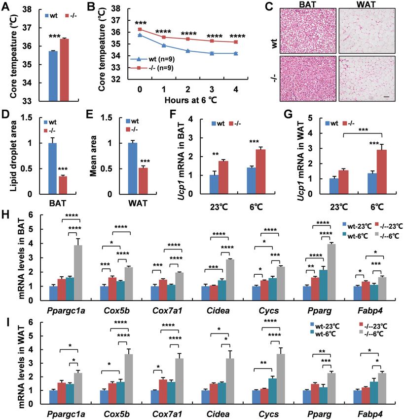
Zhang et al. Cell Death and Disease (2021)12:362                                                                    Page 2 of 14

the drugs in clinical use, but they are only a very small        therapeutic purpose if its molecular mechanisms were better
fraction among all the GPCRs2,3. Moreover, ~120 mem-             understood. However, the Adgra1 is still orphan and the
bers of GPCRs are orphans and their ligands have not             physiological function of Adgra1 remains unknown. Thus,
been identified, implying the great explored potentiality         the aim of this study is to investigate the effect of Adgra1 on
and utilized value of these GPCRs4. Therefore, further           biological process by a knockout mouse model. Our results
comprehensive and intensive study to dissect the phy-            uncover that Adgra1−/− male mice exhibit decreased body
siological function of GPCRs, especially those with              weight caused by increased energy expenditure, and
unknown function would be beneficial to us for better             increased adaptive thermogenesis mediated through SNS
understanding human diseases and drug development.               and hypothalamus–pituitary–thyroid axis. PI3K/AKT/
  ADGRA1 is a member of the adhesion GPCRs which                 GSK3β and MEK/ERK pathways are responsible for the
contain long N-termini and multiple domains that are             abnormal metabolic process. In conclusion, ADGRA1 plays
implicated in cell–cell and cell–matrix interactions. Of         an important role in regulating metabolism homeostasis and
note, the primary structure of ADGRA1 is peculiar and            negatively regulates energy expenditure and thermogenesis
differs from the other adhesion GPCRs. ADGRA1 is the             in male mice.
only member that lacks conserved domain and GPCR
autoproteolysis-inducing domain in the extracellular N-          Results
terminal region5,6. Nevertheless, ADGRA1 exhibits a long         Generation of Adgra1−/− mice
C-terminal region containing an end motif ETTV. This               To investigate the physiological function of ADGRA1,
motif in tumor endothelial marker 5 (TEM5) protein has           we generated an Adgra1−/− mouse model (Supplemen-
been reported to interact with PDZ domain of the human           tary Fig. 1a–c). The efficient deletion of Adgra1 was ver-
homolog of Drosophila discs large tumor suppressor               ified by the undetectable expression of both mRNAs and
(hDLG) protein during tumor angiogenesis7. Therefore,            proteins using reverse-transcription PCR, western blot-
these findings suggest that the interactions between              ting, and IF analysis (Supplementary Fig. 1d–f).
ADGRA1 and other proteins are likely mediated by its
conserved C-terminal region and thereby modulating               Adgra1 deficiency causes decreased body weight in chow-
signal transduction.                                             fed male mice
  Previous studies have proven that Adgra1 was pre-                 Mice were fed standard chow and their body weight was
dominantly and widely expressed in the central nervous           monitored once a week. Surprisingly, Adgra1−/− male mice
system (CNS)5. Distribution analysis on the functional circuit   exhibited significantly less weight gain than their littermate
levels suggests that Adgra1 may be involved in emotion           controls from 15 weeks on (Fig. 1A). Consistently, Adgra1−/−
regulation (amygdala, cortex, and thalamus), learning and        male mice displayed a significant reduction in fat and lean
memory (hippocampus), and body metabolism (hypothala-            mass, decreased weights of adipose tissues, and decreased
mus). Moreover, Adgra1 is well-organized in the layers of        lipid accumulation in adipocytes (Fig. 1B–F). Though the
brain sections but not in the astrocyte-like scattered pat-      serum chemistry analysis showed no significant differences
terns5, implicating that Adgra1 is mainly present in neurons     between the two genotypes, the lipoprotein cholesterol and
rather than astrocytes. Consistently, in the hippocampal         triglycerides (TG) in male mutant mice were slightly less
neurons, ADGRA1 has been proved to be perfectly co-              than that in controls (Fig. 1G). Moreover, the basal glucose
localized with HOMER1, a marker for the postsynaptic             level was lower in Adgra1−/− male mice, but the glucose
density (PSD)8. It is well known that PSD is enriched in         tolerance was intact (Fig. 1H, I). Meanwhile, both
scaffolding molecules which anchor neurotransmitter              serum insulin levels and insulin tolerance test were normal
receptors and integrate signals in response to the second        (Fig. 1J, K). Here, female mice were also monitored, but they
messenger cascades activated by the neurotransmitter             exhibited comparable body weight and similar body com-
receptors9. Furthermore, most PSD proteins contain PDZ           position between two genotypes (Supplementary Fig. 2a, b).
domains, by which they assemble specific proteins into large      Consistently, serum chemistry examination, histology ana-
molecular complexes at defined locations in the cell10.           lysis of adipose tissues, and glucose homeostasis test all
Whether ETTV motif in ADGRA1 can interact with PDZ               showed no obvious changes (Supplementary Fig. 2c–f).
domain of PSD proteins is unknown, but the accumulating          Taken together, the evidence implicated that Adgra1 defi-
evidence makes it reasonable to speculate that ADGRA1            ciency caused abnormal metabolism in male but not in
seems to be working in the CNS and controlling the neu-          female mice.
ronal signal transduction. Importantly, the ADGRA1 is
highly conserved in the vertebrate linage, indicating its cru-   Adgra1 deficiency has no obvious effect on the histology
cial role in the physiological functions in most vertebrates.    of major organs and serum chemistry in mutant male mice
  In a word, the Adgra1 may play an important role in the          We examined the gross anatomy of organs including
biological processes and it might be exploited for a             brain, heart, liver, spleen, lung, and kidney. Obviously, no

Official journal of the Cell Death Differentiation Association
Zhang et al. Cell Death and Disease (2021)12:362                                                                                         Page 3 of 14

  Fig. 1 Adgra1 deficiency causes decreased body weight in chow-fed male mice. A Body weight curves of Adgra1−/− male mice and their
  littermate controls, n = 15/group. B Body mass (total, fat, and lean weight) of Adgra1−/− male and control mice, n = 8–9/group. C Weights of adipose
  tissues in Adgra1−/− male and control mice, n = 5/group. D Representative images of HE stained BAT, and WAT from Adgra1−/− male and control
  mice. Scale bar, 50 μm, n = 5/group. E Quantify of the lipid droplet-positive area in the BAT, n = 5/group. F Quantify of the mean area of the
  adipocytes in the WAT, n = 5/group. G Serum chemistries in Adgra1−/− male and wt mice in fed state, n = 9/group. H Basal blood glucose levels after
  fasted overnight of Adgra1−/− male and control mice, n = 9/group. I GTT in Adgra1−/− male and wt mice, n = 9/group. J Serum insulin levels of
  Adgra1−/− male and control mice, n = 9/group. K ITT in Adgra1−/− male and wt mice, n = 9/group. BG blood glucose. *p < 0.05, **p < 0.01,
  ***p < 0.001 vs. controls. Data represent mean ± S.E.M.

apparent abnormalities in shapes and weights of main                          mouse and human (Supplementary Fig. 5a, b). We further
organs were found between Adgra1−/− male mice and                             investigated the exact distribution in CNS and found that
their littermate controls (Supplementary Figs. 3a–c and                       Adgra1 was highly expressed in the cortex, hypothalamus,
4a–c). Meanwhile, histological analyses by HE staining                        hippocampus, cerebellum and spinal cord. An intriguing
revealed no structure alterations in the brain (Supple-                       finding was that expression of Adgra1 in hypothalamus
mentary Fig. 3d). In addition, no significant differences                      was markedly higher than that in the other brain regions,
were observed in the serum parameters for live function,                      raising the rationality that Adgra1 plays an important role
cardiovascular function, and kidney function by the serum                     in regulating body metabolism. Furthermore, we detected
biochemical analysis (Supplementary Fig. 4d–f).                               Adgra1 mRNA levels in the key metabolic tissues
                                                                              including pituitary, thyroid, and adipose tissues, and
Adgra1 is specifically expressed in the CNS                                    found that the expression of Adgra1 was rarely detected in
  The tissue expression profile revealed that Adgra1 was                       these metabolic tissues other than hypothalamus (Sup-
exclusively and dominantly detected in brains both in                         plementary Fig. 5a). Thus, it is reasonable to speculate

Official journal of the Cell Death Differentiation Association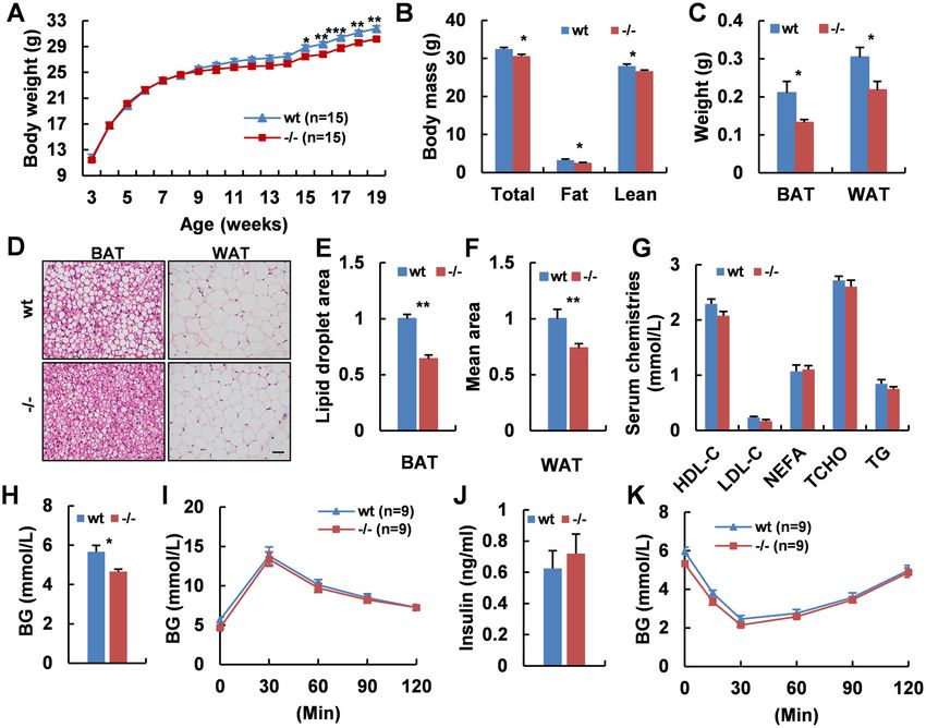
Zhang et al. Cell Death and Disease (2021)12:362                                                                Page 4 of 14

that any involvement of Adgra1 in body metabolism is            food back to the fasted mice and monitored the refeeding
possibly mediated through hypothalamus. In addition,            behaviors. As shown, food intake was similar after fasted
ADGRA1 was identified to co-localize with NEUN-                  (Fig. 3J), suggesting the unaffected appetite in Adgra1−/−
positive neuron cells rather than GFAP-positive astro-          male mice. However, the less weight gain and the higher
cytes by IF on sections of brain regions (Supplementary         ratio of food intake to body weight gain proved that the
Fig. 5c). The localization of ADGRA1 was on the mem-            ingested fuels in Adgra1−/− male mice were used for
brane by visualizing EGFP signals from ADGRA1-EGFP              expenditure rather than storage (Fig. 3K, L). But in female
fusion protein and EGFP protein only (Supplementary             mice, metabolic cage studies showed the energy metabo-
Fig. 5d). IF staining showed the comparable CNS cells           lism was unaffected (Supplementary Fig. 10a–i). Taken
between Adgra1−/− male mice and their littermate con-           together, these data demonstrate that decreased body
trols (Supplementary Figs. 6–9, a–d).                           weight is associated with the increased basal metabolic rate
                                                                and the phenotype only occurred in adulthood of male but
Adgra1−/− male mice present hypermetabolic activity             not female mice.
  To further understand how Adgra1 deficiency affected
the less body weight gain, we performed metabolic cage          Adaptive thermogenesis is increased in Adgra1−/− male
studies in both genotypes at the age of 10 weeks when their     mice
body weights were comparable and at the age of 17 weeks           To further understand the underlying mechanisms for
when Adgra1−/− male mice were significantly thinner than         the increased energy expenditure, we assessed the core
their littermate controls. The results showed that oxygen       body temperature of mice at room temperature and
consumption, carbon dioxide production, and energy              observed the mean rectal temperature in Adgra1−/− male
expenditure in Adgra1−/− male mice were significantly            mice was higher than that of their littermate controls (Fig.
elevated at the age of 10 weeks, excluding the secondary        4A). When acutely exposed to 6 °C for 4 h, Adgra1−/−
effect on the metabolic phenotypes of Adgra1−/− male            male mice still presented higher rectal temperature,
mice. Of note, the alterations of metabolic parameters          meaning cold-induced adaptive thermogenesis was
reached higher levels at the age of 17 weeks, pointing to a     increased (Fig. 4B). Meanwhile, cold stimulation reduced
progressive activation of energy expenditure in the loss of     the lipid droplets and the difference was significant (Fig.
ADGRA1, and making it clear that the decreased body             4C–E). Consistently, Ucp1, a thermogenic marker in
weight gain in Adgra1−/− male mice was a result of gradual      adipocytes, was activated at room temperature and cold
accumulation of hypermetabolic activity (Fig. 2A–E).            strengthened its expression in Adgra1−/− male mice (Fig.
Though RER was equivalent in Adgra1−/− male mice                4F, G). Similar alterations were also observed in other
compared to their littermate controls (Fig. 2F), we deeply      thermogenic genes in both BAT and WAT (Fig. 4H, I).
explored the exact destination of oxygen consumption            Collectively, ADGRA1 deficiency increases the adaptive
based on RER and VO2 according to the literature11.             thermogenesis in male mice.
Obviously, Adgra1−/− male mice preferred to consuming
O2 for fat oxidation in light and for carbohydrate oxidation    SNS is activated in Adgra1−/− male mice
in dark at both ages (Fig. 2G, H), which could be explained        BAT thermogenesis is well considered as a response to
by feeding habits of mice that they usually take food at        the stimulation of SNS. Thus, we assessed the activity of
night. Ambulating locomotor activities and food intake          SNS through analyzing the levels of TH. As expected, TH
were unchanged at the age of 17 weeks (Fig. 2I, J) as well as   was much more abundant in adipocytes of Adgra1−/−
at the 10 weeks (data not shown). Next, we removed the          male mice both at room temperature and cold enhanced
food and measured the basal metabolic rate. Consistently,       its activation by IF (Fig. 5A–C). To investigate how TH
Adgra1−/− male mice exhibited persistently hypermeta-           regulated the SNS activity, we tested the related genes
bolic activity in fasted status (Fig. 3A–F). According to RER   involved in this process. As shown, TH and β3-AR were
and VO2, oxygen consumption for fat was enhanced in             both increased in mRNAs and proteins, as a result, the
both light and dark, whereas oxygen consumption for             downstream cascades in thermogenic process were acti-
carbohydrate was similar all along (Fig. 3G, H), confirming      vated (Fig. 5F–J). Therefore, these combined results
that Adgra1−/− male mice consumed more oxygen for               demonstrate that abolition of Adgra1 increases sympa-
carbohydrate oxidation when they took in food and con-          thetic outflow into adipocytes, enhancing adaptive ther-
sumed more oxygen for fat oxidation when they did not           mogenesis in male mice. Given that cardiac SNS activity
need food or the food was taken away. Consequently, these       was also regulated by hypothalamus via sympathetic
combined results revealed that the energy expenditure is        premotor neurons, we detected the heart rate (Fig. 5D)
consistently higher in Adgra1−/− male mice regardless of        and blood pressure (Fig. 5E) of adult male mice, but no
the feeding behaviors. But whether appetite control is          differences were observed. The unaffected cardiac SNS
involved in this process was unclear. Thus, we returned the     activity may be a consequence of compensation effects

Official journal of the Cell Death Differentiation Association
Zhang et al. Cell Death and Disease (2021)12:362                                                                                        Page 5 of 14

  Fig. 2 Adgra1 deficiency induces increased energy expenditure in male mice. A–F At the age of 10 weeks and 17 weeks, the metabolic cage
  studies assess O2 consumption in curve diagram (A for 10 weeks, B for 17 weeks) and histogram (C), CO2 production (D), energy expenditure (E), and
  RER (F) during light and dark of Adgra1−/− male and control mice, n = 6/group. EE, energy expenditure. G, H O2 consumption for carbonhydrate (G)
  and fat (H) during light and dark, n = 6/group. I Locomotor activities in both genotypes at the age of 17 weeks, n = 6/group. J Food intake in both
  genotypes at the age of 17 weeks, n = 6/group. *p < 0.05, **p < 0.01, ***p < 0.001 vs. controls. Data represent mean ± S.E.M.

in vivo. In contrast, Adgra1−/− female mice exhibited                        total and free forms (Fig. 6D–G). Serum PRL levels were
comparable core temperature at both room and cold                            unchanged though the mRNA levels were increased
conditions (Supplementary Fig. 11a, b), and no significant                    (Fig. 6B, H). The similar mRNA levels of Crh and compar-
difference of the SNS activity was observed (Supplemen-                      able serum levels of corticosterone implied the normal
tary Fig. 11c–g).                                                            function of hypothalamus–pituitary–adrenal gland axis in
                                                                             male knockout mice (Fig. 6A, I). All above, Adgra1−/− male
Adgra1−/− male mice exhibit mild central hyperthyroidism                     mice exhibit mild central hyperthyroidism.
  hypothalamus–pituitary–thyroid axis participates in the
body growth and development by maintaining body energy                       Activated PI3K/AKT/GSK3β and MEK/ERK pathways in the
homeostasis12,13. Therefore, we found that mRNAs of                          hypothalamus of Adgra1−/− male mice
thyrotropin-releasing hormone (Trh) in hypothalamus and                        To clarify how ADGRA1 affects the metabolic process,
thyroid-stimulating hormone-β subunit (Tshb) in pituitary                    we analyzed the classical pathways downstream of
(Fig. 6A, B) were elevated in Adgra1−/− male mice. Mean-                     GPCRs in hypothalamus and found the PI3K/AKT
while, thyroglobulin (Tg) and Na+/K+-ATPase (Atp1b1),                        pathway was activated in Adgra1−/− male mice by
involving in the synthesis and transportation of THs, were                   western blotting (Fig. 7A) and IF staining (Fig. 7E). As a
markedly upregulated in thyroid (Fig. 6C). Moreover, serum                   result, activated AKT facilitated the phosphorylation of
THs, including T4 and T3, were also increased in both                        GSK3β (Fig. 7A). In addition, MEK/ERK was also

Official journal of the Cell Death Differentiation Association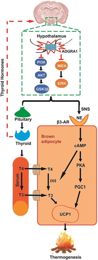
Zhang et al. Cell Death and Disease (2021)12:362                                                                                           Page 6 of 14

  Fig. 3 Basal metabolic rate is increased in fasted Adgra1−/− male mice. A–F At the age of 17 weeks, the metabolic cage studies assess O2
  consumption in curve diagram (A) and histogram (B), CO2 production (C), energy expenditure (D), RER (E), and locomotor activities (F) during light
  and dark of fasted Adgra1−/− male and control mice, n = 6/group. EE energy expenditure. G, H O2 consumption for carbonhydrate (G) and fat (H)
  during light and dark, n = 6/group. I Water intake in fasted states, n = 6/group. J Food intake after refed for 24 h, n = 15/group. K Body weight gain
  after refed for 24 h, n = 15/group. L Ratio of food intake to body weight gain in fasted-refed male mice, n = 15/group. *p < 0.05, **p < 0.01 vs.
  controls. Data represent mean ± S.E.M.

upregulated while PKA/CREB was unaffected in the                               mice and the results showed no significant differences
absence of ADGRA1 in male mice (Fig. 7A). All these                            (Supplementary Fig. 11h). To explore the possible
differences were verified by quantitative analysis                              molecular mechanism responsible for the differences
(Fig. 7B–D, F). Next, we detected these pathways in                            between male and female mice, we tested Adgra1 mRNA
transfected Neuro2A cells, in which the ADGRA1 was                             level in wt mice and found the expression of Adgra1 in
over-expressed. Results showed both PI3K/AKT/GSK3β                             male mice was higher than that in female mice (Sup-
and MEK/ERK pathways were suppressed in a dose-                                plementary Fig. 11i). While the Adgra1 mRNA in nat-
dependent association with the expression of ADGRA1,                           ural Neuro2A cells was increased under the stimulation
while the other pathways were unchanged (Fig. 7G, H).                          of testosterone with a selected range of concentrations
The signaling analyses were also conducted in female                           (Supplementary Fig. 11j).

Official journal of the Cell Death Differentiation Association
Zhang et al. Cell Death and Disease (2021)12:362                                                                                      Page 7 of 14

  Fig. 4 Adaptive thermogenesis is increased in Adgra1−/− male mice. A Rectal temperature of Adgra1−/− male and control mice, n = 9/group.
  B Rectal temperature of Adgra1−/− male and control mice when they are exposed to cold (6 °C) for 4 h, n = 9/group. C Representative images of HE
  stained BAT, and WAT from Adgra1−/− male and control mice in cold environment. Scale bar, 50 μm, n = 5/group. D Quantify of the lipid droplet-
  positive area in the BAT, n = 5/group. E Quantify of the mean area of the adipocytes in the WAT, n = 5/group. F Ucp1 mRNA levels of BAT in room
  temperature and cold environment, n = 5/group. G Ucp1 mRNA levels of WAT in room temperature and cold environment, n = 5/group. H mRNA
  levels of thermogenic genes of BAT in room temperature and cold environment, n = 5/group. I mRNA levels of thermogenic genes of WAT in room
  temperature and cold environment, n = 5/group. *p < 0.05, **p < 0.01, ***p < 0.001, ****p < 0.0001 vs. controls. Data represent mean ± S.E.M.

Testosterone treatment has a slight effect on the                           genotypes exhibited similar metabolic rate at basal con-
metabolic status in female mice                                             dition. However, after treated with testosterone for a
  Consequently, we performed testosterone treatment in                      month, Adgra1−/− female mice displayed an increasing
female mice and results showed that female mice of two                      trend in metabolic rate albeit no significant differences

Official journal of the Cell Death Differentiation Association
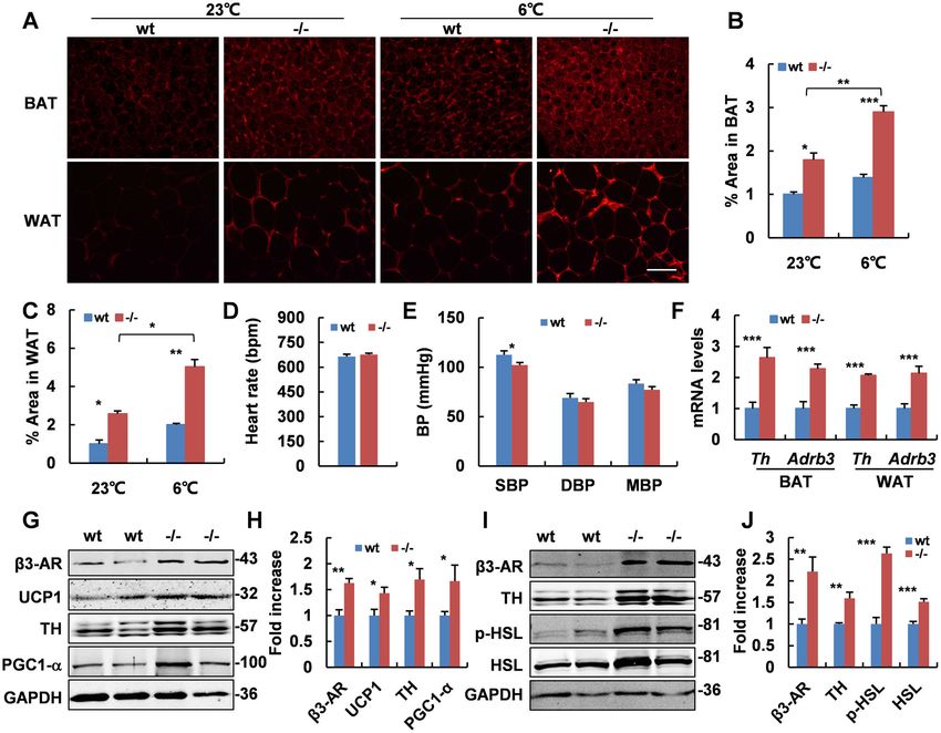
Zhang et al. Cell Death and Disease (2021)12:362                                                                                           Page 8 of 14

  Fig. 5 SNS is activated in Adgra1−/− male mice. A Representative images of IF of TH in BAT and WAT in room temperature and cold environment.
  Scale bar, 50 μm. B Quantitative analysis of positive areas of TH in BAT, n = 5/group. C Quantitative analysis of positive areas of TH in WAT, n = 5/
  group. D Heart rate in male mice of both genotypes, n = 9/group. E Blood pressure in male mice of both genotypes, n = 9/group. SBP systolic blood
  pressure, DBP diastolic blood pressure, MBP mean blood pressure. F mRNA levels of Th and Adrb3 in BAT and WAT assessed by real-time PCR, n = 5/
  group. G Protein levels in BAT of Adgra1−/− male and control mice analyzed by western blot. GAPDH is used as a loading control. H Quantitative
  analysis of relative intensities of proteins. n = 5/group. I Protein levels in WAT of Adgra1−/− male and control mice analyzed by western blot. GAPDH
  is used as a loading control. J Quantitative analysis of relative intensities of proteins, n = 5/group. Western blot analysis using anti-TH antibodies
  detects a band at ~57 kDa, at a position where the TH is expected to migrate, with two nonspecific bands running above and below. *p < 0.05,
  **p < 0.01, ***p < 0.001 vs. controls. Data represent mean ± S.E.M.

(p < 0.07) when compared with controls (Supplementary                          Discussion
Fig. 12a–i). Body weights and core temperature showed                             Metabolism homeostasis is essential for the body
no significant differences (Supplementary Fig. 13a, b).                         growth and development. Imbalance of energy metabo-
Serum detection proved the treatment effectively                               lism leads to several metabolic diseases. Typically, the
improved the testosterone levels in females (Supplemen-                        excessive intake of energy contributes to the development
tary Fig. 13c). Moreover, testosterone treatment in wt                         of obesity, which is a rapidly growing health concern for
female mice also induced increased Adgra1 mRNA in                              the modern society14,15, while hypermetabolism, char-
brain (Supplementary Fig. 13d). Androgen receptor (Ar)                         acterized by the increased energy expenditure and weight
mRNAs were unaffected both in mice models and trans-                           loss, is a manifestation of hyperthyroidism16. The patho-
fected cells (Supplementary Fig. 13e, f).                                      genesis of these metabolic diseases and the molecular

Official journal of the Cell Death Differentiation Association
Zhang et al. Cell Death and Disease (2021)12:362                                                                                        Page 9 of 14

  Fig. 6 Adgra1−/− male mice exhibit mild hyperthyroidism. A mRNA levels of Crh and Trh in hypothalamus of male mice assessed by real-time
  PCR, n = 5/group. B mRNA levels of trophic hormones in pituitary gland of male mice assessed by real-time PCR, n = 5/group. C mRNA levels of
  thyroid hormones-related genes in thyroid of male mice assessed by real-time PCR, n = 5/group. D–I Serum hormones levels of Adgra1−/− male and
  control mice. D T4 levels, n = 14/group. E Free T4 levels, n = 7–9/group. F T3 levels, n = 12/group. G Free T3 levels, n = 7–9/group. H PRL levels,
  n = 4/group. I Corticosterone levels, n = 13–14/group. *p < 0.05, **p < 0.01 vs. controls. Data represent mean ± S.E.M.

mechanisms underlying the energy balance are only par-                       thermogenesis in adipocytes through activating the
tially understood. A well-established view is that hypo-                     SNS22. Activated leptin signaling in hypothalamus
thalamus is a metabolic center which integrates                              controls the energy balance by regulating SNS outflow
information from other brain regions and provides a                          to BAT23. Consistently, Adgra1 deficiency in hypotha-
coordinated response by arousing specific metabolism                          lamus regulates BAT thermogenesis through activating
performance, including food intake, glucolipid metabo-                       the SNS evidenced by the upregulated TH and β3-AR
lism, insulin sensitivity, BAT thermogenesis, and physical                   expression levels. TH is the rate-limiting enzyme in the
activities17,18. Many neuropeptides in hypothalamus, such                    synthesis of the sympathetic neurotransmitter, nor-
as proopiomelanocortin, orexin, neuropeptide Y, agouti-                      epinephrine (NE), and β3-AR is the main receptor for
related peptide, melanin-concentrating hormone, TRH,                         NE in adipocytes. In this process, sympathetic premotor
and CRH have been proved to be vital modulators of                           neurons in hypothalamus are activated by external sti-
metabolism homeostasis19,20. In line with these findings,                     mulation, then the NE is synthesized and released in
the high expression of Adgra1 in hypothalamus together                       response to coordinated nerve impulses, binding to the
with the alterations of hypothalamic pathways suggest                        β3-AR on the adipose tissues24,25. In BAT, the interac-
that hypothalamus may be the controlling center in the                       tion activating the PGC1, which co-activates members
metabolic process of Adgra1−/− male mice.                                    of the peroxisome proliferator-activated receptor family,
   Excessive energy expenditure is the cause of thinness                     thus UCP1 is expressed, as a result, the thermogenesis is
in Adgra1−/− male mice, where the energy goes is a                           enhanced21. In WAT, HSL, a critical enzyme in lipid
point of concern. Total energy expenditure is divided                        hydrolysis, is activated by adrenergic receptors and
into three main components: obligatory energy expen-                         consumes fuel for adaptive thermogenesis26. All these
diture, locomotive energy expenditure, and adaptive                          proteins involved in thermogenic process are activated
thermogenesis-mediated energy expenditure21. BAT is a                        in Adgra1−/− male mice, confirming that these mice
mainly thermogenic tissue in rodents and has been                            devote excessive energy expenditure to thermogenesis
reported to be heavily innervated by sympathetic nerves                      via SNS.
from hypothalamus in the adaptive thermogenesis. For                           On the other hand, the hypothalamus can also affect
instance, inhibition of hypothalamic AMPK enhances                           energy expenditure and thermogenic process by means of

Official journal of the Cell Death Differentiation Association
Zhang et al. Cell Death and Disease (2021)12:362                                                                                         Page 10 of 14

  Fig. 7 ADGRA1 deficiency in male mice enhances PI3K/AKT/GSK3β and MEK/ERK pathways in the hypothalamus. A Protein levels of GPCR
  mediated pathways analyzed by western blot. GAPDH is used as a loading control. B Quantitative analysis of relative intensities of PI3K/AKT/GSK3β
  pathway. Relative intensities of p-AKT and p-GSK3β normalized to total AKT and GSK3β, respectively, n = 5/group. C Quantitative analysis of relative
  intensities of MEK/ERK pathway. Relative intensities of p-MEK and p-ERK normalized to total MEK and ERK, respectively, n = 5/group. D Quantitative
  analysis of relative intensities of PKA/CREB pathway. Relative intensities of p-PKA and p-CREB normalized to total PKA and CREB, respectively, n = 5/
  group. E Representative images of IF of phosphorylated and total AKT in the hypothalamus of both genotypes. Scale bar, 50 μm. F Quantitative
  analysis of positive areas of p-AKT and total AKT in the images, n = 5/group. G pcDNA3.1b (−) and pcDNA3.1-Adgra1 vectors are transfected into
  Neuro2A cells and the cell lysates from transfected cells are assessed by western blot. H Quantitative analysis of relative intensities of pathways.
  Relative intensities of phosphorylated proteins normalized to total proteins, respectively, n = 3 independent experiments. *p < 0.05, **p < 0.01 vs.
  controls. Data represent mean ± S.E.M.

hypothalamic–pituitary–thyroid axis. Our results illus-                       reports that either hyperthyroidism or central adminis-
trate that the activated hypothalamic–pituitary–thyroid                       tration of T3 can inhibit the activation of hypothalamic
axis in Adgra1−/− male mice results in excessive pro-                         AMPK and facilitate BAT thermogenesis by increased
duction and release of THs. The THs traveling in the                          SNS activity27. Therefore, it is reasonable to speculate that
blood, on one hand, re-enter CNS and feedback on                              the excessive THs in Adgra1−/− male mice may re-act on
hypothalamus, stimulating neural signals or regulating                        hypothalamus and activate SNS, facilitating thermogen-
TRH production; on the other hand, directly act on                            esis in adipose tissues. In addition, activated SNS initiates
thermogenic organs. The central effects of THs on energy                      a cascade of reactions to upregulate type II thyroxine
metabolism via adipose tissues are evidenced by the                           deiodinase (DII), which promotes the conversion of T4 to

Official journal of the Cell Death Differentiation Association
Zhang et al. Cell Death and Disease (2021)12:362                                                                           Page 11 of 14

                                                                            T3. T3 is a ligand for thyroid hormone receptors, which
                                                                            are assembled on the UCP1 enhancer in BAT, thus the
                                                                            expression of UCP1 is increased21. Together, these find-
                                                                            ings implicate that the increased energy expenditure and
                                                                            thermogenesis in the deficiency of Adgra1 are integrated
                                                                            reactions responded by both hormones and neural signals
                                                                            and the underlying process might be complex and multi-
                                                                            layered (Fig. 8).
                                                                              The crucial roles of neural and endocrine systems in
                                                                            controlling energy homeostasis have been largely studied,
                                                                            in which GPCR signaling pathways are increasingly dis-
                                                                            covered to be key regulators28–30. PI3K/AKT and ERK in
                                                                            hypothalamus have been proved to regulate energy bal-
                                                                            ance by stimulating SNS activity to adipocytes31,32. Simi-
                                                                            larly, our results show that activated PI3K/AKT/GSK3β
                                                                            and MEK/ERK pathways are responsible for the abnormal
                                                                            metabolic process in male mutant mice. GSK3β has been
                                                                            reported to promote dendrite formation by regulating the
                                                                            activity of the key dendrite formation effector in sympa-
                                                                            thetic neurons33, implying that PI3K/AKT/GSK3β may
                                                                            regulate the development of the hypothalamic sympa-
                                                                            thetic neurons. MEK/ERK in hypothalamus may partici-
                                                                            pate in the survival of sympathetic neurons in
                                                                            consideration of its important function in cell prolifera-
                                                                            tion and apoptosis of biological processes. Furthermore,
                                                                            both in vivo and in vitro studies suggest that Adgra1
                                                                            transmits suppressive signals to its downstream pathway.
                                                                            The other research has shown that the deletion of GPR17
                                                                            inhibits the Gαi protein and activates cAMP-PKA path-
                                                                            way, promoting energy expenditure and reducing body
                                                                            weight34. However, the PKA/CREB pathway is unchanged
                                                                            in the Adgra1−/− male mice, implying the unaffected Gαi
                                                                            levels. Nevertheless, the dissociation of the heterotrimer
                                                                            permits the free Gβγ to interact with the downstream
                                                                            effectors independent of Gαi. A classical paradigm of Gi
                                                                            signaling is that Gβγ subunits trigger the generation of
                                                                            phosphatidylinositol (3,4,5)-triphosphate (PIP3), as a
                                                                            result, the PI3K is activated35–38. Accordingly, it is plau-
                                                                            sible that Gβγ-subunit from Gi may function on the
                                                                            Adgra1 and initiate the downstream signals in our study.
                                                                            Further studies are needed to clarify how effectively the
                                                                            ADGRA1 couples with Gi protein and to define whether
                                                                            the targets of Gi signaling can be harnessed in disease
                                                                            models.
                                                                              In addition to the physiological function of Adgra1,
  Fig. 8 Schematic overview summarizing the physiological effects           another notable discovery in the current study is that
  of ADGRA1 on energy balance in male mice. ADGRA1 in male mice             abnormal metabolic phenotype is only occurred in
  modulates the PI3K/AKT/GSK3β and MEK/ERK pathways in the                  Adgra1−/− male mice but not in females. It is obvious that
  hypothalamus, negatively regulating both
                                                                            alterations of pathways in transfected cells are dose-
  hypothalamus–pituitary–thyroid axis and SNS. NE is released from SNS
  and interacts with β3-AR in brown adipocytes. On one hand, the            dependent on the expression of ADGRA1 and mRNA of
  interaction initiates a cascade of reaction activating the PGC1 and       Adgra1 is significantly higher in males than that in
  resulting in the expression of UCP1. On the other hand, the interaction   females, suggesting that the expression of Adgra1 may
  can also activate the DII, which promotes the conversion of T4 to T3,     contribute to the discrepancy of metabolic phenotypes.
  as a result, the UCP1 is expressed. Together, heat is generated.          Presumably, deletion of Adgra1 in male mice may lead to

Official journal of the Cell Death Differentiation Association
Zhang et al. Cell Death and Disease (2021)12:362 Page 12 of 14
more remarkable changes in metabolism, while the lack of Body composition
Adgra1 in female mice is not sufficient to cause the cor- Body composition (fat and lean mass) was assessed with
responding alterations. In addition, such sex-specific fea- the EchoMRI whole-body composition analyzer by
tures are often associated with gonadal hormones or their quantitative nuclear magnetic resonance relaxometry.
corresponding receptors, which may affect the energy
metabolism through regulating output of hypothalamic Glucose tolerance test (GTT) and insulin tolerance test (ITT)
neurotransmitter39–42. Consistent with these studies, we Overnight-fasted mice were given glucose, the dosage
find that testosterone promotes Adgra1 mRNA level in a was chosen based on preliminary body weight (2 mg/g
selected range of doses in Neuro2A cells. Deeply, body weight, intraperitoneal, ip). For ITT, mice were
Adgra1−/− female mice treated with testosterone exhib- fasted for 8 h and were given insulin (0.75 mIU/g, ip). Tail
ited slightly higher metabolic rate. Furthermore, testos- blood was collected, blood glucose was monitored at
terone treatment in female wt mice upregulated their indicated time points with a handheld glucometer (Roche)
Adgra1 mRNA levels, raising the possibility that after injection.
testosterone-induced expression pattern of Adgra1 was
one of the explanations for the sex-specific metabolism. Heart rate and blood pressure
However, the absence of significant difference in meta- Heart rate and blood pressure of wt and mutant mice
bolism between Adgra1−/− female mice and their litter- were monitored with a BP-98A Specimen platform
mate controls after testosterone treatment indicated that (Softron).
testosterone was not the only functional element for the
gender specificity on metabolic changes. The unaffected Body temperature and cold exposure
testosterone-AR signaling in the Adgra1 deficiency mouse Core body temperature was measured using a rectal
model suggested that testosterone could exert a direct probe thermometry connected to a digital thermometer
effect on Adgra1 gene expression in the brain. Accumu- inserted 1-cm deep at controlled room temperature
lating evidence implicated that the difference in Adgra1 (23 °C). Mice performed in cold exposure test were caged
expression levels is associated with testosterone level and individually for 4 h in a room with steady temperature of
the sex-specific metabolism may be dependent on 6 °C and provided with food and water ad libitum. Body
expression level of Adgra1 between genders. However, the temperature was monitored hourly.
molecular mechanisms should be further investigated.
In conclusion, Adgra1 deletion leads to decreased Metabolic analyses
body weight and increased energy expenditure in male Mice were individually housed and acclimatized for 48 h
but not in female mice. Adgra1 in male mice not only in the metabolic cages with ad libitum access to food and
modulates the hypothalamus–pituitary–thyroid axis but water. Next, oxygen consumption (VO2), carbon dioxide
also controls the outflow of sympathetic nerve to adi- production (VCO2), total and ambulating locomotor
pocytes, which synergistically mediates thermogenesis activities were collected continuously. Afterward, mice
in adipose tissues. The PI3K/AKT/GSK3β and MEK/ fasted for 24 h and the metabolic parameters were col-
ERK pathways initiated by deficiency of ADGRA1 in the lected as above. Energy expenditure (EE) and respiratory
hypothalamus regulate whole-body metabolic home- exchange ratio (RER) were determined by the following
ostasis. These findings provide insights into the phy- equations: EE = (3.815 + 1.232*RER)*VO2, RER = VCO2/
siological function of Adgra1 for the first time and VO2. The results were all normalized to body weight.
support Adgra1 as a new negative regulator in energy
homeostasis. Modulation of Adgra1 signaling may mRNA expression analyses
uncover new therapeutic strategies to control thermo- Total RNA was isolated from varieties of mouse tissues
genesis and combat metabolic disorders. using the Trizol method (Invitrogen, Carlsbad, CA)
according to the manufacturer’s instructions. Real-time
Materials and methods PCR was performed to evaluate mRNA levels using the
Mice SYBR Premix Ex Taq kit (Takara, Dalian, China) on an
Adgra1−/− mice were generated by a standard cre-loxp Eppendorf Mastercycler system. Samples in this study
strategy as described in Supplementary Fig. 1. Mice were were assessed in triplicate, and the results were normal-
housed in groups of 3–5 on a 12-h light/dark (7 AM/ ized to the β-actin. The primers for PCR assays were listed
7 PM) cycle under conditions of controlled temperature in Supplementary Table 1.
(23 °C) and humidity with ad libitum access to standard
laboratory chow food and water. Food was only with- Western blotting
drawn if required for an experiment. Experiments were Protein was extracted from cells and tissues using RIPA
performed in both genders. lysis buffer with protease and phosphatase inhibitor
Official journal of the Cell Death Differentiation AssociationZhang et al. Cell Death and Disease (2021)12:362 Page 13 of 14
cocktail (Roche). Proteins in equal amounts were sepa- natural Neuro2A cells were plated in a 6-well dish at a
rated by SDS-PAGE, then transferred to nitrocellulose density of 5 × 10⁵ cells/well and the medium was sup-
membranes (GE). The nitrocellulose membranes were plemented with different concentrations of testosterone
incubated overnight with specific primary antibodies at (0, 10, 20, 50, 100, and 200 ng/ml). For the transfected
4 °C and were probed with fluorescent secondary anti- cells, the medium was supplemented with PBS and tes-
bodies at room temperature for 1–2 h. Finally, western tosterone (50 ng/ml), respectively.
blot images were obtained by Odyssey infrared fluores-
cence imaging system (Li-COR). GAPDH was used as the Statistical analysis
protein loading control. All antibodies were listed in Data were presented as means ± standard error (S.E.M).
Supplementary Table 2. Quantitative analysis of band Comparison between two independent data sets was
intensity of each protein was performed using Image J determined by a two-tailed Student’s t-test. One-way and
software. two-way repeated-measures ANOVA analysis by Graph-
Pad Prism Software Version 8 were used for multiple
Biochemical assays comparisons. Post hoc statistics were conducted using
Blood samples were collected from male mice at the age Sidak’s multiple comparison test. p < 0.05 was considered
of 23 weeks, female mice without testosterone treated at to define statistical significance. Although no statistical
the age of 17 weeks, and female mice with testosterone analysis was performed to determine effect sizes afore-
treated at the age of 16 weeks. The serum was analyzed hand, sample sizes here were similar to those researched
using an automatic biochemical analyzer. Insulin and in the same type of studies34,43,44.
corticosterone levels were measured by insulin ELISA kit
(Mercodia, Sweden) and corticosterone ELISA kit (DRG, Acknowledgements
We would like to thank the Shanghai Research Center for Model Organisms for
Germany), respectively. Thyroid hormones (THs), pro-
technical assistance and help with the mouse work.
lactin (PRL), and testosterone levels were measured by
radioimmunoassay (RIA). Author details
1
School of Life Sciences and Biotechnology, Shanghai Jiao Tong University,
Histological and immunofluorescence (IF) analyses Shanghai 200240, China. 2State Key Laboratory of Medical Genomics, Research
Center for Experimental Medicine, Rui-Jin Hospital Affiliated to Shanghai Jiao
Tissues including brains, BAT, and WAT were dis- Tong University School of Medicine (SJTUSM), Shanghai 200025, China.
sected and fixed. For hematoxylin and eosin (HE) staining, 3
Shanghai Engineering and Technology Research Center for Model Animals,
it was performed routinely to administrate the histology Shanghai Model Organisms Center, Inc., Shanghai 201318, China. 4Present
address: Department of Obstetrics and Gynecology, Tang-Du Hospital Affiliated
analysis and the lipid droplet-positive areas of adipocytes to the Fourth Military Medical University, Xi’an 710038, China
were quantified using Image J software. For IF, the brain
sections were deparaffinized and antigen unmasked, Author contributions
blocked, incubated with primary antibodies at 4 °C over- X.Z., L.T., and Z.W. designed the study and researched data. X.Z. performed
night, and incubated with fluor-conjugated secondary majority of the experiments. X.W. and L.T. helped with experiments. X.Z. wrote
the manuscript. Z.W., C.S., and W.X. contributed to the discussion and reviewed
antibodies at room temperature for 2 h. Nuclei were the manuscript. W.X. and X.Z. drew the schematic diagram. All authors
visualized with 4′,6-diamidino-2-phenylindole (DAPI) approved the final version of the manuscript.
(Invitrogen) staining. Slides were observed by the fluor-
escence microscope (Nikon Eclipse 90i). Antibodies were Funding
listed in Supplementary Table 2. The fluorescence This work was supported by the grants from the National Natural Science
Foundation of China (81430028 to Z.W., 81671538 to H.Z., 81901529 to C.S.,
intensity of each section from mouse was quantified using 81900799 to L.T.), the grants from the Science and Technology Commission of
Image J software. Shanghai Municipality (19DZ2280500 to Z.W., 18ZR1423500 to C.S.,
19YF1430400 to L.T.).
Cell culture and transfection
Conflict of interest
The Neuro2A cell line was a gift from the laboratory of The authors declare no competing interests.
Shanghai Engineering and Technology Research Center
for Model Animals and was routinely cultured in standard Ethics statement
DMEM medium (Hyclone) supplemented with 10% (vol/ All research protocols involving animal experiments were approved by
vol) FBS (Gibco) within a humidified incubator containing Institutional Animal Care and Use Committee of Shanghai Research Center for
Model Organisms (Permit Number: 2017-0007). Normal human tissues were
5% CO2 at 37 °C. Adgra1 cDNA was inserted into the obtained from surgery or from autopsy in accordance with the ethical
pcDNA3.1b (−) and pEGFP-N2 (Invitrogen) vectors. guidelines established by the ethics committee of Shanghai Ruijin Hospital.
Vectors with correct splicing were transfected into Neu-
ro2A cells with Lipofectamine 3000 transfection reagent
Publisher’s note
(Invitrogen). After 48 h, cells were collected for IF and Springer Nature remains neutral with regard to jurisdictional claims in
western blot analyses. In the testosterone treatment, the published maps and institutional affiliations.
Official journal of the Cell Death Differentiation AssociationZhang et al. Cell Death and Disease (2021)12:362 Page 14 of 14
Supplementary information The online version contains supplementary 22. Martínez de Morentin, P. B. et al. Estradiol regulates brown adipose
material available at https://doi.org/10.1038/s41419-021-03634-7. tissue thermogenesis via hypothalamic AMPK. Cell Metab. 20, 41–53
(2014).
Received: 21 November 2020 Revised: 14 March 2021 Accepted: 16 March 23. Enriori, P. J., Sinnayah, P., Simonds, S. E., Garcia Rudaz, C. & Cowley, M. A. Leptin
2021 action in the dorsomedial hypothalamus increases sympathetic tone to
brown adipose tissue in spite of systemic leptin resistance. J. Neurosci. 31,
12189–12197 (2011).
24. Morrison, S. F., Madden, C. J. & Tupone, D. Central neural regulation of brown
adipose tissue thermogenesis and energy expenditure. Cell Metab. 19,
References 741–756 (2014).
1. Pierce, K. L., Premont, R. T. & Lefkowitz, R. J. Seven-transmembrane receptors. 25. Nakamura, K. et al. Identification of sympathetic premotor neurons in
Nat. Rev. Mol. Cell Biol. 3, 639–650 (2002). medullary raphe regions mediating fever and other thermoregulatory func-
2. Santos, R. et al. A comprehensive map of molecular drug targets. Nat. Rev. tions. J. Neurosci. 24, 5370–5380 (2004).
Drug Discov. 16, 19–34 (2017). 26. Harms, M. & Seale, P. Brown and beige fat: development, function and ther-
3. Davenport, A. P., Scully, C. C. G., de Graaf, C., Brown, A. J. H. & Maguire, J. J. apeutic potential. Nat. Med. 19, 1252–1263 (2013).
Advances in therapeutic peptides targeting G protein-coupled receptors. Nat. 27. López, M. et al. Hypothalamic AMPK and fatty acid metabolism mediate
Rev. Drug Discov. 19, 389–413 (2020). thyroid regulation of energy balance. Nat. Med. 16, 1001–1008 (2010).
4. Chung, S., Funakoshi, T. & Civelli, O. Orphan GPCR research. Br. J. Pharmacol. 28. Overton, H. A. et al. Deorphanization of a G protein-coupled receptor for
153, S339–346 (2008). oleoylethanolamide and its use in the discovery of small-molecule hypo-
5. Lagerstrom, M. C. et al. The evolutionary history and tissue mapping of phagic agents. Cell Metab. 3, 167–175 (2006).
GPR123: specific CNS expression pattern predominantly in thalamic nuclei 29. Vassilatis, D. K. et al. The G protein-coupled receptor repertoires of human and
and regions containing large pyramidal cells. J. Neurochemistry 100, mouse. Proc. Natl Acad. Sci. USA 100, 4903–4908 (2003).
1129–1142 (2007). 30. Xu, Y. L., Jackson, V. R. & Civelli, O. Orphan G protein-coupled receptors and
6. Bjarnadóttir, T. K. et al. The human and mouse repertoire of the adhesion obesity. Eur. J. Pharmacol. 500, 243–253 (2004).
family of G-protein-coupled receptors. Genomics 84, 23–33 (2004). 31. Plum, L. et al. Enhanced leptin-stimulated Pi3k activation in the CNS promotes
7. Yamamoto, Y. et al. Direct binding of the human homologue of the Droso- white adipose tissue transdifferentiation. Cell Metab. 6, 431–445 (2007).
phila disc large tumor suppressor gene to seven-pass transmembrane pro- 32. Rahmouni, K., Sigmund, C. D., Haynes, W. G. & Mark, A. L. Hypothalamic ERK
teins, tumor endothelial marker 5 (TEM5), and a novel TEM5-like protein. mediates the anorectic and thermogenic sympathetic effects of leptin. Dia-
Oncogene 23, 3889–3897 (2004). betes 58, 536–542 (2009).
8. Pandya, N. J. & Koopmans, F. Correlation profiling of brain sub-cellular pro- 33. Naska, S. et al. An essential role for the integrin-linked kinase-glycogen syn-
teomes reveals co-assembly of synaptic proteins and subcellular distribution. thase kinase-3 beta pathway during dendrite initiation and growth. J. Neurosci.
Sci. Rep. 7, 12107 (2017). 26, 13344–13356 (2006).
9. de Bartolomeis, A. et al. Translating preclinical findings in clinically relevant 34. Ou, Z. et al. A GPR17-cAMP-lactate signaling axis in oligodendrocytes regulates
new antipsychotic targets: focus on the glutamatergic postsynaptic density. whole-body metabolism. Cell Rep. 26, 2984–2997.e2984 (2019).
Implications for treatment resistant schizophrenia. Neurosci. Biobehav. Rev. 107, 35. Clapham, D. E. & Neer, E. J. G protein beta gamma subunits. Annu. Rev.
795–827 (2019). Pharmacol. Toxicol. 37, 167–203 (1997).
10. Kim, E. & Sheng, M. PDZ domain proteins of synapses. Nat. Rev. Neurosci. 5, 36. Viard, P. et al. Gbetagamma dimers stimulate vascular L-type Ca2+ channels
771–781 (2004). via phosphoinositide 3-kinase. FASEB J. 13, 685–694 (1999).
11. Lusk, G. Animal calorimetry. Twenty-fourth paper. Analysis of the oxidation of 37. Wang, Y. X., Dhulipala, P. D., Li, L., Benovic, J. L. & Kotlikoff, M. I. Coupling of M2
mixtures of carbohydrate and fat. J. Biol. Chem. 59, 41–42 (1924). muscarinic receptors to membrane ion channels via phosphoinositide 3-
12. Cheung, L. Y. M., Okano, H. & Camper, S. A. Sox21 deletion in mice causes kinase gamma and atypical protein kinase C. J. Biol. Chem. 274, 13859–13864
postnatal growth deficiency without physiological disruption of (1999).
hypothalamic-pituitary endocrine axes. Mol. Cell. Endocrinol. 439, 213–223 38. Stoyanov, B. et al. Cloning and characterization of a G protein-activated
(2017). human phosphoinositide-3 kinase. Science 269, 690–693 (1995).
13. Obregon, M. J. Adipose tissues and thyroid hormones. Front. Physiol. 5, 479 39. Fan, W. et al. Androgen receptor null male mice develop late-onset obesity
(2014). caused by decreased energy expenditure and lipolytic activity but show
14. Heymsfield, S. B. & Wadden, T. A. Mechanisms, pathophysiology, and man- normal insulin sensitivity with high adiponectin secretion. Diabetes 54,
agement of obesity. N. Engl. J. Med. 376, 254–266 (2017). 1000–1008 (2005).
15. Afshin, A. et al. Health effects of overweight and obesity in 195 countries over 40. Xu, Y. et al. Distinct hypothalamic neurons mediate estrogenic effects
25 years. N. Engl. J. Med. 377, 13–27 (2017). on energy homeostasis and reproduction. Cell Metab. 14, 453–465
16. Mullur, R., Liu, Y. Y. & Brent, G. A. Thyroid hormone regulation of metabolism. (2011).
Physiological Rev. 94, 355–382 (2014). 41. Musatov, S. et al. Silencing of estrogen receptor alpha in the ventromedial
17. Balthasar, N. et al. Divergence of melanocortin pathways in the control of food nucleus of hypothalamus leads to metabolic syndrome. Proc. Natl Acad. Sci.
intake and energy expenditure. Cell 123, 493–505 (2005). USA 104, 2501–2506 (2007).
18. Rezai-Zadeh, K. et al. Leptin receptor neurons in the dorsomedial hypotha- 42. Correa, S. M. et al. An estrogen-responsive module in the ventromedial
lamus are key regulators of energy expenditure and body weight, but not hypothalamus selectively drives sex-specific activity in females. Cell Rep. 10,
food intake. Mol. Metab. 3, 681–693 (2014). 62–74 (2015).
19. Parker, J. A. & Bloom, S. R. Hypothalamic neuropeptides and the regulation of 43. Rathjen, T., Yan, X., Kononenko, N. L. & Ku, M. C. Regulation of body weight and
appetite. Neuropharmacology 63, 18–30 (2012). energy homeostasis by neuronal cell adhesion molecule 1. Nat. Neurosci. 20,
20. Schwartz, M. W., Woods, S. C., Porte, D. Jr., Seeley, R. J. & Baskin, D. G. Central 1096–1103 (2017).
nervous system control of food intake. Nature 404, 661–671 (2000). 44. Turchi, R., Tortolici, F., Guidobaldi, G. & Iacovelli, F. Correction: Frataxin defi-
21. Lowell, B. B. & Spiegelman, B. M. Towards a molecular understanding of ciency induces lipid accumulation and affects thermogenesis in brown adi-
adaptive thermogenesis. Nature 404, 652–660 (2000). pose tissue. Cell Death Dis. 11, 165 (2020).
Official journal of the Cell Death Differentiation AssociationYou can also read