Developmental fates of shark head cavities reveal mesodermal contributions to tendon progenitor cells in extraocular muscles
←
→
Page content transcription
If your browser does not render page correctly, please read the page content below
Kuroda et al. Zoological Letters    (2021) 7:3
https://doi.org/10.1186/s40851-021-00170-2

 RESEARCH ARTICLE                                                                                                                                  Open Access

Developmental fates of shark head cavities
reveal mesodermal contributions to tendon
progenitor cells in extraocular muscles
Shunya Kuroda1,2* , Noritaka Adachi3 , Rie Kusakabe1                                and Shigeru Kuratani1,4

  Abstract
  Vertebrate extraocular muscles (EOMs) function in eye movements. The EOMs of modern jawed vertebrates consist
  primarily of four recti and two oblique muscles innervated by three cranial nerves. The developmental mechanisms
  underlying the establishment of this complex and the evolutionarily conserved pattern of EOMs are unknown.
  Chondrichthyan early embryos develop three pairs of overt epithelial coeloms called head cavities (HCs) in the
  head mesoderm, and each HC is believed to differentiate into a discrete subset of EOMs. However, no direct
  evidence of these cell fates has been provided due to the technical difficulty of lineage tracing experiments in
  chondrichthyans. Here, we set up an in ovo manipulation system for embryos of the cloudy catshark Scyliorhinus
  torazame and labeled the epithelial cells of each HC with lipophilic fluorescent dyes. This experimental system
  allowed us to trace the cell lineage of EOMs with the highest degree of detail and reproducibility to date. We
  confirmed that the HCs are indeed primordia of EOMs but showed that the morphological pattern of shark EOMs is
  not solely dependent on the early pattern of the head mesoderm, which transiently appears as tripartite HCs along
  the simple anteroposterior axis. Moreover, we found that one of the HCs gives rise to tendon progenitor cells of
  the EOMs, which is an exceptional condition in our previous understanding of head muscles; the tendons
  associated with head muscles have generally been supposed to be derived from cranial neural crest (CNC) cells,
  another source of vertebrate head mesenchyme. Based on interspecies comparisons, the developmental
  environment is suggested to be significantly different between the two ends of the rectus muscles, and this
  difference is suggested to be evolutionarily conserved in jawed vertebrates. We propose that the mesenchymal
  interface (head mesoderm vs CNC) in the environment of developing EOM is required to determine the processes
  of the proximodistal axis of rectus components of EOMs.
  Keywords: Head muscles, Head cavity, Head mesoderm, Extraocular muscles, Tendon

Background                                                                            oblique muscles and are innervated by three cranial
The extraocular muscles (EOMs) connect the surface of                                 motor nerves: the oculomotor, trochlear, and abducens
the eye and cranial wall and function in eyeball move-                                nerves (Fig. 1a). The primordia of EOMs in amniotes
ments. They consist primarily of four recti and two                                   emerge from the unsegmented head paraxial mesoderm,
                                                                                      in contrast to segmented trunk somites [2, 3]. Moreover,
* Correspondence: shunya.kuroda@riken.jp                                              nonmuscular tissues surrounding the primordia of
1
 Laboratory for Evolutionary Morphology, RIKEN Center for Biosystems                  EOMs in the so-called orbital region consist mainly of
Dynamics Research (BDR), 2-2-3 Minatojima-minami, Chuo-ku, Kobe
650-0047, Japan
                                                                                      mesenchymal cells derived from cranial neural crest
2
 Department of Biology, Graduate School of Science, Kobe University, Kobe             (CNC) cells [4]. The CNC cells in this region do not
657-8501, Japan                                                                       have segmental identity, as is seen in the pharyngeal
Full list of author information is available at the end of the article

                                       © The Author(s). 2021 Open Access This article is licensed under a Creative Commons Attribution 4.0 International License,
                                       which permits use, sharing, adaptation, distribution and reproduction in any medium or format, as long as you give
                                       appropriate credit to the original author(s) and the source, provide a link to the Creative Commons licence, and indicate if
                                       changes were made. The images or other third party material in this article are included in the article's Creative Commons
                                       licence, unless indicated otherwise in a credit line to the material. If material is not included in the article's Creative Commons
                                       licence and your intended use is not permitted by statutory regulation or exceeds the permitted use, you will need to obtain
                                       permission directly from the copyright holder. To view a copy of this licence, visit http://creativecommons.org/licenses/by/4.0/.
                                       The Creative Commons Public Domain Dedication waiver (http://creativecommons.org/publicdomain/zero/1.0/) applies to the
                                       data made available in this article, unless otherwise stated in a credit line to the data.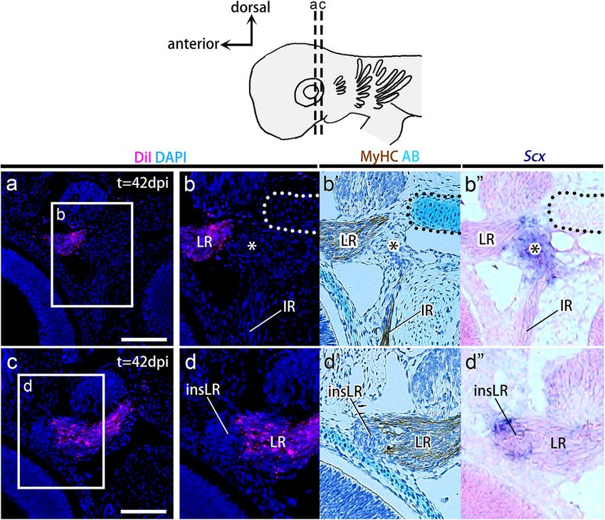
Kuroda et al. Zoological Letters        (2021) 7:3                                                                                              Page 2 of 11

 Fig. 1 Anatomy of EOMs and distributions of head cavities in the shark embryo. a Left lateral view of an adult shark (S. torazame; top) and
 schematic drawing of the left extraocular muscles (bottom). Sets of EOMs innervated by the same cranial motor nerve are shown in the same
 color: EOMs innervated by the oculomotor nerve in brown, by the trochlear nerve in orange, and by the abducens nerve in green. The blue
 dotted line indicates the outline of the left eye. b A schematic drawing of the left lateral view of the shark embryo at st. 25 showing the
 positional relationships of head cavities (HCs) to the pharyngeal and cardiac mesoderm (dark gray) and somites (light pink). Each HC is marked in
 the same color as sets of EOMs in (a), which are predicted to be derived from a single HC. c, d H&E sections in the transverse section (c) and the
 sagittal section reconstructed from serial transverse sections (d), showing the anatomical position of HCs, which are arranged in series
 dorsoventrally (c) and anteroposteriorly (d). The scheme in (a) is modified from ref. [1]. II, optic nerve; III, oculomotor nerve; IV, trochlear nerve; VI,
 abducens nerve; gV, trigeminal ganglion; ham, hyoid arch mesoderm; IO, inferior oblique muscle; IR, inferior rectus muscle; LR, lateral rectus
 muscle; mam, mandibular arch mesoderm; mnc, mandibular head cavity; MR, medial rectus muscle; op, optic cup; ot, otic vesicle; pcm, pericardial
 mesoderm; prmc, premandibular head cavity; SO, superior oblique muscle; som, somite; SR, superior rectus muscle

arches, since they are located anterior to the first                             been roughly illustrated by histological observations
pharyngeal arch [5–7].                                                           of developmental series of elasmobranch embryos
   The mesenchymal state of the head mesoderm is                                 (sharks, skates, and rays) [10, 14]. To elucidate the
commonly seen in most experimental animals                                       detailed cell fates of each HC, cell lineage tracing ex-
throughout development [8, 9]. In contrast, embryos                              periments are required. Moreover, the developmental
of chondrichthyans (cartilaginous fishes) generally                              contributions of HCs to nonmuscular tissues also re-
form three pairs of epithelial coeloms called head cav-                          main to be investigated [11, 12, 18–20].
ities (HCs) (Fig. 1b-d) [1, 10–13]. The premandibular                              In the present study, we examined the developmen-
head cavity (prmc) originates from the prechordal                                tal fates of shark HCs to determine whether each of
plate, the anteriormost element of the axial meso-                               the HCs gives rise to a different set of EOMs or
derm, that is, from the anteriormost mesoderm in the                             other musculoskeletal components. The epithelial na-
vertebrate embryo. The mandibular head cavity (mnc)                              ture of shark HCs facilitates accurate and highly re-
and hyoid head cavity (hyc), on the other hand, are                              producible labeling of a specific part of the head
derived from the head paraxial mesoderm (Fig. 1b-d)                              mesoderm, which in other animals appears only as a
[12]. Both HCs arise as schizocoels between the neu-                             cluster of mesenchymal cells without discrete histo-
rula and early pharyngula stages [12]. In later stages,                          logical boundaries [9, 21, 22]. We performed lineage
histological observations have shown that the epithe-                            tracing experiments using a lipophilic fluorescent dye
lial walls of HCs collapse and that the coelomic struc-                          in embryos of the cloudy catshark Scyliorhinus tora-
tures gradually disappear [14]. Each of the HCs was                              zame (Tanaka, 1908) [23], which has three pairs of
assumed to give rise to a distinct subset of EOMs in-                            HCs [5, 12]. We confirmed that the HCs give rise to
nervated by a single cranial nerve (Fig. 1a). Therefore,                         EOMs but also discovered that the cell fates of HCs
the morphology of HCs has long been believed to                                  were more complex than classically predicted (Fig. 1).
serve as a prepattern for EOM morphology [11, 15–                                We further revealed that HCs also give rise to tendon
17]. However, this predicted lineage of HCs has only                             progenitor cells for some of the rectus muscles.
Kuroda et al. Zoological Letters   (2021) 7:3                                                                  Page 3 of 11

Methods                                                      the embryo. The eggshell was sealed with a polycarbon-
Embryo collection                                            ate filter (GTBP01300; Merck Millipore, USA) using
Fertilized eggs of S. torazame were collected from tanks     cyanoacrylate adhesive (Aron Alpha, also known as
of adult sharks kept at 16 °C at RIKEN. Embryos were         ‘Krazy Glue’; Toagosei, Japan), to prevent air bubbles
staged according to a previous study [24]. For section in    from entering the eggshell and to prevent the contents
situ hybridization and immunohistochemistry, embryos         of the egg from protruding through the opening. The
were fixed overnight in 4% paraformaldehyde (PFA)            injected embryos were left to develop for 6–7 weeks at
(104,005; Merck KGaA, Germany) in phosphate-buffered         16 °C in seawater with 0.2% antibiotic-antimycotic mixed
saline (PBS) (pH 7.4, AM9625; Thermo Fisher Scientific,      stock solution without aeration. The incubation seawater
USA) at 4 °C.                                                was replaced multiple times weekly.

Molecular cloning and phylogenetic analysis                  Histological analysis and in situ hybridization
Total RNA of S. torazame was extracted using TRIzol          Fixed embryos were dehydrated and embedded in
Reagent (15,596,026; Thermo Fisher Scientific), and the      paraffin (Paraplast Plus, P3683; Sigma-Aldrich) at
corresponding cDNA was synthesized using SuperScript         65 °C. Sections were cut at a thickness of 7 μm. For
IV Reverse Transcriptase (18,091,050; Thermo Fisher          fluorescence detection of CM-DiI-labeled samples, the
Scientific). PCR was performed to amplify fragments of       deparaffinized sections were washed twice with PBS
the S. torazame Scleraxis (StScx) gene by LA Taq HS          and incubated for 1 h at room temperature with 4′,6-
(RR042A; TaKaRa Bio Inc., Japan) with specific primers       diamidine-2′-phenylindole dihydrochloride (DAPI)
designed based on the prospective StScx sequence ob-         (5 μg/ml, 10,236,276,001; Roche, Switzerland) in PBS.
tained from Squalomix, the elasmobranch transcriptome        Then, drops of Omnipaque300 (Daiichi-Sankyo,
database       (https://transcriptome.riken.jp/squalomix/)   Japan) were added to the sections, and the coverslips
[25]. The PCR fragments were cloned into the pGEM-T          were placed. The sections were then imaged by using
Easy vector (A1360; Promega, USA) and sequenced. For         an Axio Zoom V16 fluorescence microscope (Carl
phylogenetic analysis, amino acid sequences of ortholo-      Zeiss, Germany) with an AxioCam MRm digital cam-
gous genes from other vertebrate and invertebrate spe-       era (Carl Zeiss). For immunostaining, the sections
cies were compiled from GenBank (http://www.ncbi.            were washed with Tris-HCl-buffered saline (pH 7.8,
nlm.nih.gov/) and Ensembl (http://www.ensembl.org/).         20 mM Tris-HCl, 150 mM NaCl) containing 1% Tri-
Multiple alignments of protein sequences were per-           ton X-100 (TST), blocked with 5% skim milk in TST
formed with MAFFT [26] as implemented on the web             (TSTM) for 30 min, and incubated overnight at room
server of the European Bioinformatics Institute (http://     temperature with primary antibodies diluted in
www.ebi.ac.uk/Tools/msa/mafft/) and saved in FASTA           TSTM. Myosin heavy chain antibody (1/200, A4–
format. The resulting alignments were trimmed by tri-        1025; DSHB) was used as a primary antibody. After 3
mAl version 1.3 as implemented in Phylemon 2.0 [27]          washes in TST (5 min each), the sections were incu-
and aligned using ClustalW (http://www.clustal.org/)         bated with secondary antibodies in TSTM for 2 h at
without gaps. Phylogenetic trees were constructed using      room temperature. Anti-mouse IgG horseradish per-
the maximum-likelihood (ML) method in PhyML v.3.1            oxidase (HRP) antibody (1/400, F21453; Thermo
(http://www.atgc-montpellier.fr/phyml/) [28] to confirm      Fisher Scientific, USA) was used as a secondary anti-
the orthology of the StScx gene (Fig. S1).                   body. HRP activity was detected using 0.25 mg/ml
                                                             peroxidase substrate, 3,3′-diaminobenzidine (DAB)
Fate mapping and embryonic culture in sharks                 (D5905-50TAB; Sigma), in TST with 0.01% hydrogen
For the injection of shark embryos at stage 25, eggs were    peroxide. Hematoxylin and eosin (H&E) or Alcian
removed from the seawater tank and briefly incubated         blue staining was performed according to a standard
on ice. A small window was opened on the surface of          protocol. The sagittal section in Fig. 1d was recon-
the egg case just above the embryo. Embryos were then        structed in silico from serial transverse sections using
anesthetized with 20 μl of a mixed solution of 1% ethyl      Avizo software version 8.0.1 (Thermo Fisher Scien-
3-aminobenzoate methanesulfonate (MS-222) (E10521;           tific). In situ hybridization on paraffin sections for S.
Sigma) and 2% sodium carbonate (1:1 volume:volume).          torazame Scx (GenBank accession number LC430615)
CM-DiI (C7001; Thermo Fisher Scientific, USA) was            was performed as previously described [30]. Counter-
prepared as previously described [29] and microinjected      staining for in situ hybridization was performed with
into HCs by using a microinjector (MN-151; Narishige,        Nuclear Fast Red (Vector Laboratories, USA). Adja-
Japan). After injection, 200 μl of 0.2% antibiotic-          cent sections were analyzed to compare gene expres-
antimycotic mixed stock solution (09366–44; Nacalai          sion patterns and distributions of fluorescently labeled
Tesque, Inc., Japan) in PBS was added to the surface of      cells. Sections in brightfield images were imaged with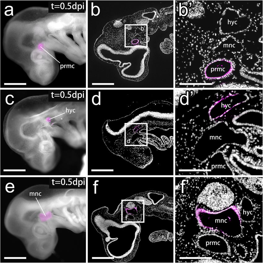
Kuroda et al. Zoological Letters       (2021) 7:3                                                                                              Page 4 of 11

a BX53 microscope (Olympus, Japan) with a DP74                                  method. We fixed embryos at 12 hours postinjection
digital camera (Olympus). Fluorescent images were                               (0.5 dpi) and confirmed that CM-DiI labeling was con-
processed by ZEN software (Carl Zeiss), and all im-                             fined to the epithelial wall of the injected coelom (num-
ages were assembled in Adobe Photoshop (Adobe                                   ber of times that DiI was specifically recovered in
Systems, USA) as previously described [31].                                     labeled embryos: prmc, n = 3/4; hyc, n = 5/7; mnc, n = 3/
                                                                                5) (Fig. 2). Although the ventral portion of the mnc con-
Results                                                                         nected with the tubular pharyngeal arch mesoderm (Fig.
Long-term cell lineage tracing in S. torazame embryos                           1b), the lumen of the pharyngeal arch canal was almost
In stage 25 (st. 25) S. torazame embryos, three pairs of                        flattened and very narrowed by this stage [12], prevent-
HCs were fully formed but had not yet begun their dif-                          ing ectopic dye labeling outside the mnc. Moreover, no
ferentiation into muscle cells (Fig. 1b-d) [12, 14]. We                         DiI-labeled cells were observed in other tissues around
attempted to label the epithelial wall of each HC by                            HCs, ensuring exclusive labeling of the HC epithelium
microinjecting CM-DiI into the coelom (Fig. 2a, c, and                          (Fig. 2b’, d′, and f′).
e). Since the left and right sides of the prmc at this stage                       In the following experiments, we incubated DiI-
are connected through a transverse canal just behind                            injected embryos until st. 31 (42 dpi; Fig. 3a), by which
Rathke’s pouch [10, 12], when DiI was injected into one                         time all EOMs were differentiated and connected to
side of the prmc, the opposite side of the prmc was also                        their attachment sites.
labeled at a certain frequency (7 cases in 12 injected em-                         We used myosin heavy chain (MyHC) antibody and
bryos). In contrast, the left and right coeloms of the mnc                      Alcian blue staining to identify muscles and skeletal tis-
and hyc were separated from each other (Fig. 1b-d), and                         sues in the developed embryos. To visualize tendon pro-
we could label each of the HCs specifically with our                            genitor cells, we isolated a shark homologue of the

 Fig. 2 DiI-labeled head cavities at the pharyngula stage. a-b′ An embryo with DiI injected into the prmc at st. 25. a Left lateral view of the DiI-
 injected embryo at 0.5 days postinjection (dpi). b-b′ A sagittal section of (a) and a magnified image in the inset (b′) showing DiI-labeled cells
 (magenta) found specifically in the epithelial wall of the prmc. c-d′ An embryo with DiI injection into the hyc at st. 25. c Left lateral view of the
 DiI-injected embryo at 0.5 dpi. (d-d′) A sagittal section of (c) and a magnified image in the inset (d′) show DiI-labeled cells (magenta) found
 specifically in the epithelial wall of the hyc. e-f′ An embryo with DiI injection into the mnc at st. 25. e Left lateral view of a DiI-injected embryo at
 0.5 dpi. f-f′ A sagittal section of (e) and a magnified image in the inset (f′) show DiI-labeled cells (magenta) found specifically in the epithelial wall
 of the mnc. Panels b, b′, d, d′, f, and f′ show sections counterstained with DAPI (gray). hyc, hyoid head cavity; mnc, mandibular head cavity; prmc,
 premandibular head cavity. Scale bars = 500 μm (a, b, c, d, e, and f), 200 μm (b′, d′, and f′)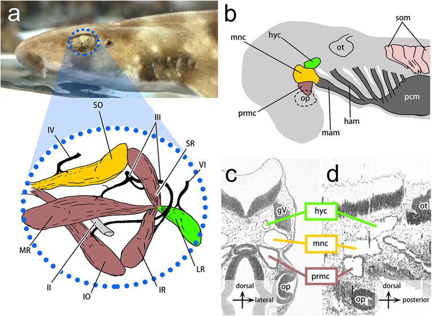
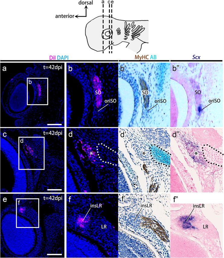
Kuroda et al. Zoological Letters        (2021) 7:3                                                                                              Page 5 of 11

 Fig. 3 Lineage tracing of the premandibular head cavities. a DiI was injected into the coelom of the prmc of a cat shark embryo at st. 25. At st.
 31 (42 days postinjection: 42 dpi), the DiI-injected embryos were fixed and histologically examined. b-h″ Transverse sections cut through the
 dashed lines in (a) DiI-injected catshark embryos at 42 dpi. Sections (b) and (f) were each aligned with adjacent sections immunostained with
 anti-myosin heavy chain (MyHC) antibody (c′, d′, e’, g’, and h′), stained with Alcian blue (AB) (c′, d′, e’, g’, and h′), and hybridized in situ with a Scx
 antisense RNA probe (g” and h″). DiI-labeled cells (magenta) are found in muscle fibers of specific sets of extraocular muscles (superior rectus,
 medial rectus, inferior oblique, and inferior rectus muscles) but not in Scx-positive tendon progenitor cells at the attachment site of all rectus eye
 muscles (asterisks) or in the pila antotica (outlined by the dotted line). IO, inferior oblique muscle; IR, inferior rectus muscle; LR, lateral rectus
 muscle; MR, medial rectus muscle; SR, superior rectus muscle. Scale bars = 200 μm

Scleraxis (Scx) gene, which is known to be expressed in                          The premandibular and hyoid head cavities give rise to
tendon progenitor cells in mice [32], chickens [33], and                         distinct sets of EOMs
zebrafish [34]. In situ hybridization analysis confirmed                         In the embryos injected with DiI into the prmc, labeled
that S. torazame Scx expression was specifically de-                             cells were observed in muscle fibers of the superior rec-
tected in cell condensations, which are presumptive                              tus (n = 17/19) (Fig. 3c), medial rectus (n = 18/19) (Fig.
tendon progenitor cells, located between the jaw                                 3d), inferior oblique (n = 14/19) (Fig. 3e), and inferior
muscle and cartilage (Fig. S2b). Using this Scx probe,                           rectus muscles (n = 19/19) (Fig. 3g) stained by anti-
MyHC antibody and Alcian blue as tissue-specific                                 MyHC antibody (Fig. 3b-h’). DiI-labeled cells were not
markers for tendon progenitor cells, differentiated                              detected in Rathke’s pouch (n = 0/12) (Fig. S3a), the tri-
muscles and cartilage, respectively, we examined the                             geminal ganglia (n = 0/19) (Fig. S3b), optic vesicles (n =
distribution of DiI-labeled cells derived from each HC                           0/19) (Fig. S3c), sclera (n = 0/19) (Fig. S3c), the chondro-
in the established musculoskeletal components, as de-                            cranium at the attachment sites of rectus muscles (pila
scribed below.                                                                   antotica [35, 36];) (n = 0/19) (Fig. 3b), or the trabecular
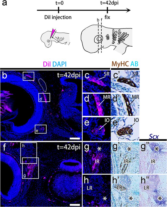
Kuroda et al. Zoological Letters       (2021) 7:3                                                                                            Page 6 of 11

 Fig. 4 Lineage tracing of the hyoid head cavities. a-d″ Transverse sections of CM-DiI-injected embryos aligned with adjacent sections
 immunostained with myosin heavy chain antibody (b′ and d′), stained with Alcian blue (AB) (b′ and d′), and hybridized in situ with a Scx
 antisense RNA probe (b″ and d″). By 42 dpi, CM-DiI-labeled cells (magenta) were found in the lateral rectus muscle but not in Scx-positive tendon
 progenitor cells at the origin of all rectus muscles (asterisks), the insertion site of the lateral rectus muscle (insLR), or the skeleton at the
 attachment sites of the rectus muscles (pila antotica; outlined by the dotted line). insLR, tendon progenitor cells at the insertion site of the lateral
 rectus muscle; IR, inferior rectus muscle; LR, lateral rectus muscle. Scale bars = 200 μm

cartilage (n = 0/19) (Fig. S3d). At the same time, DiI-                        The mandibular head cavity gives rise to tendon
labeled cells rarely emerged in polar cartilage (n = 1/19)                     progenitor cells as well as muscle fibers
(Fig. S3d) and tendon progenitor cells at the origin of                        The labeling of the mnc provided both expected and un-
the rectus muscles (n = 7/19) (Fig. 3g-h”). In the em-                         expected results. DiI-labeled cells were recovered in the
bryos injected with CM-DiI into the hyc, labeled cells                         superior oblique muscle fibers (n = 19/22) that were
were recovered in the lateral rectus muscle fibers (n =                        positive for MyHC (Fig. 5a-b’), suggesting that the mnc
15/15) (Fig. 4b, b’, d, and d′). Labeling was undetectable                     gives rise to the trochlear nerve-innervated EOM (Fig.
in the trigeminal ganglia (n = 0/15) (Fig. S4) and was                         1a). DiI-labeled cells were also detected at the point of
only rarely detected in the pila antotica (n = 1/15)                           origin of all rectus muscles (asterisks in Fig. 5d-d’) and
(Figs. 4b-b”, and S4) and two tendon progenitor ele-                           at the insertion site of the lateral rectus muscle (insLR in
ments (at the origin of rectus muscles; n = 7/15 and                           Fig. 5f-f’) at high frequencies (n = 16/22 and n = 22/22,
at the insertion of lateral rectus muscle; n = 8/15)                           respectively). These attachment sites were marked by
(Fig. 4).                                                                      Scx expression (Fig. 5d” and f″), showing the features of
  These findings demonstrate that prmc and hyc give                            tendon progenitor cells (see also Fig. S2). No labeled
rise to the oculomotor nerve- and abducens nerve-                              cells were found in other tendon progenitors (n = 0/22)
innervated components of EOMs, respectively (Fig.                              (Fig. 5), the pila antotica (n = 0/22) (Fig. 5d-d”), orbital
1a), which is in line with previous histological obser-                        cartilage (n = 0/22) (Fig. S5a), trigeminal ganglia (n = 0/
vations (Fig. 1) [17, 37]. Moreover, neither the prmc                          22) (Fig. S5b), trabecular cartilage (n = 0/22) (Fig. S5c),
nor the hyc was suggested to give rise to cartilage or                         or palatoquadrate cartilage (n = 0/22) (Fig. S5d). These
tendon.                                                                        results provide the first evidence that the epithelial wall
Kuroda et al. Zoological Letters (2021) 7:3 Page 7 of 11
Fig. 5 Lineage tracing of the mandibular head cavities. a-f″ Transverse sections of CM-DiI-injected embryos aligned with adjacent sections
immunostained with myosin heavy chain antibody (b′, d′, and f′), stained with Alcian blue (AB) (b′, d′, and f′), or hybridized in situ with an Scx
antisense probe (b″, d″, and f″). By 42 dpi, DiI-labeled cells (magenta) were found in the superior oblique muscle, and the Scx-positive tendon
progenitor cells were found at the scaffold of all rectus eye muscles (asterisks) and the insertion site of the lateral rectus muscle (insLR). insLR,
tendon progenitor cells at the insertion site of the lateral rectus muscle; LR, lateral rectus muscle; SO, superior oblique muscle.
Scale bars = 200 μm
of the mnc in shark embryos, as a part of the head progenitor cells at the origin of the rectus muscles and
mesoderm, gives rise to specific tendon progenitor cells at the insertion of lateral rectus muscles (Fig. 6d and e).
in addition to trochlear nerve-innervated EOMs (super- In contrast, we did not observe any contributions from
ior oblique muscle; Fig. 1d) [17, 37]. HCs to the cranial cartilage in our experiments.
Concerning the mnc-derived cellular condensation
Discussion at the insertion site of the lateral rectus muscle,
In this study, we reported lineage tracing analysis of given its position and developmental origin, it could
HCs in shark embryos and showed that each HC gives be the same cell population first described by Platt
rise to a set of EOMs innervated by a single cranial as ‘muscle E’ (Fig. S6, [14, 15, 38, 39]). Platt and
motor nerve (Figs. 1d and 6). In addition, our results later researchers thought that this condensation gives
provide the first evidence that HCs, which belong to the rise to muscle cells of the distal portion of the lat-
head mesoderm, give rise to dense connective tissues of eral rectus muscle (reviewed in [15]). However, in
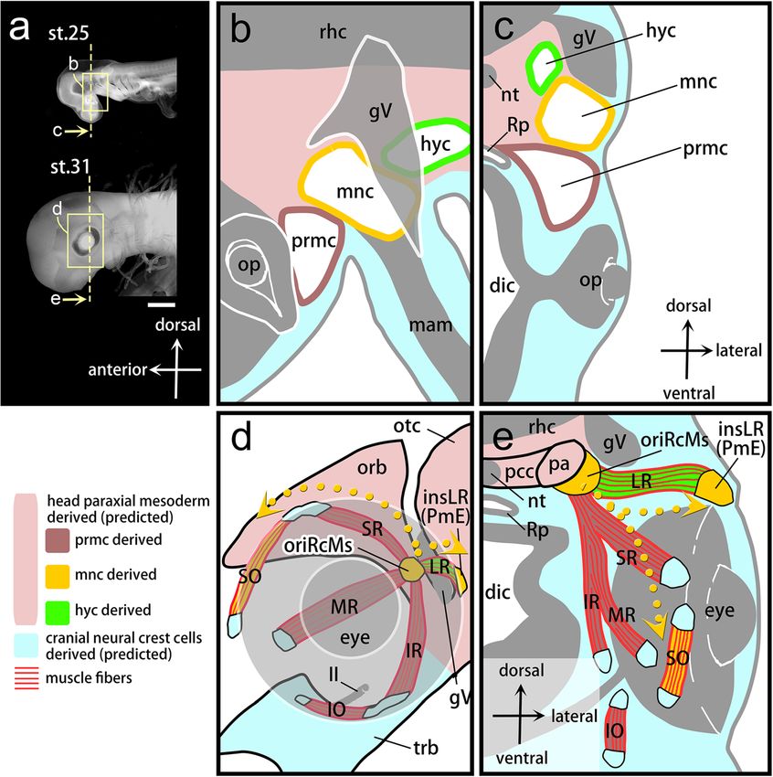
the head muscles in shark; the mnc gives rise to tendon our experiments, this condensation was not positiveKuroda et al. Zoological Letters (2021) 7:3 Page 8 of 11 Fig. 6 Developmental fates of shark head cavities in the musculoskeletal system of extraocular muscles. a Left lateral view of the shark embryos at st. 25 (top) and st. 31 (bottom). b, c Schematic drawings of a left lateral view of the inset in (a) and a transverse section at the level of the dashed line in (a) showing HCs arranged along the anteroposterior axis (b) and the dorsoventral axis (c) in the embryonic head at st. 25. d, e Schematic drawings of a left lateral view of the inset in (a) and a transverse section at the dashed line level in (a) showing musculoskeletal connections in EOMs via tendons at st.31. The colors of each musculoskeletal component correspond to its developmental origin, shown in (b and c). Yellow dotted arrows represent the dynamic migratory pathways of mnc-derived cells reconstructed based on the results of the present study. The developmental fate of CNC cell-derived tendons (light blue) and mesodermal chondrocranium (pink) are based on the prediction. II, optic nerve; dic, diencephalon; gV, trigeminal ganglion; hyc, hyoid head cavity; insLR, a tendon at the insertion of the lateral rectus muscle; IO, inferior oblique muscle; IR, inferior rectus muscle; LR, lateral rectus muscle; mam, mandibular arch mesoderm; mnc, mandibular head cavity; MR, medial rectus muscle; nt, notochord; op, optic cup; orb, orbital cartilage; oriRcMs, a tendon at the origin of four-rectus muscles; otc, otic capsule; pa, pila antotica; pcc, parachordal cartilage; pm, PmE, Platt’s ‘muscle E’; prmc, premandibular head cavity; rhc, rhombencephalon; Rp, Rathke’s pouch; SO, superior oblique muscle; SR, superior rectus muscle; trb, trabecular cartilage. Scale bars = 1 mm. Not to scale (b-e). for the MyHC antibody but did express the Scx of mnc-derived cells retain their original positions gene, suggesting that these cells are tendon progeni- throughout development and give rise to tendon pro- tor cells (Figs. 5 and S6b). genitor cells at the origin of rectus muscles (Fig. 6). In Although the classical studies that predicted the devel- contrast, the remaining mnc-derived cells show two dif- opmental fate of HCs (Fig. 1) were partially supported ferent migratory pathways, one toward the anterior por- by our experiments, the actual developmental patterns tion and the other toward the posterior portion of the and processes of the mnc-derived cells turned out to be eye (yellow dotted arrows in Fig. 6d and e). First, the su- more complex than was predicted (Fig. 6). Classical perior oblique muscle primordium, separated from the studies regarded the pattern of HCs as a prepattern of dorsal part of the mnc, passes above the eye and over- EOM morphology based on the predicted one-to-one takes the cell population derived from the prmc. At this correspondence between an HC and the innervation pat- point, the original anteroposterior arrangement of the terns of EOMs [1, 11, 15, 17]. The results of the present HCs was altered. In the posterior part of the eye, the study led us to revise the above hypothesis. One portion mnc-derived cells give rise to tendon progenitor cells at
Kuroda et al. Zoological Letters   (2021) 7:3                                                                  Page 9 of 11

the insertion of the lateral rectus muscle (Platt’s ‘muscle    cells at the proximal attachment of rectus muscles in
E’), keeping their leading position relative to the direc-     sharks. This result is consistent with the above discus-
tion of movement of the lateral rectus muscle primor-          sion about the position of the mesoderm/CNC interface
dium. It is worth noting here that mnc-derived tendons         in the orbital region. At the same time, this suggests that
are not recruited in the muscle attachment of the super-       the corresponding EOM attachment sites in mice (ala
ior oblique muscles. This may be comparable to the rela-       hypochiasmatica) and chickens (supratrabecular cartil-
tionship between the syndetome and migratory muscle            age) also adopt mesodermal tendons similar to those in
precursors, both of which are derived from a single so-        shark embryos (Fig. S7). Now, we need to reexamine the
mite [40, 41]. However, because the mnc-derived ten-           cell lineages of connective tissues of EOMs in these two
dons give scaffolds at both ends of the hyc-derived            animals, for whom the cell lineages of the structures in
lateral rectus muscles (Fig. 6), it is still unreasonable to   the head have been studied in greater detail than in any
compare the relationship between the mnc and hyc with          other vertebrate.
that of two adjacent somites. Overall, we conclude that           In the present study, we could not perform lineage tra-
the morphological pattern of HCs is not a prepattern of        cing experiments of shark CNC cells due to technical
EOMs.                                                          difficulties. Considering the results of previous experi-
                                                               ments in model animals that have shown that the perio-
Reevaluating the mesoderm/CNC boundary in the                  cular mesenchyme generally consists of CNC cells [55,
mesenchymal environment in the orbital region                  57, 58], it is reasonable to speculate that the other ten-
The majority of the mesenchymal component in the ver-          dons in shark EOMs that are not derived from HCs are
tebrate embryonic head is derived from CNC cells [4,           derived from CNC cells (Fig. 6d and e). Thus, the rectus
42]. Since CNC cells differentiate into the pharyngeal         muscles other than the lateral rectus muscles in sharks
skeleton, prechordal cranium, and connective tissues of        are suggested to have CNC-derived tendons at one at-
the head muscles, it has been presumed that musculo-           tachment site and mesodermal tendons at the other
skeletal connections in the head would be established          sites. The fact that some EOMs have tendons other than
through interactions between CNC cells and muscle              CNC-derived tendons suggests that the morphogenetic
progenitor cells [4, 42–45]. In heterotopic transplant-        process of EOMs may be partially free from the identity
ation of the trunk paraxial mesoderm into the head,            imposed by the ectomesenchymal environment. Al-
grafted cells gave rise to head muscles with nearly nor-       though our results did not support the notion that the
mal morphology [46, 47]. In Tbx1 knockout mice, bran-          morphological pattern of the HCs contributed to that of
chiomeric muscle precursors were absent, but the initial       the EOMs, the cell population boundary in the mesen-
patterning of tendon progenitor cells occurred normally        chymal environment in the orbital region might play
[48]. Thus, the morphogenetic information of the               some role in establishing the proximodistal axis in the
CNC cells that give rise to dense connective tissues in        rectus muscles.
the head can override the identity of the muscle precur-          Contexts similar to the developmental environment
sors exposed in the ectomesenchymal environment (de-           suggested in the present study have recently been re-
rived from CNC cells) [49–52].                                 ported in some neck and shoulder musculatures.
  EOM primordia first appear in the head paraxial              These muscles develop in embryonic environments
mesoderm, where they are detectable by Pitx2 expres-           with mesenchymal boundaries between the CNC and
sion, and subsequently migrate rostrally to enter ectome-      lateral plate mesoderm [59] or cardio-pharyngeal
senchymal environments in the prechordal region [50,           mesoderm [60]. The resultant muscles have hetero-
53, 54]. This migration pattern led to the belief that         genic cell populations of connective tissues. Further-
CNC cells would be the only origin of the dense con-           more, considering the recent report of mesodermal
nective tissues of EOMs, as in the case of other head          contributions to the posterior part of the pharyngeal
muscles [4, 42]. CNC cells were confirmed to contribute        skeleton in skate [61] and to the tendon progenitor
to some part of the connective tissues of EOMs using           cells for EOMs in shark (this study), we have to re-
chick/quail chimeric embryos [51] and transgenic mice          consider the rather dualistic view that the morpho-
[55]. On the other hand, the skeletal component at the         genetic processes in head and trunk musculature are
proximal attachment sites (origins) of four rectus com-        regulated strictly by CNC-derived and mesodermal
ponents of EOMs is known to be mesodermal in mice              mesenchymal environments, respectively. Further
and chickens [43, 56]. The latter results suggest that the     clarification of the developmental mechanisms shared
developing rectus muscles may be on the mesenchymal            by muscles that develop at the mesoderm/CNC
interface between the head paraxial mesoderm and               boundary will shed new light on the question of what
CNC cells. In the present study, we revealed the contri-       factors determine the evolutionary coupling or de-
butions of the head mesoderm to tendon progenitor              coupling between the mesoderm/CNC boundary and
Kuroda et al. Zoological Letters (2021) 7:3 Page 10 of 11
the morphological boundary in craniofacial and neck- University, Kobe 657-8501, Japan. 3Aix-Marseille Université, CNRS, IBDM UMR
shoulder complexes [56, 62–64]. 7288, 13288 Marseille, France. 4Laboratory for Evolutionary Morphology, RIKE
N Cluster for Pioneering Research (CPR), 2-2-3 Minatojima-minami, Chuo-ku,
Kobe 650-0047, Japan.
Conclusions
In our lineage tracing analysis in shark HCs, we con- Received: 13 November 2020 Accepted: 27 January 2021
firmed the classical view of the developmental origin of
EOMs; each HC gives rise to different subsets of EOMs
References
innervated by each cranial motor nerve. We also found 1. Goodrich ES. Studies on the structure and development of vertebrates.
that the mnc gives rise to tendon progenitor cells at the London: McMillan; 1930.
origin of the rectus muscles and the insertion of the lat- 2. Bothe I, Dietrich S. The molecular setup of the avian head mesoderm and
its implication for craniofacial myogenesis. Dev Dyn. 2006;235(10):2845–60.
eral rectus muscle. Given these newly revealed cell fates 3. Kuratani S. Craniofacial development and the evolution of the vertebrates:
of shark HCs, we conclude that the previous hypothesis the old problems on a new background. Zool Sci. 2005;22(1):1–19.
that the EOM developmental pattern was prespecified in 4. Noden DM. The embryonic origins of avian cephalic and cervical muscles
and associated connective tissues. Am J Anat. 1983;168:257–76.
HCs should be revised. Our results also suggest that the 5. Kuratani S, Horigome N. Developmental morphology of branchiomeric
developmental origins of tendon progenitor cells at ei- nerves in a cat shark, Scyliorhinus torazame, with special reference to
ther end of most rectus muscles in sharks differ from rhombomeres, cephalic mesoderm, and distribution patterns of cephalic
crest cells. Zool Sci. 2000;17(7):893–910.
each other. We speculate that the presence of the head 6. Minoux M, Rijli FM. Molecular mechanisms of cranial neural crest cell
mesoderm/CNC boundary in the mesenchymal environ- migration and patterning in craniofacial development. Development. 2010;
ment could be required for establishing the proximodis- 137(16):2605–21.
7. Kuratani S, Adachi N, Wada N, Oisi Y, Sugahara F. Developmental and
tal axis of the rectus components of EOMs. evolutionary significance of the mandibular arch and prechordal/
premandibular cranium in vertebrates: revising the heterotopy scenario of
gnathostome jaw evolution. J Anat. 2013;222(1):41–55.
Supplementary Information 8. Kuratani S, Adachi N. What are head cavities? — a history of studies on
The online version contains supplementary material available at https://doi.
vertebrate head segmentation. Zool Sci. 2016;33(3):213–28.
org/10.1186/s40851-021-00170-2.
9. Gilbert PW. The origin and development of the head cavities in the human
embryo. J Morphol. 1952;90:149–87.
Additional file 1. 10. Wedin B. The anterior mesoblast in some lower vertebrates-A comparative
study of the ontogenetic development of the anerior mesoblast in
Petromyzon, Etmopterus, Torpedo, et al. Lund: Hakan Ohlsson Boktryckeri; 1949.
Acknowledgments 11. Jarvik E. Basic structure and evolution of vertebrates, vol. 1. New York:
We would like to thank K. Shirato for his help with shark fishing; E. Momota, Academic Press; 1980.
K. Yamamoto and S. Shibuya for the maintenance of adult sharks; and K. 12. Adachi N, Kuratani S. Development of head and trunk mesoderm in the
Onimaru for helpful advice on the incubation of shark embryos. dogfish, Scyliorhinus torazame: I. embryology and morphology of the head
cavities and related structures. Evol Dev. 2012;14(3):234–56.
Authors’ contributions 13. Balfour FM. A monograph on the developmnet of elasmobranch fishes.
S. Kuroda and S. Kuratani conceived the project and designed the MacMillan; 1878.
experiments; S. Kuroda, N. A., and R. K. performed the experiments. All 14. Platt JB. A contribution to the morphology of the vertebrate head, based
authors wrote the manuscript and approved the final version of the on a study of Acanthias vulgaris. J Morphol. 1891;5:79–106.
manuscript. 15. Neal HV. The history of the eye muscles. J Morphol. 1918;30:433–53.
16. Goodrich ES. On the developmnet of the segments of the head in Scyllium.
Funding Quart J micr Sci. 1918;63:1–30.
This work was supported by a Grant-in-Aid for Scientific Research on Innova- 17. van Wijhe JW. Über die Mesodermsegmente und die Entwicklung der Nerven des
tive Areas (Research in a Proposed Research Area) no. 17H06385 to S. Kura- Selachierkopfes. Verh Kon Akad Wetensch Amsterdam. 1882;22:1–50.
tani and by a Grant-in-Aid for Scientific Research (C) no. 19K06683 to R. K. 18. Kuratani S, Ahlberg PE. Evolution of the vertebrate neurocranium: problems
of the premandibular domain and the origin of the trabecula. Zool Lett.
Availability of data and materials 2018;4(1):1.
The newly identified cDNA sequence of the S. torazame Scx gene has been 19. Bertmar G. On the ontogeny of the chondral skull in Characidae, with a
registered in GenBank under accession number LC430615. Any other discussion on the chondrocranial base and the visceral chondrocranium in
relevant data are available from the corresponding author upon reasonable fishes. Acta Zool. 1959;40(2–3):203–364.
request. 20. Jollie M. Segmentation of the vertebrate head. Am Zool. 1977;17:323–33.
21. Kuratani S, Horigome N, Hirano S. Developmental morphology of the head
Ethics approval and consent to participate mesoderm and reevaluation of segmental theories of the vertebrate head:
All animal experiments were carried out with the approval of the evidence from embryos of an agnathan vertebrate, Lampetra japonica. Dev
Institutional Animal Care and Use Committee of RIKEN, Kobe Branch. Biol. 1999;210(2):381–400.
22. Adelmann HB. The development of the eye muscles of the chick. J Morphol
Consent for publication Physiol. 1927;44(1):29–87.
Not applicable. 23. Tanaka S. Notes on some japanese fishes, with descriptions of fourteen new
species. Journ Coll Sci Imp Univ Tokyo. 1908;23:1–55.
Competing interests 24. Ballard WW, Mellinger J, Leichenault H. A series of normal stages for
The authors declare that they have no competing interests. development of Scyliorhinus canicula, the lesser spotted dogfish
(Chondrichthyes: Scyliorhinidae). J Exp Zool. 1993;267:318–36.
Author details 25. Hara Y, Yamaguchi K, Onimaru K, Kadota M, Koyanagi M, Keeley SD, Tatsumi
1
Laboratory for Evolutionary Morphology, RIKEN Center for Biosystems K, Tanaka K, Motone F, Kageyama Y, Nozu R, Adachi N, Nishimura O,
Dynamics Research (BDR), 2-2-3 Minatojima-minami, Chuo-ku, Kobe Nakagawa R, Tanegashima C, Kiyatake I, Matsumoto R, Murakumo K, Nishida
650-0047, Japan. 2Department of Biology, Graduate School of Science, Kobe K, Terakita A, Kuratani S, Sato K, Hyodo S, Kuraku S. Shark genomes provideKuroda et al. Zoological Letters (2021) 7:3 Page 11 of 11
insights into elasmobranch evolution and the origin of vertebrates. Nat Ecol 54. Sefton EM, Kardon G. Chapter Five - Connecting muscle development, birth
Evol. 2018;2(11):1761–71. defects, and evolution: an essential role for muscle connective tissue. In:
26. Katoh K, Standley DM. MAFFT multiple sequence alignment software version 7: Wellik DM, editor. Current Topics in Developmental Biology, vol. 132. United
improvements in performance and usability. Mol Biol Evol. 2013;30(4):772–80. States: Academic Press; 2019. p. 137–76.
27. Sánchez R, Serra F, Tárraga J, Medina I, Carbonell J, Pulido L, de María A, 55. Heude E, Bellessort B, Fontaine A, Hamazaki M, Treier A, Treier M, Levi G,
Capella-Gutíerrez S, Huerta-Cepas J, Gabaldón T, Dopazo J, Dopazo H. Narboux-Nême N. Etiology of craniofacial malformations in mouse models
Phylemon 2.0: a suite of web-tools for molecular evolution, phylogenetics, of blepharophimosis, ptosis and epicanthus inversus syndrome. Hum Mol
phylogenomics and hypotheses testing. Nucleic Acids Res. 2011;39(suppl_2): Genet. 2015;24(6):1670–81.
W470–4. 56. McBratney-Owen B, Iseki S, Bamforth SD, Olsen BR, Morriss-Kay GM. Development
28. Guindon S, Dufayard J-F, Lefort V, Anisimova M, Hordijk W, Gascuel O. New and tissue origins of the mammalian cranial base. Dev Biol. 2008;322(1):121–32.
algorithms and methods to estimate maximum-likelihood phylogenies: 57. Chawla B, Schley E, Williams AL, Bohnsack BL. Retinoic acid and Pitx2
assessing the performance of PhyML 3.0. Syst Biol. 2010;59(3):307–21. regulate early neural crest survival and migration in craniofacial and ocular
29. Minarik M, Stundl J, Fabian P, Jandzik D, Metscher BD, Psenicka M, Gela D, development. Birth Defects Res B Dev Reprod Toxicol. 2016;107(3):126–35.
Osorio-Perez A, Arias-Rodriguez L, Horacek I, Cerny R. Pre-oral gut contributes 58. Creuzet S, Vincent C, Couly G. Neural crest derivatives in ocular and
to facial structures in non-teleost fishes. Nature. 2017;547(7662):209–12. periocular structures. Int J Dev Bio. 2005;19(2–3):161–71.
30. Sugahara F, Murakami Y, Kuratani S. Gene expression analysis of lamprey 59. Heude E, Tesarova M, Sefton EM, Jullian E, Adachi N, Grimaldi A, Zikmund T,
embryos. In: Hauptmann G, editor. In Situ Hybridization Methods. New York: Kaiser J, Kardon G, Kelly RG, Tajbakhsh S. Unique morphogenetic signatures
Springer New York; 2015. p. 263–78. define mammalian neck muscles and associated connective tissues. eLife.
31. Bevilaqua M. Guide to image editing and production of figures for scientific 2018;7:e40179.
publications with an emphasis on taxonomy image editing for scientific 60. Adachi N, Bilio M, Baldini A, Kelly RG. Cardiopharyngeal mesoderm origins
publications. Zoosystematics Evol. 2020;96:139. of musculoskeletal and connective tissues in the mammalian pharynx.
32. Schweitzer R, Chyung JH, Murtaugh LC, Brent AE, Rosen V, Olson EN, Lassar Development. 2020;147(3):dev185256.
A, Tabin CJ. Analysis of the tendon cell fate using Scleraxis, a specific marker 61. Sleight VA, Gillis JA. Embryonic origin and serial homology of gill arches and
for tendons and ligaments. Development. 2001;128(19):3855–66. paired fins in the skate, Leucoraja erinacea. eLife. 2020;9:e60635.
33. Bonnin M-A, Laclef C, Blaise R, Eloy-Trinquet S, Relaix F, Maire P, Duprez D. 62. Matsuoka T, Ahlberg PE, Kessaris N, Iannarelli P, Dennehy U, Richardson WD,
Six1 is not involved in limb tendon development, but is expressed in limb McMahon AP, Koentges G. Neural crest origins of the neck and shoulder.
connective tissue under Shh regulation. Mech Dev. 2005;122(4):573–85. Nature. 2005;436(7049):347–55.
34. Chen JW, Galloway JL. The development of zebrafish tendon and ligament 63. Sefton EM, Piekarski N, Hanken J. Dual embryonic origin and patterning of
progenitors. Development. 2014;141(10):2035–45. the pharyngeal skeleton in the axolotl (Ambystoma mexicanum). Evol Dev.
35. De Beer GR. The development of the vertebrate skull. London: Oxford 2015;17(3):175–84.
University press; 1937. 64. Teng CS, Cavin L, Maxson REJ, Sánchez-Villagra MR, Crump JG. Resolving
36. Holmgren N. Studies on the head in fishes - embrological, morphological, homology in the face of shifting germ layer origins: lessons from a major
and phylogenetical researches. PartI: development of the skull in sharks and skull vault boundary. eLife. 2019;8:e52814.
rays. Acta Zool. 1940;21:51–267.
37. Marshall AM. On the head cavities and associated nerves of elasmobranchs. Publisher’s Note
Quart J micr Sci. 1881;21:72–97. Springer Nature remains neutral with regard to jurisdictional claims in
38. Dohrn A. Studien zur Urgeschichte des Wirbelthierkörpers. Mittheilungen published maps and institutional affiliations.
aus der Zoologischen Station yu Neapel. 1904;17:1–294.
39. De Beer GR. Memoirs: The prootic somites of heterodontus and of amia.
Quart J Micro Sci. 1924;s2–68(269):17–38.
40. Brent AE, Schweitzer R, Tabin CJ. A somitic compartment of tendon
progenitors. Cell. 2003;113:235–48.
41. Dietrich S, Schubert FR, Healy C, Sharpe PT, Lumsden A. Specification of the
hypaxial musculature. Development. 1998;125(12):2235–49.
42. Nassari S, Duprez D, Fournier-Thibault C. Non-myogenic contribution to
muscle development and homeostasis: the role of connective tissues. Front
Cell Dev Biol. 2017;5:22.
43. Couly GF, Coltey PM, Le Douarin NM. The triple origin of skull in higher
vertebrates: a study in quail-chick chimeras. Development. 1993;117:409–29.
44. Platt JB. Ectodermic origin of the cartilage of the head. Anat Anz. 1893;8:
506–9.
45. Noden DM. Interactions and fates of avian craniofacial mesenchyme.
Development. 1988;103(Supplement):121–40.
46. Borue X, Noden DM. Normal and aberrant craniofacial myogenesis by
grafted trunk somitic and segmental plate mesoderm. Development. 2004;
131(16):3967–80.
47. Noden DM. Patterning of avian craniofacial muscles. Dev Biol. 1986;116:347–56.
48. Grenier J, Teillet MA, Grifone R, Kelly RG, Duprez D. Relationship between
neural crest cells and cranial mesoderm during head muscle development.
PLoS One. 2009;4(2):e4381.
49. Tokita M, Schneider RA. Developmental origins of species-specific muscle
pattern. Dev Biol. 2009;331(2):311–25.
50. Noden DM, Francis-West P. The differentiation and morphogenesis of
craniofacial muscles. Dev Dyn. 2006;235(5):1194–218.
51. Noden DM. The role of the neural crest in patterning of avian cranial
skeletal, connective, and muscle tissues. Dev Biol. 1983;96(1):144–65.
52. Wachtler F, Jacob M. Origin and development of the cranial skeletal
muscles. Bibl Anat. 1986;29:24–46.
53. Mootoosamy RC, Dietrich S. Distinct regulatory cascades for head and trunk
myogenesis. Development. 2002;129(3):573–83.You can also read