Acid-Sensing Ion Channels and Mechanosensation - MDPI
←
→
Page content transcription
If your browser does not render page correctly, please read the page content below
International Journal of
               Molecular Sciences

Review
Acid-Sensing Ion Channels and Mechanosensation
Nina Ruan † , Jacob Tribble † , Andrew M. Peterson, Qian Jiang, John Q. Wang and Xiang-Ping Chu *

                                          Department of Biomedical Sciences, School of Medicine, University of Missouri, Kansas City, MO 64108, USA;
                                          nrgrf@umkc.edu (N.R.); jttbkh@umkc.edu (J.T.); amp8m6@umkc.edu (A.M.P.); jiangqi@umkc.edu (Q.J.);
                                          wangjq@umkc.edu (J.Q.W.)
                                          * Correspondence: chux@umkc.edu
                                          † These authors contributed equally to this work.

                                          Abstract: Acid-sensing ion channels (ASICs) are mainly proton-gated cation channels that are
                                          activated by pH drops and nonproton ligands. They are part of the degenerin/epithelial sodium
                                          channel superfamily due to their sodium permeability. Predominantly expressed in the central
                                          nervous system, ASICs are involved in synaptic plasticity, learning/memory, and fear conditioning.
                                          These channels have also been implicated in multiple disease conditions, including ischemic brain
                                          injury, multiple sclerosis, Alzheimer’s disease, and drug addiction. Recent research has illustrated
                                          the involvement of ASICs in mechanosensation. Mechanosensation is a form of signal transduction
                                          in which mechanical forces are converted into neuronal signals. Specific mechanosensitive functions
                                          have been elucidated in functional ASIC1a, ASIC1b, ASIC2a, and ASIC3. The implications of
                                          mechanosensation in ASICs indicate their subsequent involvement in functions such as maintaining
                                          blood pressure, modulating the gastrointestinal function, and bladder micturition, and contributing
                                          to nociception. The underlying mechanism of ASIC mechanosensation is the tether-gate model,

                                   which uses a gating-spring mechanism to activate ASIC responses. Further understanding of the
                                          mechanism of ASICs will help in treatments for ASIC-related pathologies. Along with the well-known
Citation: Ruan, N.; Tribble, J.;
                                          chemosensitive functions of ASICs, emerging evidence has revealed that mechanosensitive functions
Peterson, A.M.; Jiang, Q.; Wang, J.Q.;
Chu, X.-P. Acid-Sensing Ion Channels
                                          of ASICs are important for maintaining homeostasis and contribute to various disease conditions.
and Mechanosensation. Int. J. Mol.
Sci. 2021, 22, 4810. https://doi.org/     Keywords: acid-sensing ion channels; mechanosensation; neurodegenerative diseases; nociception
10.3390/ijms22094810

Academic Editors: Rashid Giniatullin,
Jian Shi and Sang-Won Suh                 1. Introduction
                                               Acid-sensing ion channels (ASICs) are mainly proton-gated cation channels [1], which
Received: 2 April 2021
                                          can be activated by a drop in extracellular pH below 7.0 and triggered by nonproton
Accepted: 30 April 2021
                                          ligands during physiological pH levels [2]. There are currently at least six identified ASIC
Published: 1 May 2021
                                          isoforms (ASIC1a, 1b, 2a, 2b, 3, and 4) encoded by four genes (Accn1, Accn2, Accn3, and
                                          Accn4) [3,4]. Activation of ASICs mostly triggers Na+ influx. As such, ASICs belong to the
Publisher’s Note: MDPI stays neutral
                                          degenerin/epithelial sodium channel (DEG/ENaC) superfamily of ion channels. These
with regard to jurisdictional claims in
                                          channels are made up of 500–560 amino acids [3,5]. The general structure of an ASIC in-
published maps and institutional affil-
                                          cludes a homotrimeric or heterotrimeric proton-gated channel [6,7]. Each of the individual
iations.
                                          subunits is shaped like a “clenched fist” with six domains: wrist, finger, β-ball, thumb,
                                          knuckle, and palm domains in the extracellular loop [3,5]. The overall structure consists
                                          of an intracellular N terminus and an intracellular C terminus with two transmembrane
                                          domains (TMs) that are voltage-independent and help recognize extracellular ligands to
Copyright: © 2021 by the authors.
                                          regulate proton-gated currents [8]. In addition to the permeability to Na+ , activation of
Licensee MDPI, Basel, Switzerland.
                                          ASICs also exhibits calcium permeability in certain subunits, such as ASIC1a, a mechanism
This article is an open access article
                                          important for the regulation of presynaptic neurotransmitter release and ultimately for
distributed under the terms and
                                          regulatory functions, such as synaptic plasticity, learning, and memory [9,10].
conditions of the Creative Commons
                                               ASIC1a is enriched in neurons and is distributed in various subcellular regions, in-
Attribution (CC BY) license (https://
creativecommons.org/licenses/by/
                                          cluding dendrites, dendritic spines, axons, neuron cell bodies, and intracellular organelles,
4.0/).
                                          such as the mitochondria [11,12]. This channel is an important mediator of acid-activated

Int. J. Mol. Sci. 2021, 22, 4810. https://doi.org/10.3390/ijms22094810                                         https://www.mdpi.com/journal/ijms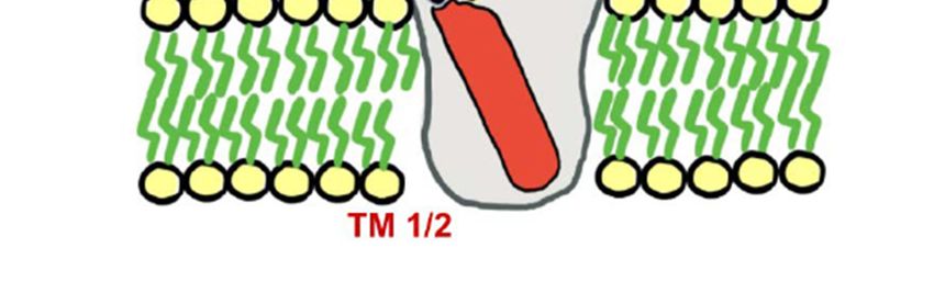
Int. J. Mol. Sci. 2021, 22, 4810                                                                                           2 of 16

                                   responses as well as acidosis-induced physiological changes in the central nervous system
                                   (CNS) and peripheral nervous system (PNS) [9,13]. Recent research has found that ASIC1a
                                   is linked to fear-related behaviors [14,15]. This linkage has been suggested by a high expres-
                                   sion of ASIC1a in the fear-related forebrain regions, such as the amygdala, dorsal striatum,
                                   and nucleus accumbens [14]. Results from numerous studies have shown decreased fear
                                   conditioning in ASIC1a knockout (KO) mice (ASIC1a−/− ) [14–16]. Furthermore, ASIC1a
                                   has been found to play a critical role in synaptic plasticity [17,18]. For example, long-term
                                   potentiation and spatial memory were decreased in ASIC1a−/− mice, while long-term
                                   depression was increased in ASIC1a KO mice [17,18]. The expansive nature of ASIC1a gives
                                   it a wide range of neuromodulation functions [19]. For instance, activation of ASIC1a facili-
                                   tated N-methyl-D-aspartate (NMDA) receptor function [20]. Apart from being expressed
                                   in the nervous system, ASIC1a is found in peripheral tissues, including the vasculature and
                                   intestines [4]. The roles these channels play in the vasculature include vasoconstriction,
                                   vascular hypertrophy, and vascular remodeling [21,22]. Recent studies have found that
                                   these effects on vascular reactivity are found within the pulmonary vasculature but not
                                   within the mesenteric vasculature [22].
                                         ASIC1b is found primarily in peripheral sensory neurons [23]. Studies have shown
                                   that ASIC1b containing channels are heterogeneous and may form heterotrimeric channels
                                   with other ASIC subtypes such as ASIC3 or ASIC1a [23]. Functional ASIC1b plays a role in
                                   peripheral nociception and pain [24,25].
                                         Like ASIC1, ASIC2 has two variants: ASIC2a and ASIC2b. ASIC2 is widely ex-
                                   pressed throughout the brain, including the hippocampus, cortex, amygdala, and olfactory
                                   bulb [26,27]. ASIC2 is also associated with other ASIC subunits, such as ASIC1a or ASIC3,
                                   to form heterotrimers [3,5]. For example, ASIC2/1a is a dominant subunit expressed in
                                   the cortex, and deletion of ASIC2 leads to decreased membrane trafficking of ASIC1a to
                                   the cell surface [28]. Consistent with this idea, both ASIC2 and ASIC1a have been impli-
                                   cated in neuroprotection during disease states [29–34]. ASIC2 has also been implicated in
                                   baroreceptor activities related to the cardiovascular system [35]. The ASIC2 that regulates
                                   baroreceptor functions is located inside the nodose ganglia [35]. In mice with a KO of
                                   the ASIC2 gene (ASIC2−/− ), there is a decreased baroreceptor function [35]. Besides the
                                   pulmonary and cardiac vasculature, immunolabeling studies reveal that ASIC2 plays a
                                   role in renal vasculature [36]. The function of ASIC2 in the kidney is to help with the
                                   myogenic regulation of renal blood flow [37]. Although there are many functions of ASIC2
                                   that need to be clarified, one of the functions of this channel is to regulate blood flow.
                                   ASIC2a also exerts its protection against acid-induced rat articular chondrocyte apoptosis
                                   through regulating ASIC1a expression and the intracellular Ca2+ levels and, at least in part,
                                   suppressing p38 and extracellular signal-regulated kinase 1/2 mitogen-activated protein ki-
                                   nase signaling pathways [38]. Different from ASIC2a [39], ASIC2b cannot form a functional
                                   channel by itself [3,19]. We do know, however, that ASIC2b has been found to modulate
                                   the properties of other ASICs, such as ASIC1a, by forming heterotrimeric channels [40,41].
                                   For example, ASIC2b forms a functional channel with ASIC1a. Such heterotrimeric channel
                                   shows calcium permeability and contributes to ischemic brain injury [40].
                                         ASIC3 is mainly located in the peripheral dorsal root ganglion (DRG) neurons [4,42].
                                   Other locations of ASIC3 in the PNS include the spiral ganglia, nodose ganglia, trigeminal
                                   ganglia, and neurons in the bladder [43]. In addition to forming functional channels with
                                   other ASIC subunits, such as ASIC1a [44], ASIC3 is also associated with the P2X3 ion
                                   channel [45]. Although ASIC3 largely contributes to pain modulation [46–48], ASIC3 also
                                   plays a role in the bladder. In mice lacking the ASIC3 gene (ASIC3−/− ), problems were
                                   found with regards to micturition, including voiding and oliguria [49].
                                         ASIC4 is primary located in the pituitary gland [50]. Other locations of ASIC4 include
                                   the olfactory bulb, hippocampus, caudate putamen, amygdala, cerebral cortices, thalamus,
                                   brainstem, spinal cord, and the preoptic area [51,52]. Like ASIC2b, ASIC4 does not form
                                   a functional channel by itself and is not activated by protons [53]. Currently, the exact
                                   physiological stimulus that activates ASIC4 is unknown [53,54]. Because of the unique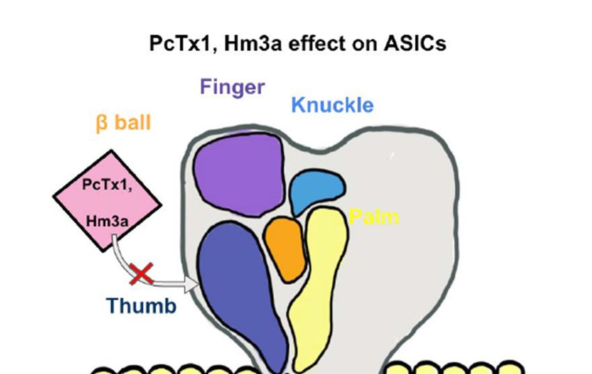
Int. J. Mol. Sci. 2021, 22, 4810                                                                                           3 of 16

                                   characteristics of ASIC4, there are a lot of unknowns when it comes to the expression
                                   and function of this channel. One other location where ASIC4 is seen and is clinically
                                   relevant is osteoblasts. ASIC4, along with ASIC2 and ASIC3, are highly expressed during
                                   osteoblastogenesis in an acid environment [55]. The function of this event is yet to be
                                   elucidated. Research has looked at correlations between ASIC1a and ASIC4 [52]. Recent
                                   studies have found that ASIC4 may modulate the innate fear response via the predator
                                   odor and anxious state [52]. The mechanism underlying the ASIC4-mediated modulation
                                   of fear responses may involve the role of ASIC4 in regulating ASIC1a in the brain [52].

                                   2. ASIC-Associated Pathologies
                                        In addition to its physiological roles, dysfunctional ASIC1a is largely linked to disease
                                   states [9,19,54]. ASIC1a is extensively involved in neurological and psychological dis-
                                   eases, such as ischemic brain injury [28,33], traumatic brain and spinal cord injury [29,30],
                                   Parkinson’s disease (PD) [31], Alzheimer’s disease (AD) [32], experimental autoimmune en-
                                   cephalomyelitis (EAE) [56], multiple sclerosis (MS) [57], seizure disorders [58], pain [59,60],
                                   and drug addiction [61–65].
                                        ASICs are implicated in drug addiction. For example, ASIC1a−/− and ASIC2−/− mice
                                   were both found to cause increases in the α-amino-3-hydroxy-5-methyl-4-isoxazolepropionic
                                   acid (AMPA):NMDA receptor ratio and dendritic spine density during cocaine addic-
                                   tion [64]. This suggests a protective role of ASIC1a and ASIC2 in drug addition by inhibit-
                                   ing cocaine-induced plasticity [62,65]. However, according to a study with overexpression
                                   of ASIC1a in the nucleus accumbens, ASIC1a plays a role in the underlying extinction and
                                   cocaine-seeking behavior, underscoring the complex roles of ASIC1a in synaptic plasticity
                                   related to drug addiction [61].
                                        Blockade of ASIC1a channels in the proximal tubule attenuated Ca2+ influx in in-
                                   stances of renal ischemic reperfusion and led to decreased levels of human proximal
                                   tubular cell apoptosis, indicating that ASIC1a contributes to reperfusion-induced injuries
                                   in the kidney [66]. In the brain, our studies have shown that disruption of either ASIC1
                                   or ASIC2 genes exerted neuroprotection against ischemic brain injury [28,67]. Deletion
                                   of ASIC2 significantly decreased the trafficking of ASIC1a to the cell membrane and re-
                                   duced the infarct volume of the brain in an experimental ischemic stroke model [28]. This
                                   enforces the functional role of ASIC1a and ASIC2 in ischemic brain injury. Along the
                                   lines of protective mechanisms in the brain, ASIC2 has also been shown to protect hu-
                                   mans from pulmonary hypertension by increasing the vasoreactivity of the pulmonary
                                   vasculature [34]. ASIC1a acidification additionally showed the capacity of recruiting the
                                   receptor-interacting serine/threonine-protein kinase 1 (RIPK1) during the development
                                   of ischemic neuronal injury, which ultimately leads to neuronal cell death [68]. Thus,
                                   when targeting 20 N-terminus residues of ASIC1a in an ischemic mouse model, it subse-
                                   quently demonstrated therapeutic potential by preventing RIPK1 activation for possible
                                   protection against neuronal cell death [68]. However, it was also found that the ASIC1a
                                   N-terminus spontaneously bound the N-ethymaleimide-sensitive fusion ATPase (NSF)
                                   during acidic conditions. As an important molecule for synaptic vesicle fusion, NSF is
                                   worth further investigation for its roles in the auto-inhibition of ASIC1a without interfering
                                   with ASIC1a’s desirable physiological functions [69]. These studies also introduce the ther-
                                   apeutic potential of ASIC1a-blocking monoclonal antibodies, such as ASC06-IgG1 [70,71].
                                   β-estradiol has also been reported for stroke treatment via a similar mechanism involving
                                   the downregulation of ASIC1a [33]. Moreover, inhibition of the neuropeptides-ASIC1a
                                   interaction reveals neuroprotection during ischemic brain injury [72,73].
                                        ASICs are involved in MS [57]. Acidotoxicity enhances the influx of Ca2+ and Na+
                                   through ASIC1a, which ultimately causes neuronal degeneration and inflammatory reac-
                                   tions in MS [57,74,75]. The finding that the expression of ASIC1a in axons and oligoden-
                                   drocytes was increased in MS patients with increased axonal injury reinforces the notion
                                   that ASIC1a is an essential player in MS [74]. ASIC2−/− mice were also observed to have a
                                   significantly decreased clinical score for MS, which identifies ASIC2 as a potential player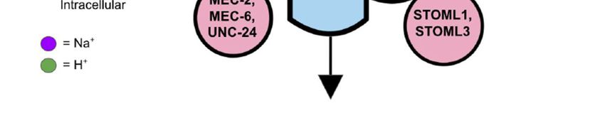
Int. J. Mol. Sci. 2021, 22, 4810                                                                                           4 of 16

                                   implicated in MS. Similar to ASIC1, ASIC2 plays a detrimental role in MS and exacerbates
                                   axon degeneration [76]. Consistent with the role of ASIC1a and ASIC2 in promoting MS,
                                   ASIC blockers, such as amiloride, have neuroprotective properties [77,78]. However, the
                                   majority of studies on ASICs and MS have been conducted either in vitro or in animal
                                   models. Studies on humans are warranted to evaluate the clinical implications of ASICs
                                   in MS [79].
                                         It has been suggested that ASICs are involved in forming the acidic environment in
                                   the pathologic AD brain, although the exact underlying mechanism remains elusive [32].
                                   For instance, upregulation of ASIC1a led to a dramatic increase in intracellular Ca2+ , which
                                   helps maintain the acidic brain environment for the ultimate degeneration of microglial
                                   cells [80,81]. Additionally, in the presence of Aβ and an agonist for group I metabotropic
                                   glutamate (mGlu) receptors, ASIC1a triggered an increase in intrinsic excitability of hip-
                                   pocampal neurons, indicating a functional coupling between ASIC1a and group I mGlu
                                   receptors in the remodeling of synaptic transmission critical for AD [32]. Interestingly, the
                                   common drug for clinical treatment of mild to moderate AD patients, memantine, has been
                                   shown to inhibit ASIC1a along with its well-known mechanism of inhibition of NMDA
                                   receptors [82]. With the further exploration of the ASIC-dependent mechanisms underlying
                                   AD, more therapeutic agents for AD by targeting ASICs are expected to be developed in
                                   the future.
                                         ASIC1b is linked to pain sensation [23–25]. Transient and long-lasting mechanical
                                   hyperalgesia in ASIC1b wild-type (ASIC1b+/+ ) mice has been shown to last much longer
                                   than in ASIC1b−/− mice, though future research is needed to delineate its mechanism
                                   further [23]. In a study carried out by Lee et al. (2018), antihyperalgesic medication at
                                   higher doses significantly reduced ASIC1b activity in rodents [25]. This reveals a correlation
                                   between the peripheral nociception and ASIC1b.
                                         Like ASIC1b, ASIC3 plays a critical role in the pain pathway [47,48]. Current studies
                                   have shown that the hyperalgesic response towards muscle inflammation was eliminated
                                   in ASIC3 KO mice [47]. The mechanism underlying the role of ASIC3 has been suggested.
                                   Namely, muscle inflammation causes a local acidosis in the affected area. This acidosis
                                   triggers the proton sensing capabilities and activation of ASIC3, which elicits a pain
                                   signal [47]. Multiple ion channels have been reported to influence the nociception of
                                   ASIC3. One receptor, in particular, the proteinase-activated receptor 2, causes systemic
                                   sensitization of ASIC3 and, in turn, increases the pain response [47]. Another location
                                   of interest where ASIC3 populates is the gastrointestinal (GI) tract [4,48]. In the GI tract,
                                   ASIC3 participates in the inflammatory response towards gastric acid secretion under
                                   conditions such as gastritis or peptic ulcers [48].
                                         There is an expanding avenue of research pertaining to pharmacological profiles
                                   of ASIC blockers in treating ASIC-associated pathologies [3,5,9]. As shown in Figure 1,
                                   spider-venom peptide psalmotoxin-1 (PcTx1) is shown to be an important inhibitor of
                                   ASIC1a by binding the thumb α-helix 5 component of ASICs in rodent models [83,84]. It
                                   works in human ASIC1a and thus becomes an important analgesic and ischemic stroke
                                   therapy [85]. Another spider venom peptide known as Hm3a has recently been identified
                                   to inhibit ASIC1a and ASIC1b with a similar half amount of excitation concentration of
                                   PcTx1 and higher levels of stability across 48 h (Figure 1). This peptide alleviates symptoms
                                   of potentially both MS and strokes [86]. Both diminazene and mambalgin-1 are also
                                   discovered to block the ASIC1a receptor, which is yet another avenue to inhibit acidosis in
                                   neuroinflammation [87,88]. APETx2, a sea anemones peptide and a selective ASIC3 blocker,
                                   have exhibited inhibition of transient ASIC3 current [89]. ASIC3 blockade significantly
                                   reduces fibromyalgia pain in mice [90]. A-317567 is a small molecule, non-amiloride ASIC
                                   blocker [91], which potently blocks ASICs, especially in the DRG [91]. Evidence shows
                                   that A-317567 is implicated in the treatment of many disorders, including chronic pain and
                                   irritable bladder conditions [92].
blocker, have exhibited inhibition of transient ASIC3 current [89]. ASIC3 blockade signif-
icantly reduces fibromyalgia pain in mice [90]. A-317567 is a small molecule, non-ami-
loride ASIC blocker [91], which potently blocks ASICs, especially in the DRG [91]. Evi-
dence shows that A-317567 is implicated in the treatment of many disorders, including
Int. J. Mol. Sci. 2021, 22, 4810 5 of 16
chronic pain and irritable bladder conditions [92].
Figure 1. An Acid-Sensing Ion Channels (ASICs) subunit has a “clenched fist” conformation with six
domains: wrist,
Figure 1. An Acid-Sensing finger, β-ball,
Ion Channels (ASICs) thumb, knuckle,
subunit has and palm
a “clenched domains. Combined,
fist” conformation these subunits
with six domains: wrist, form a
finger, β-ball, thumb, knuckle, and palm domains. Combined, these subunits form a heterotrimeric or homotrimeric struc- proton-
heterotrimeric or homotrimeric structure to help recognize extracellular ligands and regulate
ture to help recognize
gated extracellular
currents. Withligands
theand regulate proton-gated
inhibition currents.
of the “thumb” With the inhibition
component of the subunit,
of an ASICs “thumb” com-
such as with
ponent of an ASICs subunit, such as with PcTx1 or Hm3a, there will be an inhibition of certain ASICs channels. PcTx1
PcTx1 or Hm3a, there will be an inhibition of certain ASICs channels. PcTx1 leads
leads to the inhibition of ASIC1a, whereas Hm3a leads to the inhibition of both ASIC1a and ASIC1b and additionally
to the inhibition of
higher levels of ASIC1a, whereas
stability over a span Hm3a
of 48 h. leads to the inhibition of both ASIC1a and ASIC1b and additionally higher
levels of stability over a span of 48 h.
3. ASICs in Mechanosensation
3. ASICs in Mechanosensation
Mechanosensation is an integral part of ion channels and a form of signal transduc-
tion in which mechanical
Mechanosensation forcespart
is an integral are converted into neuronal
of ion channels and a signals
form of[93]. Thetransduction
signal electrical
in which signal that is created
mechanical forcesfrom mechanosensitive
are converted ion channels
into neuronal will then
signals help
[93]. Themediate numer-
electrical signal
ous bodily functions, including hearing, balance, proprioception, volume regulation of
that is created from mechanosensitive ion channels will then help mediate numerous bodily
erythrocytes, nociception, vascular function, and touch [93]. Recent research in the field
functions,ofincluding hearing,mechanosensitive
ion channel-related balance, proprioception,
functions hasvolume regulation
further elucidated theofimportance
erythrocytes,
nociception,
and vascular
widespread function,
functionsand touch [93]. Recent
of mechanosensation in ionresearch
channels.inThe
thefunctions
field ofofion channel-
mecha-
related mechanosensitive functions
nosensation in ion channels has
seem to further elucidated
be far more substantial the importance
than and widespread
simply the sensory aspect
functions of mechanosensation in ion channels. The functions of mechanosensation in
ion channels seem to be far more substantial than simply the sensory aspect that has
been elucidated for some time. The basic structure of mechanosensitive ion channels,
in general, is a transmembrane protein with a mechanical gate requiring a stimulus to
activate [94]. The neural circuits surrounding mechanosensation are not well understood,
but they help process information to cause changes in behavior and maintain physiological
homeostasis [94]. In regard to mechanosensation related specifically to ASICs, there has
also been an exponential amount of research that has been published as of late, which
has opened up many avenues for the future [4]. However, there are still many unknowns
surrounding ASICs related mechanosensation, such as the exact physiological response
that stimulates these channels. This has facilitated the research regarding ASICs and
mechanosensation to flourish.
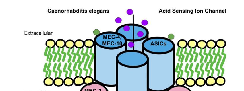
How ASICs play a role in mechanosensation remains unclear. A prevailing theory
involves the mechanotransduction complex in Caenorhabditis elegans. The complex that
senses gentle touch is composed of DEG/ENaC proteins, such as MEC-4 and MEC-10,knowns surrounding ASICs related mechanosensation, such as the exact physiological
response that stimulates these channels. This has facilitated the research regarding ASICs
and mechanosensation to flourish.
How ASICs play a role in mechanosensation remains unclear. A prevailing theory
Int. J. Mol. Sci. 2021, 22, 4810 involves the mechanotransduction complex in Caenorhabditis elegans. The complex6 of 16
that
senses gentle touch is composed of DEG/ENaC proteins, such as MEC-4 and MEC-10,
which are connected to touch receptor neurons [95] (Figure 2). The complex is additionally
supported
which by accessory
are connected subunits,
to touch receptorsuch as MEC-2,
neurons MEC-62).
[95] (Figure andTheUNC-24,
complexwhich show to
is additionally
have only a partial loss of touch sensitivity when deleted as they
supported by accessory subunits, such as MEC-2, MEC-6 and UNC-24, which show to are not associated tightly
have
with the MEC-4 and MEC-10 complex [95]. DEG/ENaC channel proteins
only a partial loss of touch sensitivity when deleted as they are not associated tightly with and accessory
proteins
the MEC-4 areand
all essential for mechanoreceptor
MEC-10 complex [95]. DEG/ENaC activity because
channel the mutation
proteins of Mec2,
and accessory Mec4,
proteins
and Mec6
are all genesfor
essential eliminated mechanoreceptor
mechanoreceptor currents
activity because the [96]. In addition,
mutation of Mec2,between
Mec4, and the ex-
Mec6
tracellular matrix (ECM) and the cytoskeleton of the organism, there
genes eliminated mechanoreceptor currents [96]. In addition, between the extracellular are ECM-linker pro-
teins,
matrixsuch
(ECM)as MEC-1
and theand MEC-9, asofwell
cytoskeleton as intracellular
the organism, there linker proteins, such
are ECM-linker as stomatin
proteins, such as
(STOM). They convey signals from the extracellular matrix to the
MEC-1 and MEC-9, as well as intracellular linker proteins, such as stomatin (STOM). cytoskeleton [97–99]. In
They
aconvey
tethersignals
model,from
MEC-4/MEC-10 act as
the extracellular a gating-spring
matrix mechanism
to the cytoskeleton in mechanosensation
[97–99]. In a tether model,
[96,100]. When compared
MEC-4/MEC-10 to nematodes,mechanism
act as a gating-spring mammals are in specifically characterized
mechanosensation by STOM
[96,100]. When
proteins,
comparedsuch as STOM-like
to nematodes, 1 (STOML1)
mammals and STOM-like
are specifically 3 (STOML3),
characterized by STOMas opposed
proteins,to nem-
such
atode accessory
as STOM-like subunits [101]
1 (STOML1) (Figure 2). 3STOM
and STOM-like is a protein
(STOML3), that istolikely
as opposed attached
nematode to the
accessory
C-terminus
subunits [101] of MEC-4/MEC-10
(Figure 2). STOM to connect withthat
is a protein the TM1 of ASIC3
is likely to suppress
attached the ion chan-
to the C-terminus of
nel [102]. Thus, in
MEC-4/MEC-10 tomice lacking
connect withSTOM,
the TM1stimulation
of ASIC3 toofsuppress
mechanosensation in D-hair
the ion channel [102].recep-
Thus,
in mice
tors on lacking
the skinSTOM, stimulation
was reduced [103].ofAdditionally,
mechanosensation in D-hair
ECM-linker receptors
proteins mayonotherwise
the skin
was
causereduced [103]. Additionally,
extracellular ECM-linker
tension and trigger proteins may
a conformational otherwise
change cause extracellular
to activate the kindlin-
tension and trigger
integrin-RhoA a conformational
pathway, which further change to activate
stimulates the kindlin-integrin-RhoA
mechanotransduction [104]. pathway,
which further stimulates mechanotransduction [104].
Figure 2. The mechanotransduction model found in Caenorhabditis elegans is the leading theory on the ASICs mechanosen-
sation model in humans. On the left, as found in Caenorhabditis elegans, MEC-4 and MEC-10 are DEG/ENaC proteins that
connect to touch receptor neurons and eventually act with a gating-spring mechanism to activate ASICs for mechanosensa-
tion. Similarly, intracellularly in nematodes, there are found to be MEC-2, MEC6, and UNC-24 accessory subunit proteins
that loosely associate with ASICs to activate mechanosensation via a similar mechanism as well. Conversely, in mammals,
ASICs are shown to contain STOML1 and STOML3 proteins which correlate with nematode accessory subunit proteins and
help activate ASICs for mechanosensation as well.
Another theory regarding the mechanism of mechanotransduction in ion channels in-
volves PIEZO proteins and their roles in the “bilayer model” [105]. Some of the mechanosen-
sitive functions that this channel is thought to carry out include proprioception, sensing
light touch, and sensing stretch in organs [105]. This “bilayer model” theory has not
been fully elucidated, and it is only linked to ion channels in general, not specifically to
ASICs [104]. The bilayer model of mechanosensation is relatively simple as the mechanical
stimulus directly modulates the gating of the ion channel [106]. This stimulus differs fromInt. J. Mol. Sci. 2021, 22, 4810 7 of 16
tissue to tissue. For example, in blood vessels, the laminar and oscillatory shear stress of
blood stimulates the ion channel, while in peripheral sensory neurons, external physical
forces stimulate the ion channel [106]. The bilayer model has been well known for some
time. However, the role that PIEZO proteins play in this theory is novel. PIEZO proteins
are large integral membrane proteins with 24–40 TMs that are implicated in converting
the mechanical force into biological signals, specifically in mammalian cells [107]. Other
functions of PIEZO proteins in humans include their promotion of various cellular de-
velopmental events, including cellular migration, elongation, and proliferation [107]. Of
the PIEZO family members, Piezo1 and Piezo2 have specifically been found to function
in mechanosensation [108,109]. Evidence proving the involvement of Piezo1 and Piezo2
proteins in mechanosensation has been found in studies with mice [109]. In these studies,
mice with disrupted Piezo1 and Piezo2 genes showed a lower level of mechanically acti-
vated cation channel activities [109]. The exact mechanism underlying the role of Piezo in
mechanosensation remains unknown. However, studies from localization and fluorescence
imaging of Piezo genes support their roles in mechanosensation [108].
ASIC1 channels are located widely in the visceral sensory ganglia [110]. In ASIC1−/−
mice, there is also a subsequent increase in mechanosensitivity in the esophageal and
colonic afferent mechanotransduction, indicating the importance of ASIC1a in visceral
mechanosensation [111]. ASIC1 also has an important visceral mechanosensation process
in urothelium and bladder compliance sensation [92,112]. However, loss of the ASIC1
did not appear to affect any cutaneous mechanoreceptors [113]. Further, ASIC1 channels
are found in human cutaneous Pacinian corpuscles and may serve as rapidly adapting
low-threshold mechanoreceptors, suggesting specific roles of ASIC1 proteins in human
mechanotransduction [114]. ASIC1 is shown to have an effect on primary hyperalgesia
during inflammation, which is a local response in the area of injury [115]. On the other hand,
ASIC1 does not particularly affect secondary hyperalgesia, which is a response outside the
area of injury [115]. ASIC1a is also involved in the pain pathway. For example, blocking
ASIC1a by PcTx1 results in the activation of the endogenous enkephalin pathway [59]. The
results suggest that ASIC1a channel is an important molecular target for treating both acute
and neuropathic pain and that PcTx1 itself could be a potential analgesic drug working
upstream of the opiate receptors. Recently, ASIC1 has been linked to migraine [116]. For
example, intravenous injection of amiloride and mambalgin-1 both exert long-lasting anti-
allodynic effects against acute and chronic cutaneous allodynia in the isosorbide dinitrate-
induced migraine model, suggesting the involvement of peripheral ASIC1 channels in
migraine cutaneous allodynia as well as in its chronification. The results shield light on
the therapeutic potential of ASIC1 inhibitors as both an acute and prophylactic treatment
for migraine [116]. Current research also demonstrates a mechanosensory role of ASIC1
in peripheral vasoconstriction and vascular remodeling [117]. More specifically, ASIC1
participates in the modulation of mechanosensation through the PNS. In ASIC1−/− mice,
the activity of mechanoreceptors on visceral afferent nerves was enhanced, indicating
that ASIC1 may, in some circumstances, decrease mechanosensation [118]. The presence
of ASIC1 in other peripheral tissues includes arteries, bone marrow, intestine, tongue,
and bladder, indicating possible involvement in mechanosensation [118–120]. Further,
ASIC1b has been tied to mechanosensation due to its apparent involvement in the pain
sensation [24,25]. When higher doses of antihyperalgesic agents were given in rodents,
the ASIC1b channel activity was decreased [24], although exact mechanisms underlying
mechanosensitive functions of ASIC1b are unclear.
ASIC2 is linked to mechanosensation in the PNS [121]. The specific neurons that have
recently been implicated in nociception and mechanosensation include the Isolectin B4-
binding DRG neurons [122]. ASIC2 is also involved in mechanosensation in the autonomic
nervous system via the nodose ganglia [123]. The autonomic regulation by ASIC2 is vital,
as ASIC2 regulates cardiac afferents to control blood pressure [124]. This function that
ASIC2 has in regard to controlling blood pressure showcases the baroreceptive functions of
ASIC2 [125]. For instance, in ASIC2 null mice, impaired baroreceptor reflex manifestationsInt. J. Mol. Sci. 2021, 22, 4810 8 of 16
were shown, such as increased blood pressure and exaggerated sympathetic response,
demonstrating the importance of ASIC2 in baroreception [35]. The underly mechanism
of ASIC2 in the regulation of mechanosensitive properties might be due to the membrane
trafficking of ASIC2 proteins [126], which needs to be examined by further study. Another
area where ASIC2 acts to regulate mechanoreceptors is the skin [121]. Although the results
are mixed, ASIC2 proteins are present in Meissner, Merkel, penicillate, reticular, lanceo-
late, and hair follicle palisades in the rat skin [110]. With regard to mechanisms, studies
have shown functional connections between ASIC2 and the tether model of mechanosen-
sation. For instance, STOML3, a protein involved with the tether model, inhibited the
acid-induced current of ASIC2 [127]. STOML3 KO mice displayed a nearly 40% reduction
in mechanoreceptor sensitivity to mechanical stimulus [127].
ASIC3 has been a heavily studied ion channel due to its wide distribution in the
PNS [128] and connections to mechanosensation [4], especially ASIC3 involved in pain
modulation. Although the data from different pain models regarding the ASIC3 involve-
ment are mixed, ASIC3 largely contributes to pain modulation (see reviews) [129–133]. In
the DRG, ASIC3 transduces mechanosensation through ECM-induced neuron stretching
instead of direct neuron indentation [134]. Different from ASIC1 in hyperalgesia, ASIC3
contributes to the development of secondary but not primary hyperalgesia [115]. In the
colon, ASIC3 is present as the most abundant subtype of mechanosensory channels among
the ASIC family and plays a critical role in visceral pain [110]. Given the abundant ex-
pression and robust roles of ASIC3 in the colon, ASIC3 is considered to be a potential
target for developing pharmacotherapies for visceral colonic pain [48]. Another location
where ASIC3 exhibits mechanosensitive properties is in sensory nerves responsible for
skeletal muscle [135]. ASIC3 has been activated during muscle ischemia. This activation
helps induce the reflex baroreceptor response, which helps vasodilate the arteries that
supply these skeletal muscles [135]. ASIC3 is also critical for maintaining proprioception
in mice [130]. Dysfunction of this process may lead to neurodevelopmental disorders with
social behavioral disorders [42]. Along with proprioception, human ASIC3 is very sensitive
to pH drops, indicating that human ASIC3 might actively modulate nociception [136]. In
studies conducted in ASIC3−/− mice, a marked reduction in acid-induced pain was no-
ticed, along with a decrease in the ability to prime nociceptors [136]. Moreover, ASIC3−/−
mice were unable to develop chronic muscle pain, establishing a clear correlation between
ASIC3 and nociception [137]. In mouse DRG neurons, recent experiments have shown
that ASIC3 expresses the dual function of mechanosensation and acid-sensation [138]. The
mechanosensing aspect of the nerves is what gives the proprioception and nociception
function [4]. This proprioceptive and nociceptive function is expressed through ASIC3
in free nerve endings of the skin and many types of cutaneous nerves projecting to me-
chanical sensory structures, including lanceolate fibers, Meissner corpuscles, and Merkel
cells [110]. ASIC3 is also heavily implicated in bladder physiology by providing sensory
signaling during the filling of the bladder [49]. In addition, ASIC3 is involved in pain
sensation caused by inflammation in the bladder [112]. Specifically, the ASIC3-mediated
mechanosensation was upregulated in the urothelium and suburothelial nerve plexus of
the bladder during cystitis [112]. Lastly, recent studies concerning lampreys have linked
ASIC3 to the mechanosensation of cerebrospinal fluid (CSF)-contacting neurons in the hy-
pothalamus [139]. The mechanosensation of these neurons is induced by fluid movement
along the walls of the third ventricle, which represents a regulatory feedback mechanism in
lampreys to protect the CNS from changes in both pH and motion [139]. The mechanosen-
sory function of ASIC3 has also been attributed to the aforementioned tether model, as has
been seen in the DRG proprioceptors [4]. Further connections between the tether model
and ASIC3 may exist since ASIC3-mediated currents were inhibited by STOML3 [127],
similar to the observations with ASIC2a.Int. J. Mol. Sci. 2021, 22, 4810 9 of 16
4. Perspective for Future Studies
One aspect of ASICs in mechanosensation is that it requires further testing on mam-
mals. Many breakthroughs in the field of mechanosensation in relation to ASICs were
made in species of non-mammalian origin. For example, the prevailing theory underlying
the mechanosensation mechanism in ASICs, the tether model, was found in nematodes [95].
Most of the studies surrounding mammalian mechanosensation in ASICs have been con-
ducted in mice. Having a more diverse set of mammals to gather data would facilitate
elucidation of the unknowns behind ASIC mechanosensation. Further efforts are also
needed to understand the mechanisms underlying the role of ASICs in mechanosensation.
Although there is a leading theory concerning ASIC and mechanosensation, there are still
many unknowns regarding the details and accuracy of this model [102]. Studies looking
at these mechanisms in detail will advance our knowledge, especially the intracellular
signaling pathway linking ASICs to mechanosensation. Another task of further studies is
the attempt to explore ASIC4. ASIC4 is by far the least understood ASIC subtype [53,54].
The physiological stimulus that activates ASIC4 is relatively unknown, and neither is its
function and expression. Further investigations of this channel are warranted to explore
whether ASIC4 plays a meaningful role in mechanosensation. Finally, more studies need to
be carried out to find additional agents that are useful for treating ASIC-related pathologies,
including MS, strokes, and others. While we are currently looking more into spider venom
peptides, e.g., PcTx1 and Hm3a, we need to illuminate underlying mechanisms in order
to enhance the efficacy of these agents and prevent possible side effects in the therapy of
ASIC-associated disorders [84,86].
5. Conclusions
In conclusion, the mechanosensitive function of ASICs is a rapidly growing field of
research. Some of the most pertinent roles of ASICs in mechanosensation include the
modulation of nociception, bladder activity, blood pressure, and activity of the GI tract, in
addition to numerous other responses (see Table 1). Both ASIC1a and ASIC1b are present in
the PNS. ASIC1a modulates mechanosensation by inhibiting the function of visceral afferent
nerves, while ASIC1b regulates the pain pathway by increasing nociception. ASIC1a and
ASIC1b can work jointly to modulate vasoconstriction of blood vessels, gastric emptying,
and bladder compliance. ASIC2a is expressed in the nodose ganglion and controls the
mechanosensitive functioning in the autonomic nervous system. The regulation through
autonomics is attributed to the modulation of cardiac afferents. The mechanosensative
function of ASIC3 is focused on pain response. Other functions include proprioception at
cutaneous nerves and sensory signaling during the filling of the bladder. In the future, we
need to clarify further the mechanisms underlying the role of ASICs in mechanosensation
and to explore ASIC4 and its contributions to mechanosensation. These research endeavors
will promote the development of a new generation of pharmacotherapies by targeting
ASICs for the treatment of ASIC-associated disorders.
Table 1. Mechanosensitive functions of ASICs.
Subtype of ASICs Mechanosensitive Function
• increases in sensitivity of mechanical forces in esophageal, colonic structures and gastric
emptying [92,112].
• has an important visceral mechanosensation process in urothelium and bladder compliance
sensation [92,112].
• is expressed in cutaneous Pacinian corpuscles and may serve as rapidly adapting low-threshold
mechanoreceptors [114].
ASIC1
• has an effect on primary hyperalgesia during inflammation, which is a local response at the area
of injury [115].
• contributes to peripheral vasoconstriction and vascular remodeling [117].
• decreases mechanosensation in PNS with peripheral tissues including arteries, bone marrow,
intestine, tongue and bladder [118–120].
• ASIC1b is involved in pain sensation [24,25].Int. J. Mol. Sci. 2021, 22, 4810 10 of 16
Table 1. Cont.
Subtype of ASICs Mechanosensitive Function
• is linked to nociception and mechanosensation in the DRG [122,124].
• is involved in mechanosensation in autonomic nervous system via the nodose ganglia [35].
ASIC2 • is modulated by cardiac afferents to control blood pressure [124].
• ASIC2a proteins have been found in Meissner, Merkel, penicillate, reticular, lanceolate, and hair
follicle palisades in rat skin [110].
• contributes to secondary hyperalgesia [115].
• is associated with visceral colonic pain [48].
• vasodilates small skeletal muscle arteries during muscle stress [135].
• is heavily associated with nociception and proprioception, these functions are specifically
channeled through Meissner corpuscles, and Merkel cells [110].
ASIC3
• is heavily implicated in bladder physiology by providing sensory signaling during the filling of
the bladder [49].
• is involved in pain sensation in the bladder associated with inflammation [112].
• contributes to neuronal mechanosensation that regulates changes in pH and motion found in
lamprey models [139].
Author Contributions: Conceptualization, X.-P.C.; Writing-Original Draft Preparation, N.R., J.T. and
X.-P.C.; Writing-Review and Editing, A.M.P., Q.J. and J.Q.W.; Visualization, N.R. and J.T.; Supervision,
X.-P.C.; Funding Acquisition, X.-P.C. All authors have read and agreed to the published version of
the manuscript.
Funding: This research was funded by American Heart Association grant number 19AIREA34470007.
Institutional Review Board Statement: Not applicable.
Informed Consent Statement: Not applicable.
Data Availability Statement: Not applicable.
Acknowledgments: We would like to thank the University of Missouri-Kansas City School of Medicine
student research program for their support of N.R., J.T., and A.M.P.’s professional studies. X.-P.C.
acknowledges the support from the American Heart Association (grant number: 19AIREA34470007).
Conflicts of Interest: The authors declare no conflict of interest.
Abbreviations
β beta
ASIC acid-sensing ion channel
ASICs acid sensing ion channels
STOML stomatin-like
AD Alzheimer’s disease
MS multiple sclerosis
NMDA N-Methyl-D-aspartic acid
Ca2+ calcium
Na+ sodium
H+ proton
PcTx1 Psalmotoxin 1
MEC mechanosensory abnormality
DRG dorsal root ganglion
mGlu metabotropic glutamate
References
1. Waldmann, R.; Champigny, G.; Bassilana, F.; Heurteaux, C.; Lazdunski, M. A proton-gated cation channel involved in acid-sensing.
Nature 1997, 386, 173–177. [CrossRef]
2. Yu, Y.; Chen, Z.; Li, W.G.; Cao, H.; Feng, E.G.; Yu, F.; Liu, H.; Jiang, H.; Xu, T.L. A nonproton ligand sensor in the acid-sensing ion
channel. Neuron 2010, 68, 61–72. [CrossRef] [PubMed]Int. J. Mol. Sci. 2021, 22, 4810 11 of 16
3. Kellenberger, S.; Schild, L. International Union of Basic and Clinical Pharmacology. XCI. Structure, function, and pharmacology
of acid-sensing ion channels and the epithelial Na+ channel. Pharmacol. Rev. 2015, 67, 1–35. [CrossRef]
4. Cheng, Y.R.; Jiang, B.Y.; Chen, C.C. Acid-sensing ion channels: Dual function proteins for chemo-sensing and mechano-sensing. J.
Biomed. Sci. 2018, 25, 46. [CrossRef] [PubMed]
5. Gründer, S.; Chen, X. Structure, function, and pharmacology of acid-sensing ion channels (ASICs): Focus on ASIC1a. Int. J.
Physiol. Pathophysiol. Pharmacol. 2010, 2, 73–94. [PubMed]
6. Yoder, N.; Yoshioka, C.; Gouaux, E. Gating mechanisms of acid-sensing ion channels. Nature 2018, 555, 397–401.
[CrossRef] [PubMed]
7. Rook, M.L.; Musgaard, M.; MacLean, D.M. Coupling structure with function in acid-sensing ion channels: Challenges in pursuit
of proton sensors. J. Physiol. 2021, 599, 417–430. [CrossRef]
8. Jasti, J.; Furukawa, H.; Gonzales, E.B.; Gouaux, E. Structure of acid-sensing ion channel 1 at 1.9 A resolution and low pH. Nature
2007, 449, 316–323. [CrossRef]
9. Zeng, W.Z.; Liu, D.S.; Xu, T.L. Acid-sensing ion channels: Trafficking and pathophysiology. Channels 2014, 8, 481–487. [CrossRef]
10. Yermolaieva., O.; Leonard, A.S.; Schnizler, M.K.; Abboud, F.M.; Welsh, M.J. Extracellular acidosis increases neuronal cell calcium
by activating acid-sensing ion channel 1a. Proc. Natl. Acad. Sci. USA 2004, 101, 6752–6757. [CrossRef]
11. Wang, Y.Z.; Zeng, W.Z.; Xiao, X.; Huang, Y.; Song, X.L.; Yu, Z.; Tang, D.; Dong, X.P.; Zhu, M.X.; Xu, T.L. Intracellular
ASIC1a regulates mitochondrial permeability transition-dependent neuronal death. Cell Death Differ. 2013, 20, 1359–1369.
[CrossRef] [PubMed]
12. Price, M.P.; Gong, H.; Parsons, M.G.; Kundert, J.R.; Reznikov, L.R.; Bernardinelli, L.; Chaloner, K.; Buchanan, G.F.; Wemmie, J.A.;
Richerson, G.B.; et al. Localization and behaviors in null mice suggest that ASIC1 and ASIC2 modulate responses to aversive
stimuli. Genes Brain Behav. 2014, 13, 179–194. [CrossRef] [PubMed]
13. Wu, J.; Xu, Y.; Jiang, Y.Q.; Xu, J.; Hu, Y.; Zha, X.M. ASIC subunit ratio and differential surface trafficking in the brain. Mol. Brain
2016, 9, 4. [CrossRef]
14. Taugher, R.J.; Lu, Y.; Fan, R.; Ghobbeh, A.; Kreple, C.J.; Faraci, F.M.; Wemmie, J.A. ASIC1A in neurons is critical for fear-related
behaviors. Genes Brain Behav. 2017, 16, 745–755. [CrossRef] [PubMed]
15. Du, J.; Price, M.P.; Taugher, R.J.; Grigsby, D.; Ash, J.J.; Stark, A.C.; Saad, M.Z.; Singh, K.; Mandal, J.; Wemmie, J.A.; et al. Transient
acidosis while retrieving a fear-related memory enhances its lability. Elife 2017, 6, e22564. [CrossRef]
16. Wang, Q.; Wang, Q.; Song, X.L.; Jiang, Q.; Wu, Y.J.; Li, Y.; Yuan, T.F.; Zhang, S.; Xu, N.J.; Zhu, M.X.; et al. Fear extinction requires
ASIC1a-dependent regulation of hippocampal-prefrontal correlates. Sci. Adv. 2018, 4, eaau3075. [CrossRef]
17. Wemmie, J.A.; Chen, J.; Askwith, C.C.; Hruska-Hageman, A.M.; Price, M.P.; Nolan, B.C.; Yoder, P.G.; Lamani, E.; Hoshi, T.;
Freeman, J.H., Jr.; et al. Welsh MJ. The acid-activated ion channel ASIC contributes to synaptic plasticity, learning, and memory.
Neuron 2002, 34, 463–477. [CrossRef]
18. Li, W.G.; Liu, M.G.; Deng, S.; Liu, Y.M.; Shang, L.; Ding, J.; Hsu, T.T.; Jiang, Q.; Li, Y.; Li, F.; et al. ASIC1a regulates insular
long-term depression and is required for the extinction of conditioned taste aversion. Nat. Commun. 2016, 7, 13770. [CrossRef]
19. Chu, X.P.; Papasian, C.J.; Wang, J.Q.; Xiong, Z.G. Modulation of acid-sensing ion channels: Molecular mechanisms and therapeutic
potential. Int. J. Physiol. Pathophysiol. Pharmacol. 2011, 3, 288–309.
20. Ma, C.L.; Sun, H.; Yang, L.; Wang, X.T.; Gao, S.; Chen, X.W.; Ma, Z.Y.; Wang, G.H.; Shi, Z.; Zheng, Q.Y. Acid-sensing ion channel
1a modulates NMDA receptor function through targeting NR1/NR2A/NR2B triheteromeric receptors. Neuroscience 2019, 406,
389–404. [CrossRef]
21. Herbert, L.M.; Resta, T.C.; Jernigan, N.L. RhoA increases ASIC1a plasma membrane localization and calcium influx in pulmonary
arterial smooth muscle cells following chronic hypoxia. Am. J. Physiol. Cell. Physiol. 2018, 314, C166–C176. [CrossRef] [PubMed]
22. Garcia, S.M.; Herbert, L.M.; Walker, B.R.; Resta, T.C.; Jernigan, N.L. Coupling of store-operated calcium entry to vasocon-
striction is acid-sensing ion channel 1a dependent in pulmonary but not mesenteric arteries. PLoS ONE 2020, 15, e0236288.
[CrossRef] [PubMed]
23. Chang, C.T.; Fong, S.W.; Lee, C.H.; Lin, S.H.; Chen, C.C. Involvement of acid-sensing ion channel 1b in the development of
acid-induced chronic muscle pain. Front. Neurosci. 2019, 13, 1247. [CrossRef] [PubMed]
24. Cristofori-Armstrong, B.; Budusan, E.; Rash, L.D. Mambalgin-3 potentiates human acid-sensing ion channel 1b under mild to
moderate acidosis: Implications as an analgesic lead. Proc. Natl. Acad. Sci. USA 2021, 118, e2021581118. [CrossRef]
25. Lee, J.Y.; Saez, N.J.; Cristofori-Armstrong, B.; Anangi, R.; King, G.F.; Smith, M.T.; Rash, L.D. Inhibition of acid-sensing ion
channels by diminazene and APETx2 evoke partial and highly variable antihyperalgesia in a rat model of inflammatory pain. Br.
J. Pharmacol. 2018, 175, 2204–2218. [CrossRef] [PubMed]
26. Ugawa, S.; Yamamoto, T.; Ueda, T.; Ishida, Y.; Inagaki, A.; Nishigaki, M.; Shimada, S. Amiloride-insensitive currents of the
acid-sensing ion channel-2a (ASIC2a)/ASIC2b heteromeric sour-taste receptor channel. J. Neurosci. 2003, 23, 3616–3622. [CrossRef]
27. Harding, A.M.; Kusama, N.; Hattori, T.; Gautam, M.; Benson, C.J. ASIC2 subunits facilitate expression at the cell surface and
confer regulation by PSD-95. PLoS ONE 2014, 9, e93797. [CrossRef]
28. Jiang, N.; Wu, J.; Leng, T.; Yang, T.; Zhou, Y.; Jiang, Q.; Wang, B.; Hu, Y.; Ji, Y.H.; Simon, R.P.; et al. Region specific contribution of
ASIC2 to acidosis-and ischemia-induced neuronal injury. J. Cereb. Blood Flow Metab. 2017, 37, 528–540. [CrossRef]
29. Yin, T.; Lindley, T.E.; Albert, G.W.; Ahmed, R.; Schmeiser, P.B.; Grady, M.S.; Howard, M.A.; Welsh, M.J. Loss of Acid sensing ion
channel-1a and bicarbonate administration attenuate the severity of traumatic brain injury. PLoS ONE 2013, 8, e72379. [CrossRef]Int. J. Mol. Sci. 2021, 22, 4810 12 of 16
30. Koehn, L.M.; Noor, N.M.; Dong, Q.; Er, S.Y.; Rash, L.D.; King, G.F.; Dziegielewska, K.M.; Saunders, N.R.; Habgood, M.D. Selective
inhibition of ASIC1a confers functional and morphological neuroprotection following traumatic spinal cord injury. F1000 Res.
2016, 5, 1822. [CrossRef]
31. Komnig, D.; Imgrund, S.; Reich, A.; Gründer, S.; Falkenburger, B.H. ASIC1a deficient mice show unaltered neurodegeneration in
the subacute MPTP model of Parkinson disease. PLoS ONE 2016, 11, e0165235. [CrossRef]
32. Mango, D.; Nisticò, R. Role of ASIC1a in Aβ-induced synaptic alterations in the hippocampus. Pharmacol. Res. 2018, 131,
61–65. [CrossRef]
33. Zhou, R.; Leng, T.; Yang, T.; Chen, F.; Hu, W.; Xiong, Z.G. β-estradiol protects against acidosis-mediated and ischemic neuronal
injury by promoting ASIC1a (acid-sensing ion channel 1a) protein degradation. Stroke 2019, 50, 2902–2911. [CrossRef]
34. Detweiler, N.D.; Herbert, L.M.; Garcia, S.M.; Yan, S.; Vigil, K.G.; Sheak, J.R.; Resta, T.C.; Walker, B.R.; Jernigan, N.L. Loss of
acid-sensing ion channel 2 enhances pulmonary vascular resistance and hypoxic pulmonary hypertension. J. Appl. Physiol. 2019,
127, 393–407. [CrossRef]
35. Lu, Y.; Ma, X.; Sabharwal, R.; Snitsarev, V.; Morgan, D.; Rahmouni, K.; Drummond, H.A.; Whiteis, C.A.; Costa, V.; Price, M.;
et al. The ion channel ASIC2 is required for baroreceptor and autonomic control of the circulation. Neuron 2009, 24, 885–897.
[CrossRef] [PubMed]
36. Yuan, L.P.; Bo, Y.; Qin, Z.; Ran, H.; Li, W.; Li, Y.F.; Ming, G. Expression of acid-sensing ion channels in renal tubular epithelial cells
and their role in patients with henoch-schönlein purpura nephritis. Med. Sci. Monit. 2017, 23, 1916. [CrossRef]
37. Gannon, K.P.; McKey, S.E.; Stec, D.E.; Drummond, H.A. Altered myogenic vasoconstriction and regulation of whole kidney blood
flow in the ASIC2 knockout mouse. Am. J. Physiol. Renal. Physiol. 2015, 308, F339–F348. [CrossRef] [PubMed]
38. Zhou, R.P.; Ni, W.L.; Dai, B.B.; Wu, X.S.; Wang, Z.S.; Xie, Y.Y.; Wang, Z.Q.; Yang, W.J.; Ge, J.F.; Hu, W.; et al. ASIC2a overexpression
enhances the protective effect of PcTx1 and APETx2 against acidosis-induced articular chondrocyte apoptosis and cytotoxicity.
Gene 2018, 642, 230–240. [CrossRef] [PubMed]
39. Lee, J.S.; Kweon, H.J.; Lee, H.; Suh, B.C. Rapid resensitization of ASIC2a is conferred by three amino acid residues in the N
terminus. J. Gen. Physiol. 2019, 151, 944–953. [CrossRef]
40. Sherwood, T.W.; Lee, K.G.; Gormley, M.G.; Askwith, C.C. Heteromeric acid-sensing ion channels (ASICs) composed of ASIC2b
and ASIC1a display novel channel properties and contribute to acidosis-induced neuronal death. J. Neurosci. 2011, 31, 9723–9734.
[CrossRef] [PubMed]
41. Kweon, H.J.; Kim, D.I.; Bae, Y.; Park, J.Y.; Suh, B.C. Acid-sensing ion channel 2a (ASIC2a) promotes surface trafficking of ASIC2b
via heteromeric assembly. Sci. Rep. 2016, 6, 1–6. [CrossRef]
42. Wu, W.L.; Cheng, S.J.; Lin, S.H.; Chuang, Y.C.; Huang, E.Y.; Chen, C.C. The effect of ASIC3 knockout on corticostriatal circuit and
mouse self-grooming behavior. Front. Cell. Neurosci. 2019, 13, 86. [CrossRef]
43. Kweon, H.J.; Cho, J.H.; Jang, I.S.; Suh, B.C. ASIC2a-dependent increase of ASIC3 surface expression enhances the sustained
component of the currents. BMB Rep. 2016, 49, 542. [CrossRef]
44. Jiang, Q.; Peterson, A.M.; Chu, Y.; Yao, X.; Zha, X.M.; Chu, X.P. Histidine residues are responsible for bidirectional effects of zinc
on acid-sensing ion channel 1a/3 heteromeric channels. Biomolecules 2020, 10, 1264. [CrossRef]
45. Stephan, G.; Huang, L.; Tang, Y.; Vilotti, S.; Fabbretti, E.; Yu, Y.; Nörenberg, W.; Franke, H.; Gölöncsér, F.; Sperlágh, B.; et al. The
ASIC3/P2X3 cognate receptor is a pain-relevant and ligand-gated cationic channel. Nat. Commun. 2018, 9, 1–8. [CrossRef]
46. Hiasa, M.; Okui, T.; Allette, Y.M.; Ripsch, M.S.; Sun-Wada, G.H.; Wakabayashi, H.; Roodman, G.D.; White, F.A.; Yoneda,
T. Bone pain induced by multiple myeloma is reduced by targeting V-ATPase and ASIC3. Cancer Res. 2017, 77, 1283–1295.
[CrossRef] [PubMed]
47. Yen, L.T.; Hsieh, C.L.; Hsu, H.C.; Lin, Y.W. Targeting ASIC3 for relieving mice fibromyalgia pain: Roles of electroacupuncture,
opioid, and adenosine. Sci. Rep. 2017, 7, 46663. [CrossRef] [PubMed]
48. Holzer, P. Acid-sensing ion channels in gastrointestinal function. Neuropharmacology 2015, 94, 72–79. [CrossRef]
49. Montalbetti, N.; Rooney, J.G.; Marciszyn, A.L.; Carattino, M.D. ASIC3 fine-tunes bladder sensory signaling. Am. J. Physiol. Renal.
Physiol. 2018, 315, F870–F879. [CrossRef]
50. Du, J.; Reznikov, L.R.; Welsh, M.J. Expression and activity of acid-sensing ion channels in the mouse anterior pituitary. PLoS ONE
2014, 9, e115310. [CrossRef] [PubMed]
51. Hoshikawa, M.; Kato, A.; Hojo, H.; Shibata, Y.; Kumamoto, N.; Watanabe, M.; Ugawa, S. Distribution of ASIC4 transcripts in the
adult wild-type mouse brain. Neurosci. Lett. 2017, 651, 57–64. [CrossRef] [PubMed]
52. Lin, S.H.; Chien, Y.C.; Chiang, W.W.; Liu, Y.Z.; Lien, C.C.; Chen, C.C. Genetic mapping of ASIC 4 and contrasting phenotype to
ASIC 1a in modulating innate fear and anxiety. Eur. J. Neurosci. 2015, 41, 1553–1568. [CrossRef]
53. Schwartz, V.; Friedrich, K.; Polleichtner, G.; Gründer, S. Acid-sensing ion channel (ASIC) 4 predominantly localizes to an early
endosome-related organelle upon heterologous expression. Sci. Rep. 2015, 5, 1–4. [CrossRef] [PubMed]
54. Storozhuka, M.; Cherninskyia, A.; Maximyuka, O.; Isaeva, D.; Krishtala, O. Acid-sensing ion channels: Focus on physiological
and some pathological roles in the brain. Curr. Neuropharmacol. 2021. [CrossRef] [PubMed]
55. Lee, C.Y.; Huang, T.J.; Wu, M.H.; Li, Y.Y.; Lee, K.D. High expression of acid-sensing ion channel 2 (asic2) in bone cells in
osteoporotic vertebral fractures. Biomed. Res. Int. 2019, 2019, 4714279. [CrossRef] [PubMed]
56. Wang, I.C.; Chung, C.Y.; Liao, F.; Chen, C.C.; Lee, C.H. Peripheral sensory neuron injury contributes to neuropathic pain in
experimental autoimmune encephalomyelitis. Sci. Rep. 2017, 7, 42304. [CrossRef] [PubMed]Int. J. Mol. Sci. 2021, 22, 4810 13 of 16
57. Vergo, S.; Craner, M.J.; Etzensperger, R.; Attfield, K.; Friese, M.A.; Newcombe, J.; Esiri, M.; Fugger, L. Acid-sensing ion channel
1 is involved in both axonal injury and demyelination in multiple sclerosis and its animal model. Brain 2011, 134, 571–584.
[CrossRef] [PubMed]
58. Ziemann, A.E.; Schnizler, M.K.; Albert, G.W.; Severson, M.A.; Howard, I.M.A.; Welsh, M.J.; Wemmie, J.A. Seizure termination by
acidosis depends on ASIC1a. Nat. Neurosci. 2008, 11, 816–822. [CrossRef] [PubMed]
59. Mazzuca, M.; Heurteaux, C.; Alloui, A.; Diochot, S.; Baron, A.; Voilley, N.; Blondeau, N.; Escoubas, P.; Gélot, A.; Cupo, A.; et al. A
tarantula peptide against pain via ASIC1a channels and opioid mechanisms. Nat. Neurosci. 2007, 10, 943–945. [CrossRef]
60. Diochot, S.; Baron, A.; Salinas, M.; Douguet, D.; Scarzello, S.; Dabert-Gay, A.S.; Debayle, D.; Friend, V.; Alloui, A.; Lazdunski, M.;
et al. Black mamba venom peptides target acid-sensing ion channels to abolish pain. Nature 2012, 490, 552–555. [CrossRef]
61. Gutman, A.L.; Cosme, C.V.; Noterman, M.F.; Worth, W.R.; Wemmie, J.A.; LaLumiere, R.T. Overexpression of ASIC1A in the
nucleus accumbens of rats potentiates cocaine-seeking behavior. Addict. Biol. 2018, 25, e12690. [CrossRef]
62. Kreple, C.J.; Lu, Y.; LaLumiere, R.T.; Wemmie, J.A. Drug abuse and the simplest neurotransmitter. ACS Chem. Neurosci. 2014, 5,
746–748. [CrossRef]
63. Zhang, G.C.; Mao, L.M.; Wang, J.Q.; Chu, X.P. Upregulation of acid-sensing ion channel 1 protein expression by chronic
administration of cocaine in the mouse striatum in vivo. Neurosci. Lett. 2009, 459, 119–122. [CrossRef] [PubMed]
64. Kreple, C.J.; Lu, Y.; Taugher, R.J.; Schwager-Gutman, A.L.; Du, J.; Stump, M.; Wang, Y.; Ghobbeh, A.; Fan, R.; Cosme, C.V.; et al.
Acid-sensing ion channels contribute to synaptic transmission and inhibit cocaine-evoked plasticity. Nat. Neurosci. 2014, 17,
1083–1091. [CrossRef] [PubMed]
65. Jiang, Q.; Wang, C.M.; Fibuch, E.E.; Wang, J.Q.; Chu, X.P. Differential regulation of locomotor activity to acute and chronic cocaine
administration by acid-sensing ion channel 1a and 2 in adult mice. Neuroscience 2013, 246, 170–178. [CrossRef] [PubMed]
66. Song, N.; Lu, Z.; Zhang, J.; Shi, Y.; Ning, Y.; Chen, J.; Jin, S.; Shen, B.; Fang, Y.; Zou, J.; et al. Acid-sensing ion channel 1a is
involved in ischaemia/reperfusion induced kidney injury by increasing renal epithelia cell apoptosis. J. Cell. Mol. Med. 2019, 23,
3429–3440. [CrossRef]
67. Xiong, Z.G.; Zhu, X.M.; Chu, X.P.; Minami, M.; Hey, J.; Wei, W.L.; MacDonald, J.F.; Wemmie, J.A.; Price, M.P.; Welsh, M.J.; et al. Neu-
roprotection in ischemia: Blocking calcium-permeable acid-sensing ion channels. Cell 2004, 118, 687–698. [CrossRef] [PubMed]
68. Wang, Y.Z.; Wang, J.J.; Huang, Y.; Liu, F.; Zeng, W.Z.; Li, Y.; Xiong, Z.G.; Zhu, M.X.; Xu, T.L. Tissue acidosis induces neuronal
necroptosis via ASIC1a channel independent of its ionic conduction. Elife 2015, 4, e05682. [CrossRef]
69. William, M.; Turnadzic, S.; Chu, X.P. Commentary: Therapeutic potential of targeting the auto-inhibition of ASIC1a for neuropro-
tection against ischemic brain injury. Front. Pharmacol. 2020, 11, 1763. [CrossRef] [PubMed]
70. Qiang, M.; Dong, X.; Zha, Z.; Zuo, X.K.; Song, X.L.; Zhao, L.; Yuan, C.; Huang, C.; Tao, P.; Hu, Q.; et al. Selection of an
ASIC1a-blocking combinatorial antibody that protects cells from ischemic death. Proc. Natl. Acad. Sci. USA 2018, 115,
E7469–E7477. [CrossRef]
71. Peterson, A.; Jiang, Q.; Chu, X.P. Commentary: Potential therapeutic consequences of an acid-sensing ion channel 1a-blocking
antibody. Front. Pharmacol. 2019, 10, 954. [CrossRef]
72. Sherwood, T.W.; Askwith, C.C. Dynorphin opioid peptides enhance acid-sensing ion channel 1a activity and acidosis-induced
neuronal death. J. Neurosci. 2009, 29, 14371–14380. [CrossRef] [PubMed]
73. Vick, J.S.; Askwith, C.C. ASICs and neuropeptides. Neuropharmacology 2015, 94, 36–41. [CrossRef]
74. Ortega-Ramírez, A.; Vega, R.; Soto, E. Acid-sensing ion channels as potential therapeutic targets in neurodegeneration and
neuroinflammation. Mediat. Inflamm. 2017, 2017, 3728096. [CrossRef]
75. Wang, J.J.; Xu, T.L. Acid-sensing ion channels as a target for neuroprotection: Acidotoxicity revisited. Sheng Li Xue Bao 2016,
68, 403–413.
76. Fazia, T.; Pastorino, R.; Notartomaso, S.; Busceti, C.; Imbriglio, T.; Cannella, M.; Gentilini, D.; Morani, G.; Ticca, A.; Bitti, P.;
et al. Acid sensing ion channel 2: A new potential player in the pathophysiology of multiple sclerosis. Eur. J. Neurosci. 2019, 49,
1233–1243. [CrossRef] [PubMed]
77. Liu, S.; Cheng, X.Y.; Wang, F.; Liu, C.F. Acid-sensing ion channels: Potential therapeutic targets for neurologic diseases. Transl.
Neurodegener. 2015, 4, 1–8. [CrossRef] [PubMed]
78. Arun, T.; Tomassini, V.; Sbardella, E.; De Ruiter, M.B.; Matthews, L.; Leite, M.I.; Gelineau-Morel, R.; Cavey, A.; Vergo, S.; Craner,
M.; et al. Targeting ASIC1 in primary progressive multiple sclerosis: Evidence of neuroprotection with amiloride. Brain 2013, 136,
106–115. [CrossRef]
79. Zhou, R.P.; Wu, X.S.; Wang, Z.S.; Xie, Y.Y.; Ge, J.F.; Chen, F.H. Novel insights into acid-sensing ion channels: Implications for
degenerative diseases. Aging Dis. 2015, 7, 491–501. [CrossRef] [PubMed]
80. Yu, X.W.; Hu, Z.L.; Ni, M.; Fang, P.; Zhang, P.W.; Shu, Q.; Fan, H.; Zhou, H.Y.; Ni, L.; Zhu, L.Q.; et al. Acid-sensing ion channels
promote the inflammation and migration of cultured rat microglia. Glia 2015, 63, 483–496. [CrossRef]
81. Karsan, N.; Gonzales, E.B.; Dussor, G. Targeted acid-sensing ion channel therapies for migraine. Neurotherapeutics 2018, 15,
402–414. [CrossRef] [PubMed]
82. Tikhonova, T.B.; Nagaeva, E.I.; Barygin, O.I.; Potapieva, N.M.; Bolshakov, K.V.; Tikhonov, D.B. Monoamine NMDA receptor
channel blockers inhibit and potentiate native and recombinant proton-gated ion channels. Neuropharmacology 2015, 89, 1–10.
[CrossRef] [PubMed]You can also read