Protease Activated Receptors and Arthritis - MDPI
←
→
Page content transcription
If your browser does not render page correctly, please read the page content below
International Journal of
               Molecular Sciences

Review
Protease Activated Receptors and Arthritis
Flora Lucena and Jason J. McDougall *

                                          Departments of Pharmacology and Anesthesia, Pain Management & Perioperative Medicine,
                                          Dalhousie University, 5850 College Street, Halifax, NS B3H 4R2, Canada; flora.lucena@dal.ca
                                          * Correspondence: jason.mcdougall@dal.ca

                                          Abstract: The catabolic and destructive activity of serine proteases in arthritic joints is well known;
                                          however, these enzymes can also signal pain and inflammation in joints. For example, thrombin,
                                          trypsin, tryptase, and neutrophil elastase cleave the extracellular N-terminus of a family of G protein-
                                          coupled receptors and the remaining tethered ligand sequence then binds to the same receptor
                                          to initiate a series of molecular signalling processes. These protease activated receptors (PARs)
                                          pervade multiple tissues and cells throughout joints where they have the potential to regulate
                                          joint homeostasis. Overall, joint PARs contribute to pain, inflammation, and structural integrity by
                                          altering vascular reactivity, nociceptor sensitivity, and tissue remodelling. This review highlights
                                          the therapeutic potential of targeting PARs to alleviate the pain and destructive nature of elevated
                                          proteases in various arthritic conditions.

                                          Keywords: arthritis; inflammation; joint damage; proteases; pain


                                          1. Introduction

                                                Musculoskeletal diseases comprise the most prevalent chronic pain conditions with
Citation: Lucena, F.; McDougall, J.J.     arthritides accounting for the majority of these disorders [1]. Although there are over
Protease Activated Receptors and          100 different types of arthritis, the most commonly studied are inflammatory rheumatoid
Arthritis. Int. J. Mol. Sci. 2021, 22,    arthritis (RA) and degenerative osteoarthritis (OA). In RA, an individual’s immune system
9352. https://doi.org/10.3390/            is dysregulated and inflammatory cells begin to destroy the host joint tissues by releasing
ijms22179352                              chemical mediators into the joint. Autoantibodies such as rheumatoid factor and antibodies
                                          directed towards activated citrullinated proteins are also prominent features of RA which
Academic Editor: Giovanni Tarantino
                                          contribute to the degenerative process and pain [2]. The RA synovium becomes hyperplas-
                                          tic and this pannus invades cartilage, bone, and menisci leading to altered biochemical
Received: 21 July 2021
                                          regulation and tissue damage. OA is characterized as an inappropriate healing response to
Accepted: 26 August 2021
                                          joint tissue injury whereby cartilage exhibits focal lesions, subchondral bone is dense and
Published: 28 August 2021
                                          fissured, osteophytes form, and the menisci can become calcified [3]. Intermittent synovitis
                                          occurs in some patients and joint inflammation is heterogeneous [4]. Pain and joint stiff-
Publisher’s Note: MDPI stays neutral
                                          ness are common to all types of arthritis and limit an individual’s ability to move which
with regard to jurisdictional claims in
                                          could put them at risk of other co-morbidities such as diabetes, obesity, or cardiovascular
published maps and institutional affil-
iations.
                                          disease [5–7].
                                                The available treatments to manage joint diseases are currently very limited and have
                                          variable efficacy between patients. Pharmacological treatments for RA include the use
                                          of non-steroidal anti-inflammatory drugs (NSAIDs), corticosteroids, methotrexate, and
                                          biologics that inhibit cytokine reactivity [8]. With respect to OA, there are no disease
Copyright: © 2021 by the authors.
                                          modifying drugs and treatments are restricted to symptom relief. Topical NSAIDs, ac-
Licensee MDPI, Basel, Switzerland.
                                          etaminophen, serotonin-noradrenaline reuptake inhibitors, and opioids are typically used
This article is an open access article
                                          to treat the disease [9]. All of the drug therapies have varying degrees of effectiveness and
distributed under the terms and
                                          can produce undesirable side effects. The search for new drug targets that can control pain,
conditions of the Creative Commons
Attribution (CC BY) license (https://
                                          inflammation, and function is therefore an important area for ongoing arthritis research.
creativecommons.org/licenses/by/
                                                The chemical mediators responsible for generating joint disease are still being defined
4.0/).                                    but are known to include prostaglandins, neuropeptides, and cytokines [10]. Emerging

Int. J. Mol. Sci. 2021, 22, 9352. https://doi.org/10.3390/ijms22179352                                          https://www.mdpi.com/journal/ijms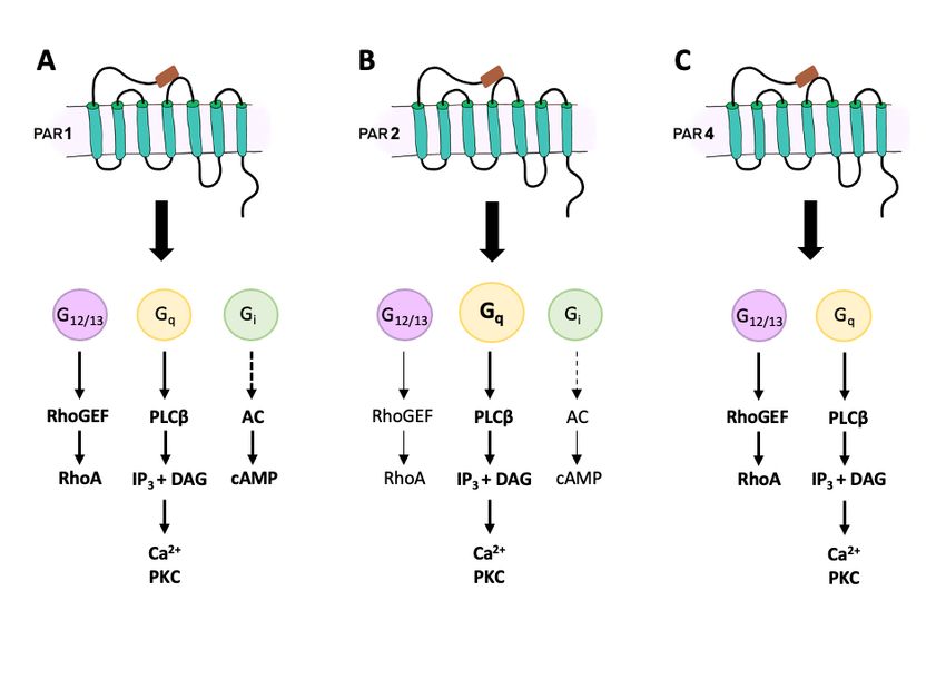
Int. J. Mol. Sci. 2021, 22, 9352                                                                                                  2 of 16

                                      evidence also indicates that proteolytic enzymes can also signal pain and inflammation
                                      in joints by cleaving a specific receptor family known as the protease activated receptors
                                      (PARs; Table 1). This group consists of four G-protein coupled receptors (PAR-1 to -4)
                                      that show a unique activation characteristic [11]. In the presence of a protease, a portion
                                      of the PAR extracellular N-terminus is cleaved which then exposes a new N-terminus
                                      sequence that acts as a tethered ligand which then binds to the second extracellular loop
                                      of the receptor [12]. This conformational change to the receptor leads to intracellular
                                      signalling that will vary depending on the receptor subtype, the cleaving protease, and
                                      the downstream pathway that becomes activated. In addition to receptor activation, some
                                      proteases can disarm or inactivate the PAR receptor by cleaving the N-terminus at an
                                      adjacent site [13]. More recently, a biased signalling of PARs has been extensively described,
                                      indicating the complexity of these receptors [14–16].
                                            PARs have been identified on the nerve terminals of nociceptors in multiple tissues
                                      suggesting that this receptor family is involved in pain control. Co-localization and phar-
                                      macological interaction between PAR-2 and pro-algesic transient receptor potential (TRP)
                                      channels in the pancreas [17], bladder [18], and oral mucosa [19] indicate that there is
                                      functional coupling between PARs and other known pain-modulating receptors. Proteases
                                      released from immunocytes, endothelial cells, or are part of the coagulation cascade are
                                      known to cleave neuronal PARs and modulate pain neurotransmission [12]. Other studies
                                      on the skin, gut, and airways have shown that PAR activation can lead to the secondary
                                      release or pro-algesic neuropeptides such as substance P and calcitonin gene-related pep-
                                      tides [20–22]. These multiple lines of evidence assert that PARs are an attractive target for
                                      the management of miscellaneous chronic pain conditions.

          Table 1. Known cleaving proteases and synthetic activating peptides for each of the PARs and their role in arthritis.

                                                             Synthetic Activating
      PAR           Activating Proteases                                                               Effect in Joints
                                                                  Peptides
                    Thrombin                                    SFLLRN-NH2                        Chondroprotection [23]
                    Granzyme A                                  TFLLRN-NH2                            Bone repair [24]
                    Plasmin                                                                        Pannus formation [25]
                    Activating protein C                                                Anti-allodynic via an opioid mechanism [26]
                    Trypsin
                    Factor Xa
     PAR-1
                    Kallikrein- 4, 5, 6, 14
                    MMP-1
                    Cathepsin G
                    Proatherocytin
                    Pen C 13
                    Chymase
                    Trypsin                                     SLIGRL-NH2                       Cartilage degeneration [27]
                                                                                            Synovial hyperaemia and increased
                    Mast cell tryptase                          FLIGRL-NH2
                                                                                                  leukocyte trafficking [28]
                                                                                         TRPV1 -dependent afferent sensitization and
                    Factor Xa: Factor VIIa
                                                                                                         pain [29,30]
     PAR-2
                    Acrosin
                    Matriptase
                    Serine 11D
                    Trypsin
                    Granzyme A
                    Kallikrein-2, 4, 5, 6, 14
     PAR-3          Thrombin                                        N/A                                     N/A
Int. J. Mol. Sci. 2021, 22, 9352 3 of 16
Table 1. Cont.
Synthetic Activating
PAR Activating Proteases Effect in Joints
Peptides
Thrombin AYPGKF-NH2 Joint damage [31]
Trypsin GYPGKF-NH2 Joint hyperaemia and oedema [32]
Afferent sensitization and pain via mast cell
Cathepsin G
degranulation and bradykinin activation [32]
PAR-4 Trypsin IV
Mannin-binding SP-1
Plasmin
Factor Xa
Kallikrein-1, 14
C4a
This review will outline the function of PARs and give an overview of the latest
studies, implicating their role in joint diseases including RA and OA.
2. PAR-1
2.1. Receptor Pharmacology
PAR-1 receptors are mainly activated by thrombin, but other mediators of the coagula-
tion cascade can also cleave it. PAR-1 is ubiquitously found in endothelial cells, platelets,
lungs, GI tract, immune cells, neurones and brain [11]. Following cleavage of PAR-1, there
is activation of the G protein subunits G12/13 , Gi , and Gq (Figure 1A) [33–36]. Signalling
via G12/13 leads stimulation of Rho guanine nucleotide exchange factor (RhoGEF), which
in turn activates GTPase RhoA and is involved in cytoskeleton reorganization. Activation
of Gi causes inhibition of adenylyl cyclase activity and hence a reduction in cyclic adeno-
sine monophosphate (cAMP) production. Finally, Gq activation initiates the intracellular
cascade starting with phospholipase-Cβ (CPLCβ) hydrolyzing phosphatidylinositol 4,5-
bisphosphate (PIP2) to produce inositol triphosphate (IP3 ) and diacylglycerol (DAG). This
leads to Ca2+ mobilization and an increase in the activity of protein kinase C (PKC) and
other intracellular signalling enzymes. Different subunits of PKC facilitate the variety of
downstream responses after PAR-1 activation [37–40].
A peculiar characteristic of PARs is that because its agonist is its own N-terminal
sequence, there is no dissociation of the agonist from the receptor, so desensitization and
termination mechanisms have to be tightly regulated. PAR-1 receptors are phosphorylated
by G-protein coupled receptor kinases (GRKs) 3 or 5 and desensitized by β-arrestin binding,
specifically β-arrestin 1, allowing the uncoupling of the G-proteins [12]. In contrast to other
GPCRs, the β-arrestin associated with PAR-1 participates only in desensitization of the
receptor, but not internalization [41]. Receptor internalization is primarily regulated by
clathrin and dynamin activity. Adaptor protein complex 2 (AP-2) is essential for PAR-1
internalization, while lysosomal sorting and receptor recycling are mediated by the R4
subfamily of regulators of G protein signalling (RGS), viz. Rab11A and Rab11B [42–45].
Curiously, there is also an agonist-dependant response where activated protein C (APC)
induces PAR-1 phosphorylation that does not result in effective internalization and the
receptor accumulates in the cellular membrane [46,47].regulated by clathrin and dynamin activity. Adaptor protein complex 2 (AP-2) is essential
for PAR-1 internalization, while lysosomal sorting and receptor recycling are mediated by
the R4 subfamily of regulators of G protein signalling (RGS), viz. Rab11A and Rab11B [42–
45]. Curiously, there is also an agonist-dependant response where activated protein C
Int. J. Mol. Sci. 2021, 22, 9352
(APC) induces PAR-1 phosphorylation that does not result in effective internalization4 of
and16
the receptor accumulates in the cellular membrane [46,47].
Figure 1. Downstream signalling pathways for PAR-1 (A), PAR-2 (B), and PAR-4 (C). Rho guanine nucleotide exchange
factor (RhoGEF); GTPase RhoA (RhoA); phospholipase-Cβ (CPLCβ); phosphatidylinositol 4,5-bisphosphate (PIP2); inositol
triphosphate (IP3 ); diacylglycerol (DAG); protein kinase C (PKC); cyclic adenosine monophosphate (cAMP).
Figure 1. Downstream signalling pathways for PAR-1 (A), PAR-2 (B), and PAR-4 (C). Rho guanine nucleotide exchange
factor (RhoGEF); GTPase RhoA (RhoA); phospholipase-Cβ (CPLCβ); phosphatidylinositol 4,5-bisphosphate (PIP2); inosi-
2.2. PAR-1 and Joints
tol triphosphate (IP3); diacylglycerol (DAG); protein kinase C (PKC); cyclic adenosine monophosphate (cAMP).
Within joints, PAR-1 has been located on fibroblasts, myoblasts, osteoblasts, chon-
drocytes,
2.2. PAR-1and
andsynoviocytes
Joints of RA and OA patients [48–50]. In joint cartilaginous tissue,
thrombin stimulates migration and proliferation of chondrocytes in a PAR-1-dependent
Within joints, PAR-1 has been located on fibroblasts, myoblasts, osteoblasts, chon-
manner [23]. Under normal circumstances, migration of chondrocytes can help with car-
drocytes, and synoviocytes of RA and OA patients [48–50]. In joint cartilaginous tissue,
tilage homeostasis and tissue healing; however, in joint diseases such as OA and RA,
thrombin stimulates migration and proliferation of chondrocytes in a PAR-1-dependent
thrombin activation of those cells can initiate cartilage degradation [51], contributing to
manner [23]. Under normal circumstances, migration of chondrocytes can help with car-
arthritis progression. Metalloproteinases, such as MMP13, are elevated in OA and RA joints
tilage homeostasis and tissue healing; however, in joint diseases such as OA and RA,
and their catabolic properties lead to degradation of type II collagen through a PAR-1- and
thrombin activation of those cells can initiate cartilage degradation [51], contributing to
PAR-3-dependent pathway [52]. The same increase in expression of MMPs is observed with
APC activity, but this seems to be through a mechanism other than PAR-1 activation [53].
In contrast, a protective role of PAR-1 in regulating osteoclast formation has also
been reported. Kanno and collaborators showed that PAR-1 engagement can initiate
a downstream response, culminating in phosphorylation of adenosine monophosphate
kinase (AMPK) and inhibition of NF-kB signalling, resulting in reduced osteoclastogenesis.
The authors showed an increase in urokinase plasminogen activating factor production
following LPS treatment, and it is believed that subsequent plasmin production activated
PAR-1 and reduced osteoclast formation [40]. These data were supported by another study
showing that osteoclastogenesis was enhanced in PAR-1 knockout mice treated with TNF-
alpha, indicating that PAR-1 acts as an inhibitor of osteoclast maturation in inflammatory
joint diseases [54]. In a model of tibial damage, bone repair was found to be inhibited in
PAR-1 knockout mice while thrombin improved bone marrow stromal cell proliferation in
a PAR-1-dependent manner [24,55]. Thrombin is also able to inhibit osteoblast apoptosis,
but in these cells this occurrence seems to be independent of PAR-1 activation [56].
In RA, synovial fibroblasts proliferate and form a hyperplastic and invasive tissue
called pannus. Through PAR-1 cleavage, thrombin upregulates the expression of RANTES
which then induces NF-kB and IL-6 expression, leading ultimately to synovial fibroblastInt. J. Mol. Sci. 2021, 22, 9352 5 of 16
differentiation and pannus formation [25,37,57,58]. In contrast, a protective role for PAR-1
in the joint has also been described. When treated with either thrombin, PAR-1-AP, or
PAR-3-AP, synovial fibroblasts taken from OA patients have been shown to release heme-
oxygenase-1 which is a protein with chondroprotective properties [38,59]. Thus, PAR-1
may be destructive or protective in the synovium depending upon whether the tissue is
taken from a joint that has RA or OA, respectively.
The role of PAR-1 in joints has also been tested pre-clinically using animal models
of arthritis. In the antigen-induced arthritis (AIA) model, it was observed that treatment
with the PAR-1 antagonist hirudin led to a reduction in joint diameter, pannus formation,
cartilage damage, fibrin deposition, and release of IL-1β [60]. Similar results were found
using the collagen-induced arthritis (CIA) model, where PEG-hirudin decreased all the
parameters cited above in addition to decreasing IL-12 expression and disease incidence
and severity [61]. Repeating the AIA model in PAR-1-knockout mice revealed that arthritic
animals had less cartilage and bone damage, diminished overall disease severity, reduced
IL-1β, IL-6 and MMP13 levels in the synovium, and consequently less synovitis [62]. Again,
it appears that PAR-1 contributes to the severity and pathophysiology of inflammatory
joint disease.
In other studies, a link between PAR-1 and psoriatic arthritis has been observed. A
transgenic mouse model that overexpresses kallikrein-related peptidase-6 (KLK6+ ) in ker-
atinocytes, psoriatic arthritis markers such as IL-17A, IL-23 and IL-6 were all elevated [63].
Moreover, overexpression of KLK6+ caused increased expression of genes and proteins
associated with psoriatic arthritis as well as pathological changes to the joint such as bone
damage, synovitis, and impaired mobility. The PAR-1 antagonist vorapaxar inhibited
the release of pro-inflammatory mediators from cultured keratinocytes, while PAR-1−/−
mice that overexpress KLK6+ did not show the same level of psoriatic arthritis [63]. These
results indicate that sustained activation of PAR-1 by KLK6 plays an important role in the
development of psoriatic arthritis.
With respect to pain, PAR-1 has divergent effects. The expression of PAR-1 in DRGs
and its co-expression with TRPV1 , SP and CGRP suggest a pro-nociceptive role for the
receptor [64]. Thrombin injected into the dorsal horn of the spinal cord promoted the release
of PGE2 , leading to a decrease in mechanical withdrawal threshold [65], and an increase
in glial cell expression [66]. Conversely, intraplantar injections of a synthetic PAR-1-AP
was able to increase the withdrawal threshold in naïve animals without causing neuronal
damage nor inflammation [67,68]. Following induction of inflammation with carrageenan,
local injection of a PAR-1-AP was able to improve the hindpaw withdrawal threshold
without affecting inflammation [67]. This analgesic effect of PAR-1 appears to involve the
endogenous opioid system since naloxone was able to block PAR-1 responses [26]. The
source of the opioids in inflamed tissues was found to be keratinocytes, as demonstrated
by a rise in proenkephalin production by these cells.
On balance, PAR-1 appears to be predominantly protective in inflammatory joint
disease but may be involved in degenerative activity in OA. Further studies are needed
to affirm the role of PAR-1 in different joint conditions as well as determining its possible
contribution to joint pain.
3. PAR-2
3.1. Receptor Pharmacology
PAR-2 is currently the most extensively characterized of all of the PARs. PAR-2 is
the only PAR that does not have thrombin as the main endogenous activator. Rather,
PAR-2 is predominantly cleaved by trypsin and tryptase, while other proteases such as
neutrophil elastase and proteinase-3 are also known PAR-2 activators [14,15]. PAR-2 is
highly expressed in epithelial cells of many systems such as lungs, liver, skin, blood vessels
and the GI tract [69–73]. Expression of PAR-2 was also seen in mast cells [74], endothelial
cells [75], different types of synoviocytes [76–79], joint sensory nerve fibres [29,30] and
dorsal root ganglia [80], showing the importance of this receptor in many systems.Int. J. Mol. Sci. 2021, 22, 9352 6 of 16
Similar to PAR-1, PAR-2 signalling involves Gq , G12/13 and Gi , although it is primarily
mediated by Gq stimulating the PLCβ-IP3 -DAG-PKC pathway (Figure 1B) [43,81]. In
some cells, activation of PAR-2 induces arachidonic acid release and fast production of
prostaglandins [73], suggesting the involvement of phospholipase A2 and cyclooxygenase-1.
Other cells show the involvement of mitogen-activated protein kinase (MAPK) and ERK1/2
pathways after stimulation of PAR-2, as well as a non-G-protein-dependent pathway medi-
ated by β-arrestin [43,82,83]. Biased signalling via the PLC-IP3 -DAG-PKC pathway leads
to Ca2+ mobilization from intracellular stores, while the MAPK pathway leads to signalling
that is Ca2+ -independent. The desensitization and termination of the PAR-2 signal is slightly
different from PAR-1. Instead of using GRKs, PAR-2 phosphorylation is accomplished by
PKC [81], which increases its affinity for clatharin, dynamin and β-arrestin 1 and 2, thereby
facilitating the uncoupling of the receptor and its eventual internalization [84–86]. The
internalization of PAR-2 involves β-arrestin with GTPases being important for endosome
formation (Rab5a) and intracellular trafficking (Rab11a) [35,87].
3.2. PAR-2 and Joints
PAR-2 is widely expressed in multiple different tissues in the joint and is therefore
likely to play a role in joint diseases such as OA and RA. In vitro studies showed that PAR-2
is expressed in osteoblasts and its activation induces calcium signalling and inhibition of
osteoclast differentiation [76,88]. Compared to wild type animals, PAR-2−/− mice have
a lower expression of osteoblasts and osteoclasts, suggesting that PAR-2 is involved in
the regulation of skeletal growth and bone repair [89]. Conversely, PAR-2 activation of
osteoblasts from OA donors led to an increase in MMP1, MMP9, IL-6 and RANKL levels,
indicating a bone resorptive effect in diseased joints [90].
Chondrocytes collected from OA patients show heightened levels of PAR-2 and expo-
sure of these cells to inflammatory cytokines increased the expression of the receptor [77,79].
In contrast, TGF-β caused a downregulation of PAR-2 expression in OA chondrocytes but
not in healthy cells [77,91], implying that various inflammatory mediators may differen-
tially regulate the expression of PAR-2 in chondrocytes during joint disease. Matriptase is
a serine protease that is rich in OA cartilage where it was shown to cause collagenolysis
through PAR-2 activation [27]. Huang and colleagues (2019) used an antagonist of PAR-2
in both in vitro and in vivo models to elucidate the activity of PAR-2 in chondrocytes
during OA as a possible treatment for the condition. They observed that AZ3451 inhibited
PAR-2 expression and many of its downstream signalling pathways in vitro, reducing the
expression of pro-inflammatory cytokines, collagen type II, and catabolic genes. In in vivo
experiments, the antagonist inhibited cartilage destruction, decreased MMP13 levels and
inhibited chondrocyte apoptosis [92]. Thus, PAR-2 may be a useful target to curtail cartilage
damage during OA progression.
In synovial fibroblasts from human RA samples, the presence of PAR-2 receptors was
detected, and stimulation of the receptor induced Ca2+ mobilization. Unlike chondrocytes,
exposure of the synoviocytes to IL-1β and TNF-α did not increase the expression of
PAR-2 in those cells [93]. In RA samples, mast cell tryptase activated PAR-2 on synovial
fibroblasts inhibiting apoptosis via a rho kinase mechanism [94]. Both mast cell tryptase
and a PAR-2-AP were able to induce synovial fibroblast proliferation in cells cultured
from RA and OA patients with RA samples showing the greatest effect [95]. Analysis
of venous blood samples from RA patients discovered that PAR-2 receptors were highly
expressed on monocytes and T-cells, and generation of IL-6 by monocytes after PAR-2
activation was more prominent in blood from RA patients. PAR-2 expression in blood
samples taken from OA patients revealed that the receptor levels were close to what was
seen in control samples [96]. However, in OA patients with concomitant synovitis, PAR-2
receptor expression was elevated in the synovium and on macrophages, lymphocytes, and
fibroblasts [97]. Measured levels of PAR-2 correlated with the severity of the synovitis,
suggesting that PAR-2 is relevant in joint disease where there is ongoing inflammation. In
other experiments, blocking PAR-2 reduced pannus invasion and proliferation, decreasedInt. J. Mol. Sci. 2021, 22, 9352 7 of 16
the release of IL-17, IL-1β, and TNF-α, and ameliorated signs of OA via regulation of the
MAPK/NF-kB pathway [50,98,99].
Direct injection of a PAR-2-AP into the joint was able to cause long lasting joint
swelling, hyperemia, and synovial vasodilation in wild type mice, but not in their lit-
termates lacking PAR-2 [100]. Degranulation of joint connective tissue mast cells with
compound 48/80 produced an inflammatory response in wild type mice but not PAR-2
knockout animals, highlighting that mast cells are involved in the PAR-2 inflammation
pathway [78]. Nociceptive behavior after an intraarticular injection of a PAR-2-AP was first
observed by Helyes et al. (2010), where they were able to show that SLIGRL-NH2 could
decrease paw withdrawal threshold and increase joint incapacitance [29]. This pro-algesic
effect of PAR-2 activation was TRPV1 -dependent since the selective TRPV1 antagonist
SB366791 blocked this response. Articular injection of the more potent PAR-2-AP FLIGRL-
NH2 induced spontaneous activity and increased firing of knee joint primary afferents
in rats that was blocked by both TRPV1 and NK1 receptor antagonists [30]. In addition
to a peripheral site of action for PAR-2, intra-thecal injection of FLIGRL-NH2 heightened
mechanonociception in naïve rats [101]. Furthermore, spinal administration of the PAR-2
antagonist GB83 reduced secondary allodynia in models of chronic arthritis, but not in
acute synovitis [101].
Cleaving serine proteases have also been shown to induce joint inflammation and pain
in a PAR-2-dependent manner. For example, local administration of neutrophil elastase
caused an increase in synovial blood perfusion, leukocyte trafficking, and nociceptive
behaviour in wild type mice but not PAR-2−/− [28]. Mast cell tryptase delivered into the
knee caused synovial hyperaemia, oedema, and pain behaviour, which was reduced by
chemical ablation of TRPV1 expressing neurones or in TRPV1 −/− animals [102]. Similarly,
another PAR-2 cleaving enzyme, matriptase, can increase synovial perfusion in wild type
mice but not PAR-2−/− [27]. In addition to activating proteases, some enzymes can silence
PAR activity by cleaving downstream of the canonical cleavage site. Calpain I, for example,
disarms PAR-2 in joints, leading to a reduction in pain behaviour [103].
Experiments investigating a role for PAR-2 in chronic inflammatory joint disease
found that in wild type mice, intra-articular injection of Freund’s complete adjuvant
produced inflammatory cellular infiltration, synovial hyperplasia, and cartilage damage;
however, none of these processes occurred in PAR-2−/− mice, confirming that PAR-2 plays
a role in joint inflammation [100]. PAR-2 has also been found to be involved in acute
joint inflammation models where either deletion of the PAR-2 gene or inhibition of the
receptor by RNA silencing reduced evidence of synovitis [100,104]. PAR-2−/− mice were
tested for their ability to develop RA in antigen-induced models and there was impaired
development of the disease, indicating that PAR-2 is involved in the adaptative immune
response to inflammatory joint disease [105,106].
PAR-2 has also been investigated in animal models of OA. Following joint destabiliza-
tion, OA gradually developed in wild type but not PAR-2−/− mice [107–110], indicating
that PAR-2 is also involved in OA development. More recently, using chemical induction of
OA, Muley and colleagues found that neutrophil elastase activity was increased in the joint
of rats after induction of OA [111]. Pharmacological inhibition of this serine protease with
alpha-antitrypsin or treatment with a PAR-2 antagonist blocked the initial inflammatory
response to neutrophil elastase and reduced pain and peripheral neuropathy [111]. In
a rodent model of chronic joint inflammation, alpha-antitrypsin reduced joint pain and
leukocyte trafficking and exerted a chondroprotective effect [112]. It is clear that PAR-2
participates in the development of both RA and OA; therefore, inhibition of this receptor
and its pathways presents a promising approach for the treatment of chronic joint diseases
and their symptoms.Int. J. Mol. Sci. 2021, 22, 9352 8 of 16
4. PAR-3
4.1. Receptor Pharmacology
The observation that platelets from PAR-1-knockout mice could still respond to throm-
bin led to the identification, cloning and characterization of PAR-3 [113,114]. A great
limitation to the study of PAR-3 is the fact that synthetic activating peptides based on the
receptor tethered ligand sequence do not activate PAR-3 [114]. It was later shown that
these so-called PAR-3-Aps actually bound to both PAR-1 and PAR-2 instead [115]. As such,
the signalling pathways of PAR-3 are still obscure, and it seems to play a more important
role as a cofactor for other PARs as a regulator of their activity through heterodimeriza-
tion [116–118].
4.2. PAR-3 and Joints
PAR-3 is co-expressed with PAR-1 on human chondrocytes but no functional activity
of PAR-3 in those cells has been observed [23]. In synovial fibroblasts from RA samples,
PAR-3 is co-expressed with PAR-1 and can cause a small Ca2+ influx itself, but not enough
to alter cell activity [58]. In contrast, thrombin and a PAR-3-AP can stimulate production of
heme oxygenase-1 in synovial fibroblasts from OA patients, and this effect can be inhibited
by siRNA blockade of PAR-3 [38]. However, since the PAR-3-AP is not selective and PAR-3
is co-expressed with PAR-1 in synovial fibroblasts, it is feasible that these responses are
actually a result of PAR-1 activation and PAR-3 is acting here as a cofactor to regulate PAR-1
activity. The main hindrance to PAR-3 biology has been the lack of selective PAR-3 agonists
and antagonists. However, the recent discovery of a PAR-3 lipid tethered binding peptide
(C660) may reveal a possible role for PAR-3 in arthritic conditions [119].
5. PAR-4
5.1. Receptor Pharmacology
PAR-4 was identified and cloned after the observation that platelets lacking PAR-1
and PAR-3 still responded to thrombin [120,121]. PAR-4 can be activated by thrombin,
cathepsin G, tissue kallikreins, trypsin, and coagulation factors, via both Gq and G12/13
signalling pathways (Figure 1C). G12/13 follows the RhoGEF, GTPase RhoA, and down-
stream signalling pathways in order to alter the conformation of platelets and initiate
aggregation. Engagement of the Gq second messenger system leads to the activation of
PLCβ, production of IP3 and DAG, leading to Ca2+ mobilization and enhancement of PKC
activity [122,123]. It has been shown that PAR-4 signalling and termination occurs at a
much slower pace than PAR-1. First PAR-1 is activated and promotes an acute Ca2+ increase
followed by a prolonged and sustained Ca2+ signal elicited by PAR-4 activation [124,125].
One of the reasons PAR-4 can sustain the signalling is based on the C-terminus being much
shorter than other PARs and it having less sites for phosphorylation than are necessary for
receptor desensitization and internalization [124]. Furthermore, PAR-4 internalization pro-
ceeds independently of β-arrestin but rather occurs via clatherin coating and endocytosis.
Nevertheless, regulators of G protein signalling (RGS) eventually recognize the receptor
and form complexes to terminate its signalling. Specifically, RGS2 interacts with Gq and
RGS4 interacts with G12/13 , inhibiting the downstream processes of these G proteins [126].
Upon internalization, PAR-4 is sorted between lysosomes for degradation or endosomes for
recycling [122]. Interestingly, the recycling of PAR-4 is more efficient when it forms a dimer
with PAR-2, resulting in a faster process of delivering PAR-4 to the cell membrane [127].
Dimerization with PAR-1 or PAR-3 can also influence PAR-4 activity by potentiating or
modulating its signalling properties [128,129].
5.2. PAR-4 and Joints
PAR-4 receptors are expressed throughout the knee joint, as seen from positive im-
munohistochemical staining in articular cartilage, subchondral bone, menisci, synovium,
mast cells, and chondrocytes. [31,32,130,131]. Intra-articular injection of the neuronal tracer
fluorogold revealed that PAR-4 is expressed on joint primary afferents, suggesting thatInt. J. Mol. Sci. 2021, 22, 9352 9 of 16
the receptor has the potential to modulate nociceptor firing. In a model of arthritis, the
level of PAR-4 in the joint positively correlated with monocyte infiltration and synovial
hypertrophy, indicating that PAR-4 plays a role in joint inflammation [97]. In contrast to
the anti-inflammatory and analgesic effect of PAR-4 activation in skin and the gut [132],
intraarticular injection of a PAR-4-AP caused a long-lasting increase in synovial blood flow,
oedema formation, and an increase in mechanical and thermal pain [32]. Recently, it has
been found that PAR-4 is involved in joint damage associated with articular bleeds. The
accumulation of blood in a joint, for example, as a result of trauma, can lead to arthropa-
thy, including synovitis and cartilage damage. Silencing of PAR-4 using siRNA reduced
synovitis scores and hyaline cartilage destruction in mice with an intra-articular bleed [31].
It was suggested that this pro-arthritic effect was in part mediated by increased plasmin
levels cleaving PAR-4 in the joint.
Electrophysiological recordings from joint primary afferents revealed an increase in
nerve firing after close intra-arterial injection of a PAR-4-AP confirming a pro-nociceptive
role for PAR-4 in joints. The inflammatory and nociceptive effects of PAR-4 activation could
be blocked by a PAR-4 antagonist or the bradykinin B2 receptor antagonist HOE140 [130].
Conversely, the TRPV1 antagonist SB366791 did not influence PAR-4 responses. Pre-
treatment of rats with a mast cell stabilizer also blocked mechanosensitization and pain
implicating a mast cell-kinin mechanism for PAR-4 activity [131]. In other studies, silencing
of PAR-4 with siRNA treatment reduced the severity of joint damage and plasmin activity
in a model of blood-induced arthropathy [31]. Together, these data imply that inhibition of
PAR-4 activity can be used to treat joint inflammation and pain.
6. Conclusions
The family of PARs are richly expressed throughout joints and are associated with
tissues associated with pain control, inflammation, and structural damage. On balance,
PARs appear to be promoters of heightened inflammation and pain, while their contribu-
tion to joint destruction may be disease-dependent and requires further exploration (for
summary, see Figure 2). Growing evidence seems to indicate that PAR-1 and PAR-2 can
cause innate and neurogenic inflammation in different arthritis models, while PAR-4 seems
to require platelet–leukocyte interactions, mast cell degranulation, and involvement of the
kallikrein-kinin systems. When it comes to pain transmission, PAR-1 imparts an analgesic
effect, whereas PAR-2 and PAR-4 support a pro-nociceptive outcome. The role of PAR-3
in joints is still unclear and we await better tools and techniques to unravel the function
of this receptor in arthritis pathogenesis and symptom development. The dual effects of
serine proteases to induce joint tissue catabolism and signal pain and inflammation make
them promising candidates to redress the destructive and painful features of arthritis.Int.
Int.J.J.Mol.
Mol.Sci. 2021,22,
Sci.2021, 22,9352
x FOR PEER REVIEW 10 of
11 of 16
17
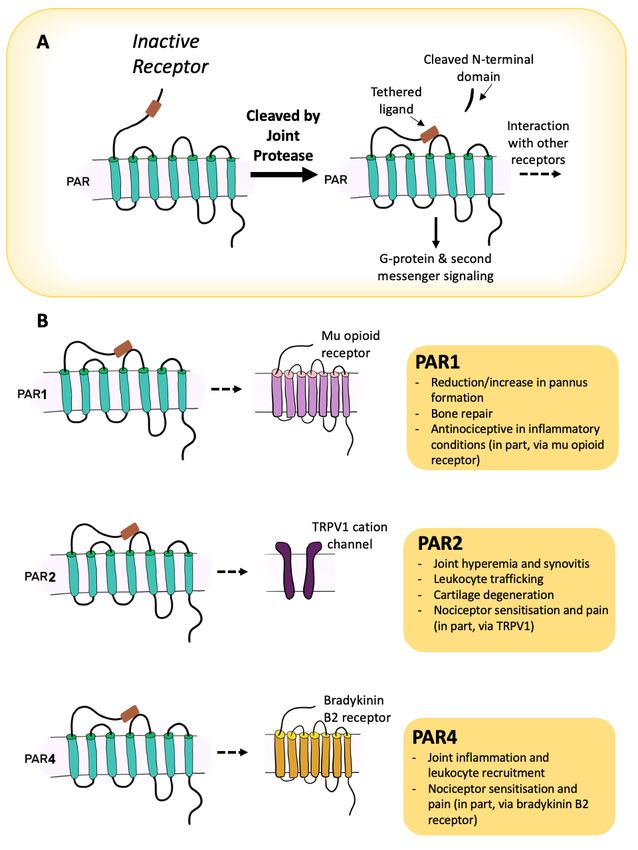
Figure2.2.Overview
Figure Overviewofofthe
therole
roleof
ofPARs
PARsininthe
thecontrol
controlofofjoint
jointinflammation
inflammationandandpain.
pain.Activation
Activation ofofaaPAR
PARisisachieved
achievedby
by
cleavage of the receptor with a proteolytic enzyme to reveal a tethered ligand that binds to the second extracellular
cleavage of the receptor with a proteolytic enzyme to reveal a tethered ligand that binds to the second extracellular loop, loop,
leadingto
leading toGGprotein
proteincoupling
couplingand
andsignalling
signalling(A).
(A).In
Injoints,
joints,PAR-2
PAR-2andand-4
-4tend
tendto
to be
be pro-inflammatory
pro-inflammatory and and pro-nociceptive,
pro-nociceptive,
whereas PAR-1 has some protective properties (B).
whereas PAR-1 has some protective properties (B).Int. J. Mol. Sci. 2021, 22, 9352 11 of 16
Author Contributions: F.L. wrote the original draft, edited, and reviewed the final version of the
manuscript. J.J.M. edited and reviewed the final version of the manuscript. Both authors have read
and agreed to the published version of the manuscript.
Funding: This research received no external funding.
Institutional Review Board Statement: Not applicable.
Informed Consent Statement: Not applicable.
Data Availability Statement: Not applicable.
Acknowledgments: Figures by M.S. O’Brien.
Conflicts of Interest: The authors declare no conflict of interest.
References
1. Woolf, A.; Pfleger, B. Burden of major muskuloskeletal conditions. Bull. World Health Organ. 2003, 81, 646–656.
2. van Delft, M.A.M.; Huizinga, T.W.J. An overview of autoantibodies in rheumatoid arthritis. J. Autoimmun. 2020, 110, 102392.
[CrossRef]
3. Grässel, S.; Zaucke, F.; Madry, H. Osteoarthritis: Novel Molecular Mechanisms Increase Our Understanding of the Disease
Pathology. J. Clin. Med. 2021, 10, 1938. [CrossRef]
4. Berenbaum, F. Osteoarthritis as an inflammatory disease (osteoarthritis is not osteoarthrosis!). Osteoarthr. Cartil. 2013, 21, 16–21.
[CrossRef] [PubMed]
5. Lee, D.M.; Weinblatt, M.E. Rheumatoid arthritis. Lancet 2001, 358, 903–911. [CrossRef]
6. Helmick, C.G.; Felson, D.T.; Lawrence, R.C.; Gabriel, S.; Hirsch, R.; Kwoh, C.K.; Liang, M.H.; Kremers, H.M.; Mayes, M.D.;
Merkel, P.A.; et al. Estimates of the prevalence of arthritis and other rheumatic conditions in the United States: Part I. Arthritis
Rheum. 2007, 58, 15–25. [CrossRef]
7. Gibofsky, A. Overview of epidemiology, pathophysiology, and diagnosis of rheumatoid arthritis. Am. J. Manag. Care 2012, 18,
S295–S302.
8. Singh, J.A.; Saag, K.G., Jr.; Akl, E.A.; Bannuru, R.R.; Sullivan, M.C.; Vaysbrot, E.; McNaughton, C.; Osani, M.; Shmerling, R.H.;
McAlindon, T.; et al. 2015 American College of Rheumatology Guideline for the Treatment of Rheumatoid Arthritis. Arthritis
Rheumatol. 2015, 68, 1–26. [CrossRef]
9. McAlindon, T.E.; Bannuru, R.R.; Sullivan, M.C.; Arden, N.K.; Berenbaum, F.; Bierma-Zeinstra, S.M.; Hawker, G.A.; Henrotin, Y.;
Hunter, D.J.; Kawaguchi, H.; et al. OARSI guidelines for the non-surgical management of knee osteoarthritis. Osteoarthr. Cartil.
2014, 22, 363–388. [CrossRef]
10. Krustev, E.; Rioux, D.; McDougall, J.J. Mechanisms and Mediators That Drive Arthritis Pain. Curr. Osteoporos. Rep. 2015, 13,
216–224. [CrossRef] [PubMed]
11. Hollenberg, M.D. Protease-mediated signalling: New paradigms for cell regulation and drug development. Trends Pharmacol. Sci.
1996, 17, 3–6. [CrossRef]
12. McDougall, J.J.; Muley, M.M. The Role of Proteases in Pain. In Pain Control; Schaible, H.-G., Ed.; Springer: Berlin/Heidelberg,
Germany, 2015; pp. 239–260. ISBN 978-3-662-46450-2.
13. Ramachandran, R.; Noorbakhsh, F.; DeFea, K.; Hollenberg, M.D. Targeting proteinase-activated receptors: Therapeutic potential
and challenges. Nat. Rev. Drug Discov. 2012, 11, 69–86. [CrossRef]
14. Zhao, P.; Metcalf, M.; Bunnett, N.W. Biased Signaling of Protease-Activated Receptors. Front. Endocrinol. 2014, 5, 67. [CrossRef]
15. Hollenberg, M.D.; Mihara, K.; Polley, D.; Suen, J.Y.; Han, A.; Fairlie, D.P.; Ramachandran, R. Biased signalling and proteinase-
activated receptors (PARs): Targeting inflammatory disease. Br. J. Pharmacol. 2013, 171, 1180–1194. [CrossRef] [PubMed]
16. Willis-Fox, O.; Preston, R.J.S. Molecular basis of protease-activated receptor 1 signaling diversity. J. Thromb. Haemost. 2019, 18,
6–16. [CrossRef]
17. Hoogerwerf, W.A.; Zou, L.; Shenoy, M.; Sun, D.; Micci, M.A.; Lee-Hellmich, H.; Xiao, S.Y.; Winston, J.H.; Pasricha, P.J. The
Proteinase-Activated Receptor 2 Is Involved in Nociception. J. Neurosci. 2001, 21, 9036–9042. [CrossRef]
18. Dattilio, A.; Vizzard, M.A. Up-regulation of protease activated receptors in bladder after cyclophosphamide induced cystitis and
colocalization with capsaicin receptor (vr1) in bladder nerve fibers. J. Urol. 2005, 173, 635–639. [CrossRef]
19. Ito, M.; Ono, K.; Hitomi, S.; Nodai, T.; Sago, T.; Yamaguchi, K.; Harano, N.; Gunjigake, K.; Hosokawa, R.; Kawamoto, T.; et al.
Prostanoid-dependent spontaneous pain and PAR2-dependent mechanical allodynia following oral mucosal trauma. Mol. Pain
2017, 13, 1–17. [CrossRef]
20. Vergnolle, N.; Bunnett, N.W.; Sharkey, K.; Brussee, V.; Compton, S.J.; Grady, E.F.; Cirino, G.; Gerard, N.; Basbaum, A.I.;
Andrade-Gordon, P.; et al. Proteinase-activated receptor-2 and hyperalgesia: A novel pain pathway. Nat. Med. 2001, 7, 821–826.
[CrossRef]
21. Grant, A.D.; Cottrell, G.S.; Amadesi, S.; Trevisani, M.; Nicoletti, P.; Materazzi, S.; Altier, C.; Cenac, N.; Zamponi, G.W.;
Bautista-Cruz, F.; et al. Protease-activated receptor 2 sensitizes the transient receptor potential vanilloid 4 ion channel to cause
mechanical hyperalgesia in mice. J. Physiol. 2007, 578, 715–733. [CrossRef] [PubMed]Int. J. Mol. Sci. 2021, 22, 9352 12 of 16
22. Obreja, O.; Rukwied, R.; Steinhoff, M.; Schmelz, M. Neurogenic components of trypsin- and thrombin-induced inflammation in
rat skin, in vivo. Exp. Dermatol. 2006, 15, 58–65. [CrossRef]
23. Kirilak, Y.; Pavlos, N.J.; Willers, C.R.; Han, R.; Feng, H.; Xu, J.; Asokananthan, N.; Stewart, G.A.; Henry, P.; Wood, D.; et al.
Fibrin sealant promotes migration and proliferation of human articular chondrocytes: Possible involvement of thrombin and
protease-activated receptors. Int. J. Mol. Med. 2006, 17, 551–558. [CrossRef]
24. Song, S.J.; Pagel, C.N.; Campbell, T.M.; Pike, R.N.; Mackie, E.J. The Role of Protease-Activated Receptor-1 in Bone Healing. Am. J.
Pathol. 2005, 166, 857–868. [CrossRef]
25. Hirano, F.; Kobayashi, A.; Hirano, Y.; Nomura, Y.; Fukawa, E.; Makino, I. Thrombin-induced expression of RANTES mRNA
through protease activated receptor-1 in human synovial fibroblasts. Ann. Rheum. Dis. 2002, 61, 834–837. [CrossRef]
26. Martin, L.; Augé, C.; Boué, J.; Buresi, M.C.; Chapman, K.; Asfaha, S.; Andrade-Gordon, P.; Steinhoff, M.; Cenac, N.;
Dietrich, G.; et al. Thrombin receptor: An endogenous inhibitor of inflammatory pain, activating opioid pathways. Pain 2009,
146, 121–129. [CrossRef]
27. Milner, J.M.; Patel, A.; Davidson, R.K.; Swingler, T.E.; Désilets, A.; Young, D.A.; Kelso, E.B.; Donell, S.T.; Cawston, T.E.;
Clark, I.M.; et al. Matriptase is a novel initiator of cartilage matrix degradation in osteoarthritis. Arthritis Rheum. 2010, 62,
1955–1966. [CrossRef] [PubMed]
28. Muley, M.M.; Reid, A.R.; Botz, B.; Bölcskei, K.; Helyes, Z.; McDougall, J.J. Neutrophil elastase induces inflammation and pain in
mouse knee joints via activation of proteinase-activated receptor-2. Br. J. Pharmacol. 2015, 173, 766–777. [CrossRef] [PubMed]
29. Helyes, Z.; Sándor, K.; Borbély, É.; Tékus, V.; Pintér, E.; Elekes, K.; Tóth, D.M.; Szolcsányi, J.; McDougall, J.J. Involvement of
transient receptor potential vanilloid 1 receptors in protease-activated receptor-2-induced joint inflammation and nociception.
Eur. J. Pain 2010, 14, 351–358. [CrossRef]
30. Russell, F.A.; Schuelert, N.; Veldhoen, V.E.; Hollenberg, M.D.; McDougall, J.J. Activation of PAR2receptors sensitizes primary
afferents and causes leukocyte rolling and adherence in the rat knee joint. Br. J. Pharmacol. 2012, 167, 1665–1678. [CrossRef]
[PubMed]
31. Nieuwenhuizen, L.; Schutgens, R.E.G.; Coeleveld, K.; Mastbergen, S.C.; Schiffelers, R.M.; Roosendaal, G.; Biesma, D.H.;
Lafeber, F.P.J.G. Silencing of protease-activated receptors attenuates synovitis and cartilage damage following a joint bleed in
haemophilic mice. Haemophilia 2015, 22, 152–159. [CrossRef]
32. McDougall, J.J.; Zhang, C.; Cellars, L.; Joubert, E.; Dixon, C.M.; Vergnolle, N. Triggering of proteinase-activated receptor 4 leads
to joint pain and inflammation in mice. Arthritis Rheum. 2009, 60, 728–737. [CrossRef] [PubMed]
33. Hung, D.T.; Wong, Y.H.; Vu, T.K.H.; Coughlin, S.R. The cloned platelet thrombin receptor couples to at least two distinct effectors
to stimulate phosphoinositide hydrolysis and inhibit adenylyl cyclase. J. Biol. Chem. 1992, 267, 20831–20834. [CrossRef]
34. Benka, M.L.; Lee, M.; Wang, G.-R.; Buckman, S.; Burlacu, A.; Cole, L.; DePina, A.; Dias, P.; Granger, A.; Grant, B.; et al. The
thrombin receptor in human platelets is coupled to a GTP binding protein of the Gαqfamily. FEBS Lett. 1995, 363, 49–52.
[CrossRef]
35. Déry, O.; Corvera, C.U.; Steinhoff, M.; Bunnett, N.W. Proteinase-activated receptors: Novel mechanisms of signaling by serine
proteases. Am. J. Physiol. Physiol. 1998, 274, C1429–C1452. [CrossRef]
36. Ossovskaya, V.S.; Bunnett, N.W. Protease-Activated Receptors: Contribution to Physiology and Disease. Physiol. Rev. 2004, 84,
579–621. [CrossRef]
37. Chiu, Y.-C.; Fong, Y.-C.; Lai, C.-H.; Hung, C.-H.; Hsu, H.-C.; Lee, T.-S.; Yang, R.-S.; Fu, W.-M.; Tang, C.-H. Thrombin-induced IL-6
production in human synovial fibroblasts is mediated by PAR1, phospholipase C, protein kinase Cα, c-Src, NF-kappaB and p300
pathway. Mol. Immunol. 2008, 45, 1587–1599. [CrossRef] [PubMed]
38. Liu, J.-F.; Hou, S.-M.; Tsai, C.-H.; Huang, C.-Y.; Yang, W.-H.; Tang, C.-H. Thrombin induces heme oxygenase-1 expression in
human synovial fibroblasts through protease-activated receptor signaling pathways. Arthritis Res. Ther. 2012, 14, R91. [CrossRef]
39. Tao, K.-M.; Tao, Y.; Chen, C.-Y.; Yang, L.-Q.; Lu, Z.-J.; Sun, Y.-M.; Huang, S.-D.; Yu, W.-F. Proteinase-activated Receptor 1
Contributed to Up-regulation of Enkephalin in Keratinocytes of Patients with Obstructive Jaundice. Anesthesiology 2014, 121,
127–139. [CrossRef]
40. Kanno, Y.; Ishisaki, A.; Kawashita, E.; Kuretake, H.; Ikeda, K.; Matsuo, O. uPA Attenuated LPS-induced Inflammatory Osteoclas-
togenesis through the Plasmin/PAR-1/Ca2+/CaMKK/AMPK Axis. Int. J. Biol. Sci. 2016, 12, 63–71. [CrossRef]
41. Paing, M.M.; Stutts, A.B.; Kohout, T.A.; Lefkowitz, R.J.; Trejo, J. β-Arrestins Regulate Protease-activated Receptor-1 Desensitization
but Not Internalization or Down-regulation. J. Biol. Chem. 2002, 277, 1292–1300. [CrossRef]
42. Paing, M.M.; Johnston, C.A.; Siderovski, D.P.; Trejo, J. Clathrin Adaptor AP2 Regulates Thrombin Receptor Constitutive
Internalization and Endothelial Cell Resensitization. Mol. Cell. Biol. 2006, 26, 3231–3242. [CrossRef]
43. Adams, M.N.; Ramachandran, R.; Yau, M.-K.; Suen, J.Y.; Fairlie, D.P.; Hollenberg, M.D.; Hooper, J.D. Structure, function and
pathophysiology of protease activated receptors. Pharmacol. Ther. 2011, 130, 248–282. [CrossRef] [PubMed]
44. Chen, B.; Siderovski, D.P.; Neubig, R.R.; Lawson, M.A.; Trejo, J. Regulation of Protease-activated Receptor 1 Signaling by the
Adaptor Protein Complex 2 and R4 Subfamily of Regulator of G Protein Signaling Proteins. J. Biol. Chem. 2014, 289, 1580–1591.
[CrossRef]
45. Grimsey, N.J.; Coronel, L.J.; Cordova, I.C.; Trejo, J. Recycling and Endosomal Sorting of Protease-activated Receptor-1 Is Distinctly
Regulated by Rab11A and Rab11B Proteins. J. Biol. Chem. 2016, 291, 2223–2236. [CrossRef] [PubMed]Int. J. Mol. Sci. 2021, 22, 9352 13 of 16
46. Schuepbach, R.A.; Feistritzer, C.; Brass, L.F.; Riewald, M. Activated protein C–cleaved protease activated receptor-1 is retained on
the endothelial cell surface even in the presence of thrombin. Blood 2008, 111, 2667–2673. [CrossRef]
47. Russo, A.; Soh, U.J.K.; Paing, M.M.; Arora, P.; Trejo, J. Caveolae are required for protease-selective signaling by protease-activated
receptor-1. Proc. Natl. Acad. Sci. USA 2009, 106, 6393–6397. [CrossRef] [PubMed]
48. Morris, R.; Winyard, P.G.; Brass, L.F.; Blake, D.R.; Morris, C.J. Thrombin receptor expression in rheumatoid and osteoarthritic
synovial tissue. Ann. Rheum. Dis. 1996, 55, 841–843. [CrossRef]
49. Shin, H.; Nakajima, T.; Kitajima, I.; Shigeta, K.; Abeyama, K.; Imamura, T.; Okano, T.; Kawahara, K.; Nakamura, T.; Maruyama, I.
Thrombin Receptor-Mediated Synovial Proliferation in Patients with Rheumatoid Arthritis. Clin. Immunol. Immunopathol. 1995,
76, 225–233. [CrossRef]
50. Xue, M.; Chan, Y.-K.A.; Shen, K.; Dervish, S.; March, L.; Sambrook, P.N.; Jackson, C.J. Protease-activated receptor 2, rather than
protease-activated receptor 1, contributes to the aggressive properties of synovial fibroblasts in rheumatoid arthritis. Arthritis
Rheum. 2011, 64, 88–98. [CrossRef]
51. Furmaniak-Kazmierczak, E.; Cooke, T.D.; Manuel, R.; Scudamore, A.; Hoogendorn, H.; Giles, A.R.; Nesheim, M. Studies of
thrombin-induced proteoglycan release in the degradation of human and bovine cartilage. J. Clin. Investig. 1994, 94, 472–480.
[CrossRef]
52. Huang, C.-Y.; Lin, H.-J.; Chen, H.-S.; Cheng, S.-Y.; Hsu, H.-C.; Tang, C.-H. Thrombin Promotes Matrix Metalloproteinase-13
Expression through the PKCδ/c-Src/EGFR/PI3K/Akt/AP-1 Signaling Pathway in Human Chondrocytes. Mediat. Inflamm. 2013,
2013, 326041. [CrossRef]
53. Jackson, M.T.; Moradi, B.; Smith, M.M.; Jackson, C.J.; Little, C.B. Activation of Matrix Metalloproteinases 2, 9, and 13 by Activated
Protein C in Human Osteoarthritic Cartilage Chondrocytes. Arthritis Rheumatol. 2014, 66, 1525–1536. [CrossRef]
54. Jastrzebski, S.; Kalinowski, J.; Mun, S.; Shin, B.; Adapala, N.S.; Jacome-Galarza, C.E.; Mirza, F.; Aguila, H.L.; Drissi, H.;
Sanjay, A.; et al. Protease-Activated Receptor 1 Deletion Causes Enhanced Osteoclastogenesis in Response to Inflammatory
Signals through a Notch2-Dependent Mechanism. J. Immunol. 2019, 203, 105–116. [CrossRef]
55. Song, S.J.; Pagel, C.N.; Pike, R.N.; Mackie, E.J. Studies on the receptors mediating responses of osteoblasts to thrombin. Int. J.
Biochem. Cell Biol. 2005, 37, 206–213. [CrossRef]
56. Pagel, C.N.; de Niese, M.R.; Abraham, L.A.; Chinni, C.; Song, S.-J.; Pike, R.N.; Mackie, E.J. Inhibition of osteoblast apoptosis by
thrombin. Bone 2003, 33, 733–743. [CrossRef]
57. Shin, H.; Kitajima, I.; Nakajima, T.; Shao, Q.; Tokioka, T.; Takasaki, I.; Hanyu, N.; Kubo, T.; Maruyama, I. Thrombin receptor
mediated signals induce expressions of interleukin 6 and granulocyte colony stimulating factor via NF-kappa B activation in
synovial fibroblasts. Ann. Rheum. Dis. 1999, 58, 55–60. [CrossRef] [PubMed]
58. Furuhashi, I.; Abe, K.; Sato, T.; Inoue, H. Thrombin-Stimulated Proliferation of Cultured Human Synovial Fibroblasts through
Proteolytic Activation of Proteinase-Activated Receptor-1. J. Pharmacol. Sci. 2008, 108, 104–111. [CrossRef] [PubMed]
59. Guillén, M.; Megías, J.; Gomar, F.; Alcaraz, M. Haem oxygenase-1 regulates catabolic and anabolic processes in osteoarthritic
chondrocytes. J. Pathol. 2007, 214, 515–522. [CrossRef]
60. Varisco, P.A.; Péclat, V.; Van Ness, K.; Bischof-Delaloye, A.; So, A.; Busso, N. Effect of thrombin inhibition on synovial inflammation
in antigen induced arthritis. Ann. Rheum. Dis. 2000, 59, 781–787. [CrossRef]
61. Marty, I.; Péclat, V.; Kirdaite, G.; Salvi, R.; So, A.; Busso, N. Amelioration of collagen-induced arthritis by thrombin inhibition. J.
Clin. Investig. 2001, 107, 631–640. [CrossRef] [PubMed]
62. Yang, Y.H.; Hall, P.; Little, C.B.; Fosang, A.J.; Milenkovski, G.; Santos, L.; Xue, J.; Tipping, P.; Morand, E.F. Reduction of arthritis
severity in protease-activated receptor-deficient mice. Arthritis Rheum. 2005, 52, 1325–1332. [CrossRef]
63. Billi, A.C.; Ludwig, J.E.; Fritz, Y.; Rozic, R.; Swindell, W.R.; Tsoi, L.C.; Gruszka, D.; Abdollahi-Roodsaz, S.; Xing, X.;
Diaconu, D.; et al. KLK6 expression in skin induces PAR1-mediated psoriasiform dermatitis and inflammatory joint disease. J.
Clin. Investig. 2020, 130, 3151–3157. [CrossRef]
64. Vellani, V.; Kinsey, A.M.; Prandini, M.; Hechtfischer, S.C.; Reeh, P.; Magherini, P.C.; Giacomoni, C.; McNaughton, P.A. Protease
Activated Receptors 1 and 4 Sensitize TRPV1 in Nociceptive Neurones. Mol. Pain 2010, 6, 61. [CrossRef] [PubMed]
65. Koetzner, L.; Gregory, J.A.; Yaksh, T.L. Intrathecal Protease-Activated Receptor Stimulation Produces Thermal Hyperal-gesia
through Spinal Cyclooxygenase Activity. J. Pharmacol. Exp. Ther. 2004, 311, 356–363. [CrossRef] [PubMed]
66. Smith, J.R.; Syre, P.P.; Oake, S.A.; Nicholson, K.J.; Weisshaar, C.L.; Cruz, K.; Bucki, R.; Baumann, B.C.; Janmey, P.A.;
Winkelstein, B.A. Salmon and Human Thrombin Differentially Regulate Radicular Pain, Glial-Induced Inflammation and Spinal
Neuronal Excitability through Protease-Activated Receptor-1. PLoS ONE 2013, 8, e80006. [CrossRef]
67. Asfaha, S.; Brussee, V.; Chapman, K.; Zochodne, D.W.; Vergnolle, N. Proteinase-activated receptor-1 agonists attenuate nociception
in response to noxious stimuli. Br. J. Pharmacol. 2002, 135, 1101–1106. [CrossRef]
68. Kawabata, A.; Kawao, N.; Kuroda, R.; Tanaka, A.; Shimada, C. The PAR-1-activating peptide attenuates carrageenan-induced
hyperalgesia in rats. Peptides 2002, 23, 1181–1183. [CrossRef]
69. Al-Ani, B.; Saifeddine, M.; Kawabata, A.; Renaux, B.; Mokashi, S.; Hollenberg, M.D. Proteinase-activated receptor 2 (PAR(2)):
Development of a ligand-binding assay correlating with activation of PAR(2) by PAR(1)- and PAR(2)-derived peptide ligands. J.
Pharmacol. Exp. Ther. 1999, 290, 753–760.Int. J. Mol. Sci. 2021, 22, 9352 14 of 16
70. Santulli, R.J.; Derian, C.K.; Darrow, A.L.; Tomko, K.A.; Eckardt, A.J.; Seiberg, M.; Scarborough, R.M.; Andrade-Gordon, P.
Evidence for the presence of a protease-activated receptor distinct from the thrombin receptor in human keratinocytes. Proc. Natl.
Acad. Sci. USA 1995, 92, 9151–9155. [CrossRef] [PubMed]
71. Hwa, J.J.; Ghibaudi, L.; Williams, P.; Chintala, M.; Zhang, R.; Chatterjee, M.; Sybertz, E. Evidence for the Presence of a Proteinase-
Activated Receptor Distinct From the Thrombin Receptor in Vascular Endothelial Cells. Circ. Res. 1996, 78, 581–588. [CrossRef]
[PubMed]
72. Saifeddine, M.; Al-Ani, B.; Cheng, C.-H.; Wang, L.; Hollenberg, M.D. Rat proteinase-activated receptor-2 (PAR-2): cDNA sequence
and activity of receptor-derived peptides in gastric and vascular tissue. Br. J. Pharmacol. 1996, 118, 521–530. [CrossRef]
73. Kong, W.; McConalogue, K.; Khitin, L.M.; Hollenberg, M.D.; Payan, D.G.; Böhm, S.K.; Bunnett, N.W. Luminal trypsin may
regulate enterocytes through proteinase-activated receptor 2. Proc. Natl. Acad. Sci. USA 1997, 94, 8884–8889. [CrossRef] [PubMed]
74. D’Andrea, M.R.; Rogahn, C.J.; Andrade-Gordon, P. Localization of protease-activated receptors-1 and -2 in human mast cells:
Indications for an amplified mast cell degranulation cascade. Biotech. Histochem. 2000, 75, 85–90. [CrossRef] [PubMed]
75. Nystedt, S.; Ramakrishnan, V.; Sundelin, J. The Proteinase-activated Receptor 2 Is Induced by Inflammatory Mediators in Human
Endothelial Cells: Comparison with the thrombin receptor. J. Biol. Chem. 1996, 271, 14910–14915. [CrossRef] [PubMed]
76. Abraham, L.A.; Chinni, C.; Jenkins, A.L.; Lourbakos, A.; Ally, N.; Pike, R.N.; Mackie, E.J. Expression of protease-activated
receptor-2 by osteoblasts. Bone 2000, 26, 7–14. [CrossRef]
77. Xiang, Y.; Masuko-Hongo, K.; Sekine, T.; Nakamura, H.; Yudoh, K.; Nishioka, K.; Kato, T. Expression of proteinase-activated
receptors (PAR)-2 in articular chondrocytes is modulated by IL-1β, TNF-α and TGF-β. Osteoarthr. Cartil. 2006, 14, 1163–1173.
[CrossRef]
78. Palmer, H.S.; Kelso, E.B.; Lockhart, J.C.; Sommerhoff, C.P.; Plevin, R.; Goh, F.G.; Ferrell, W.R. Protease-activated receptor 2
mediates the proinflammatory effects of synovial mast cells. Arthritis Rheum. 2007, 56, 3532–3540. [CrossRef]
79. Boileau, C.; Amiable, N.; Martel-Pelletier, J.; Fahmi, H.; Duval, N.; Pelletier, J.-P. Activation of proteinase-activated receptor 2 in
human osteoarthritic cartilage upregulates catabolic and proinflammatory pathways capable of inducing cartilage degradation:
A basic science study. Arthritis Res. Ther. 2007, 9, R121. [CrossRef]
80. Alier, K.A.; Endicott, J.A.; Stemkowski, P.L.; Cenac, N.; Cellars, L.; Chapman, K.; Andrade-Gordon, P.; Vergnolle, N.; Smith, P.A.
Intrathecal Administration of Proteinase-Activated Receptor-2 Agonists Produces Hyperalgesia by Exciting the Cell Bodies of
Primary Sensory Neurons. J. Pharmacol. Exp. Ther. 2007, 324, 224–233. [CrossRef]
81. Bohm, S.K.; Kong, W.; Bromme, D.; Smeekens, S.P.; Anderson, D.C.; Connolly, A.; Kahn, M.; Nelken, N.A.; Coughlin, S.R.;
Payan, D.G.; et al. Molecular cloning, expression and potential functions of the human proteinase-activated receptor-2. Biochem. J.
1996, 314 Pt 3, 1009–1016. [CrossRef]
82. Belham, C.M.; Tate, R.J.; Scott, P.H.; Pemberton, A.D.; Miller, H.R.P.; Wadsworth, R.M.; Gould, G.W.; Plevin, R. Trypsin stimulates
proteinase-activated receptor-2-dependent and -independent activation of mitogen-activated protein kinases. Biochem. J. 1996,
320, 939–946. [CrossRef] [PubMed]
83. DeFea, K.A.; Zalevsky, J.; Thoma, M.S.; Dery, O.; Mullins, R.D.; Bunnett, N.W. β-Arrestin–Dependent Endocytosis of Proteinase-
Activated Receptor 2 Is Required for Intracellular Targeting of Activated Erk1/2. J. Cell Biol. 2000, 148, 1267–1282. [CrossRef]
[PubMed]
84. Seatter, M.J.; Drummond, R.; Kanke, T.; Macfarlane, S.R.; Hollenberg, M.D.; Plevin, R. The role of the C-terminal tail in protease-
activated receptor-2-mediated Ca2+ signalling, proline-rich tyrosine kinase-2 activation, and mitogen-activated protein kinase
activity. Cell. Signal. 2003, 16, 21–29. [CrossRef]
85. Ricks, T.K.; Trejo, J.A. Phosphorylation of Protease-activated Receptor-2 Differentially Regulates Desensitization and Internaliza-
tion. J. Biol. Chem. 2009, 284, 34444–34457. [CrossRef]
86. Jung, S.-R.; Seo, J.B.; Deng, Y.; Asbury, C.L.; Hille, B.; Koh, D.-S. Contributions of protein kinases and β-arrestin to termination of
protease-activated receptor 2 signaling. J. Gen. Physiol. 2016, 147, 255–271. [CrossRef]
87. Roosterman, D.; Schmidlin, F.; Bunnett, N.W. Rab5a and rab11a mediate agonist-induced trafficking of protease-activated receptor
2. Am. J. Physiol. Physiol. 2003, 284, C1319–C1329. [CrossRef] [PubMed]
88. Smith, R.; Ransjö, M.; Tatarczuch, L.; Pagel, C.N.; Morrison, J.R.; Pike, R.N.; Mackie, E.J.; Song, S.-J. Activation of Protease-
Activated Receptor-2 Leads to Inhibition of Osteoclast Differentiation. J. Bone Miner. Res. 2003, 19, 507–516. [CrossRef]
89. Georgy, S.R.; Pagel, C.N.; Ghasem-Zadeh, A.; Zebaze, R.M.D.; Pike, R.N.; Sims, N.A.; Mackie, E.J. Proteinase-activated receptor-2
is required for normal osteoblast and osteoclast differentiation during skeletal growth and repair. Bone 2012, 50, 704–712.
[CrossRef] [PubMed]
90. Amiable, N.; Tat, S.K.; Lajeunesse, D.; Duval, N.; Pelletier, J.-P.; Martel-Pelletier, J.; Boileau, C. Proteinase-activated receptor
(PAR)-2 activation impacts bone resorptive properties of human osteoarthritic subchondral bone osteoblasts. Bone 2009, 44,
1143–1150. [CrossRef]
91. Tsai, S.-H.; Sheu, M.-T.; Liang, Y.-C.; Cheng, H.-T.; Fang, S.-S.; Chen, C.-H. TGF-β inhibits IL-1β-activated PAR-2 expression
through multiple pathways in human primary synovial cells. J. Biomed. Sci. 2009, 16, 97. [CrossRef]
92. Huang, X.; Ni, B.; Xi, Y.; Chu, X.; Zhang, R.; You, H. Protease-activated receptor 2 (PAR-2) antagonist AZ3451 as a novel
therapeutic agent for osteoarthritis. Aging 2019, 11, 12532–12545. [CrossRef]
93. Abe, K.; Aslam, A.; Walls, A.F.; Sato, T.; Inoue, H. Up-regulation of protease-activated receptor-2 by bFGF in cultured human
synovial fibroblasts. Life Sci. 2006, 79, 898–904. [CrossRef] [PubMed]You can also read