ACE2 PROTEIN-PROTEIN INTERACTION NETWORKS REVEAL POTENTIAL DRUGGABLE TARGETS FOR SARS-COV-2
←
→
Page content transcription
If your browser does not render page correctly, please read the page content below
ACE2 Protein-Protein Interaction Networks Reveal
Potential Druggable Targets For SARS-CoV-2
Qiaoxi Xia
Shantou Central Hospital
Xiao Zhou
Shantou Central Hospital
Mantong Chen
Shantou University Medical College
Ling Lin
Shantou University Medical College
Yan Zhao
Shantou Central Hospital
Xiaoqi Zheng
Shantou University Medical College
Zhongte Peng
Shantou University Medical College
Meihui Huang
Shantou Central Hospital
Shaohong Wang
Shantou Central Hospital
Zepeng Du ( zepdu@126.com )
Shantou Central Hospital https://orcid.org/0000-0002-9595-2439
Research
Keywords: ACE2, protein-protein interaction network, drug-protein network, druggable target
Posted Date: September 22nd, 2021
DOI: https://doi.org/10.21203/rs.3.rs-54096/v2
License: This work is licensed under a Creative Commons Attribution 4.0 International License.
Read Full License
Page 1/19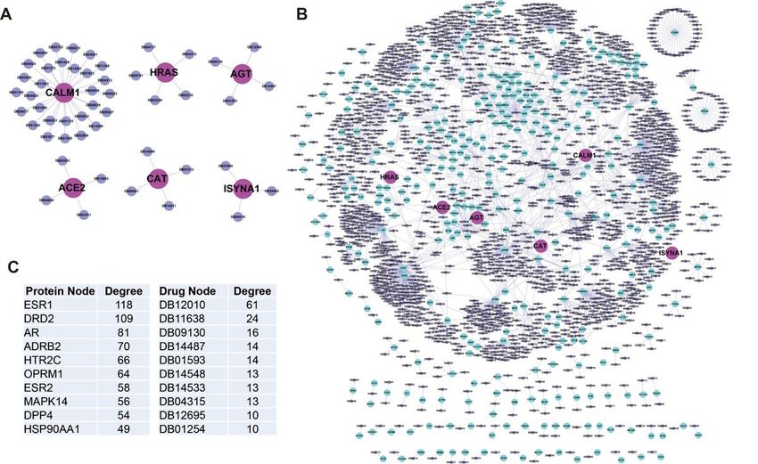
Abstract
Background: The novel coronavirus SARS-CoV-2 pandemic has infected more than 130 million people,
killed over 2.3 million so far. Currently, no effective drugs are available to treat this infectious disease, due
to limited knowledge of the molecular mechanisms of SARS-CoV-2 infection. ACE2 (angiotensin I
converting enzyme 2) has long been identified as the major receptor for coronavirus entry the host cells.

Methods: In this study, we constructed the protein-protein interaction networks (PPIN) based on ACE2 and
its interacting proteins, considering with the expression alternation and co-expression relationship. The
potential drugs targeting the proteins in the PPIN were explored.

Results: ACE2 and its interacting proteins AAMP and HRAS are obviously increased, and their PPIN show
distinguishing expression patterns during the COVID-19 progression. At least six pathways are activated
for the host cell in the response to the virus. Moreover, drug-target networks were built to provide
important clues to block ACE2 and its interacting proteins. Except the reported four drugs for ACE2, its
interacting protein CALM1 and HRAS are great potentially druggable. We also considered the path
initiated from ACE2 to nucleus by cascades of interaction, especially for the transcription factors in the
PPIN which are also druggable.

Conclusion: In summary, this study provides new insight into the disruption of the biological response to
virus mediated by ACE2, but also its cascade interacting proteins when considering of PPIN.

Introduction
The outbreak of global SARS-CoV-2 pandemic has profoundly threatened the health of billions of
individuals, overburdened local health care systems and millions of lives have lost by the end of May
2021 (https://coronavirus.jhu.edu/map.html). The most distinguishing clinic character of COVID-19
patients is a dysfunction of breath primarily due to acute respiratory distress syndrome, causing a greater
risk of mortality [1]. The first step of viral infections is the entry of the virus of the host cell, following the
replication multiple viral copies using the host cellular machinery. It has been widely acknowledged that
angiotensin-converting enzyme-2 (ACE2, EC 3.4.17.23) acts as a major receptor for COVID-19 to gain
intracellular entry into the host cells [2]. ACE2 is a membrane-bound protein, with a NH2-terminal domain
comprises the catalytic site oriented extracellularly [3]. ACE2 is normally expressed in many human
tissues, including lung, small intestine, heart, brain stem, nasal and oral mucosa [4].

The mechanism for COVID-19 causes lung dysfunction has not been fully proposed. ACE2 is considered
as a major receptor for coronavirus entry into the cells, which causes diffuse alveolar damage through
imbalances in the renin–angiotensin system. These immunologic reactions in severe COVID-19 may
characterize the cytokine storm phenomenon: a massive release of cytokines, such as interleukin (IL)-6,
IL-1, IL-8 and TNFα, from host cells and immune cells, such as monocytes and dendritic cells [5, 6]. These
inflammatory cells and factors will severe respiratory complications, like ARDS (Acute respiratory distress

                                                     Page 2/19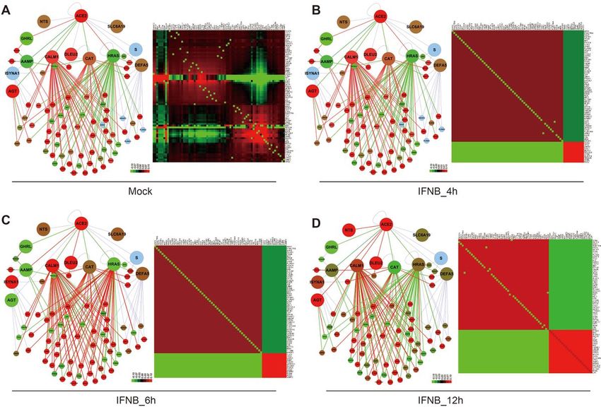
syndrome), characterizing acute diffuse alveolar damage, pulmonary oedema and formation of hyaline
membranes [7].

However, the detailed of molecular mechanism of how ACE2 is stimulated by virus and the progress of
phenotype in host cell remain unclear. Many proteins carry out multiple biological functions through the
interaction with other proteins, which maybe predicted by the interacting proteins. Analyses based on
protein-protein interaction networks (PPINs) have become a prevalent method for the high-throughput
data, especially the protein interaction data [8]. PPINs analysis can systematically integrate single or
multiple high-throughput data to obtain a global understanding of cellular functions or processes under
various conditions, e.g., different tumor sub-types, cancer progressive stages and cell sub-groups [9, 10].

In this study, the PPINs for ACE2 and its interacting proteins was constructed, and the expressed change
and expression pattern during the COVID-19 progression were integrated into the networks. Moreover, a
drug-target network was built to provide the clues to block the potential molecular path initiated from
ACE2.

Methods And Materials
Search of proteins that interacts with ACE2

The reported interacting proteins of ACE2, confirmed by previous publications, were obtained from two
databases of BioGRID (https://thebiogrid.org/) and MINT (https://mint.bio.uniroma2.it/). These proteins
were curated to obtain a unique interacting protein list.

Construction of the protein-protein interaction network (PPIN)

The latest known human protein-protein interaction data were obtained from several databases, including
BioGRID (http://thebiogrid.org/), HPRD (http://www.hprd.org/), MINT (https://mint.bio.uniroma2.it/), and
IntAct (http://www.ebi.ac.uk/intact/). These protein interaction data were integrated and obtained a
unique clean dataset after the remove of redundancy, which contained the up today human protein-
protein interactions. A total of 24,046 unique proteins and 438,656 interactions were contained in this
clean dataset, which was applied as the parental PPIN from that new or sub-PPINs were constructed by
Cytoscape [11]. Firstly, a sub-network was built by mapping ACE2 and its interacting proteins were
mapped to the parental PPIN as the seed proteins, to extract of their first class directly interacting
proteins. This subnetwork is named “ACE2 Full-PPIN”. Secondly, a smaller PPI sub-network was
illustrated, which was named “ACE2 Core-PPIN”, in which ACE2 and its interacting proteins are linked
through one or more partner proteins.

Topological parameters analyse for the PPIN

NetworkAnalyzer was applied to analyze the topological parameters, which would provide insight into the
complex network in terms of its organization and structure [12]. As one of most important property of
many large networks, the power law of distribution of node degrees was analyzed as we described
                                                  Page 3/19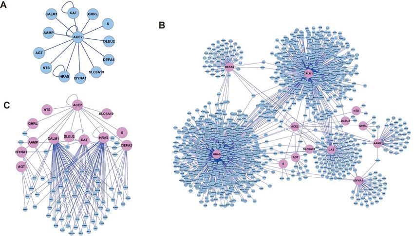
previously [13]. The node degree of a node n is defined as the number of nodes connected with n. When
considering the degree of all nodes in the network, we will have the equation of p(k)=Nk/N, with Nk
= number of nodes with degree k. Moreover, several other important topological parameters, such as
closeness centrality, shortest path length and topological coefficients, were analyzed for visualization.

Subcellular distribution of the PPIN

Subcellular locations for the proteins in the “ACE2 Full-PPIN” were downloaded from the HPRD database
and served as a node attribute in the network as we previously described [14]. If some proteins are
annotated by multiple locations, e.g., membrane and nucleus, were merged as membrane/nucleus. The
nodes in the “ACE2 Full-PPIN” were re-distributed into different layers by Cerebral plugin according to the
annotated subcellular localizations, generating a pathway-like graph remaining their physically
interactions [15].

Enrichment analyses for functions and pathways of network

Functional enrichment analysis of the “ACE2 Full-PPIN” was performed in DAVID database
(https://david.ncifcrf.gov/) to identify the enriched Gene Ontology (GO) “Biological Process” term. GO
terms with a statistically significant P-value less than 0.05 were remained for visualization using ggplot2
R package.

Expression pattern for the Core-network in COVID infected lung cells

GSE147507, an expression dataset containing SARS-CoV-2 infected normal lung cells, were obtained
from GEO database (https://www.ncbi.nlm.nih.gov/geo), which was submitted by Blanco-Melo et al. [16].
In this dataset, primary human lung epithelium (NHBE) was mock treated or infected with SARS-CoV-2 by
biological triplicates. To mimic the effect of inflammatory factor, NHBE cell was treated with human
interferon-beta (INFB) with the timepoint was set as 4h, 6h and 12h. The RNA-seq data was normalized
and the differentially expression were analyzed by SangerBox (http://soft.sangerbox.com/). The Pearson
expression correlation for each pair proteins in the “ACE2 Core-PPIN” were analyzed by an R script,
according to the expression data. The expression fold changes and correlations were integrated into the
“ACE2 Core-PPIN” as the node attribute, to illustrate the dynamic changes of the Core-PPIN under different
conditions. The correlation matrixs and their expression profile were clustered by Cluster 3.0 and
illustrated by TreeView [17].

Activated pathways analyses

The expression matrix for the proteins in the “ACE2 Core-PPIN” was submitted to the Pathview
(https://pathview.uncc.edu/) for visualization, which provides easy interactive access, and generates high
quality pathway graphs [18].

Drug-target network construction

                                                  Page 4/19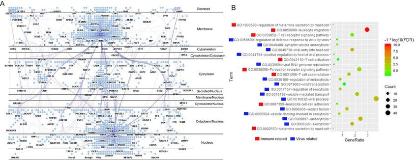
The latest release of DrugBank (version 5.1.6, https://www.drugbank.ca/) was downloaded, which
contains 13,580 drug entries, including FDA approved small molecule drugs, biologics, 131
nutraceuticals, 6,376 experimental drugs, as well as 5,223 non-redundant target proteins. An intersection
was obtained between the proteins in “ACE2 Full-PPIN” and target proteins from DrugBank, to construct a
drug-target network.

Shortest path from ACE2 to transcription factor

The shortest path problem is any possible but the minimized path(s) from one node to another given
node in the network. The proteins in the “ACE2 Full-PPIN” was compared the human transcription factors
(http://humantfs.ccbr.utoronto.ca/) to identify the transcription factors in this network. The path from
ACE2 to these transcription factors were analyzed by R “igraph” package as we previously described [19].
The visualization for the path from ACE2 to transcription factors, as well as their targeted drug were
performed by Cytoscape.

Results
Visualization of the PPIN for ACE2 and its interacting proteins

The currently known interacting proteins of ACE2 were obtained from the BioGRID and other databases.
At the beginning of this study, there are 12 reported ACE2 interacting proteins, with information shown in
Table 1 (Fig. 1A). A PPIN was built, using ACE2 and its interacting proteins as the seed nodes to extract
all their interacting proteins from the parental PPIN, which was called as the “ACE2 Full-PPIN” (Fig. 1B).
The “ACE2 Full-PPIN” contained 1,318 nodes and 1,292 edges, suggesting that ACE2 and its interacting
proteins can connect with thousands partner proteins by cascaded interactions to exceed the biological
effects of solo protein. Three top genes with the highest number of edges were HRAS (620 edges),
CALM1 (472 edges) and CAT (119 edges).

In the “ACE2 Full-PPIN”, we found multiple ACE2-interacting protein were connected by many common
partner proteins. To better illustrate their inner links, another smaller PPI sub-network, named as the “ACE2
Core-PPIN” was constructed by showing ACE2 and its interacting proteins linked by one or more partner
proteins , in which the nodes with only one interaction in the “ACE2 Core-PPIN” were removed (Fig. 1C).
This “ACE2 Core-PPIN” contained 80 nodes and 154 edges, in which ACE2 and its interacting proteins
were illustrated in light pink, while the partner proteins were indicated in light blue. Interestingly, it shown
HRAS and CALM1 have the highest number of shared nodes. This indicates that HRAS and CALM1 would
have the greatest potential to transduce the stimulus from ACE2 (Fig. 1C). Moreover, we consider these
common nodes between ACE2 and its interacting proteins might act as switch proteins, then determine
the direction of molecular signal paths by their expression level, and co-expression correlation strength as
well.

Topological structure of the “ACE2 Full-PPIN”

                                                    Page 5/19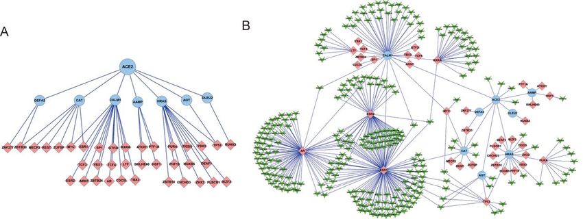
A real biological network, including PPIN, is distinguishable from any chaos or random networks by its
distinct structural properties. Many real, complex networks are characterized as “scale-free”, with a power
law degree distribution [20]. For “ACE2 Full-PPIN”, the distributions of node degree followed an
approximate power law, with the equation y = 288.18 x−1.07 and an R2 = 0.833 (Additional file 1A). The
shortest path length (the minimum number of links from one protein to another protein) of “ACE2 Full-
PPIN” was shown as mainly arranged from 2 and 3 step lengths (Additional file 1B). It also suggests that
one protein can contact another protein by only a few nodes, enabling the transformation of different
protein complexes or/and component maintenance. Topological coefficient is a measure for the extent to
which a node shares neighbors with other nodes. A topological coefficient of 0 is assigned to nodes
which have one or no neighbors (Additional file 1C). Closeness centrality measures how fast the flow of
information would be through a given node to other nodes in the network, considering the efficiency of
signal transduction in the network. The centrality curve is flat when the number of links less than 10, but
it gradually increases with more nodes have the links >10 (Additional file 1D).

Signal transduction from cell surface to nucleus through the PPIN

Evidence suggest that a protein might perform diverse functions depending on its cellular translocation,
except the interactions with other proteins [21, 22]. So, the subcellular location and/or translocation
become critical when considering its function, for the PPIN as well. After re-distribution, the “ACE2 Full-
PPIN” was divided into 11 layers and the percentage from extracellular to nucleus as follows: secreted
(2.43%), membrane (22.53%), cytoskeleton (0.15%), cytoskeleton/cytoplasm (0.23%), cytoplasm
(19.35%), secreted/nucleus (0.46%), membrane/nucleus (0.23%), cytoskeleton/nucleus (0.61%),
cytoplasm/nucleus (18.06%), nucleus (8.19%) and uncertain (27.77%, distribute near the interacting
proteins) (Fig. 2A). It shown that near 20% nodes in this PPIN have multiple subcellular locations. It also
strongly suggests that the ACE2 might transduct stimulus from the extracellular/membrane into the
cytoplasm, till to the nucleus, making up various non-classical pathways through cascades of
interactions.

Functional enrichment of the “ACE2 Full-PPIN”

As a tightly connected protein network, we presumed that ACE2 and its interacting proteins are involved in
various biological processes, especially in the pathology of COVID-19, through cascaded interactions to
amplify their biological effects. To test this speculation, GO “Biological Process” enrichment analysis
of “ACE2 Full-PPIN” was performed, acquiring more than a hundred of significantly enriched GO terms
(data not shown). Two big groups GO terms caused us a great interest (Fig. 2B). One group relates to
virus process, including “GO:0046718~viral entry into host cell”, “GO:0039694~viral RNA genome
replication”, “GO:0019083~viral transcription” and “GO:0016032~viral process”. The other interested
group is about immunity, includes “GO:0050900~leukocyte migration”, “GO:0031295~T cell
costimulation”, “GO:0050690~regulation of defense response to virus by virus” and “GO:0042110~T cell
activation”. These detailed significant GO terms with their enriched genes are listed in Additional file 2.

                                                   Page 6/19
Dynamic expression pattern for the Core-PPIN in COVID infected lung cells
In living cells, PPINs are not static, but dynamically change under different stimulus, or different stages of
disease. For this aim, the expression data of GSE147507 containing different conditions of lung cells was
obtained. Two important parameters for “ACE2 Core-PPIN” were analyzed, the expression log2(fold-
change) (logFC), and the co-expression correlation coefficient of each protein interaction. Then, the logFC
and correlation coefficient were imported into the network as a node attribute and an edge attribute,
respectively, to observe the dynamic changes in various treatments. In the Series1 of GSE147507, the
primary human lung epithelium (NHBE) was infected with SARS-CoV-2 (USA-WA1/2020) with mock
treated as a control (Fig. 3A). ACE2 is significantly increased, followed by its interacting protein AAMP
and HRAS. On the other hand, MYC in the network is also significantly upregulated (Fig. 3B). For
expression coefficient, there are near 70-80% of positive co-expression in mock NHBE, while it increases to
more than 90% positive in SARS-CoV-2 treated cells. The co-expression patters between normal and
treated are rather different, as indicated by the heatmap (Fig. 3C-D). In the Series9 of GSE147507, NHBE
were treated with human interferon-beta at the time point of 4h, 6h and 12h to mimic the stimulus of
inflammation. After the treatment, ACE2 and several interacting proteins, such as CALM1, DLEU2, ISYNA1
and NTS are obviously increased in the time series. While GHRL is consistently decrease. HRAS is
upregulated gradually from 4h to 12h, though not reach a very high expression level. For the co-
expression relationship, it is interesting to point out that both CALM1 and HRAS have positive correlation
with their interacting proteins in the control, but only CALM1 remains most positive correlation, while
HRAS mostly turns to negative correlation with its interacting proteins (Fig. 4A-D, left panel). For global
co-expression pattern as shown by the heatmaps, the total positive correlations are reduced while the
total negative correlations are increased (Fig. 4A-D, right panel). On the other hand, the expression pattern
of “ACE2 Core-PPIN” could distinguish the control group from the three treatment groups (Additional file
3).
Activated pathways in IFN stimulated lung cells
To exam what kinds of pathway are active during the mimic of COVID-19 stimulus, the expression matrix
of NHBE treated with IFN was analyzed by Pathview, which maps omics data to the KEGG pathway. There
are six significant enriched pathways, including “PI3K-Akt signaling pathway”, “Focal adhesion”, “ECM-
receptor interaction”, “Cell adhesion molecules”, “Antigen processing and presentation”, and “Regulation
of actin cytoskeleton” (Additional file 4). To our great interesting, the genes mapped to the pathways are
mostly increased, suggesting these pathways are activated during the mimic of COVID-19 inflammation
environment.
The proteins in Full-network are potential drug targets
Though many scientists and doctors are working hand to find ways to cure the COVID-19, including the
drugs targeting ACE2 itself, or the replication of SARS-CoV-2 virus. We consider it would be great helpful
to find drugs that target the ACE2 PPIN, to restrict the biological activity stimulated by the virus, thus
reduce the virus replication and spread of the SARS-CoV-2. To achieve this, we search the DrugBank
Page 7/19database and constructed drug-protein target networks. Currently, there are four registered drugs targeting
ACE2, there are DB01611 (Hydroxychloroquine), DB05203 (SPP1148), DB15643 (N-(2-Aminoethyl)-1-
aziridineethanamine), DB00608 (Chloroquine) (though some drugs are now in great controversy) (Figure
6A). On the other hand, five ACE2 interacting proteins (CALM1, HRAS, AGT, ISYNA1 and CAT) are reported
druggable. Among them, CALM1 (calmodulin 1) has the largest number of 29 drugs. The famous signal
molecular HRAS (HRas proto-oncogene, GTPase) has five tested drugs (Fig. 5A). By targeting the “ACE2
Full-PPIN”, there are 2075 nodes (1728 drugs and 347 proteins) and 2396 edges (targeting relations) in
the drug-protein network (Fig. 5B). At least 26% of proteins in “Full-PPIN” are druggable, suggesting a
great potential for treatment. The top 10 proteins have highest number of drugs, and the top 10 drugs
have the highest number of target proteins are shown in the list (Fig. 5C). ESR1 (estrogen receptor 1) has
the highest of 118 drugs, while DB12010 (Fostamatinib) targets the highest number (61) of proteins.
Some drugs have more than one targets. The detailed information about the drugs in these networks (Fig.
5) is provided in the Additional file 5.
The shortest paths from ACE2 to the downstream transcription factors
Usually an external stimulus, or the overexpression/knockdown of one gene, could cause a wide range
change in gene expression profile. We presumed the involved transcription factors play vital roles in the
alternation of gene expression profile. We applied the shortest path algorithm to illustrate how ACE2
reaches a specific transcription factor by the cascades of interaction in the PPIN. 37 transcription factors
are present in the “ACE2 Full-PPIN”. Consistent with the shortest path distribution described above, there
are only two steps from ACE2 to these transcription factors (Fig. 6A), suggesting a quick response is exist
from extracellular stimulus into nucleus, triggering the change of expression profile, then the change of
cellular activities. These transcription factors are also ideal targets for treatment. So we construct a small
PPIN, in which the drugs target the ACE2-TF. There 278 drugs in the small PPIN, targeting the 37
transcription factors (Fig. 6B). It is critical to understand why a wide range alternation of expression
profile is caused when a gene overexpression/knockdown. Shortest path algorithm was applied to
identify all possible paths from ACE2 to the transcription factor in the PPIN, which are most effective and
economical cellular information transduction.
The verification cohort from public data
During the preparation of this manuscript, Gordon et al. reported a comprehensive SARS-CoV-2 protein
interaction map [23]. They expressed 26 of the 29 SARS-CoV-2 proteins to identify their interacting human
proteins. They reported 332 high-confidence SARS-CoV-2-human protein-protein interactions, in which 66
druggable human proteins or host factors are targeted by 69 compounds[23]. To test the reliable of ACE2
based PPIN in this study, we compared the “ACE2 Full-PPIN” with the 332 SARS-CoV-2 interacting
proteins, and found 44 intersection proteins which were used to construct a co-network containing 510
nodes and 1264 edges (Additional file 6A). Among these nodes, at least six proteins (CALM1, HRAS,
DEFA5, CAT, S and ISYNA1,6/12 ACE2 interacting proteins) from “ACE2 Full-PPIN”, and fifteen SARS-CoV-
2 coding proteins are presented. After reduce the nodes with single connection, a core smaller PPIN was
Page 8/19obtained to show a clear relationship between ACE2 interacting proteins and SARS-CoV-2 coding proteins
(Additional file 6B). It also shows that HRAS and CALM1 are still the nodes with a large number of
interactions, suggesting the consistent between the networks in this study and networks from Gordon, D.
E. et al.
Discussion
COVID-19 is still a prevalent global pandemic that has already caused more than 3 million deaths
worldwide so far. Biological activities, includes stimulus response and signal transduction, are built on
protein physically interactions. In this study, several high confident PPINs were constructed based on the
ACE2 and its interacting proteins. Many proteins might link through partner proteins, which could act as
switch protein to determine the direction of signal transduction.
Many previous reported biological networks are static networks, which only present a specific state or one
time point. It has been widely aware that cellular system, including the PPIN, is highly flexible in the
response to environmental stimulus, which enables the cells adapt to different pathological stages or
physiological conditions [24]. To measure the co-expression of each pair of interacting proteins in
expression profiles, Pearson correlation coefficient is a popular correlation method [25]. In this study, we
not only constructed the static PPI network for ACE2 and its interacting proteins, but also the dynamic
networks by regarding their expression profiles in different kinds of conditions mimic COVID-19 progress.
Several proteins are consistent change during the progress, showing a dynamic co-expression pattern.
Since there is no single effective drug for COVID-19 by now, we consider whether there are multiple drugs
available for the ACE2 based PPIN to disturb the biological activities caused by the virus. Inspire by this
hypothesis, we found hundreds of drugs targeting ACE2 and its cascaded interacting proteins. With the
development of network pharmacology, the drug discovery paradigm has changed from the traditional
model “one drug → one target → one disease'" to the network mode “multi-drugs → multi-targets → multi-
diseases" [26, 27]. One successful application of drug cocktails is the clinical treatment of HIV/AIDS
patients[28]. The cocktail treatment strategy has been also applied in other diseases, such as breast
cancer and leukemia [29–31]. To overcome this combinatorial explosion problem, Zimmer et al. have
developed a model that predicted the effects of cocktails at all doses based on measuring pairs of drugs
[32]. So we believe that cocktails drug from the ACE2 drug-target network might provide important clues
to treat the virus in the clinic trial. Some of ACE2 interacting proteins are reported as drug targets in
different diseases. HRAS is a proto-oncogene which overexpresses in many tumors. Tipifarnib is a potent
and highly selective inhibitor of farnesyltransferase, a critical enzyme requisite for HRAS activation and
now a phase II trial in urothelial carcinoma is carried on [33]. CALM1 is one of kind of the EF-hand
calcium-binding protein, which modulates the function of ion channels by calmodulin regulation.
CBP501, a drug currently in phase II clinical trial for lung cancer, may sensitize tumors to the
chemotherapeutic agents bleomycin and cisplatin by inhibiting CALM1[34].
Page 9/19It usually causes a large scale of alternation in gene expression profile when the cells are stimulated. We
presumed the transcription factors or transcriptional regulators in the PPIN play crucial roles in this
expression profile change. We also speculated that the information transmitted from one protein to
another would also employ the most economical ways, through the cascaded of PPI interactions. So, we
identified there are only two steps for ACE2 to transcription factors, to cause the alternation of gene
expression profile to make the cell adapt to the stimulus. These transcription factors are considered as
drug targets. The transcription factor MYC, obviously upregulates both in the COVID-19 and IFN treated
lung cells, is a promising therapy target for multiple human cancers. Small molecules are found capable
of repression the transcriptional regulation of c-Myc, including IIA6B17, NY2267, and 28RH-NCN-1, which
inhibit MYC binds to DNA [35]. Based the above analyses, we propose a working model for ACE2 and its
interacting proteins, showing the cascaded interaction and their potential drugs for treatment (Fig. 7).
Conclusion
In summary, this study provides new insight into the disruption of the biological response to SARS-CoV-2
mediated by ACE2, but also its cascade interacting proteins when considering of PPIN.
Declarations
Funding
This work was supported by grants from the National Natural Science Foundation of China
(No.81672473, No.81502138), the Science and Technology Program of Guangdong
(No.2017A030313181).
Acknowledgement
We thank Dr. Bingli Wu for the research suggestions and proofreading the manuscript.
Authors' Contributions
Qiaoxi Xia and Mantong Chen completed the analysis, interpretation, and writing of the manuscript. Xiao
Zhou and Ling Lin performed the drawing of graphs in the manuscript. Yan Zhao, Xiaoqi Zheng, Meihui
Huang and Shaohong Wang carried out the data collection of the manuscript. Zepeng Du finished the
concepts, design and review of the manuscript.
Availability of data and materials
Public data from NCBI were applied in this article, which might involve human participants, and the
Informed content were available from the original papers.
Ethics approval and consent to participate
Page 10/19Not applicable.
Consent for publication
Not applicable
Conflicts of interest
All the authors declare no conflict of interest.
References
1. Guan WJ, Ni ZY, Hu Y, Liang WH, Ou CQ, He JX, Liu L, Shan H, Lei CL, Hui DSC, et al. Clinical
Characteristics of Coronavirus Disease 2019 in China. N Engl J Med. 2020;382(18):1708–20.
2. Li Y, Zhou W, Yang L, You R. Physiological and pathological regulation of ACE2, the SARS-CoV-2
receptor. Pharmacol Res. 2020;157:104833.
3. Chappell MC. Biochemical evaluation of the renin-angiotensin system: the good, bad, and absolute?
Am J Physiol Heart Circ Physiol. 2016;310(2):H137–52.
4. Zou X, Chen K, Zou J, Han P, Hao J, Han Z. Single-cell RNA-seq data analysis on the receptor ACE2
expression reveals the potential risk of different human organs vulnerable to 2019-nCoV infection.
Front Med. 2020;14(2):185–92.
5. Mahmudpour M, Roozbeh J, Keshavarz M, Farrokhi S, Nabipour I. COVID-19 cytokine storm: The
anger of inflammation. Cytokine. 2020;133:155151.
6. Meftahi GH, Jangravi Z, Sahraei H, Bahari Z. The possible pathophysiology mechanism of cytokine
storm in elderly adults with COVID-19 infection: the contribution of "inflame-aging". Inflamm Res
2020.
7. Pericas JM, Hernandez-Meneses M, Sheahan TP, Quintana E, Ambrosioni J, Sandoval E, Falces C,
Marcos MA, Tuset M, Vilella A, et al. COVID-19: from epidemiology to treatment. Eur Heart J.
2020;41(22):2092–112.
8. Lotfi Shahreza M, Ghadiri N, Mousavi SR, Varshosaz J, Green JR. A review of network-based
approaches to drug repositioning. Brief Bioinform. 2018;19(5):878–92.
9. Barabasi AL, Gulbahce N, Loscalzo J. Network medicine: a network-based approach to human
disease. Nat Rev Genet. 2011;12(1):56–68.
10. Zhang Q, Li J, Xue H, Kong L, Wang Y. Network-based methods for identifying critical pathways of
complex diseases: a survey. Mol Biosyst. 2016;12(4):1082–9.
11. Baryshnikova A. Exploratory Analysis of Biological Networks through Visualization, Clustering, and
Functional Annotation in Cytoscape. Cold Spring Harb Protoc 2016, 2016(6).
12. Doncheva NT, Assenov Y, Domingues FS, Albrecht M. Topological analysis and interactive
visualization of biological networks and protein structures. Nat Protoc. 2012;7(4):670–85.
Page 11/1913. Wu B, Li C, Du Z, Yao Q, Wu J, Feng L, Zhang P, Li S, Xu L, Li E. Network based analyses of gene
expression profile of LCN2 overexpression in esophageal squamous cell carcinoma. Sci Rep.
2014;4:5403.
14. Du Z, Wu B, Xia Q, Zhao Y, Lin L, Cai Z, Wang S, Li E, Xu L, Li Y, et al. LCN2-interacting proteins and
their expression patterns in brain tumors. Brain Res. 2019;1720:146304.
15. Barsky A, Gardy JL, Hancock RE, Munzner T. Cerebral: a Cytoscape plugin for layout of and
interaction with biological networks using subcellular localization annotation. Bioinformatics.
2007;23(8):1040–2.
16. Blanco-Melo D, Nilsson-Payant BE, Liu WC, Uhl S, Hoagland D, Moller R, Jordan TX, Oishi K, Panis M,
Sachs D, et al. Imbalanced Host Response to SARS-CoV-2 Drives Development of COVID-19. Cell.
2020;181(5):1036–45 e1039.
17. de Hoon MJ, Imoto S, Nolan J, Miyano S. Open source clustering software. Bioinformatics.
2004;20(9):1453–4.
18. Luo W, Pant G, Bhavnasi YK, Blanchard SG Jr, Brouwer C. Pathview Web: user friendly pathway
visualization and data integration. Nucleic Acids Res. 2017;45(W1):W501–8.
19. Du ZP, Wu BL, Wang SH, Shen JH, Lin XH, Zheng CP, Wu ZY, Qiu XY, Zhan XF, Xu LY, et al. Shortest
path analyses in the protein-protein interaction network of NGAL (neutrophil gelatinase-associated
lipocalin) overexpression in esophageal squamous cell carcinoma. Asian Pac J Cancer Prev.
2014;15(16):6899–904.
20. Santolini M, Barabasi AL. Predicting perturbation patterns from the topology of biological networks.
Proc Natl Acad Sci U S A. 2018;115(27):E6375–83.
21. Hung MC, Link W. Protein localization in disease and therapy. J Cell Sci. 2011;124(Pt 20):3381–92.
22. Wang X, Li S. Protein mislocalization: mechanisms, functions and clinical applications in cancer.
Biochim Biophys Acta. 2014;1846(1):13–25.
23. Gordon DE, Jang GM, Bouhaddou M, Xu J, Obernier K, White KM, O'Meara MJ, Rezelj VV, Guo JZ,
Swaney DL, et al. A SARS-CoV-2 protein interaction map reveals targets for drug repurposing. Nature.
2020;583(7816):459–68.
24. Chen B, Fan W, Liu J, Wu FX. Identifying protein complexes and functional modules–from static PPI
networks to dynamic PPI networks. Brief Bioinform. 2014;15(2):177–94.
25. Wang J, Peng X, Peng W, Wu FX. Dynamic protein interaction network construction and applications.
Proteomics. 2014;14(4–5):338–52.
26. Cheng F, Desai RJ, Handy DE, Wang R, Schneeweiss S, Barabasi AL, Loscalzo J. Network-based
approach to prediction and population-based validation of in silico drug repurposing. Nat Commun.
2018;9(1):2691.
27. Anighoro A, Bajorath J, Rastelli G. Polypharmacology: challenges and opportunities in drug
discovery. J Med Chem. 2014;57(19):7874–87.
Page 12/1928. Lu DY, Wu HY, Yarla NS, Xu B, Ding J, Lu TR. HAART in HIV/AIDS Treatments: Future Trends. Infect
Disord Drug Targets. 2018;18(1):15–22.
29. Marrocco I, Romaniello D, Yarden Y. Cancer Immunotherapy: The Dawn of Antibody Cocktails.
Methods Mol Biol. 2019;1904:11–51.
30. Lang T, Liu Y, Zheng Z, Ran W, Zhai Y, Yin Q, Zhang P, Li Y. Cocktail Strategy Based on Spatio-
Temporally Controlled Nano Device Improves Therapy of Breast Cancer. Adv Mater.
2019;31(5):e1806202.
31. Pollyea DA. New drugs for acute myeloid leukemia inspired by genomics and when to use them.
Hematology Am Soc Hematol Educ Program. 2018;2018(1):45–50.
32. Zimmer A, Tendler A, Katzir I, Mayo A, Alon U. Prediction of drug cocktail effects when the number of
measurements is limited. PLoS Biol. 2017;15(10):e2002518.
33. Lee HW, Sa JK, Gualberto A, Scholz C, Sung HH, Jeong BC, Choi HY, Kwon GY, Park SH. A phase II trial
of tipifarnib for patients with previously-treated, metastatic urothelial carcinoma harboring HRAS
mutations. Clin Cancer Res 2020.
34. Mine N, Yamamoto S, Saito N, Yamazaki S, Suda C, Ishigaki M, Kufe DW, Von Hoff DD, Kawabe T.
CBP501-calmodulin binding contributes to sensitizing tumor cells to cisplatin and bleomycin. Mol
Cancer Ther. 2011;10(10):1929–38.
35. Chen BJ, Wu YL, Tanaka Y, Zhang W. Small molecules targeting c-Myc oncogene: promising anti-
cancer therapeutics. Int J Biol Sci. 2014;10(10):1084–96.
Table
Table 1. ACE2 interacting proteins from database
Page 13/19Gene ID Official Full name Experimental Pubmed ID
Symbol System
801 CALM1 calmodulin 1 (phosphorylase Affinity 18070603
kinase, delta) Capture-
Western
183 AGT angiotensinogen (serpin Biochemical 10969042
peptidase inhibitor, clade A, Activity
member 8)
14 AAMP angio-associated, migratory cell Affinity 26186194
protein Capture-MS
847 CAT catalase Co- 26344197
fractionation
51477 ISYNA1 inositol-3-phosphate synthase 1 Co- 26344197
fractionation
8847 DLEU2 deleted in lymphocytic leukemia 2 Affinity 28977802
(non-protein coding) Capture-RNA
3265 HRAS Harvey rat sarcoma viral Proximity 30639242
oncogene homolog Label-MS
43740568 S Spike glycoprotein Co-crystal 32132184
Structure
340024 SLC6A19 solute carrier family 6 (neutral Co-crystal 32132184
amino acid transporter), member Structure
19
7431 VIM vimentin Affinity 26801988
Capture-
Western
1670 DEFA5 defensin, alpha 5, Paneth cell- Reconstituted DOI:10.1101
specific Complex
/2020.03.29.013490
51738 GHRL ghrelin/obestatin prepropeptide 11815627
Figures
Page 14/19Figure 1
PPINs for ACE2 and its interacting proteins. A. The reported ACE2 interacting proteins. B. The “ACE2 Full-
PPIN” is consist of ACE2 and its interacting proteins with all their public reported interacting proteins. C.
The “ACE2 Core-PPIN” was originated from the Full-PPIN by moving the nodes with only one link, to
illustrate ACE2 and its interacting proteins are connected with many partner proteins.
Figure 2
Page 15/19A. Subcellular layers of “ACE2 Full-PPIN”, which is separated into eleven layers while maintaining their
protein interactions. ACE22 and its interacting proteins are shown in pink color. B. Gene Ontology
“Biological Process” enrichment of the Full-PPIN shows significant virus activity and immune related
terms.
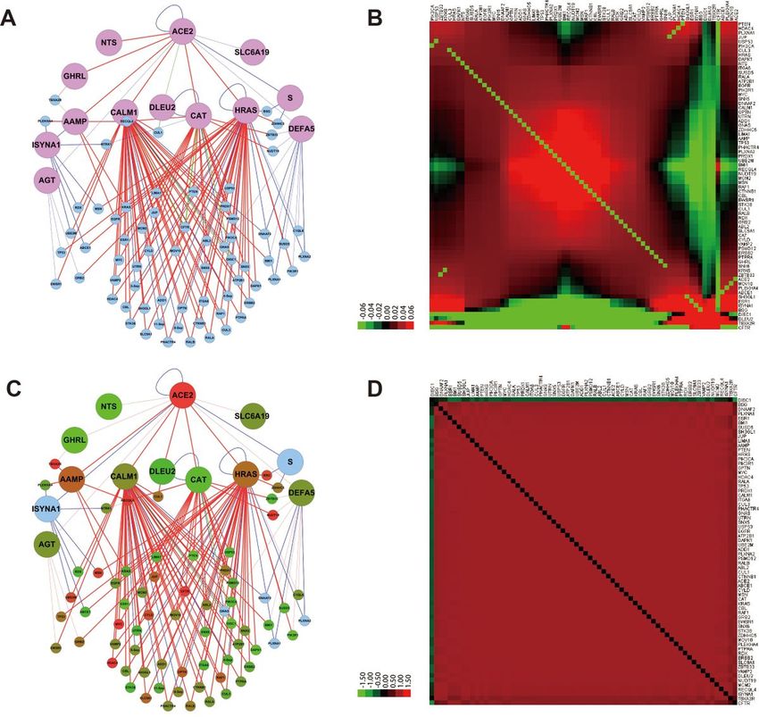
Figure 3
Expression fold-change and co-expression correlation of proteins in the “ACE2 Core-PPIN” for normal lung
and that treated with SARS-CoV-2. A. The co-expression relationship for “ACE2 Core-PPIN” in normal lung
cells. B. The heatmap of co-expression relationship for proteins in “ACE2 Core-PPIN” in normal lung cells.
Page 16/19C. The expression fold-change for the Core-PPIN in lung cells treated with SARS-CoV-2. D. The heatmap of
co-expression relationship for “ACE2 Core-PPIN” in lung cells treated with SARS-CoV-2.
Figure 4
Fold-change in expression and Pearson correlation matrix of proteins in the “ACE2 Core-PPIN” for normal
lung cells treated with INFN at different time points. (A-D, left panel)The expression fold-change for “ACE2
Core-PPIN” in normal lung cells with mock treatment or with INFN at 4h, 6h and 12h, with the mock as the
control. (A-D, right panel)The heatmap of co-expression relationship for “ACE2 Core-PPIN” in normal lung
cells.
Page 17/19Figure 5
Drug-protein network for ACE2 and its interacting proteins. A. Reported drugs for ACE2 and its interacting
proteins, respectively. B. Drugs target the proteins in “ACE2 Full-PPIN”. ACE2 and its interacting proteins
are indicated in light pink, their secondary interacting proteins are shown in light blue. Drugs are
illustrated in light purple. C. The top 10 proteins and drugs have the highest number of edges.
Figure 6
Page 18/19The shortest paths for ACE2 and transcription factors and their drugs. A. All the possible shortest paths
from ACE2 to transcription factors. The transcription factors are indicated in light pink. B. The drug
network targets the shortest paths from ACE2 to transcription factors, suggesting the possible disturbing
their signal transduction.
Figure 7
A propose model for drugs disrupt the biological activity caused by SARC-CoV-2 mediated by ACE2 and
its cascaded interacting proteins.
Supplementary Files
This is a list of supplementary files associated with this preprint. Click to download.
Additionalfile1.pdf
Additionalfile2.xlsx
Additionalfile3.pdf
Additionalfile4.pdf
Additionalfile5.csv
Additionalfile6.pdf
Page 19/19You can also read