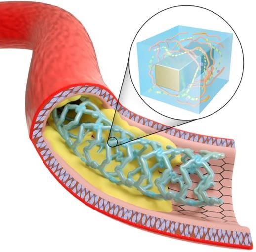
A tough endothelium-like dressing for vascular stents
←
→
Page content transcription
If your browser does not render page correctly, please read the page content below
A tough endothelium-like dressing for vascular stents Yin Chen Sun Yat-sen University https://orcid.org/0000-0002-8268-9029 Peng Gao Southwest Jiaotong University Lu Huang Sun Yat-sen University Xing Tan Southwest Jiaotong University Ningling Zhou Southwest Jiaotong University Tong Yang Southwest Jiaotong University Hua Qiu Southwest Jiaotong University Xin Dai Hong Kong University of Science and Technology Sean Michael Hong Kong University of Science and Technology Qiufen Tu Southwest Jiaotong University Nan Huang Southwest Jiaotong University Zhihong Guo Hong Kong University of Science and Technology Jianhua Zhou ( zhoujh33@mail.sysu.edu.cn ) Sun Yat-sen University Zhilu Yang Southwest Jiaotong University Hongkai Wu Hong Kong University of Science and Technology Article
Keywords: vascular stents, interventional cardiology, endothelium-like (EL) dressing Posted Date: April 23rd, 2021 DOI: https://doi.org/10.21203/rs.3.rs-431076/v1 License: This work is licensed under a Creative Commons Attribution 4.0 International License. Read Full License
A tough endothelium-like dressing for vascular stents

Yin Chen1,3†, Peng Gao2†, Lu Huang1,3, Xing Tan2, Ningling Zhou2, Tong Yang2, Hua Qiu2, Xin Dai3,
Sean Michael3, Qiufen Tu2, Nan Huang2, Zhihong Guo3, Jianhua Zhou1,4*, Zhilu Yang2* and Hongkai
Wu3*

1
 School of Biomedical Engineering, Sun Yat-sen University, Shenzhen 518107, China.
2
 Key Laboratory of Advanced Technologies of Materials, Ministry of Education, School of Materials
Science and Engineering, Southwest Jiaotong University, Chengdu 610031, China.
3
 Department of Chemistry, The Hong Kong University of Science and Technology, Hong Kong, China.
4
 Division of Engineering in Medicine, Brigham and Women’s Hospital, Harvard Medical School,
Cambridge, MA 02139, USA.
†
 These authors contributed equally to this work.
*e-mail: zhoujh33@mail.sysu.edu.cn; zhiluyang1029@swjtu.edu.cn; chhkwu@ust.hk

Vascular stent is viewed as one of the greatest advancements in interventional cardiology. However,

current approved stents suffer from in-stent restenosis associated with neointimal hyperplasia or stent

thrombosis. To address this issue, we developed an endothelium-like (EL) dressing for vascular stents

inspired by the importance and biological functions of native endothelium for cardiovascular system.

Our EL dressing is based on a de novo designed hydrogel that is mechanically tough and could preserve

integrity on stents during angioplasty. Due to its physiochemical similarities to subendothelial

extracellular matrix, the EL dressing facilitated the adhesion and growth of endothelial cells. Besides,

it is non-thrombotic and capable of inhibiting smooth muscle cells thanks to the capacity to catalyze

nitric oxide generation. Transcriptome analysis further unraveled the EL dressing could modulate the

inflammatory response and induce the relaxation of smooth muscle cells, while potentially promoting

angiogenesis by stimulating the expression of angiogenic factors. In vivo study demonstrated vascular

stents encapsulated by it promoted rapid restoration of native endothelium and persistently suppressed

in-stent restenosis in both leporine and swine models. We expect such EL dressing will open a new

avenue to the surface engineering of vascular implants for better clinical outcomes.

 1
Vascular stent, which is implanted into a narrowed blood vessel through guided balloon dilation, is

regarded as the most effective means for treating coronary artery disease1. Since its introduction in

1980s, vascular stent has been widely employed in interventional cardiology. Compared to the earlier

plain balloon angioplasty, the use of first-generation bare-metal stents (BMSs) has already presented

remarkable benefits in terms of less acute vessel closure and constrictive remodeling2. Despite these

advantages, the drawbacks of BMSs were soon reported, including acute inflammation elicited by

foreign-body reaction and in-stent restenosis (ISR) induced by neointimal hyperplasia (NIH)3. As an

alternative, drug-eluting stents (DESs) with a polymer coating carrying anti-cell proliferative drugs

were developed and became the standard of care in percutaneous coronary intervention (PCI)4.

Although DES has successfully alleviated inflammation and dramatically reduced the rate of early ISR,

the released drugs also suppress endothelial cells, thereby increasing the risk of late NIH and stent

thrombosis due to impaired endothelialization5.

 To address these complications associated with vascular stent, it is advisable to learn from nature.

The inner lining of blood vessel is a monolayer of tightly connected endothelial cells called as

endothelium6. Native endothelium is covered by a highly hydrated layer of glycocalyx that can

lubricate it and reduce its interaction with blood components7. In addition, it generates versatile

biomolecules such as nitric oxide (NO), prostacyclin, thrombomodulin, heparin-like molecules, tissue

factor pathway inhibitors and tissue plasminogen activators8. These molecules play important roles in

normal endothelial function, including prevention of thrombosis, regulation of vasomotion, promotion

of endothelial regeneration, and modulation of inflammatory response8. As a result, native endothelium

is the best antithrombotic material in nature, which maintains the patency of blood vessel.

 With the knowledge in native endothelium, we envisaged an endothelium-mimetic coating might

solve the issue of ISR for vascular stents. Such coating should be capable of preventing thrombosis,

inhibiting smooth muscles, and providing a microniche favored by endothelial cells so that native

endothelium could rapidly form to replace it. To achieve this goal, hydrogels seem to be the best

 2
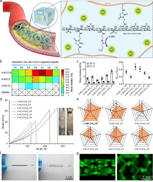
candidate compared to direct surface engineering, polymeric coatings and ceramic films because of

several reasons. First, they are three-dimensionally (3D) crosslinked aqueous materials with the best

resemblance to native tissues9,10. Second, they allow for versatile physical and chemical modifications

for special purposes. Last but not least, as bulk materials, they can be tailored as highly efficient

carriers for therapeutics. In fact, many hydrogels have been exploited as carriers of drugs11,12,

biomolecules13,14 or cells15-17 for various applications. However, developing a hydrogel coating for

vascular stents is challenging because it must be biocompatible, endothelial cell-adhesive, convenient

for handling and mechanically strong to withstand balloon dilation during angioplasty. Unfortunately,

few of the hydrogels reported so far met all these requirements.

 Herein, we developed an endothelium-like (EL) dressing for vascular stents using a de novo

designed hydrogel. This hydrogel is primarily composed of alginate and gelatin, which are analogs to

hyaluronic acid and collagen in extracellular matrix (ECM)18. Such combination enables it to resemble

the subendothelial ECM that is favorable for endothelial cells. By tuning the proportions of these two

biopolymers and the interaction between them, it can become mechanically tough. We endowed

endothelial function to it by conjugating an organoselenium species to alginate, which is capable of

persistently catalyzing the generation of NO that participates in nearly all important biological

processes of native endothelium19. We call this hydrogel-based coating an EL dressing because of its

high resemblance to native endothelium. Such EL dressing was expected to withstand the balloon

dilation during angioplasty, prevent thrombosis, promote rapid restoration of native endothelium, and

effectively suppress ISR.

Design, synthesis and optimization of the hydrogel

To generate a uniform hydrogel coating on vascular stents, a good strategy is implementing in situ

gelation. One possible approach to achieving this is dip-coating the stents with the hydrogel precursor

solution that cures subsequently. At first glance, it seems the sol-gel transition of gelatin in the solution

 3
can be utilized for that purpose, which liquefies when the temperature is above the melting point (Tm)

of gelatin and solidifies after cooling. However, the Tm of gelatin (~30 °C) is much lower than that of

our body temperature (37 °C)20, which suggests chemical crosslinking needs to be introduced in the

hydrogel. The reaction of such chemical crosslinking must be cytocompatible and mild. Considering

cytocompatibility, Michael-addition reaction is one of the best crosslinking methods as it produces no

byproducts21. However, conventional thiol-maleimide addition proceeds too fast at physiological

conditions22, which is inconvenient for our application. In our previous work, we reported Michael-

addition reaction between maleimide and amine could be leveraged for crosslinking a hydrogel23,24.

Compared to thiol-maleimide addition, the maleimide-amine addition is milder, allowing for more

maneuverability during the preparation of hydrogel. Based on our findings, we envisioned that a hybrid

hydrogel formed by the crosslinking between maleimide-modified alginate (alginate maleimide, A-M

for short) and gelatin could be tailored as the EL dressing for vascular stents (Fig. 1a).

 4
a
 Gelatin

 NO
 NO
 NO

 NO

 Alginate maleimide
 GSNO

 NO

 NO
 NO
 NO
 NO

 b Decrease in the ratio of A-M or alginate to gelatin c 15 0.6

 Shear modulus (kPa)
 25 °C
 37 °C

 G37 °C/G25 °C
 7/3 6/4 5/5 4/6 3/7 2/8 1/9
 2000
 2000 10 0.4
 A-M(15.9) 1800
 1800
 Shear modulus (Pa)
 1600
 1600

 1400
 1400 5 0.2
 A-M(9.3) 1200
 1200

 1000
 1000
 6 800 800
 0 0.0
 A-M(4.8) /6

 /7

 /5

 6

 7

 /8

 A- 9.3 4/6

 A- 5.9 3/7

 A- 5.9 5/5

 A- 5.9 4/6

 7

 /8
 4/

 3/

 3/
 _4

 3

 5

 _2

 _2
 600 600
 _

 (1 G_

 (1 G_

 (1 G_

 _

 _

 (1 /G_

 (1 /G_

 (1 /G_
 G

 /G

 /G

 G
 )/G

 /G
 A- .3)/

 /

 )/

 /

 A- .3)/
 400 400
 A- 9.3)

 A- 5.9)

 9)

 9)

 )

 )

 )
 9)
 6 6 6 6 6 6 6
 .9

 .

 5.

 5.
 Alginate
 (9

 5

 5

 (9
 200 200
 (

 (1

 (
 (1
 M

 M

 M

 M
 M

 M

 M

 M

 M

 M

 M

 M
 A-

 A-
 A-

 A-
 0 0
 d 80 e 10
 A
 10 10
 A-M(9.3)/G_4/6 8 8 8
 A-M(9.3)/G_3/7 6 6 6
 4 4 4
 A-M(15.9)/G_5/5
 2 2 2
 60 A-M(15.9)/G_4/6 0 0 0
 A-M(15.9)/G_3/7
 Stress (kPa)

 A-M(15.9)/G_2/8

 40
 A-M(9.3)/G_4/6 A-M(9.3)/G_3/7 A-M(15.9)/G_5/5
 10 10 10
 8 8 8
 6 6 6
 20 4 4 4
 2 2 2
 0 0 0

 0
 0 100 200 300
 Strain /% A-M(15.9)/G_4/6 A-M(15.9)/G_3/7 A-M(15.9)/G_2/8

 f g

 1 cm 1 mm

Fig. 1 | Development of a tough EL dressing for vascular stents using a de novo designed hydrogel.
a, Schematic for the design of our EL dressing. b, Shear moduli (at 37 °C) of the hydrogels formulated
with A-M of varying degrees of maleimidyl modification (DMM) and gelatin at different mass ratios.
c, Comparison in shear modulus among the selected hydrogels at 25 °C and 37 °C (n = 6). d, Tensile
testing of them at ambient temperature. The insets on the right exhibit the photographs of A-
M(9.3)/G_4/6 hydrogel prior to and during extension. (Scale bar: 1 cm) e, Radar charts showing their
scores in shear modulus (at 37 °C) (A), fracture strength (B), fracture strain (C), toughness (D), and
capacity for further modification (E). Dashed frameworks represent the average values. f, Photographs
of a vascular stent coated with A-M(9.3)/G_4/6 hydrogel before (left) and after (right) balloon dilation
in PBS at 37 °C. g, Fluorescence images of it before (left) and after (right) balloon dilation. The
hydrogel coating was labeled with FITC.

 5
A-M was synthesized by the coupling reaction between pristine alginate and N-(2-

aminoethyl)maleimide trifluoroacetate (AEM.TFA) via aqueous carbodiimide chemistry

(Supplementary Fig. 1). By tuning the feed ratio between alginate and AEM.TFA, A-Ms with varying

degrees of maleimidyl modification (DMM) were obtained. In our nomenclature, A-M(x) indicates

that x% structural units in alginate are coupled by maleimide. In total, three variants of A-M were

prepared, including A-M(4.8), A-M(9.3), and A-M(15.9), as characterized by proton nuclear magnetic

resonance (1H-NMR, Supplementary Fig. 2). In a preliminary trial to explore the gelation process, we

mixed the precursor solutions (10 w/v%, pH~7.5) of A-M(9.3) and gelatin at different mass ratios and

cured them at 37 °C. The cure kinetics of them were investigated by measuring the changes of their

shear moduli at 37 °C after cure for different durations. As shown in Supplementary Fig. 3, although

these hydrogels with varying formulations had different stiffness, all of them exhibited similar cure

kinetics and reached full mechanical strength within 36 h. Among them, A-M(9.3)/G_4/6 was the

stiffest hydrogel with a shear modulus of 732 Pa at 37 °C after cure for 72 h. However, we considered

even this hydrogel was a not strong enough be applied as a coating material. To further enhance their

strength, we increased the mass concentration of the precursor solutions. In the beginning, we tried to

prepare A-M solutions of 20 w/v%, but we failed because A-Ms were not fully soluble in water at such

high concentration. As a tradeoff, we ended up with using precursor solutions of 15 w/v%. At this mass

concentration, both alginate, A-Ms and gelatin can be readily manipulated. At this time, pristine

alginate and all three variants of A-M were tested together and a prolonged gelation time (72 h) was

assumed to ensure all hydrogels were fully cured.

 As anticipated, pure gelatin was unable to maintain solid state and pristine alginate could not from

a hybrid hydrogel with it at 37 °C (Fig. 1b). Nevertheless, with the use of A-M, hybrid hydrogels were

successfully generated at 37 °C. Notably, gelation only occurred when the proportions of A-M and

gelatin were in proper ranges. A-M with a higher DMM tends to give rise to gelatin in a broader range

and stiffer hydrogels. In addition, the higher the DMM of A-M is, the more gelatin is required to

 6
achieve an optimum crosslinking density. Subsequently, we selected the hydrogels with shear moduli

beyond 1.0 kPa at 37 °C (encompassed by the dashed framework in Fig. 1b) for further investigation.

Additional dynamic mechanical test (Fig. 1c) suggests that the hydrogels are much stronger at ambient

temperature (~25 °C), which is in accordance with our intuition as gelatin itself forms a physical

hydrogel below its Tm. The ratios of shear moduli at 37 °C and 25 °C (G37 °C/G25 °C) lie between 0.19

and 0.48, and A-M/G hydrogel with a higher content of A-M is consistently more refractory to

softening at 37 °C given the DMM of A-M is constant.

 To further unravel the mechanical behaviors of the hydrogels, tensile testing was conducted. The

stress-strain curves (Fig. 1d) of them are diverse that some hydrogels are weak, while others are strong

and can tolerate high strain (Supplementary Video 1). Nonetheless, stiffening at higher strain was

common in all groups. Quantitative assessment (Supplementary Fig. 4) indicated that the Young’s

modulus, fracture strength, fracture strain and toughness of them were in the ranges of [12.4, 27.3]

kPa, [10.4, 60.7] kPa, [84.3, 271.4]% and [4.2, 69.2] kJ m-3, respectively. For comparison, a

comprehensive scoring system concerning the mechanical properties of the hydrogels and their

capacity for further modification was established (Fig. 1e). Among them, A-M(9.3)/G_4/6

demonstrated the best overall performance with full scores in fracture strength, fracture strain and

toughness. In particular, this hydrogel was remarkably strong and flexible to be consecutively bent,

twisted and knotted without damage at ambient temperature (Supplementary Fig. 5). Consequently,

we selected it as the base material to make our EL dressing on vascular stents. BMSs of 316L stainless

steel were assumed as the platform for our initial attempt. Since stainless steel contains no amino

groups, a film of poly(dopamine-co-hexanediamine) (P(DA-co-HDA))25 pre-deposited onto the BMSs

to facilitate the bonding of our hydrogel to it. To assess the strength of the hydrogel on the stent, we

encapsulated it with A-M(9.3)/G_4/6 and simulated the process of angioplasty (Fig. 1f) in phosphate

buffered saline (PBS, pH 7.4) at 37 °C. Fortunately, no clear damage was identified in the hydrogel

coating (Fig. 1g; Supplementary Fig. 6 to Fig. 8) even if it had endured a very high pressure (up to 8

 7
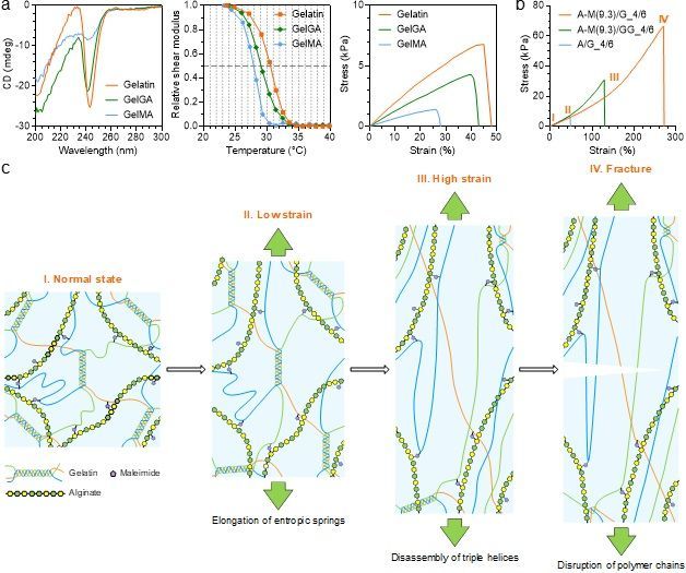
MPa) for one minute. Such preliminary result was promising and warranted further investigation.

Before preparing the EL dressing, we also noted Jayakrishnan et al.26 had reported another hybrid

hydrogel formed with alginate dialdehyde and gelatin (A-D/G). However, the crosslinking of this

hydrogel is implemented by Schiff base reaction between the aldehyde groups of oxidized alginate and

the amino groups of gelatin, which generates imine and is reversible27. Therefore, the stability of it

might be very poor. We systematically compared A-M(9.3)/G_4/6 hydrogel with A-D/G hydrogel. Our

analyses demonstrated A-M(9.3)/G_4/6 had better performance in the aspects of mechanical strength,

chemical stability, optical property and biocompatibility (Supplementary Fig. 9 to Fig. 15; see

Supplementary Information for full discussion).

Mechanism on the toughness of the hydrogel

Conventional hydrogels are normally weak because they are crosslinked by pure chemical bonds or

physical interactions. However, some of our A-M/G hydrogels displayed good mechanical properties

at ambient temperature. In particular, A-M(9.3)/G_4/6 could be extended by nearly three times with a

stiffness comparable to that of muscles28. Understanding such character is important for the application

of our materials and the design of new hydrogels with better mechanical performance. Extensive

studies have shown that a highly stretchable hydrogel generally has some mechanism to dissipate the

energy built up during its deformation. In our case, we hypothesized the physical interaction within

gelatin or between gelatin and A-M played the role of energy dissipation. To unravel this, we started

our investigation by altering the chemistry of gelatin, which generated gelatin glycinamide (GelGA or

GG in short) and gelatin methacrylate (GelMA or GM in short) (Supplementary Fig. 16 and 17).

 Gelatin exists as random coils in an aqueous solution above its Tm29. The solution spontaneously

transforms into a hydrogel when it cools. At the same time, the random coils bind to form ordered

triple helices that function as physical crosslinks for hydrogel formation29. This coil-helix transition is

reversible and readily affected by many factors, including pH, ionic strength, and chemical

 8
modification to gelatin30. Circular dichroism (CD) was employed to study the influence of chemical

modification to gelatin on the molecular structure of its hydrogel at ambient temperature. The CD

spectra show a strong negative peak around 240 nm (Fig. 2a) for pristine gelatin hydrogel, which was

assigned to the triple helices. The same peak was found for GelGA hydrogel, but the intensity of it

decreased with a 2 nm blue shift, implying the reduction of triple helices in it. For GelMA hydrogel,

the peak was almost gone, indicating the predominance of random coils in it. A direct consequence of

this phenomenon one would anticipate is the loss of mechanical strength for the generated hydrogels.

Our data reveal the shear modulus of pristine gelatin hydrogel is 8.9 kPa at ambient temperature, but

it significantly declines to 3.3 kPa (P
a 0 1.0 10 b 80

 Relative shear modulus
 Gelatin Gelatin A-M(9.3)/G_4/6 IV
 0.8 GelGA GelGA A-M(9.3)/GG_4/6
 60

 Stress (kPa)
 Stress (kPa)
 CD (mdeg)

 -10 GelMA GelMA A/G_4/6
 0.6
 5 40
 0.4 III
 -20 Gelatin
 GelGA 20
 0.2
 II
 GelMA I
 -30 0.0 0 0
 200 220 240 260 280 300 20 25 30 35 40 0 10 20 30 40 50 0 100 200 300
 Wavelength (nm) Temperature (°C) Strain (%) Strain (%)
 c III. High strain
 IV. Fracture

 II. Low strain

 I. Normal state

 1
 Gelatin Maleimide

 Alginate

 Elongation of entropic springs

 Disassembly of triple helices
 Disruption of polymer chains

Fig. 2 | Mechanism on the toughness of A-M(9.3)/G_4/6 hydrogel. a, Effects of different chemical
modifications to gelatin on the molecular structure, melting point and tensile behavior of its hydrogel.
b, Comparison in tensile behavior among A-M(9.3)/G_4/6, A-M(9.3)/GG_4/6 and A/G_4/6 hydrogels.
c, Schematic illustration for the mechanism on the toughness of A-M(9.3)/G_4/6 hydrogel.

 On top of these findings, we further mixed A-M with GelGA or GelMA to make A-M(9.3)/GG_4/6

and A-M(9.3)/GM_4/6 hydrogels. As a negative control, we prepared alginate/gelatin (A/G_4/6)

hydrogel as well. Tensile testing was conducted on these hydrogels except for A-M(9.3)/GM_4/6

because it was too brittle and repeatedly broke during demolding. Indeed, this hydrogel transformed

into a liquid upon heating above the Tm of GelMA, suggesting no chemical crosslink had formed in it.

Such phenomenon verified the necessity of amino groups for the chemical crosslinking of our hydrogel

since all of them had been amidated in GelMA (Supplementary Fig. 17). The mechanical strength of

A/G_4/6 is also very weak and almost identical to that of pristine gelatin hydrogel except that the

 10
Young’s modulus of it is even lower (P
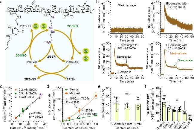
A/G_4/6 fractured at low stress upon triggering the disassembly of the triple helices. On the other hand,

due to the damage to the triple-helix structure, the capacity for energy dissipation was reduced in A-

M(9.3)/GG_4/6, resulting in lower mechanical strength of it as well. It is at the synergism between the

physical interaction and chemical crosslinking that a tough and highly stretchable hybrid hydrogel of

alginate and gelatin can be produced.

Preparation of the EL dressings with the capacity to catalyze NO generation

In cardiovascular system, NO plays critical roles in nearly all important biological functions of native

endothelium19. Consequently, we decided to prepare the EL dressings by rendering our hydrogel the

capacity to catalyze NO generation. There are two known pathways responsible for the production of

NO in vivo. One is through endothelial nitric oxide synthase (eNOS), which catalyzes the degradation

of L-arginine into NO19. The other is through glutathione peroxidase-3 (GPx-3), which metabolizes

endogenous nitrosated thiols (RSNO) to generate NO31. The pathway of eNOS is complex and involves

many components so that harnessing it is difficult. In contrast, NO generation catalyzed by GPx-3 is

relatively simple. The selenocysteine residue of this enzyme is believed to be the catalytic center32. In

fact, many selenium species have shown the capacity to catalyze the degradation of RSNO into NO.

For instance, selenocystamine (SeCA) is capable of catalyzing the production of NO in a mechanism

proposed by Meyerhoff et al.33 (Fig. 3a). This pathway can be readily exploited so that we made use

of it in our EL dressings by conjugating SeCA to A-M(9.3). Inductively coupled plasma mass

spectrometry (ICP-MS, Supplementary Fig. 24a) disclosed the content of conjugated SeCA was about

0.016 mmol g-1 in it. By tuning the proportions of SeCA-conjugated A-M(9.3) and blank A-M(9.3),

EL dressings conjugated with varying contents (0.2 to 1.0 mM) of SeCA were prepared.

 12
a b 15 15

 (×10-12 mol mg-1 min-1)

 (×10-12 mol mg-1 min-1)
 EL dressing with
 Blank hydrogel
 0.2 mM SeCA

 NO release rate

 NO release rate
 10 10

 5 5
 2R'SH + NO NO 2GSNO

 0 0
 0 10 20 30 40 50 60 0 10 20 30 40 50 60
 Time (min) Time (min)
 50 150

 (×10-12 mol mg-1 min-1)

 (×10-12 mol mg-1 min-1)
 EL dressing with EL dressing with
 0.5 mM SeCA 1.0 mM SeCA

 NO release rate

 NO release rate
 40
 NO
 100 Maximal rate
 2GSNO 2GSH 30 Sample out
 NO
 Steady rate
 2RSeH 2RSe-SG 20
 50
 10
 Sample in
 0 0
 0 10 20 30 40 50 60 0 10 20 30 40 50 60
 2R'S-SG 2R'SH
 Time (min) Time (min)
c d e f
 Flux (×10-10 mol cm-2 min-1)

 10 150 80 40

 (×10-12 mol mg-1 min-1)
 0.2 mM SeCA Steady
 (×10-12 mol mg-1 min-1)

 Immobilized SeCA (%)
 0.5 mM SeCA Maximal n.s.

 NO release rate
 NO release rate

 8 ###
 1 mM SeCA 60 30
 100 y = 45.66x 2 + 71.05x
 6 R2 = 0.9995
 **
 40 20
 4
 50 y = 27.09x
 y = 0.2287x R2 = 0.9984 20 10
 2 ###
 R2 = 0.9823
 0 0 0 0
 0.2 mM 0.5 mM 1 mM

 D 1
 D 3
 D y7
 D 14
 28
 D al
 0 10 20 30 40 0.0 0.2 0.4 0.6 0.8 1.0

 in
 ay
 ay
 a
 ay
 ay
 r ig
 Rate (×10-12 mol mg-1 min-1) Content of SeCA (mM) Content of SeCA

 O
Fig. 3 | Catalytic generation of NO from the EL dressings. a, Mechanism on the catalytic generation
of NO from NO donors (R’S-NO) by SeCA conjugated to alginate. b, Representative curves of NO
generation from GSNO (10 μM) in PBS (pH 7.4) at 37 °C with the blank hydrogel or EL dressings
conjugated with varying contents of SeCA (0.2 to 1.0 mM). c, Correlation between the fluxes and rates
of NO generation catalyzed by the EL dressings. d, Summary on the release features of NO from the
EL dressings conjugated with varying contents of SeCA (n = 6). e, Quantification of the immobilized
SeCA on the EL dressings after catalytic generation of NO. f, Release rates of NO from GSNO
catalyzed by the EL dressing conjugated with 1.0 mM SeCA after pre-incubation in PBS at 37 °C for
different durations (n = 6). One-way analysis of variance (ANOVA) with Tukey post-hoc test was
performed to determine the difference among various groups. (n.s., not significant; ####P < 0.0001
compared to other groups; **P < 0.01 between two groups)

 In most studies, the flux of NO, defined as the production of it per unit area per unit time, was

measured for a coating. Since the weight of an EL dressing on a substrate might vary significantly, we

assumed the release rate of NO (production of NO per unit mass per unit time) to accurately reflect

the NO-generating capacity of it. We detected the release rates of NO (Fig. 3b) catalyzed at 37 °C by

the EL dressings submerged in PBS containing 10 μM S-nitrosoglutathione (GSNO, an endogenous

NO donor) and 30 μM glutathione (GSH). As anticipated, the blank hydrogel was unable to catalyze

the generation of NO from GSNO. With the conjugation of SeCA, a burst release of NO followed by

 13
gradual decline until steady state was observed. To our delight, the flux of NO was nearly proportional

to the release rate of it, implying the uniform coating density of the EL dressings on the substrates (Fig.

3c). At the average coating density of 22.9 mg cm-2, the flux of NO catalyzed by the EL dressing

containing 1.0 mM SeCA was 6.19 × 10⁻¹⁰ mol cm-2 min-1, approaching the normal level from native

endothelium34. Though the steady release rate of NO was proportional to the content of conjugated

SeCA, the peak value presented a quadratic relationship with it (Fig. 3d), leading to the highest burst

release ratio of NO (3.48) in the EL dressing containing 1.0 mM SeCA (Supplementary Fig. 24b).

Nevertheless, this value is still much smaller than those of many NO-eluting coatings35. The transient

burst of NO (26.3 × 10⁻¹⁰ mol cm-2 min-1 at maximum) is unlikely to be detrimental for endothelial

cells as it only lasts for a few minutes. When an EL dressing was removed from the reaction solution,

NO release did not go back to the initial baseline, suggesting some organoselenium species had

diffused into the reaction solution. Quantitative analysis (Fig. 3e) uncovered about 41% of SeCA was

still linked to the EL dressings after catalytic generation of NO. The organoselenium species in the

solution came from several sources, including remnant free SeCA in the EL dressings, SeCA

conjugated to uncrosslinked A-M(9.3) molecules, and derivatives of SeCA as byproducts (Fig. 3a). In

the third scenario, every SeCA molecule linked to the EL dressings with merely one amino group lost

half of its constituent part after catalysis. Finally, we measured the release rates of NO from the EL

dressings after they had been pre-incubated in PBS for different durations. Our result shows that their

catalytic potency could last for more than two weeks (Fig. 3f).

Effects of the EL dressings on cellular behaviors in vitro

The integration of a vascular implant into the blood vessel is featured by the formation of neointima

predominantly consisting of smooth muscle cells and/or endothelial cells. To prevent NIH, an ideal

vascular stent must be capable of inhibiting vicinal smooth muscle cells while promoting rapid

recruitment of endothelial cells. Previous studies have reported NO at the physiological level can

 14
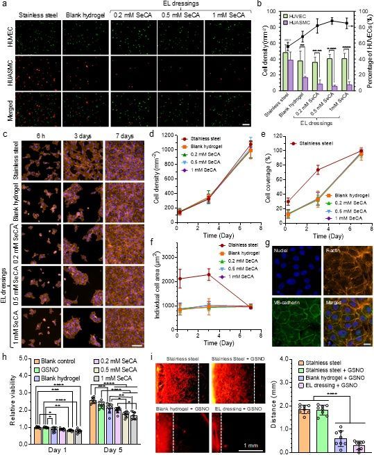
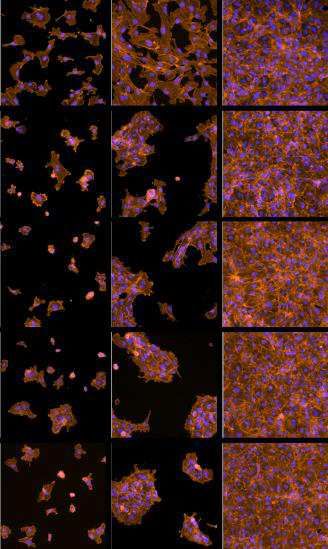
inhibit smooth muscle cells while such effect does not act on endothelial cells36,37. To corroborate this,

we started our investigation by conducting a competitive adhesion test between human umbilical vein

endothelial cells (HUVECs) and human umbilical artery smooth muscle cells (HUASMCs) on our

coatings in the medium supplemented with GSNO (10 μM) and GSH (30 μM). Our results (Fig. 4a

and 4b) demonstrate the number of HUASMCs adhering on the blank hydrogel within three hours was

less than a half of that on bare stainless steel. For the EL dressings, the cell densities were even lower.

In contrast, no significant difference in the density of adhering HUVECs was found among bare

stainless steel and these coatings. Taken together, it can be concluded the blank hydrogel coating

selectively facilitated the adhesion of endothelial cells, while NO generation catalyzed by the EL

dressings further inhibited the attachment of smooth muscle cells.

 To further evaluate the potential of our EL dressings to promote endothelial regeneration, we seeded

HUVECs onto them and cultured the cells in the presence of GSNO for prolonged time. Most of them

attached onto the substrates within 6 h (Fig. 4c). Quantitative analyses (Fig. 4d to 4f) revealed no

significant difference in the proliferation, coverage and spreading of HUVECs among the blank

hydrogel and EL dressings, suggesting NO had little influence on their behaviors. In detail, the density

and coverage of HUVECs on them increased from 147±20 cells mm-2 to 1,010±61 cells mm-2, and

12.8±2.7% to 96.9±3.0%, respectively in one week while the individual cell area almost unchanged.

Compared to these coatings, the endothelial cells were much more spread on bare stainless steel even

at early time. The individual cell area were 2,126±385 μm2 after 6 h and 2,295±243 μm2 after 3 days

on it, while the values of these indices for our coatings were barely 863±97 μm2 and 990±101 μm2,

respectively. However, no significant difference in cell proliferation rate was noted among them. The

direct consequence of better cell spreading on bare stainless steel was the higher cell coverage

(29.9±5.0% after 6 h and 73.8±6.3% after 3 days). Nonetheless, these indices became almost identical

in one week among all groups.

 15
a EL dressings b
 100 100
 Stainless steel Blank hydrogel 0.2 mM SeCA 0.5 mM SeCA 1 mM SeCA HUVEC

 Percentage of HUVECs (%)
 HUASMC
 80 80

 Cell density (mm-2)
 HUVEC
 ####
 60 60
 ** ****
 **** ****
 40 40
 HUASMC

 20 20

 0 0
 Merged

 EL dressings

 c 6h 3 days 7 days d 1200
 Stainless steel
 e 120
 0.5 mM SeCA 0.2 mM SeCA Blank hydrogel Stainless steel

 Stainless steel
 Blank hydrogel
 1000 100

 Cell density (mm-2)
 0.2 mM SeCA

 Cell coverage (%)
 800 0.5 mM SeCA
 80
 1 mM SeCA
 600 60

 400 40 Blank hydrogel
 0.2 mM SeCA
 200 20 0.5 mM SeCA
 1 mM SeCA
 0 0
 0 2 4 6 8 0 2 4 6 8
 Time (Day) Time (Day)
 f Stainless steel g
 Nuclei F-actin
 Blank hydrogel
 Individual cell area (μm2)

 3000
 0.2 mM SeCA
 0.5 mM SeCA
 EL dressings

 1 mM SeCA
 2000

 VE-cadherin Merged
 1000
 1 mM SeCA

 0
 0 2 4 6 8
 Time (Day)

 h 5.0 Blank control 0.2 mM SeCA
 i Stainless steel Stainless Steel + GSNO 4.0
 Stainless steel
 GSNO 0.5 mM SeCA Stainless steel + GSNO
 4.0
 Relative viability

 Blank hydrogel 1 mM SeCA Blank hydrogel + GSNO
 3.0
 Distance (mm)

 ****
 *** *********** EL dressing + GSNO
 3.0 ****
 **** *** *
 ** Blank hydrogel + GSNO EL dressing + GSNO 2.0
 2.0
 *
 *
 1.0 1.0

 0.0 1 mm
 Day 1 Day 5 0.0
Fig. 4 | Effects of the EL dressings on cellular behaviors in vitro. a, Fluorescence images exhibiting
the competitive adhesion between HUVECs and HUASMCs on various substrates. The cell growth
medium was supplemented with GSNO (10 μM) and GSH (30 μM). (Scale bar: 500 μm) b,
Quantitative analyses on the competitive adhesion between HUVECs and HUASMCs (n = 6). c,
Confocal laser scanning microscopy (CLSM) images displaying the adhesion, spreading and
proliferation of HUVECs seeded onto various substrates in the presence of GSNO. (Scale bar: 100 μm)
d-f, Summary of cell density, cell coverage and individual cell area on those substrates (n = 6). g,
CLSM images showing the formation of adherens junctions (VE-cadherin) between HUVECs grown
on the EL dressing containing 1.0 mM SeCA. (Scale bar: 20 μm) h, Proliferation assay of HUASMCs
co-cultured with the blank hydrogel or EL dressings containing varying contents of SeCA in the
presence of GSNO. i, Migrations of HUASMCs on bare stainless steel, the blank hydrogel and EL
dressing containing 1.0 mM SeCA. One-way ANOVA with Tukey post-hoc test was performed to
determine the difference among various substrates and two-tailed Student’s t-test was assumed to
determine the difference between the two types of cells on the same substrate. (####P < 0.0001
compared to other groups; *P
It is worthy of note HUVECs gathered as colonies and then propagated to form confluent

monolayers on our coatings whereas those grown on bare stainless steel dispersed evenly and

proliferated until the formation of an intact cell sheet. Mauck et al.38 have unraveled that cells are

regulated by the interplay between cell-cell and cell-ECM interactions. On a stiff substrate like bare

stainless steel in our case, the traction force sensed by HUVECs is relatively large, guiding them into

a more spread phenotype through activating mechanotransduction pathways such as YAP/TAZ39. On

the contrary, those grown on our soft coatings were governed by cell-cell interaction due to the

relatively low cell-ECM interaction, thereby leading to the formation of cell colonies and collective

cell migration. At this stage, we could not conclude which scenario is more favorable for endothelial

regeneration in vivo, but we observed that adherens junctions (VE-cadherin), which is necessary for

healthy endothelium, had already formed between HUVECs grown on our coatings (Fig. 4g).

 The competitive adhesion test implied our EL dressings could inhibit smooth muscle cells. To verify

this point of view, we co-cultured HUASMCs with our coatings in the presence of GSNO. Cell

proliferation assay (Fig. 4h) suggested GSNO alone had little influence on the cells, while their

viability was significantly reduced when co-cultured with an EL dressing or even the blank hydrogel.

The anti-proliferative effect of the blank hydrogel might come from uncrosslinked A-M(9.3) molecules

in it (Supplementary Fig. 13), and the cell proliferation was further inhibited upon the generation of

NO from the EL dressings. Indeed, the cell viability declined monotonically with the content of SeCA

in them. Specifically, after incubation with the EL dressing containing 1.0 mM SeCA for 5 days, the

viability of HUASMCs decreased by 30% compared to those supplemented with GSNO alone.

However, the EL dressings were unable to stop the proliferation of smooth muscle cells due to the

unsustainable generation of NO in vitro. Nonetheless, this may not be an issue in vivo since the

volumes of blood in experimental rabbits and pigs are two to three magnitudes larger.

 We continued to evaluate the migration of HUASMCs on our coatings in 24 h according to a

published protocol40. The experimental data (Fig. 4i) demonstrate GSNO alone has little influence on

 17
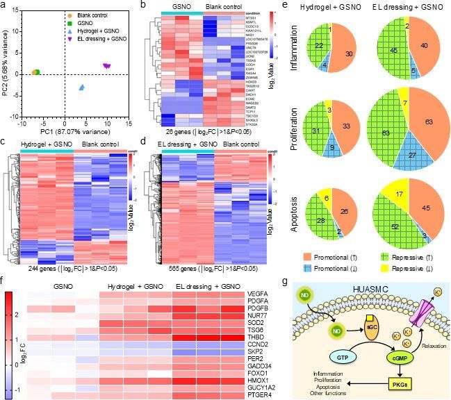
the migration of HUASMCs since the distance traveled by them on bare stainless steel was unaffected

by it (1.83±0.16 mm with GSNO vs. 1.86±0.17 mm without GSNO). To our delight, the movement of

HUASMCs on our coatings was dramatically slowed down in comparison to those on bare stainless

steel. The cells traveled 0.60±0.33 mm on the blank hydrogel and 0.31±0.17 mm on the EL dressing

containing 1.0 mM SeCA, respectively. These results indicate the hydrogel material itself possesses

some repressive effect on the migration of smooth muscles, while NO generation catalyzed by SeCA

conjugated to the EL dressing could further retard this progress.

Transcriptome analysis of HUASMCs

To figure out why HUASMCs were inhibited by the blank hydrogel and EL dressings, we performed

a transcriptome analysis after the cells had been co-cultured with them. The EL dressing containing

1.0 mM SeCA was selected as the delegate since it was most efficient in inhibiting smooth muscle

cells. Principal component analysis (PCA) and clustering assay (Fig. 5a and Supplementary Fig. 25)

revealed all three independent replicates in each group correlated well and GSNO alone barely had

any impact on the gene expression of HUASMCs. However, with the co-culture of the blank hydrogel

plus GSNO, the phenotypic change of these cells became dramatic. For those incubated with the EL

dressing plus GSNO, such change was largest. We set a threshold of │log2fold change (FC)│>1 and

P
expression profiles on cell behavior, these genes were analyzed in terms of inflammation, proliferation,

and apoptosis.

 Inflammation is a localized protective response elicited by the stimulation or injury to a tissue41,42.

However, dysregulated inflammation is disastrous as it may lead to either hyperplasia or excess

destruction. Our analysis (Fig. 5e) unraveled tens of pro-inflammatory genes were significantly up-

regulated in both cases. At the same time, numerous anti-inflammatory genes such as SOD243 and

TSG644 were also activated after both treatments (Fig. 5f). The inflammatory response was likely to

be elicited by gelatin in our coatings since it was derived from animal tissues that might contain pro-

inflammatory substances. Similar observations were also reported by others45, yet the mechanism is

not understood. In contrast, alginate might function as an anti-inflammatory mediator46 so that the

process of inflammation was constrained. In the case of HUASMCs co-cultured with the EL dressing

plus GSNO, the total number (50) of significant anti-inflammatory alterations was even larger than

that (42) of pro-inflammatory ones. Obviously, NO molecules generated from the EL dressing

contributed extra anti-inflammatory modulation since some anti-inflammatory genes such as NUR7747

were exclusively up-regulated (Fig. 5f). Regulated inflammation is beneficiary since it can help

reconstruct the damaged tissue. In this study, we noted that pro-angiogenic cytokines such as VEGF,

PDGFA and PDGFB were activated in HUASMCs after incubation with EL dressing plus GSNO,

implying such coating might accelerate endothelial regeneration in vivo.

 19
a b GSNO Blank control e Hydrogel + GSNO EL dressing + GSNO
 15
 Blank control
 GSNO 2

 Inflammation
 10 Hydrogel + GSNO 1
PC2 (5.68% variance)

 EL dressing + GSNO
 22 40
 5 45
 30

 log2Value
 4
 5
 0

 -5
 7
 3

 Proliferation
 -10
 -10 -5 0 5 10 15 63
 33
 PC1 (87.07% variance) 26 genes (│log2FC│>1&P1&P1&P
became dominant. Many anti-proliferative alterations were unique in this group, such as the down-

regulation of proto-oncogenes CCND248 and SKP249, as well as the up-regulation of tumor suppressor

genes PER250, GADD3451 and FOXO152. These results confirmed that the EL dressing exerted

intensified inhibitory effects on smooth muscle cells through the generation of NO.

 When it comes to apoptosis, pro-apoptotic alterations prevailed over anti-apoptotic ones in both

groups. Besides, the EL dressing with NO release also affected more genes. Although we did not

observe the significant up-regulation of canonical pro-apoptotic makers such as CYC and CASP3,

many anti-proliferative genes were found to be pro-apoptotic as well, including aforementioned PER2

and GADD34.

 It is well known NO affects smooth muscle cells through the canonical cGMP/PKG pathway19,36,37

(Fig. 5g). It activates soluble guanylate cyclase (sGC), which subsequently catalyzes the

transformation of guanosine triphosphate (GTP) into cyclic guanosine monophosphate (cGMP). cGMP

can induce the relaxation of smooth muscle cells by interacting with cGMP-gated ion channels or play

other biological functions through activating phosphate kinase G (PKG). In this study, we did observe

the significant and unique up-regulation of guanylate cyclase 1 soluble subunit alpha 2 (GUCY1A2)

in HUASMCs after the treatment of EL dressing plus GSNO. Besides, HMOX153 and PTGER454,

which are two mediators for vascular relaxation, were also up-regulated dramatically. These results

confirm that our EL dressing induced the relaxation of smooth muscle cells. Such phenomenon is

highly desired because it can help the blood vessel to main vasodilation, thereby preventing the

occlusion of stented artery.

Vascular stent deployment in rabbit iliac arteries

Encouraged by the results above, we continued to construct our EL dressing on BMSs, and then test

them in animals. Before that, the mechanical stability and thrombogenicity of the EL dressing were

examined. Firstly, we conducted a mock angioplasty by dilating an EL dressing-coated stent in a plastic

 21
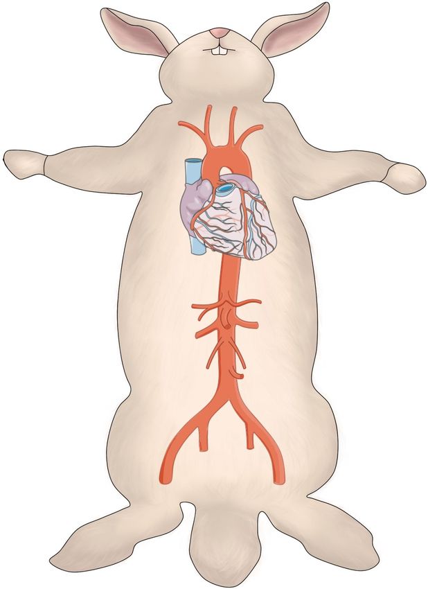
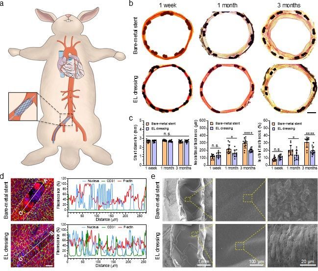
catheter perfused with PBS (37 °C). Our results (Supplementary Fig. 26) demonstrate that the EL

dressing was still intact even after being flushed by PBS for 1 week (Q = 120 mL min-1 or v = 28.3 cm

s-1). Thereafter, we carried out a thrombogenicity test in an ex vivo arteriovenous shunt model

(Supplementary Fig. 27). Our data show both bare stainless steel and the blank hydrogel triggered

severe clotting. In contrast, the EL dressing could effectively retard blood coagulation or even

completely inhibit it depending on the content of conjugated SeCA (see Supplementary Information

for detailed discussion). Indeed, the EL dressing containing 1.0 mM SeCA could be regarded as non-

thrombogenic. In view of its excellent biological performance, the EL dressing containing 1.0 mM

SeCA was selected as the coating material for BMSs.

 We started our preclinical study by implanting the EL dressing-coated stents into the right iliac

arteries of rabbits (Fig. 6a and Supplementary Fig. 28). BMSs of 316L stainless steel was used as the

control due to its wide application in clinical practice, and were implanted into their left iliac arteries.

At the designated time points, the stented arteries were harvested. Van Gieson staining of their cross-

sections showed all vascular stents were fully expanded, but neointimal growth varied dramatically

among different groups (Fig. 6b). Quantitative analyses (Fig. 6c) suggested the stent diameters were

nearly identical and matched well with the reference value (2.7 mm) provided by the manufacturer of

BMS. The neointimal thickness (NT) and ISR of them showed no difference within 1 week of

implantation. However, neointima grew fast on BMS with NT increased from 118 μm to 295 μm, and

ISR from 9.1% to 34.0% in 3 months. In contrast, these indices of EL dressing-coated stent slowly

increased to 200 μm and 21.1%, respectively, which were significantly smaller (P < 0.0001) than those

of BMS. Besides, the neointimal growth rate on EL dressing-coated stent decreased from 160 μm

month-1 in the first month to 20 μm month-1 in the next two months, whereas these values for BMS

were 217 μm month-1 and 39 μm month-1, respectively.

 22
a b 1 week 1 month 3 months Bare-metal stent EL dressing c 5 600 60 Neointimal thickness (m) Bare-metal stent Bare-metal stent Bare-metal stent In-stent restenosis (%) Stent diameter (mm) 4 EL dressing 500 EL dressing 50 EL dressing n.s. 400 **** 40 **** 3 * * 300 30 2 n.s. 200 20 n.s. 1 100 10 0 0 0 1 week 1 month 3 months 1 week 1 month 3 months 1 week 1 month 3 months d 120 Nucleus CD31 F-actin e Bare-metal stent Fluorescence (%) Bare-metal stent 100 80 60 40 20 0 0 50 100 150 200 250 Distance (m) 120 Nucleus CD31 F-actin Fluorescence (%) 100 EL dressing EL dressing 80 60 40 20 0 0 50 100 150 200 250 1 mm 100 μm 20 μm Distance (m) Fig. 6 | Vascular stent deployment in rabbits. a, Schematic illustration for vascular stent deployment in rabbit iliac arteries. b, Optical images showing the cross-sections of stented arteries after van Gieson staining. (Scale bar: 500 μm) c, Quantitative analyses on the cross-sections (n = 12). d, CLSM images unveiling the endothelialization on the stents (outlined by the dashed lines). The right panels present the fluorescence intensities of different cell components along the line segments (OA, O’B) in the images. (Blue: cell nucleus, green: CD31, red: F-actin; scale bar: 50 μm). e, SEM images showing the luminal faces of stented arteries at 3 months post stent deployment. One-way ANOVA with Tukey post-hoc test was performed to determine the difference among various groups and Student’s t-test was assumed to determine the difference between two groups. (n.s., not significant; *P < 0.05 and ****P < 0.0001) As aforementioned, our EL dressing was expected to promote rapid restoration of native endothelium. To assess that, we utilized confocal laser scanning microscope (CLSM) to examine the luminal faces of the stented arteries. CLSM (Fig. 6d and Supplementary Video 2) showed that some struts of BMS were not fully covered by endothelial cells in 1 week, which were also corroborated by 23
SEM (Supplementary Fig. 29). In addition, clusters of giant flat or small granular cells that seemed to

be inflammatory cells, were found on the non-endothelialized region. In contrast, EL dressing-coated

stent was encapsulated by intact endothelium in 1 week and hardly any sign of inflammation was

observed (Fig. 6d and Supplementary Video 3). After implantation for 1 month, both types of stents

were completely endothelialized (Supplementary Fig. 30). Nevertheless, the endothelial cells adhering

on EL dressing-coated stent presented a more mature phenotype compared to BMS, featured by

elongated morphology and high degree of orientation (Fig. 6e and Supplementary Fig. 30).

 In cardiovascular system, the coordination between coagulation and fibrinolysis are critical for

maintaining the intactness of blood vessels. During angioplasty, the vessel wall is injured inevitably,

thereby releasing tissue factors that trigger coagulation. The ensuing formation of thrombus not only

activates fibrinolysis, but also recruits inflammatory cells55 as observed on BMS in our case. However,

NO can suppress clotting cascade by preventing platelets from activation and may potentially inhibit

thrombin through up-regulation of thrombomodulin in smooth muscle cells (Fig. 5f). Besides, the EL

dressing could provide a highly hydrated lubricating interface between the stent and blood, thereby

reducing the turbulence of blood flow compared to BMS. It is reasonable to believe thrombus

formation was repressed on EL dressing-coated stent as proved by the ex vivo thrombogenicity test.

Consequently, the inflammation elicited by acute thrombosis was effectively mitigated on EL dressing-

coated stent. In addition, the EL dressing inhibited the proliferation of smooth muscle cells through

the combinational effects of NO gas and A-M molecules. At the same time, it mimicked the

subendothelial ECM, thereby providing a favorable microniche for endothelial cells. Thanks to these

factors, EL dressing-coated stent effectively suppressed ISR and presented faster restoration of native

endothelium in comparison to BMS.

Vascular stent deployment in swine coronary arteries

Although EL dressing-coated stent demonstrated satisfactory outcomes in leporine model, those data

 24
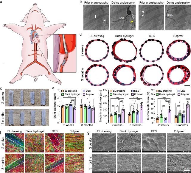
might still be inadequate in predicting its performance in human being since rabbits are herbivorous.

Among large experimental animal species, the coronary artery system and physiology of pigs are very

similar to those of human being, making them an ideal model for coronary stenting56. Consequently,

we continued to evaluate EL dressing-coated stent in a swine model. We compared it with an

everolimus-eluting DES, which represents the gold standard for coronary stents. Both of them were

constructed on the same kind of cobalt chromium alloy (CoCr) stents, and the anti-restenotic drug of

DES was loaded in its polymer coating (see Experimental Section for details). In addition, blank

hydrogel-coated and blank polymer-coated stents were included as two negative controls (see

Experimental Section for details). These four types of stents were randomly implanted in three or four

coronary arteries of individual Bama miniature pigs (Fig. 7a and Supplementary Fig. 31) under the

guidance of digital subtraction angiography (DSA).

 25
a b Prior to angiography During angiography Prior to angiography During angiography RCA LAD d EL dressing Blank hydrogel DES Polymer 2 weeks 3 months c e 4 500 70 EL dressing DES EL dressing DES EL dressing DES Neointimal thickness (m) In-stent restenosis (%) 2 weeks Stent diameter (mm) Blank hydrogel Polymer Blank hydrogel Polymer 60 Blank hydrogel Polymer ** 400 3 * * *** **** 50 **** #### *** ** *** ** 300 *** **** * * 40 2 ** * ** ** ** 200 30 3 months 1 20 100 10 0 0 0 2 weeks 3 months 2 weeks 3 months 2 weeks 3 months EL dressing Blank hydrogel DES Polymer EL dressing Blank hydrogel DES Polymer f g 2 weeks 2 weeks 3 months 3 months 100 μm 10 μm Fig. 7 | Vascular stent deployment in pigs. a, Schematic illustration for vascular stent deployment in swine coronary arteries. b, Digital subtraction angiography prior to the harvest of stented arteries. The white arrows indicate the sites of implanted stents. The yellow arrow refers to severe restenosis occurring in a polymer-coated stent. (Scale bar: 1 cm) c, Photographs displaying the luminal faces of stented coronary arteries at 2 weeks and 3 months post stent deployment. (left to right: EL dressing- coated stent, blank hydrogel-coated stent, DES and polymer-coated stent; scale bar: 5 mm) d, Optical images showing the cross-sections of stented arteries after van Gieson staining. (Scale bar: 500 μm) e, Quantitative analyses on the cross-sections (n = 6). f, CLSM images unveiling the endothelialization on the stents (outlined by the dashed lines). (Blue: cell nucleus, green: CD31, red: F-actin). g, SEM images showing the luminal faces of stented arteries at 2 weeks and 3 months post stent deployment. Student’s t-test was performed to determine the difference. (*P < 0.05, **P < 0.01, ***P < 0.001 and ****P < 0.0001 between two groups; ####P < 0.0001 compared to other groups) DSA was conducted again prior to the harvest of stented arteries, which revealed none of the stented arteries was occluded in 2 weeks. However, severe narrowing occurred in polymer-coated stents (3 out of 6) while the blood flow was almost unaffected in others at 3 months post stent deployment (Fig. 7b; 26
Supplementary Video 4 and 5). We harvested the stented arteries and examined them with naked eyes.

Our photographs (Fig. 7c) show thick neointima grew on polymer-coated stent while neointimal

formation was much slower or even negligible on other stents. The cross-sections of stented arteries

were also stained (Fig, 7d). Quantitative analyses (Fig. 7e) revealed the diameter of polymer-coated

stent was significantly smaller (0.2~0.4 mm) than those of others, suggesting the occurrence of recoil

to it due to the strong radial force produced by NIH. The NT value of it was 203±34 μm at 2 weeks

and reached 268±40 μm at 3 months since implantation, while ISR increased from 29.2±5.4% to

39.1±4.6% duration that time. Upon the loading of everolimus in the polymer coating, DES efficiently

inhibited neointimal growth. The NT and ISR for DES were barely 116±15 μm and 8.7±2.0%,

respectively after implantation for 2 weeks. However, such inhibitory effect was unsustainable as we

found these indices increased to 165±28 μm and 19.1±5.1% in 3 months. In contrast, EL dressing-

coated stent presented both efficient and sustained suppression of NIH. Though this type of stent was

slightly inferior in the short term, it defeated DES in the long run since the NT and ISR increased

merely to 135±15 μm and 13.5±1.7%, respectively in 3 months. Notably, the NIH on blank hydrogel-

coated stent was not severe when compared to polymer-coated one. In fact, the ISR of blank hydrogel-

coated stent even approached that of DES in 3 months.

 To explain the difference among various stents, we also examined the status of endothelialization

on them. CLSM and SEM images (Fig. 7f and 7g; Supplementary Fig. 32 and 33) disclose

endothelialization was dramatically delayed on DES compared to that of other stents. In fact, the

progress of endothelialization on it was still incomplete even in 3 months. In stark contrast, both EL

dressing-coated and blank hydrogel-coated stents promoted rapid restoration of endothelium in 2

weeks. However, the endothelial cells on blank hydrogel-coated stent displayed loose contact with

each other and were less oriented compared to those on EL dressing-coated one. Besides, the strut

profile was not visible for blank hydrogel-coated stent due to the thick neointima. When it comes to

polymer-coated stent, though high degree of endothelialization was observed in 2 weeks, the

 27
endothelial cells were highly elongated and oriented in the direction of blood flow. In addition, the

newly formed endothelium was dispersed with holes. At 3 months post implantation, the endothelium

on polymer-coated stent became intact, but the endothelial cells were further stretched due to the large

shear stress of blood flow caused by ISR.

 Based on the observations above, we can rationally deduce polymer-coated stent is highly pro-

inflammatory, thereby stimulating NIH in the stented artery. In contrast, blank hydrogel-coated stent

is more biocompatible so that the neointimal formation was much slower on it. In the case of DES,

though everolimus released from the polymer coating inhibited neointimal growth, such effect was

unsustainable. Besides, the anti-restenotic drug also impaired the regeneration of native endothelium.

Consequently, inflammatory response was still elicited upon the depletion of the drug, causing NIH

on DES at late stage. Fortunately, EL dressing-coated stent not only provided a temporary endothelial

function, but also promoted rapid restoration of native endothelium to replace it. As a result, EL

dressing-coated stent suppressed ISR persistently.

 To present the advancement of our EL dressing more convincingly, we did a literature review and

compared EL dressing-coated stent with other stents deployed in the iliac arteries of healthy, balloon

injured, or high-fat diet fed rabbits. Our summary (Supplementary Table 1) reflects conventional DESs

are potent in suppressing ISR in general. However, the progress of endothelialization on them was

markedly delayed, and it was still incomplete in 3 months on some DESs. BMSs are favorable for the

restoration of endothelium, but they normally possess high thrombogenicity and induced thicker

neointimal formation. The FDA-approved fully bioresorbable stent Absorb BVS® failed in all terms

of thrombogenicity, endothelialization and ISR. Other stents are either thrombogenic, inefficient in

promoting endothelialization, or incompetent in suppressing ISR. In stark contrast, EL dressing-coated

stent is non-thrombogenic and achieved complete endothelial regeneration in 1 week. In the meantime,

it is comparable to DESs in view of anti-restenosis. Taking thrombogenicity, endothelialization and

ISR into consideration together, EL dressing-coated stent is best in overall performance.

 28
Outlook

The emergence of vascular stents has saved millions of patients with coronary artery disease. However,

in-stent restenosis, as a result of neointimal hyperplasia or stent thrombosis, is a great challenge for

the approved stents. The EL dressing developed in this study shed a new light on the resolution of this

issue. It mimicked the physiological characteristics of native endothelium and promoted rapid

endothelial regeneration, while providing temporal artificial endothelial function to inhibit neointimal

hyperplasia and thrombosis. At the same time, it may also act as a carrier for therapeutic. For instance,

our EL dressing can be encapsulated with small organic or large protein-based medicine

(Supplementary Fig. 34) to afford extra therapy for coronary artery disease. Definitely, such EL

dressing should be applicable to other vascular implants such as artificial valves and blood vessels.

More importantly, the hydrogel designed for fabricating the EL dressing is self-crosslinking, highly

biocompatible, and mechanically tough. Besides, it can be conjugated with various molecules for

special goals due to the abundant functional groups. Thanks to these advantages, we expect such elastic

hydrogel will find wide applications in biomedical engineering, including scaffolds for tissue

engineering, dressings for wound healing, and interfaces for implantable medical devices.

Acknowledgements

This work was supported by the National Natural Science Foundation of China (Project No.: 32000939,

82072072, 22004135), National Key Research and Development Program of China (Project No.:

2017YFE0102400), General Research Fund from the Research Grants Council of Hong Kong (Project

No.: 16308818, 16309920) and Shenzhen Fundamental Research Program (Project No.:

JCYJ20190807160415074, JCYJ20190807160401657). We gratefully acknowledge Ms. T. You from

MOE Key Laboratory of Advanced Technologies of Materials at SWJTU for the help with SEM, and

Ms. Z. Hu from the Analytical and Testing Center at SWJTU for the aid with CLSM. We appreciate

Mr. E. M. W. Fok from the Department of Chemistry at HKUST for ICP-MS analysis. We also thank

 29
You can also read


