A study of the correlations between IVIM DWI parameters and the histologic differentiation of hepatocellular carcinoma
←
→
Page content transcription
If your browser does not render page correctly, please read the page content below
www.nature.com/scientificreports
OPEN A study of the correlations
between IVIM‑DWI parameters
and the histologic differentiation
of hepatocellular carcinoma
Yi Zhou1,3,4, Gang Yang2,4, Xue‑Qin Gong1, Yun‑Yun Tao1, Ran Wang1, Jing Zheng1, Cui Yang1,
Juan Peng1, Lin Yang1*, Jing‑Dong Li2 & Xiao‑Ming Zhang1
The present study aimed to investigate the value of intravoxel incoherent motion diffusion weighted
imaging (IVIM-DWI) in the preoperative prediction of the histologic differentiation of hepatocellular
carcinoma (HCC). Seventy HCC patients were scanned with a 3.0 T magnetic resonance scanner. The
values of apparent diffusion coefficient (ADC), slow apparent diffusion coefficient (D), fast apparent
diffusion coefficient (D*), and the fraction of the fast apparent diffusion coefficient (f) were measured.
Analysis of variance was used to compare the differences in parameters between groups with different
degrees of histologic differentiation. p < 0.05 was considered statistically significant. Receiver
operating characteristic (ROC) curves were used to analyse the efficacy of IVIM-DWI parameters for
predicting the histologic differentiation of HCC. The ADC and D values for well, moderately and poorly
differentiated HCC were 1.35 ± 0.17 × 10⁻³ mm2/s, 1.16 ± 0.17 × 10⁻³ mm2/s, 0.98 ± 0.21 × 10⁻³ mm2/s,
and 1.06 ± 0.15 × 10⁻³ mm2/s, 0.88 ± 0.16 × 10⁻³ mm2/s, 0.76 ± 0.18 × 10⁻³ mm2/s, respectively, and
all differences were significant. The D* and f values of the three groups were 32.87 ± 14.70 × 10⁻³
mm2/s, 41.68 ± 17.90 × 10⁻³ mm2/s, 34.54 ± 18.60 × 10⁻³ mm2/s and 0.22 ± 0.07, 0.23 ± 0.08, 0.18 ± 0.07,
respectively, with no significant difference. When the cut-off values of ADC and D were 1.25 × 10⁻³
mm2/s and 0.97 × 10⁻³ mm2/s, respectively, their diagnostic sensitivities and specificities for
distinguishing well differentiated HCC from moderately differentiated and poorly differentiated
HCC were 73.3%, 85.5%, 86.7%, and 78.2%, and their areas under the ROC curve were 0.821 and
0.841, respectively. ADC and D values can be used preoperatively to predict the degree of histologic
differentiation in HCC, and the D value has better diagnostic value.
Hepatocellular carcinoma (HCC) is a malignant tumour with the sixth highest morbidity rate and the fourth
highest mortality rate worldwide and causes a high disease b urden1,2. Surgical treatment is the most important
means of increasing the long-term survival of HCC p atients3. The histologic differentiation of HCC is an impor-
tant factor affecting the prognosis of patients4–8. Preoperative prediction of the histologic differentiation of HCC
using imaging methods is very important for the development of therapeutic strategies.
The traditional diffusion-weighted imaging (DWI) parameter, namely, the apparent diffusion coefficient
(ADC), can quantitatively reflect the diffusion motion of water molecules in active tissues9. However, ADC
values include two types of information: the molecular diffusion of water and microcirculation p erfusion10.
Intravoxel incoherent motion (IVIM) has emerged in recent years and can distinguish between the diffusion of
pure water molecules and microcirculation perfusion-related information in tissues, thus compensating for the
deficiency of traditional DWI11,12. Currently, IVIM-DWI studies of hepatic lesions mainly focus on the staging
brosis13–16, identification of hepatic n
of hepatic fi odules17–22, and evaluation of the treatment response of liver
cancer23–27. However, studies of IVIM-DWI and histologic differentiation in HCC are rare, and the results of the
few available studies are inconsistent.
1
Medical Imaging Key Laboratory of Sichuan Province, Medical Research Center, Department of Radiology,
Affiliated Hospital of North Sichuan Medical College, Nanchong 637000, Sichuan, People’s Republic of
China. 2Institute of Hepato‑Biliary‑Intestinal Disease, Department of Hepatobiliary Surgery, Affiliated Hospital
of North Sichuan Medical College, Nanchong 637000, Sichuan, People’s Republic of China. 3Department of
Radiology, People’s Hospital of Deyang City, Deyang 618000, Sichuan, People’s Republic of China. 4These authors
contributed equally: Yi Zhou and Gang Yang. *email: linyangmd@163.com
Scientific Reports | (2021) 11:10392 | https://doi.org/10.1038/s41598-021-89784-2 1
Vol.:(0123456789)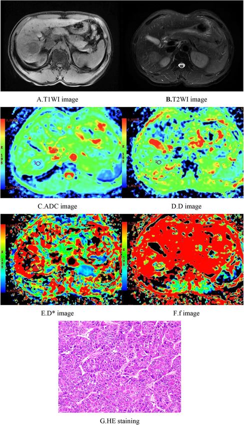
www.nature.com/scientificreports/
Therefore, this study analysed correlations between IVIM-DWI parameters and histologic differentiation
in 70 HCC patients who underwent surgical resection at our hospital between September 2017 and September
2019. We aimed to investigate the value of IVIM-DWI for preoperative prediction of the histologic differentia-
tion of HCC.
Materials and methods
Case collection. The present study was approved by the Ethics Committee of the Affiliated Hospital of
North Sichuan Medical College. Written informed consent was obtained from all individual participants, and
all methods were performed in accordance with the relevant guidelines and regulations. The records of the 70
patients who underwent surgical resection of HCC between September 2017 and September 2019 at our hos-
pital were collected. The patients included 65 males and 5 females aged from 24 to 70 years, with a mean age of
50.64 ± 10.51 years. The findings were validated in an independent cohort (22 patients, age: 53.31 ± 12.49 years).
Magnetic resonance imaging (MRI) scans. All patients were scanned with the same GE Discov-
ery MR750 3.0 T magnetic resonance scanner (GE Medical Systems, Milwaukee, Wis., USA). Body-specific
32-channel phased-array coils were used. Plain liver MRI, IVIM-DWI, and dynamic enhancement scans were
performed. The scanning sequences and parameters were the same as those used in our previous s tudies22.
Image postprocessing. MRI results were sent to a GE ADW4.6 postprocessing workstation, and Function-
MADC software was used for image analysis. The region of interest (ROI) was placed in the largest solid area
of the tumour showing the main properties of the tumour and was compatible with the pathological evaluation
area while avoiding haemorrhagic and necrotic areas. Pseudocolour maps of the ADC, slow apparent diffusion
coefficient (D), fast apparent diffusion coefficient (D*), and the fraction of the fast apparent diffusion coefficient
(f) were generated, and the ADC, D, D*, and f values were measured. The values of all parameters were measured
three times, and the average value was recorded.
Histologic grading. All surgically resected tumours were fixed with formalin, embedded in paraffin,
and stained with haematoxylin–eosin (HE). Histologic differentiation was classified according to the criteria
of the World Health Organization (WHO) Classification of Tumors: Well differentiated: tumour cells resem-
bling mature hepatocytes with minimal to mild atypia, cytoplasm ranging from abundant and eosinophilic to
moderate and basophilic, and nuclei showing minimal to mild nuclear atypia; moderately differentiated: clearly
malignant cells according to HE staining with a morphology strongly suggesting hepatocellular differentiation,
cytoplasm ranging from abundant and eosinophilic to moderate and basophilic, and nuclei showing moderate
nuclear atypia; and poorly differentiated: clearly malignant cells on HE staining with a morphology consistent
with the broad spectrum of poorly differentiated carcinomas, cytoplasm ranging from moderate to scarce, which
is usually basophilic, and nuclei with marked nuclear pleomorphism, which may include anaplastic giant cells.
The number of slides for pathological study was determined according to the tumour volume (one slide per cen-
timetre). When heterogeneous areas exist in one tumour nodule or between different nodules, the tumour grade
is defined according to the major grade of tumour tissue.
Statistical analysis. Analysis of variance (ANOVA) was used to compare differences in parameters among
groups with different degrees of histologic differentiation. The Spearman rank correlation test was used to ana-
lyse the correlations between the IVIM-DWI parameters and the histologic differentiation of HCC tissues. A
value of p < 0.05 was considered statistically significant. Receiver operating characteristic (ROC) curves were
used to analyse the efficacy of IVIM-DWI parameters for predicting the histologic differentiation of HCC.
Results
Plain MRI scans of HCC showed slightly longer T1-weighted imaging (T1WI) and T2WI values with hetero-
geneous signals, and some lesions showed long T1 and T2 values for necrotic liquefaction areas. Most of the
lesions showed "fast-in fast-out" intensification on the enhanced scan, while a few lesions showed mild, uneven
intensification or no obvious intensification in the arterial phase.
Among the 70 cases of HCC, 15 were well differentiated, 46 were moderately differentiated, and 9 were
poorly differentiated. The ADC values for well, moderately, and poorly differentiated HCC were 1.35 ± 0.17 × 10⁻³
mm2/s, 1.16 ± 0.17 × 10⁻³ mm2/s, and 0.98 ± 0.21 × 10⁻³ mm2/s, and the D values were 1.06 ± 0.15 × 10⁻³ mm2/s,
0.88 ± 0.16 × 10⁻³ mm2/s, and 0.76 ± 0.18 × 10⁻³ mm2/s, respectively; all differences were significant. The well
differentiated group had significantly higher ADC and D values than the moderately differentiated and poorly
differentiated groups, and the moderately differentiated group had significantly higher ADC and D values than
the poorly differentiated group. ADC and D values were significantly associated with histologic differentiation
(r = − 0.510, p < 0.05 and r = − 0.512, p < 0.05, respectively). The D* values of the well, moderately, and poorly
differentiated HCC groups were 32.87 ± 14.70 × 10⁻³ mm2/s, 41.68 ± 17.90 × 10⁻³ mm2/s, and 34.54 ± 18.60 × 10⁻³
mm2/s, and the f values were 0.22 ± 0.07, 0.23 ± 0.08, and 0.18 ± 0.07, respectively, with no significant differences;
the D* and f values were not significantly correlated with histologic differentiation. When the cutoff values of
the ADC and D were 1.25 × 10⁻³ mm2/s and 0.97 × 10⁻³ mm2/s, respectively, their diagnostic sensitivities and
specificities for distinguishing well differentiated HCC from moderately differentiated and poorly differentiated
HCC were 73.3% and 85.5% (ADC) and 86.7% and 78.2% (D), and their areas under the ROC curves (AUCs)
were 0.821 and 0.841, respectively. The AUC was larger for D (Tables 1, 2) (Figs. 1, 2, 3, 4, 5).
Scientific Reports | (2021) 11:10392 | https://doi.org/10.1038/s41598-021-89784-2 2
Vol:.(1234567890)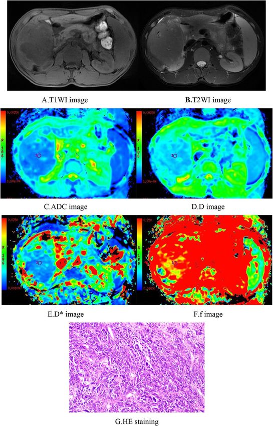
www.nature.com/scientificreports/
Parameters Well differentiated group Moderately differentiated group Poorly differentiated group F p
ADC 1.35 ± 0.17a 1.16 ± 0.17b 0.98 ± 0.21c 13.156 0.000
D 1.06 ± 0.15a 0.88 ± 0.16b 0.76 ± 0.18c 11.586 0.000
D* 32.87 ± 14.70 41.68 ± 17.90 34.54 ± 18.60 1.777 0.177
f 0.22 ± 0.07 0.23 ± 0.08 0.18 ± 0.07 1.207 0.306
Table 1. Comparison of IVIM-DWI parameters in different HCC histologic differentiation groups. The units
for ADC, D, and D* were all × 10−3 mm2/s. Different superscript letters indicate significant differences in the
parameters between various groups.
Parameters Cutoff Sensitivity (%) Specificity (%) AUC 95% CI
ADC 1.25 73.3 85.5 0.821 0.702–0.939
D 0.97 86.7 78.2 0.841 0.727–0.954
Table 2. The efficacy of IVIM parameters for distinguishing well differentiated HCC from moderately and
poorly differentiated HCC. CI confidence interval.
In the independent validation cohort, the ADC and D values of the three groups were also significantly
different and showed the value of distinguishing well differentiated HCC from moderately differentiated and
poorly differentiated HCC; the D value was high and had an AUC of 0.896. The results are consistent with those
of the previous cohort.
Discussion
Studies have shown that the ADC values of traditional DWI and the histologic grade are significantly negatively
correlated in HCC5,28,29. However, reports show inconsistent conclusions. Nasu et al.10 showed that the mean
ADC values of well differentiated, moderately differentiated, and poorly differentiated HCC were 1.45, 1.46, and
1.36 × 10⁻³ mm2/s, respectively. The ADC had no significant correlation with the histologic grade; however, as
the pathological grade of HCC increased, the DWI signal also increased.
Few studies are available on the correlations between IVIM-DWI parameters and histologic differentiation
in HCC; in the few available studies, the results are inconsistent. Some studies have shown that ADC and D
values have significant negative correlations with the histologic grade in HCC and that ADC and D values can
be used for histologic grading, while D* and f values did not significantly correlate with the histologic grade.
For example, Woo et al.5 investigated the IVIM-DWI results of 40 HCC patients and found that the D value and
ADC value of HCC in the high-grade group were significantly lower than those in the low-grade group; ROC
analysis demonstrated a higher AUC value for D than for ADC for differentiating high-grade HCC from low-
grade HCC. Wei et al.30 also reported similar results.
Other studies showed that in addition to ADC and D values, D* and/or f values were also associated with
the histologic grade. Zhu et al.31 studied the IVIM-DWI results in 62 cases of HCC and found significant dif-
ferences in the ADC and D values among the three groups (G1, G2, and G3), and ADC, D, and D* values were
significantly correlated with the histologic grade. Shan32 used ADC and IVIM-DWI parameters to evaluate
histologic differentiation in 106 cases of hepatitis B virus-related HCC. The results showed that the ADC, D,
and f values of HCC were significantly different among the well differentiated, moderately differentiated, and
poorly differentiated groups and were significantly correlated with histologic differentiation; the AUCs for the
diagnosis of well differentiated HCC by the ADC, D, and f were 0.903, 0.84, and 0.782, respectively, while the
AUCs for the diagnosis of poorly differentiated HCC were 0.787, 0.726, and 0.624, respectively. Sokmen33 and
Granata et al.34 obtained similar results. In addition, Li35 studied IVIM-DWI in rat models of HCC induced by
diethylnitrosamine (DEN). The results showed that, compared with the low histologic grade group, the ADC and
D in the high-grade group were significantly decreased, while D* and f were significantly increased. This study
suggested that the ADC and IVIM parameters had equal value for predicting the histologic grade of HCC. The
ADC and all IVIM parameters can be used for the pathological grading and preoperative assessment of HCC.
In this study, the ADC values of HCC tissues were all higher than the D values, which is similar to previous
literature reports5,17,18. The reason may be that the ADC value obtained using the single exponential model not
only includes information about water molecule diffusion but also contains information related to microcircula-
tion perfusion. In the present study, significant differences in the ADC and D values were found among differ-
ent HCC histologic differentiation groups; histologic differentiation was significantly correlated with the ADC
and D values, and poorer histologic differentiation resulted in lower ADC and D values. The possible reason
for these results may be that poorer tumour differentiation led to faster proliferation, resulting in increases in
the number of tumour cells and the tumour density and a decrease in the amount of intercellular substance;
therefore, the diffusion of water molecules within the tumour tissue is more constrained, which is reflected by
significant decreases in ADC and D values11. IVIM-DWI distinguishes between simple water molecule diffusion
information and microcirculatory perfusion diffusion information within the tissues, thereby better reflecting the
Scientific Reports | (2021) 11:10392 | https://doi.org/10.1038/s41598-021-89784-2 3
Vol.:(0123456789)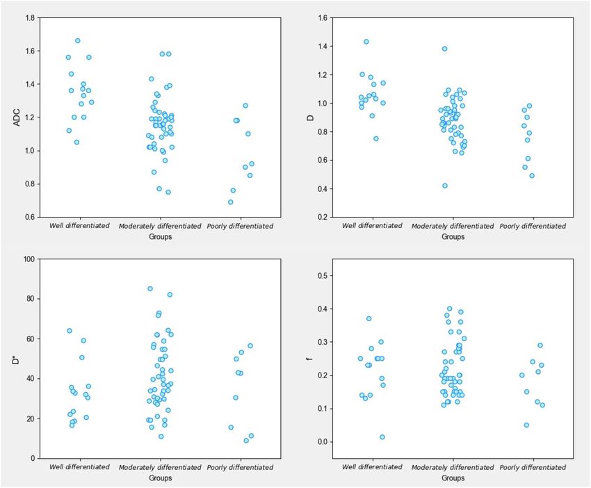
www.nature.com/scientificreports/
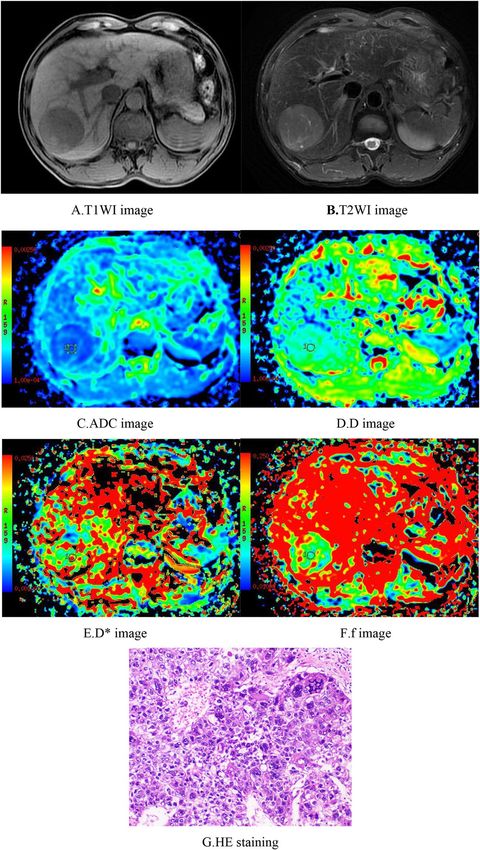
Figure 1. Female, 50 years old, HCC in the right lobe, well differentiated. (A) T1WI image. Low-signal area is
visible in the right hepatic lobe with a clear boundary. (B) T2WI images. The lesion showed a heterogeneous,
slightly high signal and a clear boundary. (C–F) ADC, D, D*, and f images; the measured values were 1.56 × 10⁻³
mm2/s, 1.04 × 10⁻³ mm2/s, 15.50 × 10⁻³ mm2/s, and 0.29, respectively. (G) HE staining (*100) showed well
differentiated HCC tissues.
microstructural changes within the tissue36. The results of this study showed that the D value was more effective
for identifying the histologic differentiation of HCC.
Scientific Reports | (2021) 11:10392 | https://doi.org/10.1038/s41598-021-89784-2 4
Vol:.(1234567890)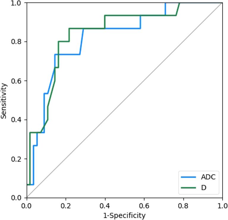
www.nature.com/scientificreports/
Figure 2. Male, 61 years old, HCC in the right hepatic lobe. (A) T1WI image. The lesion showed a low signal
and a relatively clear boundary. (B) T2WI image.The lesion showed a slightly high signal, with a relatively clear
boundary. (C–F) ADC, D, D*, and f images, respectively. The ADC value was 1.18 × 10⁻³ mm2/s, the D value was
0.92 × 10⁻³ mm2/s, the D* value was 30.8 × 10⁻³ mm2/s, and the f value was 0.18. G. HE staining (*100) showed
moderately differentiated HCC tissues.
Both the D* and f values can reflect microcirculatory perfusion-related information in tissues. The D* value
reflects the diffusion motion of microcirculation perfusion in the tissue capillary network, which is related to the
morphology of capillaries and the flow velocity of blood inside the capillaries. The f value represents the volume
ratio of microcirculation perfusion-related diffusion to total diffusion. It is affected by the capillary vascular
Scientific Reports | (2021) 11:10392 | https://doi.org/10.1038/s41598-021-89784-2 5
Vol.:(0123456789)www.nature.com/scientificreports/
Figure 3. Male, 29 years old, HCC in the right hepatic lobe. (A) T1WI image. The lesion showed a
heterogeneous low signal, an irregular morphology, and an unclear boundary. (B) T2WI image. The lesion
showed a heterogeneous, slightly high signal, an unclear boundary, and patchy necrosis inside. (C–F) ADC, D,
D*, and f images, respectively. The measured ADC was 1.10 × 10⁻³ mm2/s, the D value was 0.86 × 10⁻³ mm2/s, the
D* value was 29.90 × 10⁻³ mm2/s, and the f value was 0.16. (G) HE staining (*100) showed poorly differentiated
HCC tissues.
capacity within the tissues37. The D* and f values in this study were not significantly correlated with histologic
differentiation, which is consistent with the results of Woo et al.5, whereas some differences from the results of
Li35 and Granata et al.34 were noted.
The inconsistency of the above results may be related to the following factors: (1) different case composi-
tions: Yoon et al.18 showed that the D* and f values of hepatic lesions with a rich blood supply were higher than
those of lesions with a poor blood supply. Woo et al.5 found that the f value was positively correlated with the
enhancement ratio in the arterial phase of the tumours. In this study, some of the HCC lesions were character-
ized by a lack of blood supply, with no obvious enhancement or only mild heterogeneous enhancement in the
hepatic arterial phase, which may offset the high D* and f value effects of lesions with a rich blood supply. (2)
Tumour heterogeneity: A higher tumour histologic grade results in greater variability and a more complicated
Scientific Reports | (2021) 11:10392 | https://doi.org/10.1038/s41598-021-89784-2 6
Vol:.(1234567890)www.nature.com/scientificreports/
Figure 4. Scatter plots showing IVIM-DWI parameters in different HCC histologic differentiation groups.The
graphs were created by using Python 3.6 (https://www.python.org/).
Figure 5. The efficacy of IVIM-DWI parameters for the diagnosis of well differentiated HCC. The ROC curves
were created by using Python 3.6 (https://www.python.org/).
Scientific Reports | (2021) 11:10392 | https://doi.org/10.1038/s41598-021-89784-2 7
Vol.:(0123456789)www.nature.com/scientificreports/
microstructure, leading to greater differences in the D* and f values. (3) The microcirculation perfusion repre-
sented by the D* value might also include other physiological processes, such as physiological activities including
glandular secretion, as well as fluid flow inside the glandular ducts and c atheters38; the activities of these fluids
are difficult to differentiate from blood perfusion, which may affect the measurement of the D* and f values. (4)
The D* and f values have poor r epeatability39.
The limitations of this study are as follows: (1) the sample sizes were quite different among groups. In par-
ticular, the small number of cases in the poorly differentiated group may have led to bias in the results. The
sample size should be increased in the future to continue the study. (2) Manual delineation of the ROIs led to
certain unavoidable measurement errors. (3) A consensus regarding the number and distribution of b values is
still lacking. Different b values and distributions may result in differences in parameter m easurements12,13,40,41.
In summary, the ADC and D values of HCC tissues were significantly correlated with histologic differentia-
tion. ADC and D values can be used to preoperatively predict the degree of histologic differentiation of HCC,
and the D value has better diagnostic value.
Received: 7 January 2021; Accepted: 30 April 2021
References
1. Fitzmaurice, C. et al. Global, regional, and national cancer incidence, mortality, years of life lost, years lived with disability, and
disability-adjusted life-years for 32 cancer groups, 1990 to 2015: a systematic analysis for the Global Burden of Disease Study.
JAMA Oncol. 3, 524–548 (2017).
2. Yang, J. D. et al. A global view of hepatocellular carcinoma: trends, risk, prevention and management. Nat. Rev. Gastroenterol.
Hepatol. 16, 589–604 (2019).
3. [Diagnosis, management, and treatment of hepatocellular carcinoma (V2017)]. Zhonghua Gan Zang Bing Za Zhi 25, 886–895
(2017).
4. Zhou, L. et al. Factors predictive for long-term survival of male patients with hepatocellular carcinoma after curative resection. J.
Surg. Oncol. 95, 298–303 (2007).
5. Woo, S. et al. Intravoxel incoherent motion diffusion-weighted MR imaging of hepatocellular carcinoma: correlation with enhance-
ment degree and histologic grade. Radiology 270, 758–767 (2014).
6. Chen, J. et al. Preoperative evaluation of the histological grade of hepatocellular carcinoma with diffusion-weighted imaging: a
meta-analysis. PLoS ONE 10, 2015 (2015).
7. Li, X. et al. Combined application of gadoxetic acid disodium-enhanced magnetic resonance imaging (MRI) and diffusion-weighted
imaging (DWI) in the diagnosis of chronic liver disease-induced hepatocellular carcinoma: a meta-analysis. PLoS ONE 10, 2015
(2015).
8. Court, C. M. et al. Determination of hepatocellular carcinoma grade by needle biopsy is unreliable for liver transplant candidate
selection. Liver Transpl. 23, 1123–1132 (2017).
9. Taouli, B. & Koh, D. M. Diffusion-weighted MR imaging of the liver. Radiology 254, 47–66 (2010).
10. Nasu, K. et al. Diffusion-weighted imaging of surgically resected hepatocellular carcinoma: imaging characteristics and relationship
among signal intensity, apparent diffusion coefficient, and histopathologic grade. AJR Am. J. Roentgenol. 193, 438–444 (2009).
11. Le Bihan, D. et al. MR imaging of intravoxel incoherent motions: application to diffusion and perfusion in neurologic disorders.
Radiology 161, 401–407 (1986).
12. Yamada, I., Aung, W., Himeno, Y., Nakagawa, T. & Shibuya, H. Diffusion coefficients in abdominal organs and hepatic lesions:
evaluation with intravoxel incoherent motion echo-planar MR imaging. Radiology 210, 617–623 (1999).
13. Luciani, A. et al. Liver cirrhosis: intravoxel incoherent motion MR imaging—pilot study. Radiology 249, 891–899 (2008).
14. Chen, C. et al. Initial study of biexponential model of intravoxel incoherent motion magnetic resonance imaging in evaluation of
the liver fibrosis. Chin. Med. J. (Engl.) 127, 3082–3087 (2014).
15. Yoon, J. H. et al. Evaluation of hepatic fibrosis using intravoxel incoherent motion in diffusion-weighted liver MRI. J. Comput.
Assist. Tomogr. 38, 110–116 (2014).
16. Ichikawa, S. et al. MRI-based staging of hepatic fibrosis: comparison of intravoxel incoherent motion diffusion-weighted imaging
with magnetic resonance elastography. J. Magn. Reson. Imaging 42, 204–210 (2015).
17. Ichikawa, S. et al. Intravoxel incoherent motion imaging of focal hepatic lesions. J. Magn. Reson. Imaging 37, 1371–1376 (2013).
18. Yoon, J. H. et al. Evaluation of hepatic focal lesions using diffusion-weighted MR imaging: comparison of apparent diffusion coef-
ficient and intravoxel incoherent motion-derived parameters. J. Magn. Reson. Imaging 39, 276–285 (2014).
19. Watanabe, H. et al. Characterizing focal hepatic lesions by free-breathing intravoxel incoherent motion MRI at 3.0 T. Acta Radiol.
55, 1166–1173 (2014).
20. Klauss, M. et al. IVIM-diffusion-MRI for the differentiation of solid benign and malign hypervascular liver lesions—evaluation
with two different MR scanners. Eur. J. Radiol. 85, 1289–1294 (2016).
21. Choi, I. Y. et al. Intravoxel incoherent motion diffusion-weighted imaging for characterizing focal hepatic lesions: correlation with
lesion enhancement. J. Magn. Reson. Imaging 45, 1589–1598 (2017).
22. Peng, J. et al. Intravoxel incoherent motion diffusion-weighted imaging to differentiate hepatocellular carcinoma from intrahepatic
cholangiocarcinoma. Sci. Rep. 10, 7717 (2020).
23. Wagner, M. et al. Diffusion-weighted MR imaging for the regional characterization of liver tumors. Radiology 264, 464–472 (2012).
24. Shirota, N. et al. Intravoxel incoherent motion MRI as a biomarker of sorafenib treatment for advanced hepatocellular carcinoma:
a pilot study. Cancer Imaging 16, 1 (2016).
25. Wu, L. et al. ADCtotal ratio and D ratio derived from intravoxel incoherent motion early after TACE are independent predictors
for survival in hepatocellular carcinoma. J. Magn. Reson. Imaging 46, 820–830 (2017).
26. Peng, J. et al. Intravoxel incoherent motion diffusion weighted imaging for the therapeutic response of transarterial chemoembo-
lization for hepatocellular carcinoma. J. Cancer Ther. 10, 591–601 (2019).
27. Park, Y. S. et al. Using intravoxel incoherent motion (IVIM) MR imaging to predict lipiodol uptake in patients with hepatocellular
carcinoma following transcatheter arterial chemoembolization: a preliminary result. Magn. Reson. Imaging 32, 638–646 (2014).
28. Nishie, A. et al. Diagnostic performance of apparent diffusion coefficient for predicting histological grade of hepatocellular car-
cinoma. Eur. J. Radiol. 80, e29–e33 (2011).
29. Nakanishi, M. et al. Relationship between diffusion-weighted magnetic resonance imaging and histological tumor grading of
hepatocellular carcinoma. Ann. Surg. Oncol. 19, 1302–1309 (2012).
30. Wei, Y. et al. Preliminary study of whole-tumor volume analysis of mono-exponential and intravoxel incoherent motion models
in the preoperative histologic grading of hepatocellular carcinoma. Zhonghua Yi Xue Za Zhi 98, 2460–2465 (2018).
Scientific Reports | (2021) 11:10392 | https://doi.org/10.1038/s41598-021-89784-2 8
Vol:.(1234567890)www.nature.com/scientificreports/
31. Zhu, S. C. et al. Intravoxel incoherent motion diffusion-weighted magnetic resonance imaging for predicting histological grade of
hepatocellular carcinoma: comparison with conventional diffusion-weighted imaging. World J. Gastroenterol. 24, 929–940 (2018).
32. Shan, Q. et al. Evaluating histologic differentiation of hepatitis B virus-related hepatocellular carcinoma using intravoxel incoherent
motion and AFP levels alone and in combination. Abdom. Radiol. (N. Y.) 42, 2079–2088 (2017).
33. Sokmen, B. K. et al. Value of intravoxel incoherent motion for hepatocellular carcinoma grading. Transpl. Proc. 51, 1861–1866
(2019).
34. Granata, V. et al. Intravoxel incoherent motion (IVIM) in diffusion-weighted imaging (DWI) for hepatocellular carcinoma: cor-
relation with histologic grade. Oncotarget 7, 79357–79364 (2016).
35. Li, M., Zheng, X. J., Huang, Z. X. & Song, B. Predicting histological grade of HCC in rats using intravoxel incoherent motion
imaging. Sichuan Da Xue Xue Bao Yi Xue Ban 49, 243–247 (2018).
36. Le Bihan, D. et al. Separation of diffusion and perfusion in intravoxel incoherent motion MR imaging. Radiology 168, 497–505
(1988).
37. Le Bihan, D. & Turner, R. The capillary network: A link between IVIM and classical perfusion. Magn. Reson. Med. 27, 171–178
(1992).
38. Dm, K., Dj, C. & Mr, O. Intravoxel incoherent motion in body diffusion-weighted MRI: reality and challenges. Am. J. Roentgenol.
196, 1351–1361 (2011).
39. Kakite, S. et al. Hepatocellular carcinoma: short-term reproducibility of apparent diffusion coefficient and intravoxel incoherent
motion parameters at 3.0 T. J. Magn. Reson. Imaging 41, 149–156 (2015).
40. Dyvorne, H., Jajamovich, G., Kakite, S., Kuehn, B. & Taouli, B. Intravoxel incoherent motion diffusion imaging of the liver: optimal
b-value subsampling and impact on parameter precision and reproducibility. Eur. J. Radiol. 83, 2109–2113 (2014).
41. Patel, J. et al. Diagnosis of cirrhosis with intravoxel incoherent motion diffusion MRI and dynamic contrast-enhanced MRI alone
and in combination: preliminary experience. J. Magn. Reson. Imaging 31, 589–600 (2010).
Acknowledgements
This study was supported by the projects of the Department of Science and Technology of Sichuan Province
(2016JY0105).
Author contributions
Y.Z., G.Y., J.Z. and C.-Y. conducted the measurements, analysed the results, and prepared the manuscript; L.Y.
wrote and revised the manuscript; J.-D.L. and X.-M.Z. designed the study; X.-Q.G., Y.-Y.T., R.W. and J.P. collected
the data and edited the manuscript.
Competing interests
The authors declare no competing interests.
Additional information
Correspondence and requests for materials should be addressed to L.Y.
Reprints and permissions information is available at www.nature.com/reprints.
Publisher’s note Springer Nature remains neutral with regard to jurisdictional claims in published maps and
institutional affiliations.
Open Access This article is licensed under a Creative Commons Attribution 4.0 International
License, which permits use, sharing, adaptation, distribution and reproduction in any medium or
format, as long as you give appropriate credit to the original author(s) and the source, provide a link to the
Creative Commons licence, and indicate if changes were made. The images or other third party material in this
article are included in the article’s Creative Commons licence, unless indicated otherwise in a credit line to the
material. If material is not included in the article’s Creative Commons licence and your intended use is not
permitted by statutory regulation or exceeds the permitted use, you will need to obtain permission directly from
the copyright holder. To view a copy of this licence, visit http://creativecommons.org/licenses/by/4.0/.
© The Author(s) 2021
Scientific Reports | (2021) 11:10392 | https://doi.org/10.1038/s41598-021-89784-2 9
Vol.:(0123456789)You can also read