A Mouse Model of Ulcerative Cutaneous Leishmaniasis by Leishmania (Viannia) panamensis to Investigate Infection, Pathogenesis, Immunity, and ...
←
→
Page content transcription
If your browser does not render page correctly, please read the page content below
ORIGINAL RESEARCH
                                                                                                                                                  published: 13 June 2022
                                                                                                                                         doi: 10.3389/fmicb.2022.907631

                                              A Mouse Model of Ulcerative
                                              Cutaneous Leishmaniasis by
                                              Leishmania (Viannia) panamensis to
                                              Investigate Infection, Pathogenesis,
                                              Immunity, and Therapeutics
                                              Natalia Muñoz-Durango 1†, Alexander Gómez 1†, Natalia García-Valencia 1, Miguel Roldán 2,
                                              Marcela Ochoa 3, David E. Bautista-Erazo 1 and José R. Ramírez-Pineda 1*
                                              1
                                               Grupo Inmunomodulación (GIM), Instituto de Investigaciones Médicas, Facultad de Medicina, Corporación Académica para
                                              el Estudio de Patologías Tropicales (CAEPT), Universidad de Antioquia, Medellín, Colombia, 2 Instituto de Patología, Facultad
                                              de Medicina, Universidad de Antioquia, Medellín, Colombia, 3 Programa de Estudio y Control de Enfermedades Tropicales
                                              (PECET), Facultad de Medicina, Universidad de Antioquia, Medellín, Colombia

                          Edited by:
                        Daniel Pletzer,       A mouse model of cutaneous leishmaniasis (CL) by Leishmania (Viannia) panamensis (L(V)p)
    University of Otago, New Zealand          that reproduces the characteristics of the human disease remains elusive. Here we report the
                      Reviewed by:            development of a CL model that uses a mouse-adapted L(V)p isolate to reproducibly induce
                Morad-Remy Muhsin,
                                              a dermal disease with a remarkable similarity to human CL. BALB/c mice infected intradermally
   University of Otago, New Zealand
           Fabienne Tacchini-Cottier,         in the ear with 105 stationary UA-946 L(V)p promastigotes develop a progressive cutaneous
 University of Lausanne, Switzerland          disease that exhibits the typical ulcerated lesions with indurated borders observed in CL
                 Fátima Ribeiro-Dias,
Universidade Federal de Goiás, Brazil         patients. Although most of parasites in the inoculum die within the first week of infection, the
                     *Correspondence:         survivors vigorously multiply at the infection site during the following weeks, paralleling disease
                 José R. Ramírez-Pineda       appearance and aggravation. Regional lymphadenopathy as well as lymphatic dissemination
         jrobinson.ramirez@udea.edu.co
                                              of parasites to draining lymph nodes (dLN) was evidenced early after infection. Viable parasites
     †
         These authors have contributed
                                              were also isolated from spleen at later timepoints indicating systemic parasitic dissemination,
                    equally to this work
                                              but, strikingly, no signs of systemic disease were observed. Increasing numbers of myeloid
                      Specialty section:      cells and T lymphocytes producing IFNγ and IL-4 were observed in the dLN as disease
            This article was submitted to
                                              progressed. A mixed adaptive L(V)p-specific T cell-mediated response was induced, since ex
         Infectious Agents and Disease,
                  a section of the journal    vivo recall experiments using dLN cells and splenocytes revealed the production of type 1
                Frontiers in Microbiology     (IFNγ, IL-2), type 2 (IL-4, IL-13), regulatory (IL-10), and inflammatory (GM-CSF, IL-3) cytokines.
             Received: 30 March 2022          Humoral adaptive response was characterized by early production of IgG1- followed by IgG2a-
              Accepted: 25 May 2022
             Published: 13 June 2022          type of L(V)p-specific antibodies. IFNγ/IL-4 and IgG2a/IgG1 ratios indicated that the initial
                             Citation:        non-protective Th2 response was redirected toward a protective Th1 response. In situ studies
        Muñoz-Durango N, Gómez A,             revealed a profuse recruitment of myeloid cells and of IFNγ- and IL-4-producing T lymphocytes
        García-Valencia N, Roldán M,
                                              to the site of infection, and the typical histopathological changes induced by dermotropic
    Ochoa M, Bautista-Erazo DE and
  Ramírez-Pineda JR (2022) A Mouse            Leishmania species. Evidence that this model is suitable to investigate pharmacological and
       Model of Ulcerative Cutaneous          immunomodulatory interventions, as well as for antigen discovery and vaccine development,
         Leishmaniasis by Leishmania
  (Viannia) panamensis to Investigate         is also presented. Altogether, these results support the validity and utility of this novel mouse
   Infection, Pathogenesis, Immunity,         model to study the pathogenesis, immunity, and therapeutics of L(V)p infections.
                    and Therapeutics.
         Front. Microbiol. 13:907631.         Keywords: cutaneous leishmaniasis, Leishmania (Viannia) panamensis, mouse model, vaccine, immunopathology,
    doi: 10.3389/fmicb.2022.907631            forward vaccinology

Frontiers in Microbiology | www.frontiersin.org                                       1                                              June 2022 | Volume 13 | Article 907631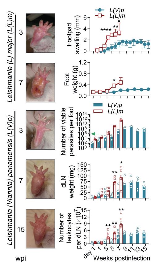
Muñoz-Durango et al.                                                                                                      Ulcerative Cutaneous Leishmaniasis Mouse Model

INTRODUCTION                                                                                  studies have revealed intriguing characteristics of the natural history
                                                                                              of L(V)p infection in endemic regions, such as the variable
Mouse models have been extremely useful to dissect the immune                                 frequency of asymptomatic infections and spontaneous healing,
mechanisms mediating leishmaniasis pathogenesis and host                                      latency, evolution to chronic disease and/or recurrence, and
resistance to Leishmania parasites, as prototypic intracellular                               metastatic behavior (Weigle and Saravia, 1996; World Health
microorganisms. Inbred mouse strains have been infected with                                  Organization, 2010). It is generally assumed that this broad spectrum
different Leishmania species, and a variety of outcomes, ranging                              of outcomes upon infection is the result of the interactions between
from asymptomatic infection to fatal systemic disease, have been                              parasite- and host-derived factors in a particular environment.
observed. The resulting picture of the reported experimental                                      Syrian hamsters have been claimed as a suitable animal model
work is that whereas some pathways are used by the host immune                                to investigate host response to L(V)p and other Leishmania
system to deal with all tested Leishmania species, other pathways                             species of the Viannia subgenus, since persistent cutaneous lesions
operate in a species-specific manner. For instance, whereas a                                 are induced upon promastigote inoculation in those animals
sustained Th1 IFNγ-mediated response is required for protection                               (Hommel et al., 1995; Osorio et al., 2003). Interestingly, as
in all tested models, a non-healing disease might be the result                               observed in humans, parasites persist for long periods and
of a dominant Th2 response, a weak Th1 response, a prominent                                  disseminate not only to draining lymph nodes (dLN) but also
regulatory response (e.g., IL-10- or TGFβ-mediated) or even                                   to distant tissues where metastatic lesions are observed (Travi
from B cell responses, depending on the species involved                                      et al., 1988; Saravia et al., 1990; Martinez et al., 1991). Cytokine
(McMahon-Pratt and Alexander, 2004). Innate immune cells and                                  mRNA measurements in the site of infection, dLN, and spleen
pathways are also differently modulated by Leishmania species,                                evidenced the expression of IFNγ, IL-10, IL-4, IL-12, and TGFβ
contributing to disease or protection in a different manner (Hurrell                          (Travi et al., 2002; Osorio et al., 2003; Espitia et al., 2010),
et al., 2016; Zamboni and Sacks, 2019). Moreover, host response                               which resembles the mixed Th1/Th2/regulatory profile observed
to Leishmania can further diverge at the strain level within the                              in humans (Bosque et al., 2000; Trujillo et al., 2002; Castilho
same species, as indicated by the inflammasome/neutrophil-                                    et al., 2010; Díaz et al., 2010). Whereas this model has been
mediated non-healing response observed with the Seidman strain                                useful to investigate mechanisms of parasite virulence (Acestor
of Leishmania (Leishmania) major (L(L)m) even in the presence                                 et al., 2006), the unavailability of genetically homogeneous inbred
of a robust Th1 response (Charmoy et al., 2016). Conversely,                                  strains, together with the scarcity of immunologic/molecular
many virulence factors that have been identified in some species                              tools and genetic manipulation systems in hamsters, has limited
(e.g., the lipophosphoglycan, the A2 protein, or cysteine protease                            its use to map the host immune pathways that are relevant for
enzymes) are not expressed or are non-operative in other                                      pathogenesis or immunity. Although murine models would
(McMahon-Pratt and Alexander, 2004; Rossi and Fasel, 2018a).                                  be convenient for this purpose, species of Viannia subgenus
This species-specific nature of the mammalian host–Leishmania                                 were early recognized as poorly infective in mice (Hommel
interaction implies that information gained with a particular                                 et al., 1995; Travi et al., 2002; McMahon-Pratt and Alexander,
species cannot be assumed to apply across the whole genus                                     2004), probably due to the difficulties to reproducibly induce
or subgenus.                                                                                  measurable and sustained cutaneous lesions in these animals.
    Leishmania (Viannia) panamensis (L(V)p) is one of the most                                Even in BALB/c mice, where most of mammalian-infecting
important causal agents of American cutaneous leishmaniasis (CL)                              Leishmania species can grow leading to overt disease, Leishmania
and is responsible for a large proportion of leishmaniasis cases                              (Viannia) guyanensis (L(V)g) and Leishmania (Viannia) braziliensis
in Central and South America (Alvar et al., 2012; Aronson et al.,                             (L(V)b) infection are generally asymptomatic or induce a mild
2020). L(V)p has been isolated from patients with different                                   disease that resolves within few weeks with almost complete
manifestations of the disease, including the localized (LCL), mucosal                         elimination of parasites (de Moura et al., 2005; Sousa-Franco
(ML), and disseminated (DisCL) forms (World Health Organization,                              et al., 2006; de Oliveira and Brodskyn, 2012). Although McMahon-
2010; Vélez et al., 2015; Scorza et al., 2017). Clinical–epidemiological                      Pratt’s group reported the injection of putative infective forms
                                                                                              in the foot of BALB/c mice, the resulting disease consists of
                                                                                              small non-ulcerative and poorly characterized lesions (Castilho
Abbreviations: Ag, Antigen; BCA, Bicinchoninic acid assay; CD, Cluster of
                                                                                              et al., 2010), which appear very limited to model the tissue-
differentiation; CL, Cutaneous leishmaniasis; ConA, Concanavalin A; CpG, CpG
Oligodeoxynucleotide; DC, Dendritic cell(s); DisCL, Disseminated CL; dLN, Draining            damaging characteristics of human CL by L(V)p. Thus, to our
lymph node; ELISA, Enzyme-linked immunosorbent assay; GM-CSF, Granulocyte-                    knowledge, a suitable murine model of CL by L(V)p that mimics
macrophage Colony-stimulating factor; HRP, Horseradish Peroxidase; id, Intradermal;           the characteristics of the human disease has not been developed
IFNγ, Interferon Gamma; Ig, Immunoglobulin; IL, Interleukin; ip, Intraperitoneal;             and characterized. Here, we report a clinical isolate of L(V)p
L(V), Leishmania (Viannia); L(V)b, Leishmania (Viannia) braziliensis; L(V)g, Leishmania
(Viannia) guyanensis; L(V)p, Leishmania (Viannia) panamensis; L(L)m, Leishmania
                                                                                              able to reproducibly induce chronic ulcerative lesions upon
(Leishmania) major; LCL, Localized CL; LN, Lymph node; mAb, Monoclonal                        intradermal (id) injection in the ear of BALB/c mice that closely
antibody; ML, Mucosal leishmaniasis; MW, Molecular weight; NNN, Novy-MacNeal-                 resembles the human disease caused by this species. The kinetics
Nicolle medium; PBMC, Peripheral blood mononuclear cell; PBS, Phosphate-buffered              of clinical, parasitological, immunological, and histopathological
saline; PMA, Phorbol 12-myristate 13-acetate; RCL, Recurrent CL; SC, Subcutaneous;            response to the infection was systematically investigated, and
SDM, Schneider’s drosophila medium; SDS, Sodium dodecyl sulfate; SPF, Specific
pathogen-free; TGFβ, Transforming growth factor beta; Th, T helper; TNFα, Tumor
                                                                                              the resulting model was used to evaluate the efficacy of
necrosis factor alpha; Tregs, Regulatory T cells; VEGF, Vascular endothelial growth           pharmacological and immunological interventions. Our findings
factor; wpi, Weeks postinfection.                                                             demonstrate that BALB/c mice are susceptible to infection with

Frontiers in Microbiology | www.frontiersin.org                                           2                                         June 2022 | Volume 13 | Article 907631
Muñoz-Durango et al. Ulcerative Cutaneous Leishmaniasis Mouse Model
L(V)p, providing a suitable new model to study the pathogenesis Anderson et al., 2008) and myeloid and T cells analyzed. For
and immunity to Leishmania parasites and to assess drug and further details on methodology and data analysis, see
vaccine candidates. Supplementary Materials and Methods.
MATERIALS AND METHODS RESULTS
BALB/c mice (Charles River, United States) were used in all
Selection of a Suitable L(V)p Human
experiments, and procedures were approved by the institutional
ethical animal committee. From the twelve clinical L(V)p isolates
Isolate for BALB/c Mouse Infections
Although our previous efforts to induce a cutaneous disease
tested in mice (Supplementary Table S1), the UA-946 stock
in mice with L(V)p were disappointing, the finding of viable
(MHOM/CO/93/UA-946) was chosen for the establishment of
parasites and a robust IgG L(V)p-specific Ab response in
the model. The genome sequence of the UA-946 L(V)p isolate
chronically infected asymptomatic animals (not shown) indicated
has been reported (Urrea et al., 2018). L(L)m (MHOM/IL/81/
that L(V)p was able to infect, survive, and persist in mouse
BNI; Ramírez-Pineda et al., 2004) was used in some experiments
tissues and to elicit a parasite-specific adaptive response. On
for comparison. Total Leishmania antigen (Ag) was prepared
the bases of the significant heterogeneity exhibited by field
by freezing/thawing cycles, and fractionation methods were
isolates of the same Leishmania species on mouse pathogenicity
used for some procedures. Animals were infected into the
(Monroy-Ostria et al., 1994; Barral et al., 1996; de Oliveira
right hind footpad, the base of the tail, or the right ear,
and Brodskyn, 2012; Loeuillet et al., 2016) and the reports
subcutaneously (sc) or id, with stationary promastigotes, and
using serial passages in vivo to adapt microbial pathogens to
a clinical follow-up was performed consisting in the assessment
grow in mouse tissues and produce successful infections
of body weight and lesion development. The footpad thickness
(Marchetti et al., 1995; Xu et al., 2011; Li and McCray, 2020),
and lesion size were registered weekly with the help of a digital
we implemented a two-step strategy of selection-adaptation.
caliper (precision: 0.02 mm, model MT-00855, Uyustools, China).
The strategy consisted in the initial identification of clinical
The footpad swelling was calculated as the difference in the
isolates that better infected mouse tissues followed by a
thickness (in mm) between the infected and the contralateral
second step of in vivo adaptation (as described in
non-infected foot. Lesion size in the base of the tail and the
Supplementary Materials and Methods). Thus, we tested 12
ear was reported as the area (in mm2) by measuring the two
human isolates representing different clinical forms of
crossed diameters of lesions and calculating with the formula
2 leishmaniasis caused by L(V)p (localized, mucosal, disseminated,
D1 + D 2 or recurrent cutaneous leishmaniasis; LCL, ML, DisCL or RCL,
A=π . At different timepoints after infection, mice
4 respectively; Supplementary Table S1), and although most of
were euthanized to obtain tissues for parasitological,
the L(V)p infections were asymptomatic, three isolates produced
histopathological, and immunological analysis. Infected tissues
mild symptomatic infections in the footpad of BALB/c mice
were removed, weighted, and cultured to determine the parasite
(Supplementary Figure S1). As reported for other species
burden per organ (footpad and ear) or mg of tissue (base of
(Monroy-Ostria et al., 1994; Alves-Ferreira et al., 2015; Loeuillet
the tail) by using a limiting dilution protocol (Buffet et al.,
et al., 2016), there was no relation between human and mouse
1995; Lima et al., 1997). Draining LNs and spleens were
pathogenicity/virulence. As expected, parasites could be cultured
weighted and cell suspensions seeded in promastigote culture
from infected tissue and the dLN of animals infected with
medium and incubated at 26oC to determine the presence of
the three pathogenic isolates 3 or 8 months after infection
viable parasites. In chemotherapeutic experiments, mice received
(Supplementary Table S2). This indicated that some human
treatment: a weekly intraperitoneal (ip) injection of Glucantime®,
L(V)p isolates successfully infect BALB/c mice, provoking a
500 mg/kg, for 4 weeks, or Miltefosine, 20 mg/kg/day, orally for
measurable inflammation in the footpad. One isolate, coded
five consecutive days, administered after disease establishment
UA-946 and exhibiting the most consistent results in independent
(4–5 weeks postinfection). In immunomodulatory interventions,
experiments using several routes/sites, was selected for further
the synthetic CpG-containing oligodeoxynucleotide 1826 (CpG;
in vivo adaptation via serial passages in BALB/c mice and for
5′-TCCATG
subsequent characterization.
ACGTTCCTGACGTT-3′; phosphorothioate-modified; Integrated
DNA Technologies - IDT, United States) was used for co-delivery
or as a vaccine adjuvant with total Ag or protein fractions. Localized Non-ulcerative Chronic Lesions
Schemes, doses, and administration routes employed for each in the Footpads of Mice Infected With
independent experiment are specified in the corresponding L(V)p
figure legend. Cytokine concentrations in supernatants of dLN We first evaluated the kinetics of lesion development and
or spleen cells stimulated with 10 μg L(V)p Ag for 72 h were parasite replication after UA-946 L(V)p inoculation in the
determined by ELISA and/or Luminex. L(V)p-specific IgG1 footpad of BALB/c mice and compared it to that of L(L)m,
and IgG2a antibodies in serum were quantified by ELISA. Flow a well-documented model of leishmaniasis susceptibility.
cytometry of cell suspensions from infected tissue and Although infection with L(V)p was asymptomatic during the
dLN was performed as reported (Duthie et al., 2007; first 3 weeks, it progressed to a nodular non-ulcerated
Frontiers in Microbiology | www.frontiersin.org 3 June 2022 | Volume 13 | Article 907631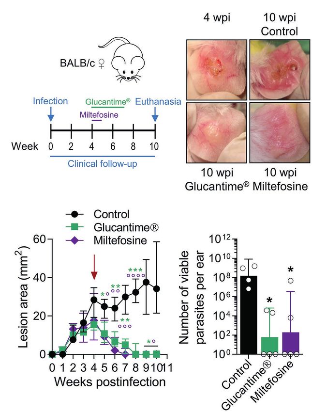
Muñoz-Durango et al.                                                                                             Ulcerative Cutaneous Leishmaniasis Mouse Model

inflammatory lesion that reached maximal size around 8–9 weeks                        footpad swelling tended to reduce toward weeks 14 and 15
postinfection (Figures 1A,B). No further aggravation of the                           postinfection (Figure 1B). Mice looked healthy, and body weight
inflammatory lesions was observed throughout the study, instead                       and behavior were comparable to non-infected animals. In
                                                                                      contrast and as expected, mice infected with L(L)m exhibited
                                                                                      footpad inflammation as early as 2 weeks postinfection that
   A                              B
                                                                                      rapidly progressed to ulcers and aggravated to necrosis by the
                                                                                      7th week postinfection (Figures 1A,B). At this time, L(L)
                                                                                      m-infected mice displayed weight loss and symptoms of systemic
                                                                                      disease (hypoactivity and piloerection), forcing humane sacrifice.
                                                                                      Foot weight measurement confirmed that whereas both species
                                                                                      promoted a progressive inflammatory process in BALB/c mice,
                                                                                      inflammation induced by L(V)p was slower and limited
                                  C                                                   (Figure 1C). From the 106 L(V)p parasites injected into BALB/c
                                                                                      mice only hundreds could be recovered 1 day postinfection;
                                                                                      however, this surviving fraction was able to actively multiply
                                                                                      within the following 11 weeks in a 6-log factor (Figure 1D).
                                                                                      Interestingly, for the remaining observation period, viable parasite
                                                                                      loads stabilized indicating an effective control of parasite
                                  D
                                                                                      replication by the host during the chronic phase. From a similar
                                                                                      inoculum of L(L)m, a bigger fraction (thousands) survived the
                                                                                      first day, and surviving parasites rapidly replicated (8-log factor)
                                                                                      to reach levels of hundreds of billions within few weeks. These
                                                                                      results indicate that L(V)p provokes in the footpads of BALB/c
                                                                                      mice a non-ulcerative, self-limited, and localized chronic lesion
                                                                                      that contrasts with the severe, systemic, and potentially lethal
                                                                                      disease caused by L(L)m in these mice.
                                  E

                                                                                      Infection With L(V)p in the Ear Dermis
                                                                                      Induces Chronic Ulcers That Closely
                                                                                      Resemble Human CL
                                                                                      The confirmation of active parasite replication in the footpads
                                  F
                                                                                      of mice infected with L(V)p prompted us to examine the
                                                                                      response to infection in other anatomical locations. In pilot
                                                                                      experiments, we found that injection of 105 parasites in the
                                                                                      base of the tail or the ear was sufficient to induce lesions
                                                                                      within 4 weeks postinfection (data not shown). Therefore, this
                                                                                      inoculum was chosen for further experiments. BALB/c mice
                                                                                      infected sc in the base of the tail exhibited an early nodular
                                                                                      small lesion that progressed to either larger nodules or
                                                                                      ulcerative lesions within the following weeks postinfection
  FIGURE 1 | Localized non-ulcerative chronic lesions in the footpads of mice
                                                                                      (Supplementary Figures S2A,B). Although some animals
  infected with L(V)p. BALB/c mice were infected subcutaneously (sc) with 106
  Leishmania (Viannia) panamensis (L(V)p) stationary promastigotes in the right       developed typical ulcers, other never developed ulcerated lesions.
  footpad and monitored weekly. In parallel, a group of mice were infected with       Among different experiments, lesions (either nodules or ulcers)
  a similar inoculum of Leishmania (Leishmania) major (L(L)m) promastigotes as        tended to be heterogeneous in size and appearance and less
  a control with a highly pathogenic Leishmania species in BALB/c mice.               reproducible (Supplementary Figures S2A–D). The kinetics
  Representative photographs at the indicated time postinfection are presented
                                                                                      of parasitic load at the infection site, again, evidenced the
  (A). The severity of lesions in mice infected with L(L)m at the 7th week
  postinfection obligated immediate euthanasia. Footpad swelling was                  presence of few viable parasites at the asymptomatic early
  individually measured and graphed (B). At the indicated time, mice were             phase, followed by an active phase of parasite multiplication,
  sacrificed and the weight (C) and the number of viable parasites (D) of the         which coincides with the onset of disease, and then by a late
  foot determined. Draining lymph nodes (dLN) were also removed and the               chronic phase where the control of parasite replication was
  weight (E) and cellularity (F) determined. Results are expressed as the
  median ± interquartile range (B), median ± range (C), geometric mean (D) or
                                                                                      observed (Supplementary Figure S2E).
  median (E,F) from 2–7 mice per time/group. The arrow in (D) indicates the              In humans, L(V)p promastigotes are deposited in the dermis
  infective inoculum. wpi, weeks postinfection. *p < 0.05, **p < 0.01, and            during sandfly biting, and after an incubation period that ranges
  ****p < 0.0001 [mixed-effects analysis (B); two-way ANOVA (C) with Geisser–         from 2 weeks to several months, the infiltration of immune
  Greenhouse correction and Bonferroni’s multiple comparisons post-hoc test
                                                                                      cells leads to the formation of a small papule that progresses
  for multiple comparison through time; Mann–Whitney U-test (E,F)].
                                                                                      to form the typical ulcerated lesion that characterizes LCL by

Frontiers in Microbiology | www.frontiersin.org                                   4                                        June 2022 | Volume 13 | Article 907631
Muñoz-Durango et al.                                                                                                  Ulcerative Cutaneous Leishmaniasis Mouse Model

L(V) species. Although spontaneous healing is observed in a                               inoculum typically ranged from 2 to 3 weeks but can be extended
significant proportion of CL patients, skin lesions might last                            up to 7 weeks when lower infective challenges are used
from months to years (Weigle and Saravia, 1996). We therefore                             (Figure 2B; Supplementary Figure S3). From week 3 to 6
switched to a more physiological route of infection by injecting                          postinfection, most of animals (usually over 90%) presented
BALB/c mice id in the ear with UA-946 L(V)p promastigotes                                 ulcers and lesions that looked similar in appearance and size
and monitoring the animals clinically and parasitologically. As                           among mice. By week 7, the average of lesion size stabilized,
shown in Figure 2A, mice developed inflammatory lesions                                   with some animals maintaining their lesion or even improving,
that progressed to small nodules and subsequently to ulcers                               and others exhibiting aggravation (Figure 2B), as indicated
with an appearance that was strikingly similar to the typical                             by the presence of larger ulcers with necrotic foci. Although
ulcerative lesions observed in human CL by L(V)p, consisting                              overall few live parasites (0.1% of the inoculum) were isolated
of a well-circumscribed ulcer, with a granular and crusted                                at day 1 and 7 postinfection, the vigorous parasite multiplication
base, and indurated borders (Weigle and Saravia, 1996; Aronson                            that took place in the following weeks preceding disease onset
et al., 2020). The incubation period after a 105 promastigotes                            and progression demonstrate that survivors were sufficient to
                                                                                          establish a productive infection (Figures 2A–D). Parasite growth
                                                                                          stabilized at later timepoints (7 to 9 week postinfection), indicating
   A                                B                                                     effective control of replication by host (Figure 2D). Interestingly,
                                                                                          in spite that the slope of parasite growth significantly flattened
                                                                                          after 5 weeks postinfection, ulcerative lesions remained or
                                                                                          aggravated in many of the animals (Figures 2B,D), indicating
                                                                                          that the pathological processes that maintain ulcers and tissue
                                                                                          damage persist even in the presence of an effective antiparasitic
                                                                                          response during this chronic phase of the disease. Independent
                                                                                          experiments in which extended follow-up was performed
                                                                                          demonstrated the chronic, yet auto-resolutive, nature of this
                                                                                          model (Supplementary Figure S4). While early (up to week
                                                                                          10 postinfection) lesion resolution is observed in very few
                                    C
                                                                                          animals, the majority exhibits large, often mutilating, chronic
                                                                                          lesions that resolve slowly and require up to 24 weeks to heal
                                                                                          completely (Figure 2B; Supplementary Figure S4).
                                                                                              Collectively, these results demonstrate that BALB/c mice
                                                                                          infected id in the ear with UA-946 L(V)p develop an evident
                                                                                          cutaneous disease that closely replicates human CL lesions.
                                                                                          This adapted strain has been maintained and used in our
                                                                                          laboratory for years with a remarkable reproducibility
                                                                                          (Supplementary Figure S5). These two reasons prompted us
                                    D                                                     to further characterize other aspects of the host response at
                                                                                          this anatomical location.

                                                                                          Lymphadenopathy and Parasite
                                                                                          Dissemination in the Absence of Systemic
                                                                                          Disease
                                                                                          Regional lymphadenopathy is a common early clinical feature
                                                                                          of CL by L(V) that can precede lesion appearance (Palma
                                                                                          et al., 1991; Weigle and Saravia, 1996; Bomfim et al., 2007;
                                                                                          World Health Organization, 2010). Compared to non-infected,
                                                                                          L(V)p-infected mice had dLNs that progressively increased
  FIGURE 2 | Infection with L(V)p in the ear dermis induces chronic ulcers that
  closely resemble human CL. BALB/c mice were infected with 105 L(V)p                     in weight along infection, which parallels an increase in the
  stationary promastigotes id in the right ear and monitored weekly.                      number of leukocytes (Figure 3A), suggesting progressive
  Representative photographs at the indicated time postinfection are presented            cell recruitment and/or proliferation in this organ. Maximal
  (A). The size of the lesion was measured and graphed as the median [thick               growth of dLN was observed at later timepoints of infection,
  line] and for each individual mouse [thin lines] (B). At the indicated time, mice
  were sacrificed, and the ears removed to measure the weight (C) as an
                                                                                          which coincides with higher disease severity and parasitic
  indicator of inflammation. Results are presented as the median ± interquartile          loads (Figures 2, 3A). Mice infected with L(V)p in the footpad
  range in (C). The number of viable parasites (D) in the ears was also                   (Figures 1E,F) or the base of the tail (not shown) presented
  determined by limiting dilution and graphed, with lines representing the                a similar kinetics of dLN swelling. Notably, viable parasites
  geometric mean. The arrow in (D) indicates the infective inoculum. n = 3–5
                                                                                          could be isolated from the dLN of most of mice as soon as
  mice per time. wpi: weeks postinfection.
                                                                                          the week 1 postinfection, which has been also documented

Frontiers in Microbiology | www.frontiersin.org                                       5                                         June 2022 | Volume 13 | Article 907631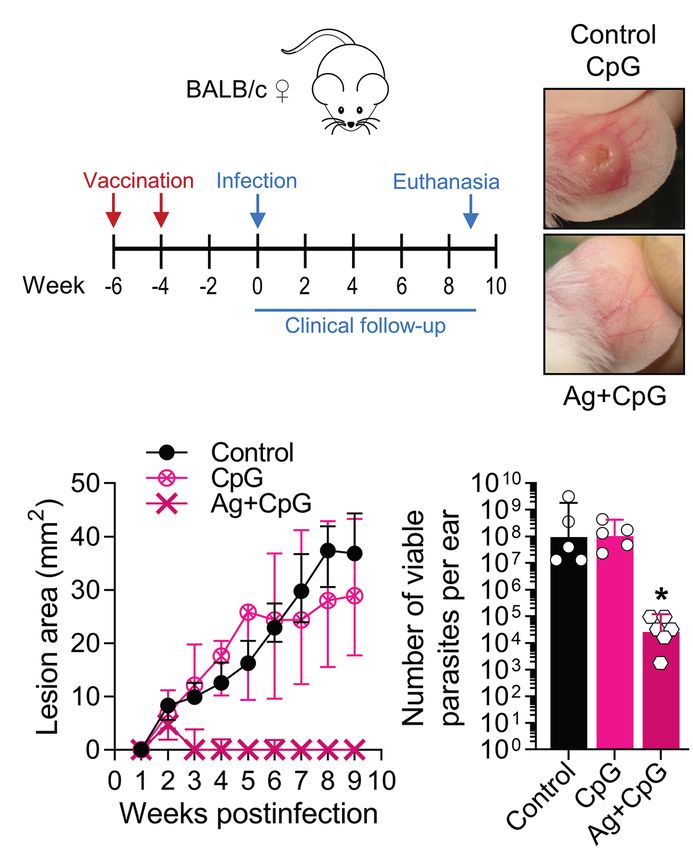
Muñoz-Durango et al.                                                                                              Ulcerative Cutaneous Leishmaniasis Mouse Model

                                                                                         numbers of CD3+ CD4+, CD3+ CD8+, and CD3+ double-
            A                                                                            negative T cells observed during the study period
                                                                                         (Supplementary Figures S6B, S7B) that most likely were
                                                                                         the result of massive T cell expansion upon L(V)p Ag
                                                                                         presentation. Although less accentuated, the weight and
                                                                                         cellularity of the spleen also increased after infection, and
                                                                                         maximal values were observed at the latest timepoint evaluated
                                                                                         (Figure 3B). Furthermore, parasites also reached the spleen
                                                                                         of some mice by the week 3 postinfection and were detectable
                                                                                         in all mice at 9 weeks postinfection (Figure 3B), suggesting
                                                                                         hematogenous besides lymphatic dissemination. Notably, the
                                                                                         evident parasite dissemination to proximal and distal lymphoid
                                                                                         organs was not associated with signs of systemic disease,
            B
                                                                                         since animals looked healthy and total body weight gain was
                                                                                         normal and comparable to non-infected mice (Figure 3C).
                                                                                         Mice infected at the footpad, or the base of the tail also
                                                                                         remained free of clinical signs of systemic disease. Altogether,
                                                                                         these findings demonstrate that cutaneous lesions in L(V)
                                                                                         p-infected BALB/c mice are accompanied by regional
                                                                                         lymphadenopathy and parasite dissemination but not by
                                                                                         systemic clinical involvement, which again was reminiscent
                                                                                         of the features of leishmaniasis by L(V) in immunocompetent
            C
                                                                                         humans as a systemic but non-life-threatening infection with
                                                                                         a localized cutaneous presentation (Weigle and Saravia, 1996;
                                                                                         Scorza et al., 2017; Burza et al., 2018).

                                                                                         Antigen-Specific Adaptive Immune
                                                                                         Response to L(V)p Infection
                                                                                         The presence of parasites and the accumulation of immune
                                                                                         cells in the dLN and spleen of L(V)p-infected mice indicated
                                                                                         that active Ag presentation and lymphocyte activation/
  FIGURE 3 | Lymphadenopathy and parasite dissemination in the absence of                proliferation were taking place in secondary lymphoid organs
  systemic disease. Mice infected as indicated in Figure 2 were used to remove           after infection. Moreover, T cells compatible with Th1 (IFNγ-
  the dLN in order to determine the weight and cellularity of the organ (A). The
                                                                                         producing) and Th2 (IL-4-producing) were already present
  spleens were also obtained for a similar analysis (B). Blue bars indicate the
  weights, and white bars show the number of leukocytes (A,B). Cell
                                                                                         in the dLN as early as week 3 postinfection and its
  suspensions from both organs were also cultured to determine the percentage            abundancy increased to peak at week 5–7 postinfection
  of mice harboring viable parasites at the indicated timepoint, which is shown in       (Supplementary Figure S7C). It was therefore important to
  the top of the bars in parenthesis (A,B). As an indicator of overall health, the       characterize the cytokine response upon Ag re-stimulation in
  total body weight per mouse was also determined at the indicated time (C). A
                                                                                         vitro, as a surrogate of parasite-specific T cell response in
  group of non-infected mice was also analyzed as controls for comparison
  purposes. Healthy BALB/c mice of the same age gained approximately 0.5 g of            vivo. By using a multi-analyte Luminex system, we screened
  body weight per week, which lies within the normal ranges reported by the              for the secretion of cytokines and growth factors
  commercial supplier. Data correspond to individual analysis of 3–6 mice per            (Supplementary Table S3). As shown in Figures 4A, a mixed
  time. The results are expressed as median ± interquartile range.                       immune response was observed in the dLN. Although the
                                                                                         levels of cytokine secretion at early times of infection (week
                                                                                         1 postinfection) were low or no detectable, the pattern remained
in human cases as a demonstration of the early L(V) spread                               with a more robust response at later timepoints. Type 1 (IL-2,
via lymphatics during primary infections (Weigle and Saravia,                            IFNγ), type 2 (IL-4 and IL-13), and regulatory (IL-10) cytokine
1996). All animals were positive for parasite growth by week                             responses were observed upon Ag re-stimulation during the
3 postinfection, and this status persisted for the rest of the                           whole observation period. Interestingly, other cytokines, such
study (Figure 3A). A preliminary characterization of the dLN                             as IL-3 and GM-CSF, were also detected in appreciable amounts.
cells by flow cytometry demonstrated a rapid, progressive,                               Splenocyte Ag stimulation induced a cytokine profile with a
and sustained recruitment of CD11c + CD11b- Gr1- and to                                  remarkable similarity to that observed in dLN. A notable
a lesser extent of CD11c + CD11b + Gr1- myeloid cells, likely                            exception was the strong induction of IL-6 in splenocytes
corresponding to dendritic cell subsets migrating from the                               compared to the very weak response observed in the dLN
periphery (Supplementary Figures S6A, S7A). This kinetics                                (Supplementary Figure S8; Supplementary Table S3). Although
remarkably coincides with a tenfold increase in the absolute                             some differences in the kinetics and relative abundance of

Frontiers in Microbiology | www.frontiersin.org                                      6                                      June 2022 | Volume 13 | Article 907631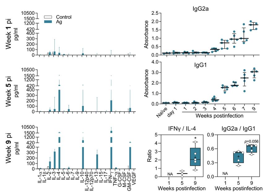
Muñoz-Durango et al.                                                                                                 Ulcerative Cutaneous Leishmaniasis Mouse Model

               A                                                                       B

                                                                                       C

  FIGURE 4 | Antigen-specific cytokine and antibody response of L(V)p-infected BALB/c mice. Draining LNs from L(V)p-infected mice were obtained at the indicated
  timepoints to prepare cell suspensions. Cells were cultured in the absence (Control) or presence (Ag) of L(V)p total lysate. The concentration of the indicated
  cytokine secreted by dLN cells was determined by a multi-analyte Luminex platform and graphed as the median ± interquartile range (A). Serum samples were also
  used to quantify the antigen-specific IgG1 and IgG2a response by ELISA (B). Ab levels in individual mice are shown and the bars are the median ± interquartile
  range. IFNγ/IL-4 and IgG2a/IgG1 ratios were calculated and graphed as “box and whiskers” plots (C). IFNγ/IL-13 and IFNγ/(IL-4 + IL-13) ratios exhibited a similar
  pattern to that presented in (C). NA: not applicable. pi: postinfection. n = 3–6 mice/group. *p < 0.05 (Mann–Whitney U-test in (C) comparing week 5 vs. 9).

certain cytokines (such as IL-4, IL-13, and IL-5) were observed,                      suggested that the early non-protective Ag-specific Th2 response
a mixed Th1/Th2/regulatory/inflammatory response (IL-2, IFNγ,                         is shifted toward a Th1-dominated response that enables parasite
IL-4, IL-13, IL-10, and GM-CSF) was also present in mice                              control (Figure 4C; Supplementary Figure S9D).
infected in the footpad (Supplementary Figures S9A,B). The
L(V)p-specific antibody response was also analyzed in animals
infected in the ear or the footpad. We observed an early IgG1                         In situ Changes Related to L(V)p Infection
response that increased along with disease progression until                          and Disease Development
stabilization (or even decline at the late chronic stage) when                        Studies with human biopsy specimens taken from L(V)
the parasite multiplication is controlled (Figure 4B;                                 p-infected patients reported epidermal hyperplasia/
Supplementary Figure S9C). In contrast, the Th1-related IgG2a                         hyperkeratosis; diffuse dermal infiltrate of lymphocytes,
response was delayed but steadily increased until the end                             histiocytes, neutrophils, eosinophils and plasmocytes; and
the experiment.                                                                       the presence of amastigotes, necrotic foci, granulomas and
   In summary, L(V)p-infection elicited a mixed adaptive immune                       resolution-related epithelioid and giant cells as usual
response in BALB/c mice, involving T cells that produce Th1,                          anatomopathological characteristics (Palma et al., 1991;
Th2, Treg, and inflammatory lymphokines, which replicates                             Weigle and Saravia, 1996; Palma and Saravia, 1997; González
the combined Th1/Th2/Treg cytokine pattern observed in                                et al., 2018). Immunohistological phenotyping further
stimulated PBMCs from L(V)p-infected humans (Figure 4;                                confirmed the cellular identity of the major infiltrators and
Supplementary Figure S8; Bosque et al., 2000; Trujillo et al.,                        additionally discriminated the lymphocyte compartment into
2002; Castilho et al., 2010; Díaz et al., 2010). Moreover, the                        B and T cells, CD4+ and CD8+ T cells, and Th1, Th2,
IFNγ/IL-4 and IgG2a/IgG1 ratios calculated at late weeks                              Th17, and Treg cell subpopulations as potential drivers of
postinfection compared to those calculated at earlier timepoints                      pathology and/or immunity (Isaza et al., 1996; Palma and

Frontiers in Microbiology | www.frontiersin.org                                   7                                             June 2022 | Volume 13 | Article 907631
Muñoz-Durango et al. Ulcerative Cutaneous Leishmaniasis Mouse Model
Saravia, 1997; Gonzalez et al., 2020a,b). Although the (Supplementary Figure S10A). Flow cytometry also allowed
magnitude and pattern of infiltrates might be heterogeneous to evidence the emigration of myeloid cells phenotypically
in human samples, our histopathological and flow cytometric compatible with skin resident dendritic cells, which seem
analysis in the ears of L(V)p-infected mice evidenced most to be the cells appearing at the same times in the dLN
of those characteristics (Figure 5; Supplementary Figure S10). (Supplementary Figure S7), presumably transporting parasites
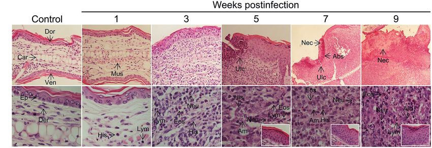
Ear sections evidenced a progressive recruitment of and parasite antigens (see section “Lymphadenopathy and
inflammatory cells (Figure 5) that paralleled parasite Parasite Dissemination in the Absence of Systemic Disease”
multiplication and clinical lesion appearance and aggravation above). CD4+ and CD8+ T cells also infiltrate the infected
(Figure 2). Neutrophils, eosinophils, parasitized histiocytes, ears with maximal amounts observed at 5 weeks postinfection
lymphocytes/lymphoplasmocytes, as well as acanthosis, were (Supplementary Figure S10B). Both IFNγ- and IL-4-
consistently observed and its abundance accentuated as producing CD4+ T cells, as well as IFNγ-producing CD8+
disease progressed. Most severe pathological changes, such T cells, were detected in infected but not in control mice
as massive leukocyte infiltration, abundant presence of heavily (Supplementary Figure S10C), further suggesting that a
parasitized macrophages, epidermal hyperplasia/ mixed Th1/Th2 response also characterizes the in situ response
hyperkeratosis, and the appearance of necrotic areas, were to L(V)p in BALB/c mice. Histopathological analysis of
observed at 7–9 weeks postinfection (Figure 5), timepoints footpad sections at week 7 postinfection showed that L(L)
at which the adaptive response was already vigorous (Figure 4) m induces massive inflammation and tissue destruction,
and right after the temporal window in which robust parasite whereas L(V)p did not alter the global tissue architecture
multiplication took place (Figure 2). These histopathological and induces only moderated and limited changes (see
findings were in line with our preliminary flow cytometry Supplementary Figure S11 and legend for details).
analysis from ear macerates, which revealed the massive Collectively, results presented thus far demonstrate that the
and progressive infiltration of myeloid cells phenotypically development of a progressive inflammatory response and the
compatible with granulocytes, and mononuclear phagocytes establishment of a robust mixed Ag-specific adaptive immune
A
B
FIGURE 5 | Histopathology of L(V)p-infected BALB/c mice ears. Representative photographs of hematoxylin/eosin-stained ear sections from the infected mice
at the indicated week postinfection are shown. A photograph of a section from a normal non-infected mouse was also included (Control) for comparison.
Magnifications are 40X for control and 1–5 weeks postinfection, and 10X for 7 and 9 weeks postinfection in the upper panel (A). In the lower panel, a 100X
magnification was used for all photographs (B), to document the appearance of the dermis. Insets in the lower panel show the appearance of the epidermis.
Note that no major changes were observed in the ears at the 1st week postinfection in the dermal compartment (no parasites, and very few histiocytes,
granulocytes or lymphocytes could be identified), and no alterations were detected in the epidermis, which is consistent with the clinical, parasitological, and
immunological findings at this time (Figures 2–4). At week 3 postinfection, however, an evident inflammatory infiltrate was observed in the dermis predominantly
represented by granulocytes (primarily neutrophils but also eosinophils), amastigote-containing histiocytes and lymphoplasmocytes. Hypertrophic/hyperplasic
muscle fibers could be observed in the dermis, and irregular acanthosis in the epidermis was also present in the areas where the inflammatory reaction was
taking place. More pronounced changes that indicated aggravation of the inflammatory process were observed at subsequent times (5th–9th weeks
postinfection). Heavily parasitized histiocytes were observed starting at the 5th week postinfection until the end of the experiment. A profuse lymphoplasmacytic
and granulocytic infiltrate was also evident in the dermis, with granulocytes forming microabscesses, which were also present in the epidermis, presumably
provoking ulcerations. Marked and progressive acanthosis and hyperkeratosis, the presence of necrotic areas in the dermis and epidermis and accentuated
forms of all previously described changes, were common anatomopathological characteristics of the chronic phase (7–9 weeks) of the disease. All
histopathological changes were restricted to affected skin areas since the zones of the ear where neither parasite replication occurred, nor leukocyte infiltrated
was present, looked histopathologically normal (with only some evidence of muscle hypertrophy). Eos, polymorphonuclear eosinophil; Neu, polymorphonuclear
neutrophil; Ep, epidermis; Der, dermis; Mus, muscle, Abs, abscess; Ulc, ulcer; His, histiocyte; Am, amastigote; Dor, dorsal face; Ven, ventral face; Car, cartilage;
Nec, necrosis; Lym, lymphoplasmocyte.
Frontiers in Microbiology | www.frontiersin.org 8 June 2022 | Volume 13 | Article 907631Muñoz-Durango et al. Ulcerative Cutaneous Leishmaniasis Mouse Model
response are two key events associated with L(V)p infection
and pathogenesis in BALB/c mice. A B
Exploiting the Model to Evaluate
Pharmacological and Immunological
Interventions
Since antimonials and Miltefosine are first-line drugs used
for leishmaniasis (Reithinger et al., 2007; Aronson et al.,
2020), we tested the clinical and parasitological response
to these agents in BALB/c mice with established L(V)p-induced
LCL. Mice treated with antimonials were cured (as defined
by the complete disappearance of the ulcer and the
re-epithelialization of the affected skin) within 3–7 weeks
and had about a 105–106-fold reduction in the parasitic C D
load when infected in the ears (Figures 6A–D), as well as
in the footpad (Supplementary Figure S12A) or the base
of the tail (Supplementary Figure S12B). Miltefosine was
also leishmanicidal in vivo and induced cure of L(V)p-infected
mice (Figures 6A–D). This indicates that common drugs
used to treat L(V)p infection in humans also promote disease
resolution in our animal model.
We also tested CpG-containing oligodeoxynucleotides, a
synthetic immunomodulatory agent that has been shown to
deviate the immune response toward a protective Th1-type
cytokine pattern and promote parasite control when used
at low dose (Zimmermann et al., 1998, 2008; Raman et al.,
2012). We found that the dermal co-delivery of CpG with
L(V)p infective promastigotes prevented the development of FIGURE 6 | Using the L(V)p-BALB/c model to investigate pharmacological
cutaneous lesions in BALB/c mice and served also as a interventions. BALB/c mice were infected id in the ear with 105 stationary
protective adjuvant when combined with total or membrane promastigotes and lesion development monitored weekly (A). Once lesions were
Ag (Supplementary Figure S13). Interestingly, CpG alone established at the week 4 postinfection (B, left upper corner; C, arrow), mice were
was sufficient to significantly protect BALB/c mice from ®
treated ip with Glucantime (500 mg/kg, once a week, 4 weeks) or orally with
Miltefosine (20 mg/kg/day, five consecutive days) as indicated (A). Representative
an infective challenge when administered 2 weeks before
photographs taken at the indicated timepoints are shown (B). The therapeutical
infection, suggesting that CpG pre-treatment triggered a effect of the agents was clinically monitored measuring the size of the lesions
protective effect in the absence of leishmanial Ag lasting during the subsequent weeks (C); asterisks and small circles correspond to p-
for at least 2 weeks (Supplementary Figure S13). Therefore,
we performed vaccination experiments in which the
®
values from the statistical test comparing control versus Glucantime and
Miltefosine, respectively. The burden of viable parasites in the ears at the end of
immunizing Ag + CpG formulation was administered following the experiment (week 10 postinfection) was analyzed by a limiting dilution assay
(D). Data in (C,D) are presented as the median ± interquartile range (n = 5–12
a homologous prime-boost scheme, and the infective challenge
mice) and the geometric mean ± 95%IC (n = 4–5 mice), respectively. wpi: weeks
administered 4 weeks after the boost (Figure 7A). A strong postinfection. *p < 0.05, **p < 0.01, ***p < 0.001, ****p < 0.0001 (Mixed-effects
clinical and parasitological protection was observed that analysis with Geisser–Greenhouse correction and Bonferroni’s multiple
required the presence of both the adjuvant and the antigen comparisons post-hoc test for multiple comparison through time; Kruskal–Wallis
(Figures 7B–D), indicating the potential of a TLR9-mediated test with Dunn’s multiple comparison post-hoc test). A repetition chemotherapy
pathway of protective adjuvanticity for vaccine development ®
experiment with Glucantime in which an extended clinical follow-up was
performed, indicated that remaining mild erythema/swelling observed in some
against this species. Collectively, these results confirm the
animals at week 10 postinfection completely resolved within the following
utility of this model for the preclinical assessment of potential 2–3 weeks and only a rough/bright clear appearance, most likely representing
chemo/immunotherapeutic and prophylactic agents against scarification, was observed. Results are representative of three independent
L(V)p CL. ®
experiments performed with Glucantime , whereas the experiment with
Miltefosine was performed only once.
A 28–30 kDa Protein Fraction of the L(V)p
Proteome Protects From an Infective proteomics. Total L(V)p Ag was fractioned by electroelution
Challenge (Supplementary Figure S14A), and although sufficient protein
Having demonstrated CpG as a protective adjuvant and the yield for in vivo testing was obtained only for few fractions,
utility of our model in a vaccination setting, we initiated efforts results indicated that a fraction containing proteins in the
to identify defined Ag as vaccine candidates using preparative 29–30 kDa range reproduced (Supplementary Figures S14B,C)
Frontiers in Microbiology | www.frontiersin.org 9 June 2022 | Volume 13 | Article 907631Muñoz-Durango et al. Ulcerative Cutaneous Leishmaniasis Mouse Model
apart with F9 + CpG (Figure 8B) were sufficient to protect mice
A B clinically and parasitologically from a L(V)p challenge 4 weeks
after the boost (Figures 8B–F), and protection required the presence
of both parasite antigens (F9) and CpG adjuvant (see groups
CpG alone and F9 alone in Figures 8C–F). While the level of
protection induced by F9 + CpG was comparable to that induced
by total Ag + CpG, the F14 + CpG vaccine, as anticipated, did not
confer significant protection. F9 + CpG-induced protection was
related to a particular L(V)p-specific cytokine and antibody profile
(Figures 8G,H). Counterintuitively, IFNγ production seemed to
be lower in vaccinated groups (Figure 8G); however, when
normalized to the lesion size (as surrogate of antigenic load
that strongly correlates with viable parasite burden;
C D Supplementary Figure S16A), it emerged that vaccination with
Ag + CpG induced significant amounts of this key antimicrobial
cytokine (Supplementary Figure S16B). It was interesting that
Ag + CpG also induced IL-13 but no IL-4 and that the pattern
of F9 + CpG-vaccinated mice resembled that from
Ag + CpG-vaccinated animals, whereas the pattern induced by
F14 + CpG resembled more that observed in control non-
vaccinated mice (Supplementary Figure S16B). In a similar
manner, IFNγ/IL-4 and IgG2a/IgG1 ratios also indicated that
vaccination with F9 + CpG, but not with F14 + CpG, promoted a
significant shift toward a Th1-type of response which characterizes
protected animals vaccinated with Ag + CpG (Figures 8G,H, right
panel). Finally, in a separated set of experiments, we used the
model to confirm that mice vaccinated with F9 are protected
FIGURE 7 | Using the L(V)p-BALB/c model to investigate immunological
interventions. BALB/c mice were vaccinated with Ag + CpG (12.5 μg/6.25 μg
not only from a primary, but also from a secondary infection
sc in the base of the tail) and boosted with the same dose 2 weeks later, to (Supplementary Figure S17), and that fine dosing of F9/
be infected in the ear dermis with 105 L(V)p stationary promastigotes 4 weeks adjuvant is critical for optimal induction of protection
after the boost (A). Mice were followed weekly to monitor lesion growth. Mice (Supplementary Figure S18). Collectively, in these experiments
injected with CpG (6.25 μg) or PBS were used as controls. Representative
we exploited the L(V)p-BALB/c model to identify a fraction of
photographs of the ears from Ag + CpG-vaccinated mice and controls at the
week 6 postinfection are shown (B). The kinetics of lesion progression after
the L(V)p proteome encompassing potential antigenic candidates
infection is graphed (C) as the median ± interquartile range. From week 2 for the preclinical development of a molecularly defined vaccine
postinfection, there was a statistically significant difference between control and to explore immunological surrogates of protection.
and Ag + CpG. The number of viable parasites at the site of infection was
determined at the end of the experiment (week 9 postinfection) by a limiting
dilution assay and graphed (D) as the geometric mean ± 95%CI. n = 5–6 mice.
A similar efficacy of Ag + CpG has been observed in repetition experiments,
DISCUSSION
with more than 90% of mice protected from clinical disease and a 103–106-
fold reduction in parasitic loads. wpi: weeks postinfection. *p < 0.05 (Two-way Although mouse models of human leishmaniasis caused by
ANOVA with Geisser–Greenhouse correction and Bonferroni’s multiple most Leishmania species have been implemented in many
comparisons post-hoc test for multiple comparison through time; Kruskal– laboratories worldwide permitting significant advance of science
Wallis test with Dunn’s multiple comparison post-hoc test).
in the field, attempts to develop a murine model of L(V)p
infection that reproduces the manifestations of the human
disease have been relatively unfruitful. In this article,
the protective effect of the total lysate (Figure 7). We then shifted we presented the first ulcerative mouse model of L(V)p
to a manual fractionation method to obtain fractions in amounts infection, the characterization of the disease at the stages of
sufficient to perform a complete L(V)p proteome screening. From parasite multiplication and control, and examples of its use
the 16 fractions obtained (Supplementary Figure S15A), 15 for preclinical pharmacological testing and vaccine development.
were tested in vivo using our mouse model, and although several The availability of this new model warrants further research
fractions induced a significant clinical protection, the F9 (which that could expand the understanding of the immunobiology
contains proteins in the 28–30 kDa range) was again the most of Leishmania infections and promote translational work for
protective (Supplementary Figure S15B). a better control.
In a third experiment, we compared the protective F9 with In pioneering efforts seeking to establish a murine model
the less-protective F14 fraction (Supplementary Figure S15; of L(V)p infection, researchers injected extremely high amounts
Figure 8A) and used PBS and total Ag as negative and positive of parasites (107–108 parasites) and reported inflammatory lesion
controls, respectively. Two vaccine injections separated 2 weeks formation (Neal and Hale, 1983; Guevara-Mendoza et al., 1997).
Frontiers in Microbiology | www.frontiersin.org 10 June 2022 | Volume 13 | Article 907631Muñoz-Durango et al. Ulcerative Cutaneous Leishmaniasis Mouse Model
A B C
D E F
G H
FIGURE 8 | A 28–30 kDa protein fraction of the L(V)p proteome that protects against an infective challenge. In an independent experiment but following the same
strategy presented in Supplementary Figure S15, the fractions F9 and F14 from the L(V)p promastigote proteome were obtained and run in SDS/PAGE gels. The
photograph of a silver-stained gel presented in (A) confirmed that yield, purity, and integrity of the fractions were appropriated for in vivo testing. Mice were then
vaccinated sc on the back with 6 μg of the protein fraction in combination with 3.5 μg CpG and boosted 2 weeks later with the same preparation to be infected with
L(V)p 4 weeks after the boost (B). PBS- or Ag + CpG-injected mice were used as negative and positive controls, respectively. Representative photographs illustrating
the appearance of the infected ears in the different experimental groups at the 6th week postinfection are shown (C). Lesion size was registered weekly and
graphed as the kinetics of lesion growth (D) or the size of the lesion in individual mice at the 6th week postinfection (E). Animals were sacrificed at the 6th week
postinfection, and the ears used to quantify the numbers of viable parasites by limiting dilution assay (F). Cell suspensions from dLN were stimulated with L(V)p Ag
and the amounts of the indicated cytokine determined by ELISA (G). The IFNγ/IL-4 ratios were calculated and graphed (G, right). Serum samples were also obtained
and used to quantify the circulating levels of L(V)p-specific IgG1 and IgG2a antibodies (H). IgG2a/IgG1 ratios were also calculated and graphed (H, right). Data are
shown as the median ± interquartile range (D,E,G,H; n = 4–6 mice per group), or the geometric mean ± 95%CI (F; n = 3 pools from 5 to 6 mice per group). Ratios
presented in (G,H) are graphed as “box and whiskers” plots. MW: molecular weight. wpi: weeks postinfection. *p < 0.05, **p < 0.01, and ***p < 0.001 (Kruskal–Wallis
test with Dunn’s multiple comparison post-hoc test).
Subsequent attempts, however, were discouraging, since no and self-limited, measurable and reproducible skin lesions after
reproducible and predictable lesion formation could be induced infection with L(V) isolates (de Moura et al., 2005; Sousa-
(Hommel et al., 1995; Travi et al., 2002; McMahon-Pratt and Franco et al., 2006; Castilho et al., 2010; de Oliveira and
Alexander, 2004; and our laboratory observations). This led Brodskyn, 2012; Novais et al., 2013; Pena DaMata et al., 2015;
to the perception that mice were not reliable to model infections Borges et al., 2018; Hartley et al., 2018; de Carvalho et al.,
with L(V)p and with L(V) organisms more generally. More 2019). The selection-adaptation strategy implemented in our
recently, efforts in several laboratories indicated that under laboratory allowed the establishment of a suitable in vivo system
particular experimental conditions mice can develop, yet small that recapitulates the essential human clinicopathological and
Frontiers in Microbiology | www.frontiersin.org 11 June 2022 | Volume 13 | Article 907631Muñoz-Durango et al. Ulcerative Cutaneous Leishmaniasis Mouse Model immunoinflammatory responses to L(V)p as a member of the be convenient to further investigate LRV1/cytotoxicity-dependent L(V) subgenus and that follows three phases: (1) an acute or LRV1/cytotoxicity-independent mechanisms of pathogenesis phase (from day 0 to week 5–6) in which a short early silent and elucidate the relative contributions of microbial- and host- period of parasite establishment takes place, followed by a derived factors to the intriguing and distinct pathophysiology period of robust parasite multiplication, lesion appearance, and of L(V) infections. Moreover, given the known link between progression to ulcer. (2) An intermediate chronic phase, lasting disease chronicity and refractoriness to treatment, the preclinical from week 5–6 to week 11–12, in which massive tissue damage testing of host- and parasite-directed strategies aimed to reduce is observed often leading to partial or total mutilation. (3) A deleterious inflammation is necessary, and this model appears final stage of lesion resolution, lasting up to 22–24 weeks, in suited for this purpose. which lesion is healed and inflammation resolved. While most We observed that infection in three different anatomic of available murine models of L(V) infections in BALB/c and locations leads to effective establishment and multiplication C57BL/6 mice exhibit an inflammatory acute phase with variable of L(V)p, although id/ear delivery was optimal both at magnitude and duration followed by rapid resolution/healing, mimicking the human disease and at exhibiting consistency our model is unique in presenting the intermediate phase, and reproducibility. While the influence of the route/site of making it particularly useful to investigate the chronic disease Leishmania infection on the clinical outcomes has been long and immunopathology. Moreover, C57BL/6 mice were resistant documented (Kirkpatrick et al., 1987; Loeuillet et al., 2016), to L(V)p as indicated by the very small lesions accompanied the underlying mechanisms are incompletely understood. by low parasitic loads observed after infection in the ear (not Ribeiro-Gomes et al. showed that id but not sc delivery of shown). This clear cut of mouse susceptibility/resistance to L(L)m leads to the rapid recruitment of high amounts of UA-946 L(V)p resembles the classical model of L(L)m but neutrophils that efficiently capture parasites, providing without the disadvantage of the systemic, visceralizing, and appropriate host cells that assure successful parasite lethal disease observed in BALB/c mice, and thus, L(V)p appears establishment and subsequent multiplication (Ribeiro-Gomes to better model not only L(V) infections particularly, but CL et al., 2014). The study additionally indicated that it is the in general. route (id) rather than the anatomical site (ear or footpad), Importantly, progression to the mutilating chronic phase in what dictates the higher neutrophil response to dermal injury our model occurs in the absence of further parasite multiplication and that the effective dose achieved after id/ear injection because parasite loads after week 10 postinfection were always could be reached via sc/footpad infection by increasing below the maximal 109 and usually in the 104–108 range (not (approximately tenfold) the size of the inoculum. Our L(V) shown), a consistent observation in many independent p promastigote inoculum was 105 for id/ear injection and experiments performed in our laboratory during years, indicating 106 for sc/footpad injection, and yet tissue destructive versus that persistence/progression of disease is the result of an mild non-ulcerative inflammation, respectively, were observed. immunopathological process. That ulceration was the result of This, together with the different outcomes after UA-946 L(V) lymphocyte-driven immunopathology was indicated by the p delivery using the same route (sc) but different site (base slow-progressing non-ulcerative lesions observed in SCID BALB/c of the tail and footpad), which was also observed in hamsters mice infected with a similar UA-946 L(V)p inoculum (not infected with L(V)p id in the foot versus id in the snout shown). This is of importance since a hallmark of L(V) infections (Osorio et al., 2003), and the similar outcome after using in humans is the propensity to induce chronic tissue-damaging similar site (foot) but different routes (sc route in the present immunopathology in the context of effective parasite control. study and id route in the study by Castilho et al., 2010), While the immunopathological mechanisms operating in L(V) indicate that site-related effects are major contributors to the infections have been enigmatic for many years, two relevant different disease presentation. While our results indicated that pathways have been recently described. In one, cytotoxic activities the overall pattern of T cell response in the LN draining involving CD8+ T and NK cells linked to inflammatory executors the ear and the footpad was similar, detailed analysis revealed such as NLRP3 inflammasome, IL-1β, and neutrophils mediate some differences, such as the kinetics of the Th2 cytokines exacerbated tissue damage in response to L(V)b infection (Novais IL-4 and IL-13, with footpad infection exhibiting early transient et al., 2021; Carvalho et al., 2022). In the other, the innate high levels and ear infection inducing high levels that are immune recognition of the viral RNA present in L(V)g isolates sustained and even increasing at the chronic phase, which harboring the Leishmania virus LRV1 (or via exogenous viral is consistent with the abundant literature pointing to the coinfections) triggers a type I IFN response, immunopathology, type of adaptive immune response induced (whether biased chronicity, and metastasis (Rossi and Fasel, 2018b). Whether toward Th1, Th2, or regulatory) as a determining factor of these two mechanisms also operate during pathogenic response the distinct site-related outcomes (Kirkpatrick et al., 1987; to L(V)p is an open question to be addressed in the future. Nabors and Farrell, 1994; Nabors et al., 1995; The L(V)p model presented here, in which no special Baldwin et al., 2003; Tabbara et al., 2005; Mahmoudzadeh- manipulation of the host (such as the CD8+ T cell reconstitution Niknam et al., 2013). A recent report showed that TLR7- of immunodeficient mice or the use of IFNγ-deficient mice) dependent effector functions of neutrophils control early or additional innate inflammatory trigger in the infective parasite replication and subsequent disease progression without inoculum (such as the viral endosymbiont) are required to apparent alteration in the CD4+ T cell response after id/ear exhibit the chronic and tissue damaging phenotype, could (but not sc/footpad) L(L)m infection (Regli et al., 2020), Frontiers in Microbiology | www.frontiersin.org 12 June 2022 | Volume 13 | Article 907631
You can also read