A five year retrospective study on the epidemiology of hand, foot and mouth disease in Sabah, Malaysia - Nature
←
→
Page content transcription
If your browser does not render page correctly, please read the page content below
www.nature.com/scientificreports
OPEN A five‑year retrospective study
on the epidemiology of hand,
foot and mouth disease in Sabah,
Malaysia
Siat Yee Fong1,2, Daisuke Mori3, Christina Rundi4, Jun Fai Yap4, Muhammad Jikal4,
A. L. Liza Binti Abd Latip5, Victor Johnny4 & Kamruddin Ahmed1,3*
Hand, foot, and mouth disease (HFMD) is endemic in Malaysia, with the number of cases increasing.
Sabah has experienced several HFMD outbreaks, but information on the epidemiology and molecular
characteristics of responsible viruses is scarce. In this study, data of 17,574 reports of HFMD cases
in Sabah from 2015 to 2019 were extracted from a public health disease surveillance system and
analyzed. Twenty-one swab samples from 13 children were collected from Beaufort, Sabah, during
an outbreak in August 2018 for detection and serotyping of causative viruses by semi-nested reverse
transcription-polymerase chain reaction (snRT-PCR) of the VP4–VP2 region and consensus degenerate
hybrid oligonucleotide primer PCR of the VP1 region, respectively. Nucleotide sequencing and
phylogenetic analysis were conducted by the neighbor-joining method. The average annual incidence
of HFMD was 94.3 per 100,000 people, with the greatest yearly increase between 2017 and 2018.
Swabs from six children were tested positive for enterovirus, of which five were positive for CVA16 and
one for EV71. All CVA16 strains belonged to sub-genotype B1a, and the EV71 strain belonged to sub-
genotype B5. Phylogenetic analyses indicate that enterovirus genotype shift might be responsible for
the increasing trend of HFMD in Sabah, however, further study is needed.
Hand, foot, and mouth disease (HFMD) is a contagious viral infection. It commonly affects children under five
years of age, but it can occasionally affect adults1. Symptoms of HFMD include fever, sore throat, and maculo-
papular or vesicular rashes on hands, feet, and mouth. Although the disease is generally mild, severe HFMD can
lead to fatal neurologic and systemic complications2. HFMD is caused by a few members of the genus Enterovirus,
belonging to the family Picornaviridae, particularly coxsackievirus A16 (CVA16) and enterovirus 71 (EV71) of
the enterovirus A s pecies3.
EV71 is divided into seven genotypes (A-G) based on the nucleotide diversity of the viral protein 1 (VP1)
gene. Genotype A contains the prototype strain BrCr, which was first isolated in the United States in 1969.
Genotypes B and C are more widely circulated throughout the world, while genotypes E–F and D–G were iden-
tified more recently in Africa and India, r espectively4. CVA16 was first isolated in South Africa in 1 9515. Due
to the diversity of the VP1 gene, it is classified into two genotypes, A and B, where the latter is the predominant
circulating genotype responsible for CVA16 infection w orldwide5. Genotype B can be divided into B1 and B2,
and genotype B1 can be further divided into B1a, B1b, and B1c6.
Major EV71-associated HFMD outbreaks have been occurring across the Asia–Pacific region since the late
1990s7. In Malaysia, the first outbreak due to EV71 infection was documented in Sarawak (East Malaysia) in April
1997 and subsequently spread to Peninsular Malaysia in June 1997, where more than 4000 children were affected
and 41 deaths were reported8,9. CVA16 was also isolated from the HFMD outbreak in 1997, but only EV71 was
isolated from fatal cases9. CVA16 infection was seen to occur during the outbreak or inter-outbreak of E V7110.
1
Borneo Medical and Health Research Centre, Faculty of Medicine and Health Sciences, Universiti Malaysia Sabah,
Jalan UMS, 88400 Kota Kinabalu, Sabah, Malaysia. 2Department of Biomedical Sciences, Faculty of Medicine
and Health Sciences, Universiti Malaysia Sabah, Jalan UMS, 88400 Kota Kinabalu, Sabah, Malaysia. 3Department
of Pathology and Microbiology, Faculty of Medicine and Health Sciences, Universiti Malaysia Sabah, Jalan UMS,
88400 Kota Kinabalu, Sabah, Malaysia. 4Sabah State Health Department, Ministry of Health Malaysia, Jalan
Mat Salleh, 88590 Kota Kinabalu, Sabah, Malaysia. 5Beaufort Health Office, Ministry of Health Malaysia, Pekan
Beaufort, 89800 Beaufort, Sabah, Malaysia. *email: ahmed@ums.edu.my
Scientific Reports | (2021) 11:17814 | https://doi.org/10.1038/s41598-021-96083-3 1
Vol.:(0123456789)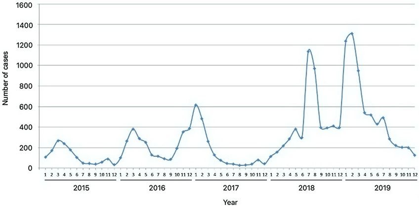
www.nature.com/scientificreports/
Figure 1. Monthly distribution of hand, foot, and mouth disease cases in Sabah from 2015 to 2019.
2019
Year 2015 2016 2017 2018 Total
No. of cases 1390 2644 1872 5157 6511
Incidence rate (per 100,000) 39.9 75.9 51.1 138.6 166.1
Gender
Male 769 (55.3) 1464 (55.4) 1054 (56.3) 2930 (56.8) 3671 (56.4)
Female 621 (44.7) 1180 (44.6) 818 (43.7) 2227 (43.2) 2840 (43.6)
Age in years
5 171 (12.3) 355 (13.4) 204 (10.9) 822 (15.9) 748 (11.5)
Transmission classification
Clusters of cases 121 (8.7) 143 (5.4) 88 (4.7) 990 (19.2) 696 (10.7)
Sporadic cases 1269 (91.3) 2501 (94.6) 1784 (95.3) 4167 (80.8) 5815 (89.3)
Table 1. Demographic characteristics of hand, foot, and mouth disease cases in Sabah, Malaysia, from 2015 to
2019. Percentage of total is shown in parenthesis.
Several researchers have characterized EV71 and CVA16 strains from HFMD cases in Peninsular Malaysia
and Sarawak10–13. Although a number of HFMD outbreaks have been reported in Sabah, information on the
genetic characteristics of these strains in Sabah is limited. So far, only two studies have been conducted on EV71
strains isolated from Sabah in 1 99911 and 2 00812, but none on CVA16. Since then, no studies have been conducted
on HFMD in Sabah for almost a decade. The limited research could be due to the assumption of low burden
of HFMD in Sabah and a lack of standardized sample collection protocols that would maximize the yield of
viruses for molecular characterization. More surveillance is necessary to understand and monitor the emergence
and spread of new strains. Besides, understanding the epidemiological pattern of HFMD is important for the
implementation of appropriate intervention strategies. Therefore, this five-year retrospective study explored
the epidemiological pattern of HFMD in Sabah using the data of 17,574 reports of HFMD cases recorded in a
Malaysian public health disease surveillance system, eNotifikasi, and investigated the genotype characteristics
of seven enterovirus-positive samples that caused HFMD in the region.
Results
Temporal and demographic trends. From the eNotifikasi system (http://enotifikasi.moh.gov.my), a total
of 17,574 reports of HFMD cases were recorded in Sabah during the five-year study period, with no deaths
reported. While cases were notified year-round, seasonal analysis showed that reported cases peaked annually
between January and March, except in 2018, where HFMD peaked in July (Fig. 1). The number of cases gradu-
ally increased from 2015 to 2019, but there was a slight decrease in 2017 from the previous year (Table 1). The
annual incidence rate from 2015 to 2019 varied from 39.9 per 100,000 people to 166.1 per 100,000 people, with a
Scientific Reports | (2021) 11:17814 | https://doi.org/10.1038/s41598-021-96083-3 2
Vol:.(1234567890)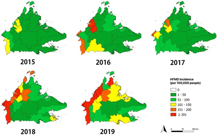
www.nature.com/scientificreports/

                                                   Year                                Total
                                   Districts       2015    2016   2017   2018   2019
                                   Kota Kinabalu   427     771    431    1029   1328   3986
                                   Papar           12      9      3      326    642    992
                                   Tuaran          152     288    200    344    380    1364
                                   Ranau           7       26     65     185    224    507
                                   Kota Belud      3       8      13     150    242    416
                                   Penampang       201     352    189    484    582    1808
                                   Putatan         44      111    46     146    198    545
                                   Kudat           58      64     29     142    77     370
                                   Kota Marudu     4       3      11     37     71     126
                                   Pitas           2       16     5      29     109    161
                                   Sandakan        33      217    236    365    535    1386
                                   Beluran         48      92     44     97     158    439
                                   Tongod          6       3      5      1      4      19
                                   Kinabatangan    13      20     16     31     27     107
                                   Tawau           125     166    84     435    697    1507
                                   Semporna        4       2      5      160    88     259
                                   Kunak           0       3      0      15     17     35
                                   Lahad Datu      14      66     108    150    212    550
                                   Beaufort        66      49     88     515    219    937
                                   Sipitang        50      70     31     147    204    502
                                   Kuala Penyu     20      36     39     118    105    318
                                   Keningau        4       74     97     168    253    596
                                   Tenom           49      49     53     26     56     233
                                   Tambunan        43      112    46     45     60     306
                                   Nabawan         5       37     28     12     23     105

                                  Table 2.  The number of hand, foot, and mouth disease cases in each district of Sabah from 2015 to 2019.

                                  five-year average annual incidence of 94.3 per 100,000 people. The greatest year-to-year increase, approximately
                                  2.7-fold, was observed from 2017 to 2018. Notably, similar trends for gender and age groups were observed every
                                  year. Males accounted for most of the cases (male-to-female ratio of 1.3:1), and one-year-old children had the
                                  highest number of cases for both genders.

                                  Spatial trends across districts. Among the 25 districts, Kota Kinabalu reported the highest number of
                                  HFMD cases every year (total 3986 cases), with a peak of 1328 cases in 2019 (Table 2). On the contrary, Tongod
                                  district had the lowest number of cases, with just 19 cases reported throughout the five years. Of the 25 districts,
                                  a peak in the number of cases was observed in 17 (68%) districts in 2019, five (20%) districts in 2018, two (8%)
                                  districts in 2016, and one (4%) district in 2015. The annual spatial trend dynamics are shown in Fig. 2, indicat-
                                  ing that there was a yearly variation across districts. Throughout the five-year study period, the district with the
                                  largest variation in annual incidence rate was Beaufort (58 per 100,000 people in 2016 to 657 per 100,000 people
                                  in 2018) while Kinabatangan had the smallest variation in annual incidence rate, ranging from 8 per 100,000
                                  people in 2015 to 18 per 100,000 in 2018. A prominent spatial change was observed in 2018, when incidence
                                  increased markedly in the western regions of Sabah from the previous year. High incidence rates were not only
                                  found in densely populated districts, such as Kota Kinabalu, Putatan and Penampang, but also in low population
                                  density districts, including Sipitang, Beaufort, Kuala Penyu and Kota Belud. The highest incidence district was
                                  Kuala Penyu with a five-year average annual incidence of 257 per 100,000 people, while the lowest were Tongod
                                  and Kunak districts, both with 9 per 100,000 people.

                                  Phylogenetic analysis of enteroviruses. A total of 21 swab samples were collected from 13 children
                                  during an outbreak reported in Beaufort, Sabah, in August 2018. Swabs from six children comprising of four
                                  females and two males, ranging from one to seven years old were tested positive for enteroviruses using snRT-
                                  PCR.
                                     Sequence analysis of the VP1 gene of the positive snRT-PCR samples by BLAST identified five (83.3%) CVA16
                                  and one (16.7%) EV71 serotypes. The partial VP1 sequences of the CVA16 and EV71 viruses were 254 bp and
                                  359 bp in length, respectively. These partial VP1 sequences were used to construct the phylogenetic trees of the
                                  two serotypes.
                                     This is the first study that shows the genetic characteristics of CVA16 strains from Sabah. According to the
                                  phylogenetic analysis, all five CVA16 strains detected in this study were classified as genotype B, specifically sub-
                                  genotype B1a. Three CVA16 strains clustered with Thai strains from 2017, while the other three clustered with

Scientific Reports |   (2021) 11:17814 |                  https://doi.org/10.1038/s41598-021-96083-3                                                 3

                                                                                                                                                Vol.:(0123456789)
www.nature.com/scientificreports/
Figure 2. Annual spatial trend of hand, foot, and mouth disease across the 25 districts in Sabah from 2015 to
2019. The maps were generated using QGIS version 3.14.1 Pi (https://www.qgis.org/).
another Thai strain from 2016 (Fig. 3). None of the CVA16 strains in this study belong to the same sub-genotypes
as the other Malaysian strains, namely B1b, B1c and B2. The circulating strains in Malaysia in the early 2000s
were of sub-genotypes B1b and B2, which were later replaced with B1c as the predominant sub-genotype in
the late 2000s. However, more studies are needed to determine the evolutionary change of CVA16 in Malaysia.
The only EV71 strain detected in this study was classified as a genotype B, precisely sub-genotype B5 (Fig. 4).
The strain was clustered closely together with five EV71 strains from Thailand detected in 2012, 2014, and 2017,
and a strain from France detected in 2013. However, it did not cluster with any previously detected Malaysian
strains. Strains isolated from Sarawak and Sabah in 2003 and 2008, respectively, also belonged to the same sub-
genotype but of different lineage. Sub-genotype B4 was found circulating in Sabah and Sarawak in 1999 and
2000, respectively, while B3 was the predominant sub-genotype during the 1997 outbreak in Sarawak. From
this phylogenetic analysis, genotypic changes of EV71 have been observed over time even in the same region,
for instance Sarawak.
Discussion
There has been a considerable increase in reports of HFMD cases in Sabah throughout the five-year study period.
HFMD cases peaked in the early months, between January and March every year, except in 2018, where the
highest number of reported cases was in July. The exact causes of the increment of cases and the notable seasonal
characteristic of HFMD as well as the different peak period in 2018 are unclear. However, there are several factors
that could possibly affect HFMD incidence.
A study by Zhao and Hu (2019) found that school terms and a major public holiday in China, the Chinese
Spring Festival, affected the HFMD transmission seasonality in mainland China, contributing to two major
waves in the r egion14. The study revealed that the Chinese Spring Festival reflected the seasonal contact rate in
the population, which dominantly caused the larger peak in March, while the school terms reflected the sea-
sonal contact rate in children, resulting in the smaller peak in a utumn14. The school year in Malaysia starts in
early January and it is divided into two school terms: from January to May for the first term and from June to
November for the second term. Besides, Chinese New Year, which is the same as the Chinese Spring Festival, is
one of Malaysia’s biggest holidays celebrated in the beginning of the year, between late January and mid-February.
The first day of school and the Chinese New Year celebration in Malaysia when combined, could be a driving
factor for the seasonal peaks (between January and March every year, except for 2018) revealed in this study.
However, further in-depth research is necessary to determine the effects of school terms and holidays on HFMD
transmission in Malaysia.
In 2018, a large HFMD outbreak was reported in Malaysia, which affected over 76,000 children nationwide15.
Selangor recorded the highest number of HFMD cases on August 4, 2018 with 12,868 cases, followed by Kuala
Lumpur with 4996 cases and Sarawak with 4988 cases16. Although the number of HFMD cases in Sabah was not
as high as these states, the outbreak undoubtedly had a significant impact on the local community as the region
in general, experienced a sudden spike in HFMD cases in 2018. As shown in this study, cases in 2018 continued
to soar upwards until July of the same year, unlike the previous years and 2019, where cases started to decrease
after March, which could be due to exhaustion of susceptible hosts14 or effect of control measures. A possible
cause for the unusual pattern in 2018 might be due to the movement of people traveling to their respective loca-
tions for polling during Malaysia’s 14th General Election on May 9, 2018. This was later followed by two major
celebrations, the Harvest Festival, which is an important regional event and Eid-ul-Fitr (Festival of Breaking the
Fast) celebrated in late May and mid-June, respectively.
Meteorological factors, such as air temperature, rainfall, relative humidity, air pressure and wind speed, have
been shown to impact HFMD differently depending on the climatic zones and latitudes17,18. A meta-analysis
by Duan et al. (2019), revealed that HFMD incidences in subtropical and temperate regions were significantly
associated with meteorological factors but not in tropical r egions17. On the contrary, other studies of tropical
Scientific Reports | (2021) 11:17814 | https://doi.org/10.1038/s41598-021-96083-3 4
Vol:.(1234567890)www.nature.com/scientificreports/
Figure 3. Phylogenetic tree constructed using partial VP1 sequences (254 bp) of coxsackievirus A16 strains by
neighbor-joining method. The strains analyzed in this study are marked with a filled circle. The number adjacent
to the node represents the bootstrap value; values < 70% are not shown. The scale bar at the bottom indicates the
genetic distance expressed as nucleotide substitutions per site. The nucleotide sequences of our strains have been
submitted to the DNA Data Bank of Japan (DDBJ) with accession nos. LC567884–LC567887 and LC567889.
climate found significant correlation between HFMD and weather variables, including air temperature, rainfall,
pressure, and wind s peed19,20. These show that meteorological factors have inconsistent and varying effects even
in regions with the same climate, However, the effects of meteorological factors on HFMD incidence in this
study were not examined and thus, the question on whether these factors contribute to the seasonal peaks in
Sabah remains an enigma.
Scientific Reports | (2021) 11:17814 | https://doi.org/10.1038/s41598-021-96083-3 5
Vol.:(0123456789)www.nature.com/scientificreports/
Figure 4. Phylogenetic tree constructed using partial VP1 sequences (359 bp) of enterovirus 71 strains by
neighbor-joining method. The strain analyzed in this study is marked with a filled square. The number adjacent
to the node represents the bootstrap value; values < 70% are not shown. The scale bar at the bottom indicates the
genetic distance expressed as nucleotide substitutions per site. The nucleotide sequence of our strain has been
submitted to the DNA Data Bank of Japan (DDBJ) with accession no. LC567890.
Scientific Reports | (2021) 11:17814 | https://doi.org/10.1038/s41598-021-96083-3 6
Vol:.(1234567890)www.nature.com/scientificreports/
In this study, CVA16 and EV71 were detected from the swab samples collected during the HFMD outbreak
in Beaufort in August 2018, where the former was the predominant serotype. Other recent studies of HFMD
in Peninsular Malaysia also reported the presence of CVA16 and EV71 in their samples, besides CVA6, which
was not detected in this study. Ling et al. (2014) identified CVA6, CVA16 and EV71 from specimens collected
from patients in Seri Kembangan, Selangor, where CVA6 was the most common among the three types of
enterovirus10. Another study by Lee et al. (2021) reported that only CVA6 and CVA16 were found in clinically
diagnosed HFMD patients in Kuala Lumpur and Selangor, where CVA6-infected cases were more common in
children under 12 months o ld15.
EV71 and CVA16 are regarded as the two major etiological agents of HFMD in M alaysia9 but only in recent
years has CVA6 become one of the predominant serotypes in the country, as reported in the other studies
mentioned previously. In fact, the emergence of CVA6 as the pathogen responsible for HFMD outbreaks has
been observed long ago in other countries, dating back to 2 00821, but not in Malaysia. This might be due to the
limited number of studies on the characterization and identification of enterovirus types that caused HFMD in
Malaysia, especially in Sabah.
Four sub-genotypes, B3, B4, C1 and C2 of the neurovirulent EV71 were found co-circulating and caused
the 1997 HFMD outbreak in Peninsular Malaysia, while sub-genotypes B3 and C1 co-circulated in Sarawak in
the same year9. Sub-genotype B3 was the predominant type that was associated with fatal cases in 19977. Later
in 2000, sub-genotypes B4, B5 and C1 co-circulated during the outbreaks in Peninsular Malaysia and Sarawak,
where B4 emerged as the predominant sub-genotype7,9. From 2003 onwards, EV71 in Malaysia was mainly of sub-
genotype B5, with minor contribution from sub-genotype C1 until 20057–9. The predominance of sub-genotype
B5 in Malaysia remains even after a decade, as shown in Ling et al. (2014)10. The EV71 strain in the present study
was also classified as sub-genotype B5 but with the limited number of samples in this study, further investigations
with a larger sample size are necessary to determine the prevalence of sub-genotype B5 in Malaysia.
Genotype B of CVA16 was identified in samples from Sarawak and Peninsular Malaysia from 1997 to 2014.
Sub-genotype B2a was found circulating from 1997 to 2007 in Selangor, Kuala Lumpur and Johor, while sub-
genotype B2c was found in Selangor from 2006 to 200711. In 2014, CVA16 strains identified in samples from Seri
Kembangan, Selangor were sub-genotypes B2b and B 2c10. In Sarawak, several isolates from 1998 to 2000 were
classified as sub-genotype B111. Meanwhile, sub-genotype B2a was the main type of CVA16 circulating in the
region in 1997–2003, 2005 and 2007 and sub-genotype B2c only emerged after 200511. According to the sequence
analysis of VP1 gene in this study, all CVA16 strains were classified as sub-genotype B1a, which has never been
reported from Malaysia and is distinct from those isolated from Sarawak and Peninsular Malaysia. Phylogeneti-
cally, our CVA16 strains were close to the strains from Thailand, suggesting that they share a common ancestor.
The first characterization of CVA16 and the identification of sub-genotype B1a in Sabah can shed some light on
the genomics of HFMD-causing enterovirus in the region.
As in our study, EV71 and CVA16 have been identified circulating in several countries, including China,
Japan, and Thailand6. Further, co-circulation of both CVA16 and EV71 serotypes during outbreaks has been
reported in various countries, including Malaysia, Taiwan, and China22, which is a significant public health
concern. It has been suggested that co-circulation of multiple serotypes of enterovirus may cause genetic recom-
bination and co-infection, leading to increased risk of severe d isease23.
In support of this notion, the present study found that HFMD mainly affected children under-five years of
age, especially one-year-old children. This may be attributed to a decrease in maternal antibodies when they start
weaning and more interaction with other children. A similar trend has been reported in other countries showing
that HFMD cases were mostly found in children under the school-age24. However, infants less than one-year-
old had a lower infection rate, presumably due to the lack of contact with other children or the protective effect
of maternal antibodies from breast m ilk25. In agreement with other s tudies24,26, this study also revealed male
predominance in HFMD cases, possibly attributed to the different degree of attention and treatment given by
caregivers between boys and girls, especially in Asian countries. Caregivers are more likely to seek medical care
for boys than girls24. On the contrary, in Singapore and Taiwan, gender did not significantly affect the infection
rate27,28.
As shown in this study, other countries in Asia, such as C hina29, Singapore, and J apan7, also observed an incre-
ment of more than two-fold in the number of cases throughout the last decade. Besides, this study also found that
areas with high population density and more developed economies, such as Kota Kinabalu and Penampang had
high number of HFMD cases, which is consistent with previous s tudies30. This may be attributed to the economic
development and urbanization in these areas leading to more frequent communication and close contact with
individuals, resulting in a higher risk of HFMD t ransmission31. However, high incidence rates were also found
in low populated rural areas, such as Sipitang and Kuala Penyu, which may be due to inadequate sanitary condi-
tions, lack of clean water supply, poor hygiene practices and limited knowledge on the d isease32.
In conclusion, HFMD in Sabah is often overlooked, and the disease warrants attention since the number of
cases has increased remarkably over the last five years. Knowledge of the epidemiological and molecular char-
acteristics of HFMD in Sabah is essential to assist in developing appropriate prevention and effective treatment
strategies. This will contribute to the overall success of reducing the burden of communicable diseases in Malay-
sia. This study, for the first time, described the epidemiological features of HFMD in Sabah and investigated the
enterovirus serotypes that might be responsible for the disease in the area. Due to the limited number of samples
in this study, further studies are necessary to include more HFMD samples from different areas of Sabah for
molecular analysis to gain a better understanding of the etiologic causes of HFMD in the region.
Scientific Reports | (2021) 11:17814 | https://doi.org/10.1038/s41598-021-96083-3 7
Vol.:(0123456789)www.nature.com/scientificreports/
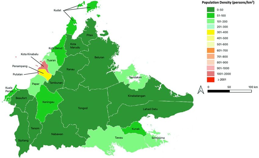
Figure 5. Map of Sabah, showing its 25 districts, which are colored according to their population density,
expressed as number of persons per square kilometer. The map was generated using QGIS version 3.18.1 Zurich
(https://www.qgis.org/).
Materials and methods
Ethics statement. HFMD notifications are captured under the Public Health Infectious Disease Surveil-
lance System for the purpose of analysis of data and determining the epidemiological pattern and characteristics.
The study was registered under the National Medical Research Registry and the study protocol was approved by
the Medical Ethics and Research Committee, Ministry of Health Malaysia (NMRR-20-3286-56537). Permission
was obtained from Director-General of Health Malaysia to publish this article (Ref. no. 800-4/4/1 Jld.91(56)
dated February 4, 2021). All procedures were performed in accordance with the relevant guidelines, protocols,
and regulations of the Ministry of Health, Malaysia (https://www.moh.gov.my/moh/resources/auto%20dow
nload%20images/589d71f714d23.pdf). Clinical samples were taken as part of outbreak control measures and
informed consents were obtained from guardians of the children. All information was kept anonymous.
Study site. Sabah is a region in Malaysia, located at the northern portion of the Borneo island. The region,
with a geographical area of 73,904 k m2, consists of 25 d
istricts33, with an estimated population of 3.9 million in
34
2019 (Fig. 5). Kota Kinabalu is the capital city of Sabah, and it has the highest population density of 1397 per
square kilometer. Sabah has a relatively low urban population compared to other Malaysian states, where only
54% of Sabah’s population live in urban areas33. Sabah has an equatorial climate with high humidity and high
annual rainfall.
Epidemiological data. From January 2015 to December 2019, a total of 17,574 clinically diagnosed and
PCR-confirmed HFMD cases in Sabah were reported and recorded in a Malaysian public health disease surveil-
lance system called eNotifikasi (also known as Communicable Disease Control Information System; http://enoti
fikasi.moh.gov.my). Data of all reports of HFMD cases were extracted from the database, including demographic
information, date of onset, date of diagnosis, and classification of transmission.
Sample collection for molecular analysis. Thirteen children ranging from one to eight years old with
clinical symptoms of HFMD, such as fever, mouth ulcers, maculopapular rashes, and vesicles on palms and soles,
were included in the study. Twenty-one samples from these children, including rectal, mouth, vesicle, and sole
swabs, were collected from Beaufort District Health Office, Beaufort, Sabah, from August 9, 2018, to August 13,
2018. The swabs were kept in viral transport medium (VTM), labeled, and refrigerated before being transported
to the Borneo Medical and Health Research Centre in Universiti Malaysia Sabah for molecular analysis.
Sample processing. The collected samples were vortexed and filtered through a 0.22 µm syringe filter. The
filtrate of each sample was stored in a 2 mL screw-cap tube, labeled, and kept at − 80 °C until further analysis.
Scientific Reports | (2021) 11:17814 | https://doi.org/10.1038/s41598-021-96083-3 8
Vol:.(1234567890)www.nature.com/scientificreports/
Primer Sequence (5′–3′) Gene Location
AN32 GTYTGCCA VP1 3009–3002
AN33 GAYTGCCA VP1 3009–3002
AN34 CCRTCRTA VP1 3111–3104
AN35 RCTYTGCCA VP1 3009–3002
MD91 CCTCCGGCCCCTGAATGCGGCTAAT VP4 444–468
OL68-1 GGTAAYTTCCACCACCANCC VP4 1178–1197
EVP4 CTACTTTGGGTGTCCGTGTT VP4 541–560
224 GCIATGYTIGGIACICAYRT VP3 1977–1996
222 CICCIGGIGGIAYRWACAT VP1 2969–2951
AN89 CCAGCACTGACAGCAGYNGARAYNGG VP1 2602–2627
AN88 TACTGGACCACCTGGNGGNAYRWACAT VP1 2977–2951
Table 3. Primers used for cDNA synthesis, PCR amplification, and sequencing.
Viral RNA extraction and first‑strand cDNA synthesis. Viral RNA was extracted from samples using
the QIAamp Viral RNA Mini Kit (Qiagen, Germany) according to the manufacturer’s instructions. The RT-PCR
procedure for each amplification was performed in a 10 µL reaction mixture with 2.5 µL of the extracted RNA
template, cDNA primers AN32, AN33, AN34, and AN35 (Table 3), and the SuperScript III First-Strand Syn-
thesis System (Invitrogen, USA). The mixture was incubated at 50 °C for 50 min, then at 85 °C for 5 min before
storing the cDNA synthesis reaction at − 20 °C for further analysis.
Enterovirus detection using semi‑nested reverse‑transcription polymerase chain reaction
(snRT‑PCR). Enteroviral RNA was detected in samples by snRT-PCR of the VP4–VP2 region. The first
primer pair, MD91-OL68-1 (Table 3), was used to amplify the partial 5’ non-coding region, VP2, and complete
VP4. 3 µL of RNA was used in the PCR using the AccessQuick RT-PCR System (Promega, USA). The PCR con-
dition consisted of 1 cycle of initial denaturation at 95 °C for 2 min, 40 cycles of denaturation at 95 °C for 30 s,
annealing at 55 °C for 30 s, and extension at 72 °C for 1 min, followed by 1 cycle of final extension at 72 °C for
7 min.
snRT-PCR with the second primer pair, EVP4-OL68-1 (Table 3), was performed by using 2 µL of the PCR
product under the following PCR condition: 1 cycle of initial denaturation at 95 °C for 6 min, 40 cycles of dena-
turation at 95 °C for 30 s, annealing at 55 °C for 30 s, and extension at 72 °C for 1 min, followed by 1 cycle of
final extension at 72 °C for 7 min.
Enterovirus serotyping using consensus degenerate hybrid oligonucleotide primer (CODE‑
HOP). The enterovirus VP1 gene sequence was amplified using a CODEHOP protocol described by Nix,
Oberste, and Pallansch (2006)35. Briefly, the cDNA generated by the four different primers (AN32–AN35) was
used for the first round of PCR with primers 224 and 222 (Table 3). The condition for the first PCR consisted of
1 cycle of initial denaturation at 95 °C for 6 min, 40 cycles of denaturation at 95 °C for 30 s, annealing at 42 °C
for 30 s, and extension at 60 °C for 1 min. The first PCR generated a product of 992 bp, which was used for the
second round of PCR with primers AN89 and AN88 (Table 3) for nested amplification. The condition for the
second PCR consisted of 1 cycle of initial denaturation at 95 °C for 6 min, 40 cycles of denaturation at 95 °C for
30 s, annealing at 60 °C for 30 s, and extension at 72 °C for 30 s, followed by 1 cycle of final extension at 72 °C for
5 min. The second PCR generated a product of 375 bp.
Sequence analysis. Partial nucleotide sequences of the VP1 gene of CVA16 (254 bp) and EV71 (359 bp)
were used for phylogenetic analyses. The amplified DNA of each positive sample was sequenced using the Big-
Dye Terminator v3.1 Cycle Sequencing Kit (Applied Biosystems, Foster City, CA) according to the manufac-
turer’s instructions, and the product was run on an ABI Prism 3100 Genetic Analyzer (Applied Biosystems).
Nucleotide sequences of VP1 were checked against the NCBI database by BLAST to find the enterovirus serotype
with the highest identity. The nucleotide sequences of other CVA16 and EV71 strains were extracted from Gen-
Bank. Multiple sequence alignment was performed using Clustal W, and phylogenetic trees were constructed
using MEGA 6.0, applying the neighbor-joining method based on the Tamura–Nei substitution m odel36. Boot-
strap analysis of 1000 replicates was conducted to determine the significance of the branching of the constructed
tree. Nucleotide sequences analyzed in this study have been submitted to the DNA Data Bank of Japan (DDBJ).
Data analysis. Data analysis was performed using Microsoft Excel for Mac 2017 (Version 15.30). QGIS
v3.14.1 (Pi) was used to generate spatial maps of HFMD cases in Sabah.
Received: 12 February 2021; Accepted: 27 July 2021
Scientific Reports | (2021) 11:17814 | https://doi.org/10.1038/s41598-021-96083-3 9
Vol.:(0123456789)www.nature.com/scientificreports/
References
1. Yu, L., He, J., Wang, L. & Yi, H. Incidence, aetiology, and serotype spectrum analysis of adult hand, foot, and mouth disease patients:
A retrospective observational cohort study in northern Zhejiang, China. Int. J. Infect. Dis. 85, 28–36. https://doi.org/10.1016/j.ijid.
2019.05.016 (2019).
2. Xu, M. et al. Genotypes of the enterovirus causing hand foot and mouth disease in Shanghai, China, 2012–2013. PLoS ONE 10,
e0138514. https://doi.org/10.1371/journal.pone.0138514 (2015).
3. Takahashi, S. et al. Hand, foot, and mouth disease in China: Modeling epidemic dynamics of enterovirus serotypes and implica-
tions for vaccination. PLoS Med. 13, e1001958. https://doi.org/10.1371/journal.pmed.1001958 (2016).
4. van der Sanden, S. M. et al. Prediction of protection against Asian enterovirus 71 outbreak strains by cross-neutralizing capacity
of serum from Dutch donors, the Netherlands. Emerg. Infect. Dis. 22, 1562–1569. https://doi.org/10.3201/eid2209.151579 (2016).
5. Mao, Q. et al. Coxsackievirus A16. Hum. Vaccine Immunother. 10, 360–367. https://doi.org/10.4161/hv.27087 (2014).
6. Chen, X. et al. Molecular epidemiology of coxsackievirus A16: Intratype and prevalent intertype recombination identified. PLoS
ONE 8, e82861. https://doi.org/10.1371/journal.pone.0082861 (2013).
7. Puenpa, J., Wanlapakorn, N., Vongpunsawad, S. & Poovorawan, Y. The history of enterovirus A71 outbreaks and molecular epi-
demiology in the Asia-Pacific region. J. Biomed. Sci. 26, 75. https://doi.org/10.1186/s12929-019-0573-2 (2019).
8. NikNadia, N. M. N. et al. Cyclical patterns of hand, foot and mouth disease caused by enterovirus A71 in Malaysia. PLoS Negl.
Trop. Dis. 10, e0004562. https://doi.org/10.1371/journal.pntd.0004562 (2016).
9. Chua, K. B. & Kasri, A. R. Hand foot and mouth disease due to enterovirus 71 in Malaysia. Virol. Sin. 26, 221–228. https://doi.org/
10.1007/s12250-011-3195-8 (2011).
10. Ling, B. P., Jalilian, F. A., Harmal, N. S., Yubbu, P. & Sekawi, Z. Detection and characterization of viruses causing hand, foot and
mouth disease from children in Seri Kembangan, Malaysia. Trop. Biomed. 31, 654–662 (2014).
11. Chan, Y. F. et al. Comparative genetic analysis of VP4, VP1 and 3D gene regions of enterovirus 71 and coxsackievirus A16 circulat-
ing in Malaysia between 1997–2008. Trop. Biomed. 29, 451–466 (2012).
12. Yusof, M. A. et al. Molecular epidemiology of human enterovirus71 (HEV71) strains isolated in Peninsular Malaysia and Sabah
from year 2001 to 2009. J. Gen. Mol. Virol. 3, 18–26 (2011).
13. Ooi, M. H. et al. Human enterovirus 71 disease in Sarawak, Malaysia: A prospective clinical, virological, and molecular epidemio-
logical study. Clin. Infect. Dis. 44, 646–656. https://doi.org/10.1086/511073 (2007).
14. Zhao, J. & Hu, X. The complex transmission seasonality of hand, foot, and mouth disease and its driving factors. BMC Infect. Dis.
19, 521. https://doi.org/10.1186/s12879-019-4153-6 (2019).
15. Lee, M. H. P. et al. Detection of enteroviruses during a 2018 hand, foot and mouth disease outbreak in Malaysia. Trop. Biomed. 38,
150–153. https://doi.org/10.47665/tb.38.1.026 (2021).
16. Menon, P. HFMD cases at its highest in Petaling. The Star https://www.thestar.com.my/metro/metro-news/2018/08/10/hfmd-
cases-at-its-highest-in-petaling-dr-siti-mariah-over-90-of-those-infected-in-selangor-are-child (2018).
17. Duan, C. et al. Meteorological factors and its association with hand, foot and mouth disease in Southeast and East Asia areas: A
meta-analysis. Epidemiol. Infect. 147, e50. https://doi.org/10.1017/S0950268818003035 (2019).
18. Lee, C. C. D., Tang, J. H., Hwang, J. S., Shigematsu, M. & Chan, T. C. Effect of meteorological and geographical factors on the
epidemics of hand, foot, and mouth disease in island-type territory, East Asia. Biomed, Res. Int. 2015, 805039. https://doi.org/10.
1155/2015/805039 (2015).
19. Hii, Y. L., Rocklöv, J. & Ng, N. Short term effects of weather on hand, foot and mouth disease. PLoS ONE 6, e16796. https://doi.
org/10.1371/journal.pone.0016796 (2011).
20. Leong, P. F., Labadin, J., Rahman, S. B. A. & Juan, S. F. S. Quantifying the relationship between the climate and hand-foot-mouth
disease (HFMD) incidences. In 2011 Fourth International Conference on Modeling, Simulation and Applied Optimization 1–5 (IEEE,
2011). https://ieeexplore.ieee.org/document/5775582.
21. Bian, L. et al. Coxsaclievirus A6: A new emerging pathogen causing hand, foot and mouth disease outbreaks worldwide. Expert
Rev. Anti Infect. Ther. 13, 1061–1071. https://doi.org/10.1586/14787210.2015.1058156 (2015).
22. Perera, D. et al. Molecular phylogeny of modern coxsackievirus A16. Arch. Virol. 152, 1201–1208. https://doi.org/10.1007/s00705-
006-0934-5 (2007).
23. Klein, M. H. EV71 vaccines: A first step towards multivalent hand, foot and mouth disease vaccines. Expert Rev. Vaccines 14,
337–340. https://doi.org/10.1586/14760584.2015.993385 (2015).
24. Koh, W. M. et al. The epidemiology of hand, foot and mouth disease in Asia: A systematic review and analysis. Pediatr. Infect. Dis.
J. 35, e285–e300. https://doi.org/10.1097/INF.0000000000001242 (2016).
25. Lin, H. et al. Protective effect of exclusive breastfeeding against hand, foot and mouth disease. BMC Infect. Dis. 14, 645. https://
doi.org/10.1186/s12879-014-0645-6 (2014).
26. Wang, Y. et al. Epidemiology and clinical characteristics of hand foot, and mouth disease in a Shenzhen sentinel hospital from
2009 to 2011. BMC Infect. Dis. 13, 539. https://doi.org/10.1186/1471-2334-13-539 (2013).
27. Ang, L. W. et al. The changing seroepidemiology of enterovirus 71 infection among children and adolescents in Singapore. BMC
Infect. Dis. 11, 270. https://doi.org/10.1186/1471-2334-11-270 (2011).
28. Chang, L. Y. et al. Risk factors of enterovirus 71 infection and associated hand, foot, and mouth disease/herpangina in children
during an epidemic in Taiwan. Pediatrics 109, e88. https://doi.org/10.1542/peds.109.6.e88 (2002).
29. Qiu, J. et al. The clinical and epidemiological study of children with hand, foot, and mouth disease in Hunan, China from 2013 to
2017. Sci. Rep. 9, 11662–11662. https://doi.org/10.1038/s41598-019-48259-1 (2019).
30. Xu, C. et al. Effects of temperature fluctuations on spatial-temporal transmission of hand, foot, and mouth disease. Sci. Rep. 10,
2541. https://doi.org/10.1038/s41598-020-59265-z (2020).
31. Neiderud, C. J. How urbanization affects the epidemiology of emerging infectious diseases. Infect. Ecol. Epidemiol. 5, 27060. https://
doi.org/10.3402/iee.v5.27060 (2015).
32. NikNadia, N. et al. Risk factors for enterovirus A71 seropositivity in rural indigenous populations in west Malaysia. PLoS ONE
11, e0148767. https://doi.org/10.1371/journal.pone.0148767 (2016).
33. Murphy, A. et al. Incidence and epidemiological features of dengue in Sabah, Malaysia. PLoS Negl. Trop. Dis. 14, e0007504. https://
doi.org/10.1371/journal.pntd.0007504 (2020).
34. Kaur, N. et al. The east coast districts are the possible epicenter of severe dengue in Sabah. J. Physiol. Anthropol. 39, 19. https://doi.
org/10.1186/s40101-020-00230-0 (2020).
35. Nix, W. A., Oberste, M. S. & Pallansch, M. A. Sensitive, seminested PCR amplification of VP1 sequences for direct identification of
all enterovirus serotypes from original clinical specimens. J. Clin. Microbiol. 44, 2698–2704. https://d oi.o
rg/1 0.1 128/J CM.0 0542-0 6
(2006).
36. Tamura, K., Stecher, G., Peterson, D., Filipski, A. & Kumar, S. MEGA6: Molecular evolutionary genetics analysis version 6.0. Mol.
Biol. Evol. 30, 2725–2729. https://doi.org/10.1093/molbev/mst197 (2013).
Scientific Reports | (2021) 11:17814 | https://doi.org/10.1038/s41598-021-96083-3 10
Vol:.(1234567890)www.nature.com/scientificreports/
Acknowledgements
We would like to thank the Director-General of Health Malaysia for his permission to publish this article. The
study was supported in part by a Launching Grant for Centre of Excellence from Universiti Malaysia Sabah
(Grant Number: AM18006).
Author contributions
K.A., and M.J. conceptualized the study. S.Y.F., D.M., J.F.Y., A.L.L.B.A.L., and V.J. curated data. S.Y.F., D.M.,
C.R., J.F.Y., M.J., A.L.L.B.A.L., V.J., and K.A. analyzed data. K.A. acquired funds. C.R., J.F.Y., M.J., A.L.L.B.A.L.,
and V.J. performed investigations. K.A., C.R., and M.J. performed project administrations. K.A., C.R., and M.J.
supervised. S.Y.F. wrote the original draft. S.Y.F., D.M., C.R., J.F.Y., M.J., A.L.L.B.A.L., V.J., and K.A. reviewed,
edited and wrote the final manuscript.
Competing interests
The authors declare no competing interests.
Additional information
Correspondence and requests for materials should be addressed to K.A.
Reprints and permissions information is available at www.nature.com/reprints.
Publisher’s note Springer Nature remains neutral with regard to jurisdictional claims in published maps and
institutional affiliations.
Open Access This article is licensed under a Creative Commons Attribution 4.0 International
License, which permits use, sharing, adaptation, distribution and reproduction in any medium or
format, as long as you give appropriate credit to the original author(s) and the source, provide a link to the
Creative Commons licence, and indicate if changes were made. The images or other third party material in this
article are included in the article’s Creative Commons licence, unless indicated otherwise in a credit line to the
material. If material is not included in the article’s Creative Commons licence and your intended use is not
permitted by statutory regulation or exceeds the permitted use, you will need to obtain permission directly from
the copyright holder. To view a copy of this licence, visit http://creativecommons.org/licenses/by/4.0/.
© The Author(s) 2021
Scientific Reports | (2021) 11:17814 | https://doi.org/10.1038/s41598-021-96083-3 11
Vol.:(0123456789)You can also read


