25I seeds implantation for lymph nodes metastasisfrom radioactive iodine-refractory differentiated thyroid carcinoma: A short-term ecacy and ...
←
→
Page content transcription
If your browser does not render page correctly, please read the page content below
25I seeds implantation for lymph nodes
metastasisfrom radioactive iodine-refractory
differentiated thyroid carcinoma: A short-term
efficacy and dosimetry study
Guoxu Zhang ( zhangguoxu502@sina.com )
General hospital of northern theater command
Wenwen Zhang
General hospital of northern theater command
Shanshan Hao
General hospital of northern theater command
Zhiguo Wang
General hospital of northern theater command
Liqiu Ji
General hospital of northern theater command
Xiangyan Ge
General hospital of northern theater command
Gen Li
General hospital of northern theater command
Youchao Wang
General hospital of northern theater command
Research Article
Keywords: Thyroid tumor,Tumor metastasis,Lymph nodes,Brachytherapy,Iodine radioisotope,
Radiation dose
Posted Date: February 1st, 2022
DOI: https://doi.org/10.21203/rs.3.rs-1310697/v1
License: This work is licensed under a Creative Commons Attribution 4.0 International License.
Read Full License
Page 1/12Abstract Purpose: To evaluate the efficacy and safety of 125I seeds implantation for lymph nodes metastasis (LNM) from radioactive iodine-refractory differentiated thyroid carcinoma (RAIR-DTC), and to verify the computer three-dimensional treatment planning system (TPS) from the dosimetry accuracy in assisting seeds implantation to treat LNM. Methods and materials: Retrospective analysis was performed on 17 RAIR-DTC patients with LNM admitted to the General Hospital of Northern Theater Command from December 2016 to January 2019 (8 males, 9 females, median age 58 years). All patients underwent preoperative TPS planning design, CT- guided puncture and 125I seeds implantation (seed activity 14.8-25.9 MBq). The dosimetric results of postoperative validation were compared with those of preoperative planning, including the dosimetric parameters such as target volume before and after surgery and the dose received by 90% and 100% gross tumor volume (GTV) (D90, D100),the percentage of GTV receiving 100% and 150% prescription dose (V100, V150), homogeneity index (HI). All patients underwent CT after 6 months to compare the LNM size, serum thyroglobulin (Tg) level, and the improvement of complications before and after treatment. Efficacies were divided into complete remission (CR), partial remission (PR), stable disease (SD), progressive disease (PD). Paired t test or Wilcoxon signed rank test were used to analyze the data. Results: Among 17 patients, a total of 226 125I radioactive seeds were implanted. Among them, 1 achieved CR, 10 achieved PR, 4 were with SD, 2 were with PD. The median diameter of LNM was 1.40(0.65, 3.05) cm before treatment and 0.40(0.21,0.91) cm 6 months after treatment (z=-3.95, P
RAIR-DTC), the 10-year survival rate of this part of patients is less than 10% [2]. Finding an effective
treatment for RAIR-DTC has always been a hot topic in the field of thyroid cancer. At present, the
commonly used methods for the treatment of RAIR-DTC include targeted drug therapy, local reoperation,
chemotherapy, external radiation therapy, immunotherapy, gene therapy, etc. Part of lymph node
metastases are often due to hidden locations or limitations of important anatomical structures (such as
main trachea, large blood vessels, etc.), causing patients to lose the opportunity for surgery. In recent
years, radioactive seed implantation has been widely used in the local treatment of malignant tumors.
Considering that dose distribution is the most direct and important factor affecting the efficacy of seed
implantation [3], A reasonable pre-implantation plan can be carried out according to the treatment
planning system (TPS) to achieve the expected dose distribution. This study explored the short-term
efficacy and adverse effects of 125I seed implantation in the treatment of RAIR-DTC withlymph node
metastases, and we compared the preoperative and postoperative dosimetry results of the computerized
three-dimensional TPS-assisted seed implantation therapy, and evaluated the accuracy of the technology
in guiding the dose distribution of seed implantation therapy.

Materials And Methods
Research object
A total of 17 RAIR-DTC patients with lymph node metastasis who were hospitalized at the Northern
Theater Command General Hospital and received 125I seed implantation from December 2016 to
January 2019 were collected, including 8 males and 9 females, with a median age of 58 years. The range
was 22 to 70 years old,andall ofthemwere digonisedaspapillary thyroid carcinoma (PTC). 22 metastatic
lymph nodes were diagnosed pathologically by needle biopsy, of which 5 patients had 2 lymph node
metastases, 12 cases of them has 1 lymph node metastasis. Each patient took 131I with a cumulative
dose of >22.2 GBq and received 125I seed implantation at least half a year after 131I treatment. (This
study was reviewed and approved by the Medical Ethics Committee of our hospital, and all patients
signed an informed consent form.)Inclusion criteria: (1) Complete thyroidectomy or near-total resection
before operation, according to functional neck dissection and extended radical resection of cervical
lymph nodes, confirmed by pathology as DTC metastasis, it was identified as RAIR-DTC according to the
2015 American Thyroid Association (ATA) guidelines [4]; (2) WBC count≥3.5×10⁹/L, Hb≥90 g/L,
prothrombin activity>40 %, PLT>100×109/L, normal electrocardiogram, serum alanine aminotransferase
and serum aspartate aminotransferase in liver function test Enzyme value does not exceed 3 times the
normal upper limit; (3) Karnofsky performance status (KPS) scor≥70, with detailed clinicopathological
data and follow-up data, survival time of more than 1 year; (4) no further transfer. Exclusion criteria: (1)
Thyroglobulin antibody (TgAb) positive; (2) Severe heart, lung, liver and kidney dysfunction; (3) Severe
coagulation dysfunction; (4) General condition is very poor or cachexia.

                                                   Page 3/12
Instrument
A 64-slice CT with Discovery VCT PET/CT produced by GE in the United States was used for routine
scanning. The implant gun and the push thimble are flat-head general-purpose types produced by
Zhejiang Xiangshan Jinheng Machinery Manufacturing Co., Ltd. The 15-20 cm×18 G puncture needle
produced by Yako Co.; Ltd. is used. The particle TPS is provided by Beijing Feitian Zhaoye Technology
Co., Ltd. The 125I radioactive particle source is produced by Beijing Atom Technology Co., Ltd. The
particle length is 4.5 mm, the diameter is 0.8 mm, and the fully enclosed titanium shell. The particle
activity is 14.8-25.9 MBq, the half-life is 59.6 d, and the tissue penetration distance is 17 mm. Steam
sterilization. The whole body 131I imaging adopts the Symbia T16 SPECT/CT instrument of Siemens
company in Germany. Five items of thyroid function (free triiodothyronine, free thyroxine, TSH, TgAb, anti-
thyroid peroxidase autoantibody) and thyroglobulin (Tg) are detected by chemiluminescence method.
The instrument and kit are composed of Soling Diagnostics (Italy) Co., Ltd. provides: Soling-LIAISON
Chemiluminescence Analyzer, Soling-LIAISON Kit.
Preparation before surgery
Complete various laboratory tests before surgery, including blood routine, ABO and RH blood types,
coagulation, liver and kidney function, four items of infectious diseases, five items of thyroid function, Tg,
electrocardiogram, etc.; inform patients and their families complications and precautions that may occur
during and after the operation and sign the informed consent; Stopdrinking for 2~4 hours before local
anesthesia, establishing thevenous access; performing breathing training to reduce the intervention
caused by the patient’s respiratory activity during the operation Implant deviation; patients with pain
intolerance can be given tramadol analgesia (oral 50-100 mg/time) 30 min before surgery, and codeine
phosphate tablets (oral, 15-30 mg/time) can be given for irritation cough.
TPS plan
Import the enhanced or plain CT scan images 1 week before the operation into the TPS system for
design, avoiding important tissues and organs (such as large blood vessels, heart, spinal cord, etc.) as
much as possible. Delineate the gross tumor volume (GTV) and planning target volume (PTV). The PTV
range includes the tumor itself and surrounding tissues that may infiltrate (such as burr signs around the
tumor). Set an appropriate prescription dose of 110~140 Gy and particle activity (14.8~25.9 MBq) [5],
follow the principle of 0.5~1.0 cm between particles, 1 cm between needle tracks, Making 90% of GTV
received prescription dose (D90)> matched peripheral dose (matched peripheral dose, MPD), and then
calculate the number of particles and particle source distribution to simulate the best way to enter the
needle. Obtain the preoperative planning parameters D90, D100, the volume percentage (V100, V150) of
100% and 150% of the prescribed dose received by GTV, and the dose-volume histogram (DVH).
Page 4/12Intraoperative operations
Select different positions according to the lesion location, place an external positioning grating on the
skin area corresponding to the lesion, select the puncture point by CT scan, and intramuscularly inject 2%
lidocaine for local infiltration anesthesia, determine the depth and angle of the puncture path, and follow
the TPS plan Needle cloth layer by layer. During the operation, the position and direction of the implanted
needle were adjusted according to the CT scan, so that the depth of the needle into the lesion was
through the center of the tumor and 0.5 cm from the edge of the tumor. Then, one particle was implanted
every 0.5 to 1.0 cm in the backward way, and the particles were supplemented from the periphery to the
center, and the needle was placed again referring to the position of the implanted needle, and the
intraoperative real-time optimization until the implantation was completed. Care should be taken to avoid
important blood vessels and nerves during implantation. During the operation, the patient's heart rate,
blood pressure, respiration and blood oxygen saturation were monitored. At the same time, the patient's
consciousness, pain, breathing, cough, hemoptysis and other conditions were observed, and timely
symptomatic treatment was performed. After the operation is completed, press the puncture point for 10-
20 minutes.
Post-operative verification plan
After the operation is completed, the CT images are immediately transferred to TPS for postoperative
verification and particle identification, and D90, D100, V100, V150, DVH, and homogeneity index (HI) are
counted. HI=(VT, ref-VT, 1.5ref)/VT, ref, VT, ref, VT, 1.5ref is the volume of the target area receiving the
prescribed dose, and the volume of the target area receiving 150% of the prescribed dose (cm3); Larger,
indicating that the target dose distribution is more uniform [6].
Postoperative review and short-term efficacy evaluation
(1) Efficacy judgment. Neck color Doppler ultrasound and neck CT were reviewed before treatment and 6
months after treatment to evaluate the effect of 125I seed implantation. According to the solid tumor
efficacy evaluation standard (Response Evaluation Criteria in Solid Tumors, RECIST) 1.1 for efficacy
evaluation [7]. ①Complete remission (CR): The lesion completely disappeared, and there is no imaging
suggestion of recurrence, tumor residual and metastasis, or only 125I particle metallic shadow; ②Partial
remission (PR): The sum of the short diameter of the target lesion is at least The baseline short diameter
of the lesion (the sum of the smallest diameters measured before treatment) was reduced by 30%; ③stable
disease (SD): those who did not meet the criteria for either PR or progressive disease (PD); ④PD : Based on
the minimum sum of the short diameters of the target lesions during the entire study, the diameter sum
increased by ≥20%; or the absolute value of the diameter sum increased by ≥5 mm (the appearance of
one or more new lesions is also regarded as PD).(2) Adverse reactions. After the operation, closely
observe whether the patient has adverse reactions such as bleeding, hematoma, particle displacement,
Page 5/12needle implantation, etc., and refer to the United States Tumor Radiotherapy Cooperation Group/European Organization for Research and Treatment of Cancer (Radiation Therapy Oncology Group/European Organization for Research and Treatment of Cancer). (RTOG/EORTC) Acute Radiation Response Scoring Standard evaluates skin radiation damage caused by radioactive seed implantation [8], observes and evaluates complications during operation, postoperative and follow-up. Statistical analysis Use IBM SPSS 19.0 software to process data. Quantitative data conforming to the normal distribution are expressed as ±s, quantitative data not conforming to the normal distribution are expressed as M (P25, P75); qualitative data are expressed as frequency or percentage. The quantitative data before and after treatment were compared by paired t test or Wilcoxon signed rank test. P
Time                                     Pre-                Post-operative     T/Z        P
                                          operative           verification

                                          planning

 Number of implanted                      15.5                14.5(2.0~30.0)     Z          P=0.553>0.05
 particles[grain;M(P25,P75)]              (2.0~32.0)                             =-0.593

 PTV[cm3;M(P25,P75)]                      1.4                 1.4(0.6~10.6)      Z          P=0.109>0.05
                                          (0.6~9.8)                              =-1.604

 D90(cGy;x±s)                             12                  12                 t          P=0.402>0.05
                                                                                 =0.251
                                          497.8±1             378.8±3 182.0
                                          686.4

 D100(cGy;x±s)                            8                   6                  t          P=0.0130.05
                                          (0.2~0.4)                              =-0.663

 Table1: D90 and D100 are the prescribed doses received by 90% and 100% of the gross tumor volume
 (GTV), HI is the uniformity index, PTV is the planned target volume, and V100 and V150 are the
 percentages of the GTV receiving 100% and 150% of the prescribed dose respectively.
No serious bleeding or vascular embolism occurred during the operation. One case had a small amount
of bleeding at the surgical site during the operation, which was relieved after intravenous injection of
hemostatic drugs and compression to stop the bleeding; 2 cases had a skin reaction grade II reaction,
which was improved after anti-inflammatory and external application of drugs, and no radioactive
particles migrated to other tissues or organs Case.

Discussion
Cervical lymph node metastasis is the most common after PTC. Lymph node metastases are mostly
located near the large blood vessels in the neck, around the trachea, and near the important nerves. The
rapid growth of the tumor will cause invasion of the surrounding adjacent tissues. Patients often
experience facial edema, pain, and voice. Clinical symptoms such as hoarseness and numbness. In
recent years, the advantages of 125I particle therapy have been gradually explored clinically. 125I
continuously attenuates between tissues and releases 27.4~31.4 keV X-rays and 35.5 keV γ-rays [9],
among which γ-rays directly ionize tumor DNA single-strand or double-strand breaks, and X-rays generate
oxygen free from indirect ionization. Base inactivation of tumor cells [10-11]. In addition, 125I particles
act on some cancer cells in the quiescent phase (G0) phase, making them transformed into proliferation

                                                  Page 7/12
phase (G2 and M phase) cells that are easily killed after entering the division phase [10-11]. Recently,
Chen Wei et al. [12] performed 125I particle therapy on 15 patients with neck recurrence after DTC. The
volume reduction rate was (83.5±16.9)% after 12 months, and the postoperative serum Tg was 4.9 (0.7,
13.2) μg/L, which is significantly lower than preoperative [57.0(8.6, 114.8) μg/L], 22 high-enhancement
lesions turned into low- or no-enhancement lesions, suggesting that the implantation of 125I particles in
neck lesions has a large scope of application and strong operability , Good tolerance. In this study, 17
patients received 125I seed implantation, including 1 CR, 10 PR, and 4 SD. After treatment, the size and Tg
of the lesion were significantly lower than those before surgery, which were similar to the results of
previous studies [13]. After treatment, the symptoms of 3 cases of hoarseness and 5 cases of local
tenderness were significantly relieved. In this study, a fixed head rest or a body membrane was used to
avoid and reduce patient displacement during the implantation process. The results of this study show
that 125I radioactive particle therapy is minimally invasive, safe, and can significantly improve the quality
of life of patients in the short term.
Dosage determination is an important part of 125I seed implantation. Before TPS, the ideal dose
distribution is automatically designed according to the tumor boundary, important tissue location,
prescription dose, particle activity, etc., after the operation, the target area dose and the prescription dose
are verified after the operation. Errors etc. evaluate the quality of radioactive seed implantation. In this
study, according to the "Specifications for the Clinical Application of Radioactive Particles in Tumor
Therapy", the prescribed dose of radiotherapy was set to 110-140 Gy [5]. If the dose is too small, the
purpose of killing the tumor will not be achieved; if the dose is too large, it will Cause damage to normal
tissues and organs around the lesion. In this study, D90, V100, HI and other dosimetry parameters were
not statistically significant before and after surgery, and D90 reached (12 378.8±3 182.0) cGy and V100
reached (92.2±14.3)% after surgery, indicating that radioactive seed implantation The expected plan
distribution is basically achieved, and the dosimetry requirements can be better met according to the TPS
preoperative plan. However, the HI before and after the operation in this study was low, and the
postoperative verification V150 was more than 50%, suggesting that the dose distribution in the target
area is uneven and the volume of the high-dose area is too large. Analyze the possible reasons for the
error: (1) Postoperative changes in the size of the lesion and rapid reduction of edema around the lesion
accelerate the dispersion and regression of radiation doseand reduce the conformation index of the dose
distribution within the lesion [14-19]; and The verification dose is not the actual absorbed dose of the
tumor. In this study, the postoperative verification time was within 24 hours. Local tissue edema that did
not resolve within a short period of time after surgery would affect the assessment of the biological
effect dose [18]; (2) due to postoperative CT Artifacts, imaging of a particle on two levels and overlapping
of some particles make it difficult to identify the precise position of the particle, leading to a deviation in
the postoperative verification and measurement dose; (3) The density of the tumor is different due to
hemorrhage or necrosis due to particle radiation effects. This produces a change in particle position. In
addition, postoperative verification found that 2 patients had progressed to the target lesion about 4
months after implantation treatment. Further research found that their postoperative D90 was
significantly lower than the prescribed dose planned before surgery. The main considerations are as
Page 8/12follows: (1)2 The structure of the operation area was disordered, and the patient’s age was too old, and
the neck tissue was soft, which made implantation deviations during the operation; (2) Before and after
the operation, the local muscles contracted due to posture examination, intraoperative anesthesia, and
puncture stimulation. The shape of the volume changes.
RAIR-DTC lymph node metastasis is mostly close to the body surface, and the γ-rays released by 125I
particles can cause DNA double-strand breaks and affect skin cell metabolism [20]. Two patients in this
study developed painful erythema, pigmentation after 1 week of anti-inflammatory treatment, and no skin
ulceration. The analysis was due to multiple lesions being superficial and close to each other. With the
elimination of tumor tissue in the coverage area, particles occurred Caused by the formation of local "hot
spots" after aggregation. This suggests that when implanting particles, the distance between the particles
and the skin surface should be at least 1 cm, and the activity and number of particles should be
appropriately reduced according to the anatomical part of the lesion.
In summary, 125I seed implantation for the treatment of RAIR-DTC lymph node metastasis has the
advantages of good curative effect and fewer complications. The use of TPS can make the target dose
conformity better, effectively kill the tumor and better protect the surrounding normal tissues. However,
this study is a single-center retrospective study with a small sample size and a short follow-up period. It is
necessary to expand the sample size and analyze the effects of factors such as dose and tumor size on
the efficacy to provide better guidance for the standardized diagnosis and treatment of 125I particles.
Declarations
Acknowledgements
None.
Authors’contributions
ZGX carried out the studies,HSS and WGZ participated in collecting data, and drafted themanuscript.
ZWW and JLQ performed the statistical analysis and participated in its design. GXY 、LG and WYC helped
to draft the manuscript. All authors read and approved the final manuscript.
Funding
Natural Science Foundation of Liaoning Province (20180551010)
Availability of data and materials
No additional data are available.
Page 9/12Ethics approval and consent to participate
The study protocol was approved by the General hospital of northern theater command, and the
participants provided written informed consent.
Consent for publication
Not applicable
Competing interests
All authors declared that there are no conflicts of interest.
References
1. Fahiminiya S, de Kock L, Foulkes WD. Biologic and clinical perspectives on thyroid cancer[J]. N Engl
J Med, 2016, 375(23): 2306-2307. DOI:10.1056/NEJMc1613118.
2. Jin Y, Van Nostrand D, Cheng L, et al. Radioiodine refractory differentiated thyroid cancer[J].Crit Rev
Oncol Hematol, 2018, 125: 111-120. DOI:10.1016/j.critrevonc.2018.03.012.
3. Chasseray M, Dissaux G, Bourbonne V, et al. Dose to the penile bulb and individual patient anatomy
are predictive of erectile dysfunction in men treated with 125I low dose rate brachytherapy for
localized prostate cancer[J]. Acta Oncol, 2019, 58(7): 1029-
1035.DOI:10.1080/0284186X.2019.1574981.
4. Schmidt A, Iglesias L, Klain M, et al. Radioactive iodine-refractory differentiated thyroid cancer: an
uncommon but challenging situation[J]. Arch Endocrinol Metab, 2017, 61(1): 81-89. DOI:
10.1590/2359-3997000000245.
5. Wang JJ. Standard for clinical application of radioactive particles in the treatment of tumor[M].
Beijing: Peking University Medical Press, 2011: 24.
6. Shiraishi Y, Yorozu A, Ohashi T, et al. A dose-response analysis of biochemical control outcomes after
125I monotherapy for patients with favorable-risk prostate cancer[J].Int J Radiat Oncol Biol Phys,
2014, 90(5): 1069-1075. DOI:10.1016/j.ijrobp.2014.08.340.
7. Eisenhauer EA, Therasse P, Bogaerts J, et al. New response evaluation criteria in solid tumours:
revised RECIST guideline (version 1.1)[J]. Eur J Cancer, 2009, 45(2): 228-247.
DOI:10.1016/j.ejca.2008.10.026.
8. Bradley JD, Hope A, El Naqa I, et al. A nomogram to predict radiation pneumonitis, derived from a
combined analysis of RTOG 9311 and institutional data[J].Int J Radiat Oncol Biol Phys, 2007, 69(4):
985-992. DOI:10.1016/j.ijrobp.2007.04.077.
Page 10/129. Zheng W. Changes in PD-1/PD-L1 pathway function in non-small-cell lung cancer tissue before and
after 125I seed implantation[J]. J Hainan Med Univ, 2017, 23(13): 1829-1832.
DOI:10.13210/j.cnki.jhmu.20170809.028.
10. Zhang J, Zhu Y, Dong M, et al. Iodine-125 interstitial brachytherapy reduces tumor growth via
Warburg effect inhibition in non-small cell lung cancer A549 xenografts[J]. Oncol Lett, 2018, 16(5):
5969-5977. DOI:10.3892/ol.2018.9346.
11. Zhu Y, Dong M, Yang J, et al. Evaluation of iodine-125 interstitial brachytherapy using micro-positron
emission tomography/computed tomography with 18F-fluorodeoxyglucose in hepatocellular
carcinoma HepG2 xenografts[J].Med Sci Monit, 2019, 25: 371-380.DOI:10.12659/MSM.912590.
12. Chen W, Xie F, Zhang MB, et al. Ultrasound guided 125I brachytherapy in iodine refractory
differentiated thyroid cancer[J]. Chin J Med Imaging, 2020, 28(1): 26-30. DOI:10.3969/j.issn.1005-
5185.2020.01.006.
13. Zhang WW, Hao SH, Wang ZG, et al. Clinical value of 125I seeds implantation in treatment of lymph
nodes metastases from 131I refractory differentiated thyroid carcinoma[J]. Chin J Nucl Med Mol
Imaging, 2018, 38(1): 9-13. DOI:10.3760/cma.j.issn.2095-2848.2018.01.003.
14. Xing C, Zhang KX, Yuan QQ, et al. Dosimetry verification of radioactive seed implantation for
malignant tumor assisted by 3D-printing coplanar template[J]. Chin J Radiol Med Prot, 2017, 37(7):
514-517. DOI:10.3760/cma.j.issn.0254-5098.2017.07.008.
15. Du P, Xiao Y, Lu W. Modified fan-shaped distribution technology for computed tomography (CT)-
guided radioactive seed implantation in lung cancer patients with lung dysfunction[J]. Med Sci
Monit, 2017, 23: 4366-4375. DOI:10.12659/msm.902105.
16. Mukai Y, Hayashi N, Koike I, et al. Acute and late toxicities in localized prostate cancer patients
treated with low-dose 125I brachytherapy (110 Gy) in combination with external beam radiation
therapy versus brachytherapy alone (160 Gy)[J]. J Contemp Brachytherapy, 2018, 10(5): 397-404.
DOI:10.5114/jcb.2018.79379.
17. Wu J, Wang J, Sui AX, et al. Effect of tumor target volume reduction on dose after 125I seeds
implantation[J]. Chin J Exp Surg, 2015, 32(2): 309. DOI:10.3760/cma.j.issn.1001-9030.2015.02.033.
18. Wang JJ. Radioactive seeds implantation therapy in the era of precision medicine[J]. Chin J Nucl
Med Mol Imaging, 2018, 38(1): 1-3. DOI:10.3760/cma.j.issn.2095-2848.2018.01.001.
19. Zhao XZ, Zhang HT, Di XM, et al. Relationship between the peripheral dose and radioactive counts of
125I seeds detected by SPECT/CT[J]. Chin J Nucl Med Mol Imaging, 2017, 37(6): 351-354.
DOI:10.3760/cma.j.issn.2095-2848.2017.06.007.
20. Lin Q, Zhang Y, Dai JJ, et al. CT-guided 125I seed brachytherapy for superficial lymph node
metastases: a report of 23 cases[J]. J Shandong Univ (Health Sci), 2017, 55(2): 38-44.
DOI:10.6040/j.issn.1671-7554.0.2016.1350.
Figures
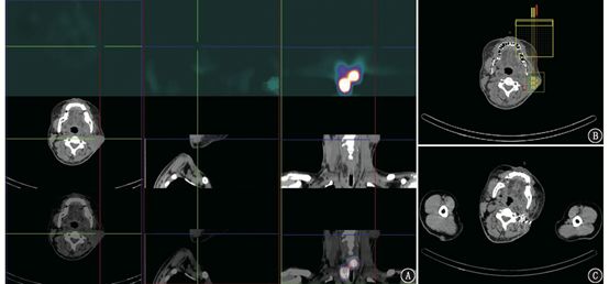
Page 11/12Figure 1
131I SPECT/CT imaging of a patient with 131I refractory differentiated thyroid cancer with metastasis to
the left neck lymph node (male, 37 years old) before and after 125I seed implantation (the cross and the
arrow show the lesion). A. 131I SPECT/CT neck fusion imaging showed no iodine uptake in the left
cervical lymph node metastases; B. Treatment planning system (TPS) preoperative needle placement; C.
Reexamination of the left cervical lymph node 6 months after treatment Significantly shrink, particle
aggregation, local skin pigmentation
Page 12/12You can also read