Ten-year survival in glioblastoma patient with neurofibromatosis type 1: illustrative case
←
→
Page content transcription
If your browser does not render page correctly, please read the page content below
J Neurosurg Case Lessons 3(4): CASE21630, 2022
DOI: 10.3171/CASE21630

Ten-year survival in glioblastoma patient with neurofibromatosis type
1: illustrative case
Sarah Basindwah, MD,1 Hisham Alkhalidi, FRCPath,2 Ahmed Abdelwarith, MD,3 and Sherif Elwatidy, PhD, FRCS(SN)1
1
Department of Surgery, Division of Neurosurgery, 2Department of Pathology, and 3Department of Oncology, College of Medicine, King Saud University, Riyadh, Saudi Arabia

    BACKGROUND Gliomas are commonly detected in patients with neurofibromatosis type 1 (NF1) at an early age. Few patients with NF1 are
    diagnosed with glioblastoma. The course of management, response to therapy, and prognosis of such patients are unknown. Few reports have shown
    longer-than-average survival rates for patients with NF1 with glioblastoma.
    OBSERVATIONS A 27-year-old man with NF1 presented with symptoms of high intracranial pressure. Imaging and pathology showed left
    frontotemporal glioblastoma. Gross total resection was achieved, and concurrent chemoradiotherapy was administered. Recurrence of tumor was
    detected 48 months later, and the patient underwent tumor debulking and concurrent chemoradiotherapy. The patient received first-, second-, and third-
    line chemotherapy (temozolomide, bevacizumab, bevacizumab/irinotecan) with good tolerance and has survived >10 years since then with good
    functional status.
    LESSONS This case demonstrates >10 years overall survival of glioblastoma in a patient with NF1. Reports of patients with NF1 with longer survival
    may be attributed to the young age at diagnosis and relatively better tolerance for therapy. It might also support the growing evidence of a unique
    subset of glioblastoma associated with NF1 and opens the door for a more molecular targeted therapy in the future.

    https://thejns.org/doi/abs/10.3171/CASE21630
    KEYWORDS glioblastoma survival; neurofibromatosis type 1 glioma; long-term survival; IDH wild-type glioma

    Neurofibromatosis type 1 (NF1) is an autosomal dominant tumor                        glioblastoma in patients with NF1 and perhaps the need for different or
predisposition syndrome. Patients with NF1 are diagnosed clinically                     combined therapy in this population.
and radiologically with pathognomonic cutaneous and neural axis                             In this case report, we present a case of 10-year overall survival in
tumors.1 Gliomas are commonly detected early in life and are usually                    a patient with glioblastoma with NF1 along with the disease course,
low-grade tumors.2 Although patients with NF1 are at a higher risk of                   and we review the literature on similar reported cases and their overall
glioblastoma than the general population, they are relatively uncommon                  survival.
compared with low-grade gliomas in patients with NF1.3 The epidemi-
ology, pathogenesis, prognosis, and survival of such patients are                       Illustrative Case
poorly understood. There are few reports on gliomas that are more                          A 27-year-old man with a known case of NF1 diagnosed after
aggressive than predicted in patients with NF1,4 whereas other reports                  meeting the clinical diagnostic criteria presented in 2010 with acute
have shown prolonged glioblastoma survival in the same population,5                     onset of severe headache, confusion, and bilateral high-grade papil-
with the longest reported overall survival being 104.4 months (8.7                      ledema. His physical examination revealed multiple cafe-au-lait
years).5 In pediatric patients with NF1, although they are at a higher                  spots on the trunk. Computed tomography (CT) showed a large het-
risk of developing glioblastoma, they have a better prognosis than their                erogeneous mass lesion in the left frontotemporal region measuring
non-NF1 peers.6 This might indicate a different genetic landscape of                    approximately 6.4  4.3 cm associated with extensive surrounding

ABBREVIATIONS CCRT = concurrent chemoradiotherapy; CNS = central nervous system; CT = computed tomography; IDH-1 = isocitrate dehydrogenase 1;
MEK = mitogen-activated protein kinase kinase; MRI = magnetic resonance imaging; NF1 = neurofibromatosis type 1; WHO = World Health Organization.
INCLUDE WHEN CITING Published January 24, 2022; DOI: 10.3171/CASE21630.
SUBMITTED November 3, 2021. ACCEPTED December 6, 2021.
© 2022 The authors, CC BY-NC-ND 4.0 (http://creativecommons.org/licenses/by-nc-nd/4.0/).

                                                                                                             J Neurosurg Case Lessons | Vol 3 | Issue 4 | January 24, 2022 | 1

                                                                                                                      Unauthenticated | Downloaded 02/10/22 09:22 AM UTC
FIG. 1. A–C: Axial T2, axial fluid-attenuated inversion recovery, and coronal T2-weighted images showing a
left frontotemporal mass with heterogeneous enhancement measuring 6 6 3 cm with significant sur-
rounding edema and 1.6-cm midline shift and uncal herniation.
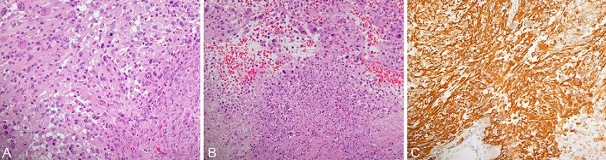
vasogenic edema and causing mass effect in the form of efface- 6 7.2 7.5 cm and extending into the left T2/T3 neural foramina.
ment of the adjacent cortical sulci and approximately 1.6 cm midline Six months later, the patient underwent thoracotomy and total excision
shift to the right side. Magnetic resonance imaging (MRI) showed a of the chest lesion. Histopathology (Fig. 3) revealed features of malig-
left frontotemporal large mass measuring 6 3 6 cm, with inter- nant nerve sheath tumor. These included cellular fascicles of spindle
nal areas of cysts and necrosis. It exhibited heterogeneous signal cell proliferation with thin hyperchromatic or vesicular nuclei. The
intensity with significant thick postcontrast enhancement. Significant nuclear pleomorphism was moderate to focally marked. The mitotic
surrounding edema and contralateral midline shift by 1.6 cm as well count exceeded 5/10 high-power field in areas. Atypical mitotic figures
as uncal herniation were seen (Fig. 1). Spinal MRI showed multiple were noted. The tumor cells were positive for S100, with scattered
neurofibromas scattered all over the spinal axis. axons that exhibited neurofilament immunostaining reactivity. No necro-
The patient underwent an urgent left pterional craniotomy and total sis was identified. The tumor remained stable throughout follow-up.
excision of the lesion. Histopathology (Fig. 2) revealed cystic high- The patient continued regular clinical and radiological follow-up
grade glioma. The tumor was cellular and showed a variety of cell pat- with MRI that showed no signs of recurrence or progression. He
terns. These included anaplastic or giant cells with marked nuclear aty- remained in good health and stable condition until the summer of
pia and pleomorphism and sheets of smaller round to oval cells. The 2015, when he underwent a redo craniotomy and tumor debulking
cytoplasm amount was variable, from scant to abundant, and many for a recurrent tumor. The tumor pathology was glioblastoma multi-
cells were exhibiting a minigemistocyte appearance. Microvascular pro- forme, and the patient received CCRT. A follow-up MRI in the fall of
liferation and areas of necrosis were evident. The Ki-67 proliferative 2016 showed extensive high-grade recurrence in the left cerebral
index was high and estimated to exceed 20% in multiple areas. hemisphere, and the patient received 6 cycles of temozolomide as
Mitoses were easily found, including atypical figures. The result of iso- palliative chemotherapy.
citrate dehydrogenase 1 (IDH-1) immunohistochemistry was negative. In the spring of the following year, the patient presented to the
Cytogenetic analysis showed the presence of 1p/19q codeletion. emergency department with bilateral lower limb weakness, and CT
The patient’s postoperative course was uneventful, and he recei- of his brain showed recurrent tumor. The patient received 3 cycles
ved the standard treatment for glioblastoma (concurrent chemora- of a single-agent palliative chemotherapy (bevacizumab) with stable
diotherapy [CCRT] and adjuvant temozolomide). disease, followed by 4 cycles of a bevacizumab/irinotecan regimen,
Routine postoperative chest radiography showed a large shadow in and then he sought a second opinion elsewhere.
the left upper mediastinum. Chest CT showed a heterogeneously The patient remined in stable status until the fall of 2019, when he
enhancing mass in the apex of the left lung/left paravertebral region showed recurrence in his follow-up MRI with no clinical neurological
extending to the left supraclavicular region, measuring approximately deterioration. The patient received 4 cycles of bevacizumab/irinotecan.
FIG. 2. A: The biopsies showed a high-grade glial tumor that exhibited a variety of cell patterns. These
included anaplastic glial cells with marked nuclear atypia and pleomorphism and cell shapes that varied from
small rounded to giant multinucleated (hematoxylin-eosin [H&E], original magnification 200). B: Multiple
foci of necrosis (lower half of the field) are evident (H&E, original magnification 200). C: Glial fibrillary acidic
protein (GFAP) confirms the glial nature of the tumor (GFAP, original magnification 200).
2 | J Neurosurg Case Lessons | Vol 3 | Issue 4 | January 24, 2022
Unauthenticated | Downloaded 02/10/22 09:22 AM UTCFIG. 3. A: Coronal T2-weighted images showing a large left supraclavicular mass with inhomogeneous
enhancement that has intraforaminal but no intraspinal extension at T2–3 level. B: Sections from the lung
mass show a sarcoma that exhibits high cellularity and marked nuclear pleomorphism. Atypical mitosis is
noticeable in this field (hematoxylin-eosin, original magnification 200). C: The tumor cells exhibit patchy
S100 reactivity, which is compatible with a malignant peripheral nerve sheath tumor (S100, original magnifica-
tion 400).
One year later, the patient showed further progression of his tumor on brain tumors in adults and 50% of gliomas across all age groups.14 The
MRI, with personality changes but no other symptoms, and he refused global incidence of glioblastoma is 10 per 100,000 people.15
further treatment. There are limited data on glioblastoma in patients with NF1. Few
The patient was referred to psychiatry and is following up with cases have been reported in the literature.8,16 In the few reported
our service with no evidence of clinical compromise, is communicat- cases (Table 1),5,7,16–30 it is noted that the mean age of patients
ing, and is able to walk with assistance. with NF1 with glioblastoma at diagnosis is much younger (mean
age 34 years) than that of patients with sporadic glioblastoma
Discussion (mean age 55 years).31 In a systematic review of patients without
Observations NF1 with glioblastoma and >10-year survival, there was an inverse
Our patient is a 27-year-old man with NF1 diagnosed with glioblas- relationship between age at diagnosis and years of survival, where
toma, wild type. He had a prolonged disease course with multiple lines younger age at diagnosis by 4.7 years resulted in 1 year longer
of chemotherapy and radiotherapy and has survived for 10 years with overall survival after 10 years of survival.32
no evidence of clinical compromise and with a good quality of life.
Molecular Pathophysiology
The 2016 WHO classification of tumors in the CNS classifies gli-
NF1-Associated Gliomas
NF1 is an autosomal dominant familial tumor syndrome with omas on the basis of both histological and molecular features,
including 1p/19q codeletion and IDH mutation.33 In sporadic glio-
mutation of the NF1 gene located on chromosome 17q11.2. The
mas, an IDH mutation is detected in most cases, regardless of the
incidence of NF1 is 1 in 2,000 to 5,000 individuals.1 Alongside the
grade.34 Individuals harboring IDH mutation have a better prognosis
characteristic cafe-au-lait spots and cutaneous nodules, low-grade
than those with IDH wild type.35,36
brain tumors such as pilocytic astrocytomas and optic gliomas rep-
Among the few reported glioblastoma cases in NF1, only 7
resent the majority of intracranial neoplasms in NF1.7
reported the molecular features of the tumors, with no IDH mutation
It is thought that the neurofibromin, the protein encoded by the
detected (IDH wild type) except for 2 patients with IDH-1 mutation
NF1 gene, acts to inhibit the Ras signaling pathway. A mutation in (Table 1). This might indicate that the NF1-associated glioblastomas
the NF1 gene leads to uncontrolled cell growth, manifesting as mul- have a different landscape from sporadic glioblastoma.37
tiple peripheral and central nervous system (CNS) tumors.8 Gliomas A comprehensive genomic study included whole-exome sequ-
are seen in approximately 20% of patients with NF1.2 Although encing for tumor and germline cells to further understand the rule of
patients with NF1 are at a higher risk of developing low-grade glio- somatic mutations in tumorigenesis in patients with NF1. The data
mas (World Health Organization [WHO] CNS grade 1 and 2), the showed that there are no somatic mutations that clustered around
prevalence of high-grade gliomas in patients with NF1 is 10–50 the NF1 protein domain and supported the theory of a “second hit”
times higher than in the general population.9 of the heterozygous NF1 allele needed to develop a tumor.37 In our
Optic pathway gliomas are the most common gliomas in patients patient, 1p/19q codeletion was present with no IDH mutation
with NF1. They usually present early in childhood (mean presenting detected (glioblastoma, IDH wild type).
age is 4.5 years). Nonoptic pathway gliomas usually present later in
childhood or in early adulthood (mean presenting age is 7 years).10 Treatment of Glioblastoma in Patients With NF1
Gliomas presenting in adults with NF1 are usually high grade, arising All reported cases of glioblastoma in patients with NF1, including
in cerebral hemispheres.11 A study on 100 NF1 individuals showed our case, received the standard therapy for glioblastoma (gross
that 7% of patients with NF1 were diagnosed with grade IV gliomas.12 total resection followed by fractionated radiotherapy, with concurrent
and adjuvant temozolomide), with second- and third-line administra-
Glioblastoma in Adult Patients With NF1 tion at the time of recurrence as indicated.
In the general population, glioblastoma is the most common and most The NF1 mutation is a marker for treatment-resistant gliomas. It
fatal primary brain tumor in adults.13 It accounts for >60% of primary leads to loss of neurofibromin and subsequent increase in RAS
J Neurosurg Case Lessons | Vol 3 | Issue 4 | January 24, 2022 | 3
Unauthenticated | Downloaded 02/10/22 09:22 AM UTCTABLE 1. Summary of 24 adult patients with NF1 diagnosed with glioblastoma, with treatment, recurrence, and survival
Age (yrs)/ Molecular Survival Functional
Authors & Year Sex Location Features Treatment Recurrence (mos) Status
Miaux et al., 32/F Occipital NA NA NA NA NA
199717
Miyata et al., 30/F Right frontal NA SR 1 RT 1 10 mos 12 mos 1 NA
200518 chemotherapy (PCV)
Mehta et al., 63/M Parietal NA Biopsy NA 2 mos NA
200819
Hakan et al., 28/F Frontal NA SR 1 RT 1 NA 41 mos 1 NA
200820 chemotherapy (TMZ)
Broekman 28/F Right NA SR 1 RT 1 6 mos 12 mos Nystagmus, diplopia,
et al, cerebellar chemotherapy (TMZ) facial numbness, ataxia
200921
Theeler et al., 59/M Right temporal NA SR 1 RT 1 24 mos 104.4 mos 1 Stable
20145 chemotherapy (TMZ)
1 17 cycles
bevacizumab 1
irinotecan
Theeler et al., 25/M Thalamus NA RT 1 chemotherapy 2 mos 13.9 mos Multiple ischemic
20145 (TMZ) strokes, sepsis
1 10 cycles
bevacizumab 1 TMZ
Theeler et al., 32/M Cerebellar IDH wild RT 1 chemotherapy 3 mos 72.6 mos Cardiac thrombosis
20145 hemispheres type (TMZ)
1 adjuvant erlotinib 9
cycles
1 37 cycles
bevacizumab 1
irinotecan
Jeong et al., 32/M Right frontal NA SR 1 RT 1 NA 9 mos 1 NA
20147 chemotherapy (TMZ)
Varghese 60/M Right frontal NA SR 1 RT 1 NA NA Hemiparesis improved
et al., chemotherapy (TMZ)
201522
Ameratunga 24/M Left cerebellar IDH wild Tumor debulking 1 RT 24 mos 24 mos 1 Improved clinically,
et al., type 1 chemotherapy (TMZ) functional
201623 1 SR 1 everolimus1
TMZ
1 MEK inhibitor
(trametinib)
Shibahara 52/M Occipital NA SR 1 RT 1 NA 49 mos NA
et al., chemotherapy
201824
Shibahara 34/M Frontal NA SR 1 RT 1 NA 106 mos 1 NA
et al., chemotherapy
201824
Shibahara 28/M Insula NA SR 1 RT 1 NA 60 mos 1 NA
et al., chemotherapy
201824
Shibahara 53/M Frontal NA SR 1 RT 1 NA 87 mos 1 NA
et al., chemotherapy
201824
CONTINUED ON PAGE 5 »
4 | J Neurosurg Case Lessons | Vol 3 | Issue 4 | January 24, 2022
Unauthenticated | Downloaded 02/10/22 09:22 AM UTC» CONTINUED FROM PAGE 4
TABLE 1. Summary of 24 adult patients with NF1 diagnosed with glioblastoma, with treatment, recurrence, and survival
Age (yrs)/ Molecular Survival Functional
Authors & Year Sex Location Features Treatment Recurrence (mos) Status
Singla et al., 25/M Right frontal NA SR 1 RT 24 mos 36 mos 1 Back to baseline,
201825 functional
Fortunato 23/M Brainstem IDH wild SR 1 RT 1 NA 1 mos Adrenal insufficiency
et al., type chemotherapy (TMZ)
201826
Wong et al., 27/M Multiple IDH1 Subtotal resection 1 RT NA 39 mos NA
201927 mutation 1 chemotherapy (TMZ)
Narasimhaiah 21/F Right NA SR 1 RT 1 NA 32 mos 1 NA
et al., frontoparietal chemotherapy (TMZ)
201916 lobe
Narasimhaiah 26/M Right NA Subtotal resection 120 mos Lost follow up NA
et al., paraventricular 1 total resection 10
201916 years later
Flower & 23/M Cerebellar NA SR 1 RT 1 17 mos 18 mos Acute neurological
Gallo, chemotherapy (TMZ) decline, memory loss,
201928 unsteady gait,
dysarthria, and
dysphasia
Awada et al., 19/M Brainstem IDH 1 RT 1 chemotherapy 6 mos 48 mos 1 Back to baseline,
202029 mutation (TMZ) functional
1 VEGF inhibitor
(axitinib) 1 PCDL-1
inhibitor (avelumab)
1 axitinib 1 lomustine,
2 cycles
1 MEK inhibitor
(trametinib)
Cai et al., 51/F Right temporal IDH wild SR 1 RT 1 NA 13 mos 1 Headache and
202130 type chemotherapy (TMZ) weakness improved,
functional
Present case 27/M Frontoparietal IDH wild SR 1 RT 1 48 mos 121 mos 1 Functional
type chemotherapy (TMZ)
1p/19q 1 bevacizumab
codeletion 1 bevacizumab/
irinotecan
NA = not available; PCDL-1 = programmed cell death 1 ligand; PVC = procarbazine, nimustine hydrochloride, and vincristine sulfate; RT = radiotherapy; SR = gross
total resection; TERT = telomerase reverse transcriptase; TMZ = temozolomide; VEGF = vascular endothelial growth factor.
activity and other RAS effectors. This has led to the development tolerated. He was also diagnosed with a malignant nerve sheath
of combination therapies targeting multiple steps of the RAS sig- tumor shortly after diagnosis; the tumor was managed with surgi-
naling pathway over the past 2 decades. Therapies such as mito- cal resection alone, and follow-up images showed a stable lesion
gen-activated protein kinase kinase (MEK) inhibitors, BRAF throughout the decade.
inhibitors, and checkpoint inhibitors are under trial.38,39 One report
of a 19-year-old with a mesencephalic IDH-1 mutant glioblastoma Prognosis of Glioblastoma in Patients With NF1
did not undergo surgical resection because of the location of the Despite all advances in research technologies, glioblastomas
tumor and received radiotherapy with 3 lines of chemotherapy remain incurable, with a median survival of only 15 months in spo-
with either recurrence or no improvement. The patient then radic cases.15,40 Very few patients have long-term survival beyond
received a MEK inhibitor (trametinib 2 mg once daily) with a sub- 2.5 years.31 Stupp et al.41 reported 2-year survival figures for
sequent BRAF inhibitor (dabrafenib 50 mg twice daily) and was patients who underwent concurrent adjuvant temozolomide and
alive with complete response at 4-year follow-up.29 Our patient radiotherapy. Only 5% of patients diagnosed with glioblastoma sur-
received CCRT twice, with multiple cycles of temozolomide, beva- vive for >5 years, and this measure decreases to 2% among
cizumab, and bevacizumab/irinotecan, all of which were well patients aged 65 years or older.42
J Neurosurg Case Lessons | Vol 3 | Issue 4 | January 24, 2022 | 5
Unauthenticated | Downloaded 02/10/22 09:22 AM UTCHuttner et al.6 reported that pediatric patients with NF1 are at 13. Wirsching HG, Galanis E, Weller M. Glioblastoma. Handb Clin
higher risk of developing glioblastoma and have a better prognosis Neurol. 2016;134:381–397.
than children without NF1, with no reports on adult patients. The 14. Rock K, McArdle O, Forde P, et al. A clinical review of treatment
outcomes in glioblastoma multiforme – the validation in a non-trial
median survival rate of such cases with surgical resection, radio-
population of the results of a randomised phase III clinical trial: has
therapy, and chemotherapy is 36 months, with maximum survival of a more radical approach improved survival? Br J Radiol. 2012;
106 months. 85(1017):e729–e733.
Although it is challenging to conclude the survival rate from the 15. Iacob G, Dinca EB. Current data and strategy in glioblastoma multi-
few reported cases, available data (Table 1) show that the survival forme. J Med Life. 2009;2(4):386–393.
rate in adult patients with NF1 diagnosed with glioblastoma is 16. Narasimhaiah D, Sridutt BS, Thomas B, Vilanilam GC. Glioblastoma
approximately 34 months, with maximal survival of 104.4 months in adults with neurofibromatosis type I: a report of two cases. Neu-
from the time of diagnosis, with conventional treatment for primary ropathology. 2019;39(5):368–373.
17. Miaux Y, Guermazi A, Cornu P, et al. High-intensity lesion on
and recurrent tumor. In our case, our patient was alive at 121- T1-weighted MR images in neurofibromatosis type 1: a case of
month follow-up with radiological evidence of recurrence but overall premalignant lesion. Acta Neurochir (Wien). 1997;139(11):
good functional status and no neurological symptoms. 1085–1087.
18. Miyata S, Sugimoto T, Kodama T, et al. Adenoid glioblastoma aris-
Lessons ing in a patient with neurofibromatosis type-1. Pathol Int. 2005;
This case report describes >10 years of overall survival of glio- 55(6):348–352.
blastoma in a patient with NF1, which, to the best of our knowl- 19. Mehta RS, Abraham M, Plesa C, Ennis P. Glioblastoma multiforme
edge, has not been reported in the literature. Reports cases of in an adult with von Recklinghausen disease. Commun Oncol.
2008;5(10):544–548.
patients with NF1 with longer survival rates may be attributed to the
20. Hakan T, Aker FV. Case report on a patient with neurofibromatosis
young age at diagnosis and relatively better tolerance of therapy. type 1 and a frontal cystic glioblastoma. Neurol Neurochir Pol.
Larger molecular studies on patients with NF1 with glioblastoma are 2008;42(4):362–365.
needed to better understand the molecular differences and how 21. Broekman ML, Risselada R, Engelen-Lee J, Spliet WG, Verweij
they may apply to future treatment plans and prognosis. BH. Glioblastoma multiforme in the posterior cranial fossa in a
patient with neurofibromatosis type I. Case Rep Med. 2009;2009:
References 757898.
1. Rasmussen SA, Friedman JM. NF1 gene and neurofibromatosis 1. 22. Varghese P, Abdul Jalal MJ. A rare case of neurofibromatosis -
Am J Epidemiol. 2000;151(1):33–40. type 1. Asian J Neurosurg. 2015;10(4):344–347.
2. Listernick R, Charrow J, Gutmann DH. Intracranial gliomas in neuro- 23. Ameratunga M, McArthur G, Gan H, Cher L. Prolonged disease
fibromatosis type 1. Am J Med Genet. 1999;89(1):38–44. control with MEK inhibitor in neurofibromatosis type I-associated
3. Rasmussen SA, Yang Q, Friedman JM. Mortality in neurofibromato- glioblastoma. J Clin Pharm Ther. 2016;41(3):357–359.
sis 1: an analysis using U.S. death certificates. Am J Hum Genet. 24. Shibahara I, Sonoda Y, Suzuki H, et al. Glioblastoma in neurofibro-
2001;68(5):1110–1118. matosis 1 patients without IDH1, BRAF V600E, and TERT promoter
4. Guillamo JS, Creange A, Kalifa C, et al. Prognostic factors of CNS mutations. Brain Tumor Pathol. 2018;35(1):10–18.
tumours in neurofibromatosis 1 (NF1): a retrospective study of 104 25. Singla N, Kapoor A, Radotra BD, Chatterjee D. Malignant conver-
patients. Brain. 2003;126(Pt 1):152–160. sion to glioblastoma in neurofibromatosis type I-associated pleomor-
5. Theeler BJ, Ellezam B, Yust-Katz S, Slopis JM, Loghin ME, de phic xanthoastrocytoma: unknown predictors of favorable outcome.
Asian J Neurosurg. 2018;13(3):826–829.
Groot JF. Prolonged survival in adult neurofibromatosis type I
26. Fortunato JT, Reys B, Singh P, Pan E. Brainstem glioblastoma mul-
patients with recurrent high-grade gliomas treated with bevacizu-
tiforme in a patient with NF1. Anticancer Res. 2018;38(8):
mab. J Neurol. 2014;261(8):1559–1564.
4897–4900.
6. Huttner AJ, Kieran MW, Yao X, et al. Clinicopathologic study of glio-
27. Wong WH, Junck L, Druley TE, Gutmann DH. NF1 glioblastoma
blastoma in children with neurofibromatosis type 1. Pediatr Blood clonal profiling reveals KMT2B mutations as potential somatic onco-
Cancer. 2010;54(7):890–896. genic events. Neurology. 2019;93(24):1067–1069.
7. Jeong TS, Yee GT. Glioblastoma in a patient with neurofibromatosis 28. Flower H, Gallo P. Cerebellar glioblastoma in an NF1 patient. Is it
type 1: a case report and review of the literature. Brain Tumor Res surgical debulking really necessary? Br J Neurosurg. 2020;34(6):
Treat. 2014;2(1):36–38. 669–671.
8. Ratner N, Miller SJ. A RASopathy gene commonly mutated in 29. Awada G, Serruys D, Schwarze JK, Van De Voorde L, Duerinck J,
cancer: the neurofibromatosis type 1 tumour suppressor. Nat Rev Neyns B. Durable complete response of a recurrent mesencephalic
Cancer. 2015;15(5):290–301. glioblastoma treated with trametinib and low-dose dabrafenib in a
9. Gutmann DH, Rasmussen SA, Wolkenstein P, et al. Gliomas pre- patient with neurofibromatosis type 1. Case Rep Oncol.
senting after age 10 in individuals with neurofibromatosis type 1 2020;13(2):1031–1036.
(NF1). Neurology. 2002;59(5):759–761. 30. Cai JW, Chen XY, Chen JY, et al. Glioblastoma in a female neurofi-
10. Mahdi J, Shah AC, Sato A, et al. A multi-institutional study of brain- bromatosis 1 patient without IDH1, BRAF V600E, and TERT pro-
stem gliomas in children with neurofibromatosis type 1. Neurology. moter mutations: a case report. Medicine (Baltimore).
2017;88(16):1584–1589. 2021;100(13):e25346.
11. D’Angelo F, Ceccarelli M, Tala, et al. The molecular landscape of 31. Ostrom QT, Gittleman H, Farah P, et al. CBTRUS statistical report:
glioma in patients with neurofibromatosis 1. Nat Med. 2019;25(1): primary brain and central nervous system tumors diagnosed in the
176–187. United States in 2006-2010. Neuro Oncol. 2013;15(2 Suppl):
12. Rodriguez FJ, Perry A, Gutmann DH, et al. Gliomas in neurofibro- ii1–ii56.
matosis type 1: a clinicopathologic study of 100 patients. J Neuro- 32. Tykocki T, Eltayeb M. Ten-year survival in glioblastoma. A system-
pathol Exp Neurol. 2008;67(3):240–249. atic review. J Clin Neurosci. 2018;54:7–13.
6 | J Neurosurg Case Lessons | Vol 3 | Issue 4 | January 24, 2022
Unauthenticated | Downloaded 02/10/22 09:22 AM UTC33. Louis DN, Perry A, Reifenberger G, et al. The 2016 World Health 41. Stupp R, Mason WP, van den Bent MJ, et al. Radiotherapy plus
Organization classification of tumors of the central nervous system: concomitant and adjuvant temozolomide for glioblastoma. N Engl J
a summary. Acta Neuropathol. 2016;131(6):803–820. Med. 2005;352(10):987–996.
34. Yan H, Parsons DW, Jin G, et al. IDH1 and IDH2 mutations in glio- 42. Ostrom QT, Gittleman H, Liao P, et al. CBTRUS statistical report:
mas. N Engl J Med. 2009;360(8):765–773. primary brain and central nervous system tumors diagnosed in the
35. Dubbink HJ, Taal W, van Marion R, et al. IDH1 mutations in low- United States in 2007-2011. Neuro Oncol. 2014;16(4 Suppl):
grade astrocytomas predict survival but not response to temozolo- iv1–iv63.
mide. Neurology. 2009;73(21):1792–1795.
36. Cheng HB, Yue W, Xie C, Zhang RY, Hu SS, Wang Z. IDH1 muta- Disclosures
tion is associated with improved overall survival in patients with The authors report no conflict of interest concerning the materials or
glioblastoma: a meta-analysis. Tumour Biol. 2013;34(6):3555–3559. methods used in this study or the findings specified in this paper.
37. Laycock-van Spyk S, Thomas N, Cooper DN, Upadhyaya M. Neu-
rofibromatosis type 1-associated tumours: their somatic mutational
spectrum and pathogenesis. Hum Genomics. 2011;5(6):623–690.
Author Contributions
38. Gross AM, Wolters P, Baldwin A, et al. SPRINT: Phase II study of Conception and design: all authors. Acquisition of data: all authors.
the MEK 1/2 inhibitor selumetinib (AZD6244, ARRY-142886) in chil- Analysis and interpretation of data: Basindwah, Alkhalidi, Elwatidy.
dren with neurofibromatosis type 1 (NF1) and inoperable plexiform Drafting the article: all authors. Critically revising the article: all authors.
neurofibromas (PN) [abstract]. J Clin Oncol. 2018;36(15 Suppl): Reviewed submitted version of manuscript: Basindwah, Alkhalidi,
10503. Elwatidy. Approved the final version of the manuscript on behalf of all
39. Rodriguez EF, Scheithauer BW, Giannini C, et al. PI3K/AKT path- authors: Basindwah. Statistical analysis: Basindwah, Elwatidy.
way alterations are associated with clinically aggressive and histo- Administrative/technical/material support: Basindwah, Elwatidy. Study
logically anaplastic subsets of pilocytic astrocytoma. Acta supervision: all authors.
Neuropathol. 2011;121(3):407–420.
40. Koshy M, Villano JL, Dolecek TA, et al. Improved survival time Correspondence
trends for glioblastoma using the SEER 17 population-based regis- Sarah Basindwah: College of Medicine, King Saud University, Riyadh,
tries. J Neurooncol. 2012;107(1):207–212. Saudi Arabia. sarah.basindwah@gmail.com.
J Neurosurg Case Lessons | Vol 3 | Issue 4 | January 24, 2022 | 7
Unauthenticated | Downloaded 02/10/22 09:22 AM UTCYou can also read