Early postnatal interactions between beige adipocytes and sympathetic neurites regulate innervation of subcutaneous fat
←
→
Page content transcription
If your browser does not render page correctly, please read the page content below
RESEARCH ARTICLE

                                        Early postnatal interactions between
                                        beige adipocytes and sympathetic
                                        neurites regulate innervation of
                                        subcutaneous fat
                                        Jingyi Chi1, Zeran Lin1†, William Barr1†, Audrey Crane1‡§, Xiphias Ge Zhu2,
                                        Paul Cohen1*
                                        1
                                         Laboratory of Molecular Metabolism, The Rockefeller University, New York, United
                                        States; 2Laboratory of Metabolic Regulation and Genetics, The Rockefeller
                                        University, New York, United States

                                        Abstract While beige adipocytes have been found to associate with dense sympathetic neurites
                                        in mouse inguinal subcutaneous white fat (iWAT), little is known about when and how this
                                        patterning is established. Here, we applied whole-tissue imaging to examine the development of
                                        sympathetic innervation in iWAT. We found that parenchymal neurites actively grow between
                                        postnatal day 6 (P6) and P28, overlapping with early postnatal beige adipogenesis. Constitutive
*For correspondence:
                                        deletion of Prdm16 in adipocytes led to a significant reduction in early postnatal beige adipocytes
pcohen@rockefeller.edu                  and sympathetic density within this window. Using an inducible, adipocyte-specific Prdm16
†
                                        knockout model, we found that Prdm16 is required for guiding sympathetic growth during early
 These authors contributed
                                        development. Deleting Prdm16 in adult animals, however, did not affect sympathetic structure in
equally to this work
                                        iWAT. Together, these findings highlight that beige adipocyte-sympathetic neurite communication
Present address: ‡Tufts                 is crucial to establish sympathetic structure during the early postnatal period but may be
University School of Medicine,          dispensable for its maintenance in mature animals.
Boston, United States; §Maine
Medical Center, Portland, United
States
Competing interests: The                Introduction
authors declare that no
                                        The development of adipose tissue was a critical adaptation for our ancestors. White adipose tissue
competing interests exist.
                                        enables the safe storage and rapid mobilization of energy in response to nutritional needs, while
Funding: See page 19                    brown adipose tissue defends body temperature by dissipating energy as heat (Rosen and Spiegel-
Received: 07 November 2020              man, 2014). In modern times, however, excess high-calorie foods, a sedentary lifestyle, and thermal
Accepted: 15 February 2021              comfort have contributed to an overexpansion of white fat and a relative paucity of brown fat
Published: 16 February 2021             (Heymsfield and Wadden, 2017; van Marken Lichtenbelt et al., 2018). This has resulted in a signif-
                                        icant increase in the prevalence of obesity and associated diseases including type 2 diabetes, hyper-
Reviewing editor: Peter
                                        tension, cardiovascular disease, and many types of cancer (Kopelman, 2000). Obesity now affects
Tontonoz, University of
California, Los Angeles, United
                                        over 40% of adults in the United States and over 600 million adults worldwide (CDC, 2020a;
States                                  The GBD 2015 Obesity Collaborators, 2017). Excess adiposity is at the center of the leading
                                        causes of morbidity and mortality, and obesity-related medical care costs the United States health
    Copyright Chi et al. This article
                                        care system nearly $150 billion each year (CDC, 2020b). Addressing this public health emergency
is distributed under the terms of
                                        will therefore require new approaches based on a deeper understanding of the tissues and pathways
the Creative Commons
Attribution License, which              involved in energy homeostasis.
permits unrestricted use and                The crucial role of adipose tissue in energy balance has driven great interest in investigating this
redistribution provided that the        tissue as a target for the treatment of obesity. While white adipocytes store excess energy, thermo-
original author and source are          genic brown and beige adipocytes convert lipids and glucose into heat, thereby increasing energy
credited.                               expenditure (Rosen and Spiegelman, 2014). Unlike classical brown adipocytes which are

Chi et al. eLife 2021;10:e64693. DOI: https://doi.org/10.7554/eLife.64693                                                                1 of 21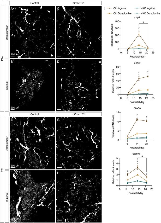
Research article                                                                                Developmental Biology Medicine

                                    eLife digest Mammals have two types of fatty tissue: white fat that mainly stores energy, and
                                    brown and beige fat, also known as thermogenic fat, which burns energy to generate heat. In
                                    humans, brown fat is associated with potent anti-obesity and anti-diabetes effects. A better
                                    understanding of how this type of fat develops and functions could lead to therapeutic strategies to
                                    treat these conditions.
                                       Adult human brown fat is similar to rodent inducible brown fat, also known as beige fat. In adult
                                    mice, beige fat cells need stimulation from the environment to form. Cold can lead to the
                                    generation of beige fat cells by activating a part of the nervous system known as the sympathetic
                                    nervous system. In order for this cold-induced formation of beige fat cells to take place, nerves from
                                    the sympathetic nervous system must first innervate the fatty tissue. Beige fat cells themselves are
                                    important for establishing this innervation, but it was not well understood when and how this occurs.
                                       To study the role of beige fat cells in the establishment of nerve innervation, Chi et al. used
                                    genetically modified mice whose beige fat cells are removed when they are treated with an
                                    antibiotic called doxycycline. If mice that had not been genetically modified were treated with
                                    doxycycline, they developed beige fat cells soon after birth, and these cells shortly became densely
                                    innervated by the sympathetic nervous system. However, if the mutant mice were treated with
                                    doxycycline around birth, these mice could not make beige fat cells during the treatment and failed
                                    to develop dense innervation even when they grew older. These results showed that beige fat cells
                                    that form soon after birth are necessary to establish sympathetic nervous system innervation. But are
                                    beige fat cells required to maintain this innervation as the mice grow older? To test this, Chi et al.
                                    removed them after the innervation was fully established. These mice maintained their innervation,
                                    showing that beige fat cells appear to only be required during the establishment of innervation.
                                       Understanding how the sympathetic nervous system establishes its connection to fat so cold can
                                    stimulate beige fat formation is a first step to finding new treatments for conditions such as diabetes
                                    or obesity. Exploring the timing that underlies the interactions between the sympathetic nervous
                                    system and beige fat cells may provide therapeutic targets in this direction.

                                    thermogenic in basal conditions, murine beige adipocytes, which resemble human brown adipocytes
                                    in their molecular signature (Shinoda et al., 2015), reside in white adipose depots and need to be
                                    activated by external stimuli such as the sympathetic nervous system to drive thermogenesis
                                    (Rosen and Spiegelman, 2014; Wang and Seale, 2016). Recent studies have shown that activation
                                    of thermogenic adipocytes in both rodents and humans is associated with increased whole-body
                                    energy expenditure, improved glucose homeostasis, and enhanced insulin sensitivity (Becher et al.,
                                    2021; Cypess et al., 2009; Lee et al., 2014; Seale et al., 2011; van Marken Lichtenbelt et al.,
                                    2009; Virtanen et al., 2009), suggesting a new approach to defend against obesity.
                                        The sympathetic nervous system plays a key role in enhancing thermogenic function of brown
                                    and beige adipocytes. Although located in distinct fat depots, both brown and beige adipocytes are
                                    surrounded by dense sympathetic neurites, termed parenchymal innervation (Blaszkiewicz et al.,
                                    2019; Chi et al., 2018b; Guilherme et al., 2019; Jiang et al., 2017; Wirsen, 1964). Norepineph-
                                    rine, a neurotransmitter released by these parenchymal neurites, activates b-adrenergic signaling in
                                    thermogenic adipocytes, resulting in enhanced thermogenesis and lipolysis (Cannon and Neder-
                                    gaard, 2004; Hsieh and Carlson, 1957). The important role of sympathetic stimulation in thermo-
                                    genesis has driven great interest in understanding the structural and molecular details of
                                    sympathetic control of thermogenic adipocytes. Adipocyte-derived factors have been shown to act
                                    on the sympathetic nervous system to regulate its structure and activity. Recent studies have identi-
                                    fied S100B and TGFb1 in brown adipocytes as important molecular determinants of sympathetic
                                    innervation in brown fat (Hu et al., 2020; Zeng et al., 2019).
                                        However, it remains largely unclear how beige adipocytes, which are embedded in white fat
                                    depots, modulate their sympathetic innervation. Assisted by a whole-adipose immunolabeling and
                                    clearing method, called Adipo-Clear, we recently found that the density of sympathetic parenchymal
                                    neurites in close apposition to beige adipocytes is dependent on PRDM16 (PR domain containing
                                    16), the transcriptional determinant of beige adipocyte identity and function (Chi et al., 2018b).

Chi et al. eLife 2021;10:e64693. DOI: https://doi.org/10.7554/eLife.64693                                                           2 of 21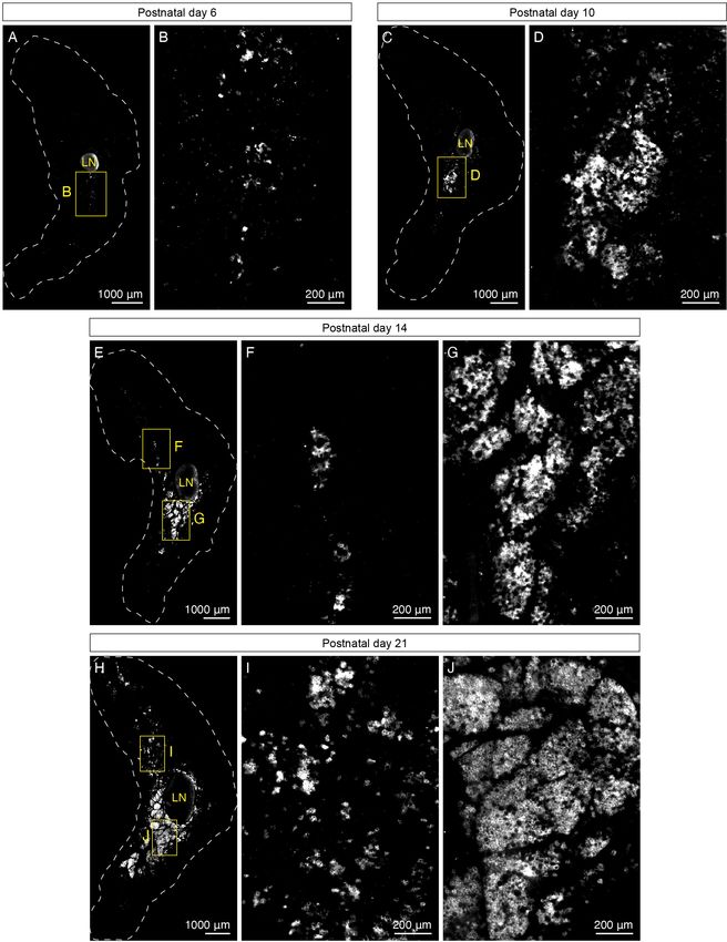
Research article Developmental Biology Medicine
Specifically, deletion of Prdm16 in adipocytes led to ablation of beige adipocyte function and dra-
matically reduced parenchymal innervation density, suggesting that beige adipocyte-associated fac-
tors regulate the structure of sympathetic innervation. As neural projections and circuits can be
regulated during development and by physiological stimuli in adult animals (Glebova and Ginty,
2005; Holtmaat and Svoboda, 2009), it is important to determine when the sympathetic innerva-
tion surrounding beige adipocytes is established.
Using 3D whole-tissue imaging, we have begun to decipher the timing of the interactions
between sympathetic neurites and beige adipocytes in mouse inguinal subcutaneous white fat
(iWAT). We found that sympathetic parenchymal innervation in iWAT actively grows during the early
postnatal period. Interestingly, we observed that the establishment of dense parenchymal innerva-
tion closely follows the development of early postnatal beige adipocytes. To our surprise, using an
inducible, adipocyte-specific Prdm16 knockout mouse model, we found that Prdm16 in beige adipo-
cytes is required for sympathetic axon growth during early development, but not necessary for main-
taining sympathetic structure in adulthood.
Results
Sympathetic innervation of iWAT is established during early postnatal
development
To better understand adipocyte-sympathetic neurite interactions, we investigated whether the asso-
ciation between beige adipocytes and dense sympathetic innervation is developmentally deter-
mined. We first mapped the developmental timing of the sympathetic nervous system in iWAT using
Adipo-Clear coupled with light sheet fluorescent imaging. Given that iWAT undergoes active tissue
morphogenesis during late embryonic and early postnatal stages (Wang et al., 2013), we first per-
formed whole tissue immunostaining and imaging in iWAT isolated from postnatal day (P) two mice
using an antibody targeting tyrosine hydroxylase (TH), a maker for sympathetic fibers, which acts as
the rate-limiting enzyme in the catecholamine biosynthesis pathway. At this stage, adipocytes
appeared fully vascularized and organized into distinct lobular structures, as shown by the endothe-
lial cell marker PECAM (also known as CD31) (Figure 1—figure supplement 1A,C and F), consistent
with previous reports (Hong et al., 2015). While we could detect TH-positive (TH+) signals resem-
bling nerve fascicles as well as fibers wrapping around large blood vessels, dense parenchymal inner-
vation in close apposition to adipocytes, which was reported in adult iWAT (Chi et al., 2018b;
Jiang et al., 2017), was not obvious at this age (Figure 1—figure supplement 1A–F).
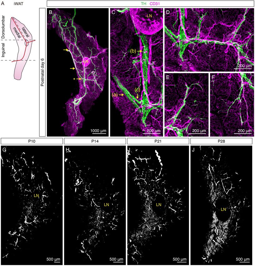
At P6, more distinct features of sympathetic innervation in iWAT were observed: (a) travelling in
parallel within nerve fascicles and (b) wrapping around main blood vessels in a dense mesh-like mor-
phology (Figure 1A–C, Figure 1—figure supplement 2A–D). Upon further analyzing the innervation
pattern across the entire tissue, we observed that these structures were all interconnected to form a
continuous sympathetic network. Specifically, we found several convergence points where TH+
nerve fibers within a nerve fascicle deviated from the bundle and merged with the innervation of the
central blood vessel (Figure 1B–C, Figure 1—figure supplement 2A and C, Figure 1—video 1),
suggesting that sympathetic fibers leave the nerve fascicle and wrap around the main blood vessel
as the first order of innervation. Subsequently, the main blood vessel innervation extended around
branching arterioles and venules as the second order of innervation (Figure 1D, Figure 1—figure
supplement 2E–F, Figure 1—video 1). Lastly, discrete nerve fibers became apparent at the termi-
nals of the second-order innervation to project into tissue parenchyma, where adipocytes are
located. Notably, the majority of these nerve fibers appeared to follow capillaries to arrive in the tis-
sue parenchyma (Figure 1E–F, Figure 1—figure supplement 2G–J). Although nerve endings were
visible in the tissue parenchyma at P6, we did not observe any extensive innervation surrounding adi-
pocytes. In addition, both the dorsolumbar and inguinal regions of iWAT showed similar innervation
patterns at this stage (Figure 1—figure supplement 3A–B, Figure 1—video 2). The results from P2
and P6 samples indicate that the sympathetic axons in iWAT first grow along the vasculature before
reaching the tissue parenchyma, consistent with previous findings showing that developing sympa-
thetic axons follow the vasculature to reach their target organs (Glebova and Ginty, 2005).
Remarkably, adipocyte-innervating neurites became apparent four days later. In the inguinal
region, dense parenchymal neurites surrounding adipocytes were first found at P10, in particular
Chi et al. eLife 2021;10:e64693. DOI: https://doi.org/10.7554/eLife.64693 3 of 21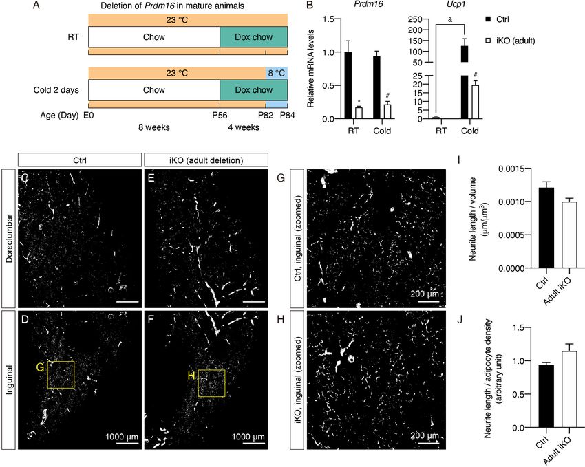
Research article                                                                                            Developmental Biology Medicine

Figure 1. Sympathetic innervation of iWAT is established during early postnatal development. (A) Schematic view of iWAT. Red lines represent main
blood vessels. Using the lymph node and blood vessels as landmarks, iWAT depot is divided into the inguinal and dorsolumbar regions. Dorsolumbar
region is further divided into medial and lateral subregions, hereafter referred to as the dorsomedial and dorsolateral regions. Dotted lines indicate
boundaries of each region. (B–F) Representative images of iWAT from a P6 C57BL6/J mouse immunolabeled with TH (green) and CD31 (magenta). (B)
Maximum intensity projection (MIP) from a 1000 mm z-stack. Arrows indicate convergence points where nerve fibers deviate from nerve bundles to
establish blood vessel innervation. (C) High-magnification image of the indicated (*) convergence point in (B). Arrows indicate distinct features of
sympathetic innervation in iWAT: (a) nerve fascicle, (b) blood vessel innervation, (c) a nerve fiber departing from a nerve fascicle to join blood vessel
innervation. (D) High-magnification image showing sympathetic innervation from main blood vessel extending to arterioles or venules. (E–F) High-
magnification images showing discrete nerve endings project into tissue parenchyma. (G–J) Representative whole-tissue images of iWAT from (G) P10,
Figure 1 continued on next page

Chi et al. eLife 2021;10:e64693. DOI: https://doi.org/10.7554/eLife.64693                                                                          4 of 21
Research article                                                                                      Developmental Biology Medicine

Figure 1 continued
(H) P14, (I) P21, and (J) P28 C57BL6/J mice immunolabeled with TH. MIPs from 50 mm z-stacks are shown. Lymph nodes are indicated as LN. Scale bars
are indicated. All imaging studies were performed in at least three independent animals, and representative images are shown.
The online version of this article includes the following video and figure supplement(s) for figure 1:
Figure supplement 1. Organization of sympathetic nervous system in iWAT at P2.
Figure supplement 2. Organization of sympathetic nervous system in iWAT at P6.
Figure supplement 3. Development of sympathetic parenchymal innervation in iWAT.
Figure 1—video 1. Corresponding to Figure 1B–C.
https://elifesciences.org/articles/64693#fig1video1
Figure 1—video 2. Corresponding to Figure 1—figure supplement 3.
https://elifesciences.org/articles/64693#fig1video2

                                    within lobules at the core of this region (Figure 1G, Figure 1—figure supplement 2K, Figure 1—
                                    figure supplement 3D, Figure 1—video 2). At P14, the number of lobules that contain dense
                                    parenchymal neurites dramatically increased, spreading outwards from the core of the inguinal
                                    region (Figure 1H, Figure 1—figure supplement 3F, Figure 1—video 2). From P21 and onwards,
                                    more inguinal lobules were found to harbor dense parenchymal innervation (Figure 1I–J, Figure 1—
                                    figure supplement 3H & J, Figure 1—video 2), with the pattern comparable to that of adult iWAT
                                    (Chi et al., 2018b). Interestingly, the emergence of dense parenchymal neurites in the dorsomedial
                                    region lagged behind. While parenchymal neurites were detectable in the dorsomedial region at
                                    P14 and P21 (Figure 1H–I, Figure 1—figure supplement 2K–L, Figure 1—figure supplement 3E &
                                    G, Figure 1—video 2), we did not observe densely innervated lobules that resemble the adult inner-
                                    vation pattern in this region until P28 (Figure 1J, Figure 1—figure supplement 3I, Figure 1—video
                                    2). Notably, the dorsolateral region of iWAT remained sparsely innervated relative to the inguinal
                                    region and the dorsomedial region throughout the early postnatal period (Figure 1G–J, Figure 1—
                                    figure supplement 2K–L, Figure 1—figure supplement 3A,C,E,G & I, Figure 1—video 2).

                                    UCP1+ beige adipocytes and dense sympathetic parenchymal
                                    innervation emerge together during early postnatal development
                                    As our previous findings suggest that beige adipocytes interact with sympathetic projections and
                                    modulate the density of sympathetic parenchymal innervation (Chi et al., 2018b), we next investi-
                                    gated whether early postnatal development of sympathetic innervation may also be regulated by
                                    beige adipocytes. We analyzed the localization of beige adipocytes using an antibody against
                                    uncoupling protein 1 (UCP1), a widely accepted marker for thermogenic adipocytes, and compared
                                    their distribution in relation to the sympathetic parenchymal innervation in iWAT using whole-tissue
                                    imaging. As expected, we observed a strong association between beige adipocytes and parenchy-
                                    mal innervation, even during early postnatal development.
                                       Specifically, we found that beige adipocytes first emerge in iWAT of P6 animals that were born
                                    and housed at room temperature, as shown by a few UCP1+ adipocytes sparsely distributed in the
                                    core of the inguinal region, close to the inguinal lymph node (Figure 2A–B, Figure 2—figure sup-
                                    plement 1D). At P10, we detected clusters of UCP1+ adipocytes located in distinct lobules in the
                                    core of the inguinal region (Figure 2C–D, Figure 2—figure supplement 1E). Four days later, at P14,
                                    the lobules containing UCP1+ adipocytes further expanded from the core (Figure 2E & G, Fig-
                                    ure 2—figure supplement 1F). At P21 and P28, extensive UCP1+ lobules occupied a significant
                                    portion of the inguinal region, comparable to the extent of UCP1+ cells only seen in adult animals
                                    after cold exposure (Figure 2H and J, Figure 2—figure supplement 1A,C,G, & H). On the other
                                    hand, the emergence of UCP1+ adipocytes in the dorsolumbar region again lagged behind. UCP1+
                                    adipocytes in the dorsomedial region first emerged in small clusters at P14 and then as distinct
                                    lobules at P21 (Figure 2E–F & and H–I). At P28, the same region contained a large number of
                                    lobules harboring UCP1+ adipocytes (Figure 2—figure supplement 1A–B). Interestingly, the dorso-
                                    lateral region was devoid of UCP1+ adipocytes at all stages analyzed (Figure 2A,C,E and H, Fig-
                                    ure 2—figure supplement 1A & D–H).
                                       To obtain a quantitative measure of these early postnatal beige adipocytes, we also examined
                                    mRNA levels of brown and beige adipocyte-enriched genes in the inguinal and dorsolumbar regions

Chi et al. eLife 2021;10:e64693. DOI: https://doi.org/10.7554/eLife.64693                                                                   5 of 21
Research article Developmental Biology Medicine Figure 2. UCP1+ beige adipocytes emerge during early postnatal development. (A–J) Representative optical sections of iWAT from P6, P10, P14, and P21 C57BL6/J mice immunolabeled with UCP1. (A) Whole-tissue optical section of a P6 iWAT. (B) High-magnification view of the boxed inguinal region in (A). (C) Whole-tissue optical section of a P10 iWAT. (D) High-magnification view of the boxed inguinal region in (C). (E) Whole-tissue optical section of a P14 iWAT. (F) High-magnification view of the boxed dorsolumbar region in (E). (G) High-magnification view of the boxed inguinal region in (E). (H) Figure 2 continued on next page Chi et al. eLife 2021;10:e64693. DOI: https://doi.org/10.7554/eLife.64693 6 of 21
Research article                                                                                        Developmental Biology Medicine

Figure 2 continued
Whole-tissue optical section of a P21 iWAT. (I) High-magnification view of the boxed dorsolumbar region in (H). (J) High-magnification view of the
boxed inguinal region in (H). Lymph nodes are indicated as LN. Dotted lines indicate tissue boundaries based on tissue autofluorescence signals shown
in Figure 2—figure supplement 1D–H. Scale bars are indicated. All imaging studies were performed in at least three independent animals, and
representative images are shown.
The online version of this article includes the following figure supplement(s) for figure 2:
Figure supplement 1. UCP1+ beige adipocytes emerge during early postnatal development.
Figure supplement 2. UCP1+ beige adipocytes emerge during early postnatal development.
Figure supplement 3. UCP1+ beige adipocytes and dense sympathetic parenchymal innervation emerge together during early postnatal development.

                                    of iWAT. In line with the imaging results, Ucp1 mRNA expression showed a gradual increase from
                                    P6 to P14 in the inguinal region (Figure 2—figure supplement 2A). To our surprise, although we
                                    observed more extensive UCP1+ adipocytes in the inguinal region at P21 and P28 by imaging, Ucp1
                                    mRNA levels peaked around P12-P16, with the expression level at P14 being fourfold and eightfold
                                    higher than that of P21 and P28, respectively (Figure 2—figure supplement 2A). Other thermo-
                                    genic genes (Cidea and Cox8b) also gradually increased their mRNA expression from P6 to P14, fol-
                                    lowed by a small downward trend after P21 (Figure 2—figure supplement 2A). These quantitative
                                    transcriptional data suggest that the beige adipocytes arising in the inguinal region of iWAT during
                                    early postnatal development may exhibit peak thermogenic potential around 2–3 weeks of age and
                                    gradually become less active as animals mature. On the other hand, all thermogenic genes showed
                                    significantly lower mRNA expression in the dorsolumbar region compared to the inguinal region at
                                    most time points (Figure 2—figure supplement 2A). Of note, Prdm16, the transcriptional coregula-
                                    tor that determines beige adipocyte phenotype, showed a consistent 1.5- to twofold increase in
                                    mRNA levels in the inguinal relative to the dorsolumbar region across all time points (Figure 2—fig-
                                    ure supplement 2B). Other transcriptional regulators of beige adipocyte development, such as
                                    Cebpb and Ppargc1a, showed similarly consistent regional differences (around 1.5-fold for Cebpb
                                    and twofold for Ppargc1a) during early development (Figure 2—figure supplement 2B). Further-
                                    more, we did not observe any significant regional differences in markers of adipocyte maturation
                                    and function (Fabp4, Pparg, and Adipoq) (Figure 2—figure supplement 2C). Taken together, these
                                    data suggest that adipocytes from the two regions of iWAT are equally differentiated, but the ingui-
                                    nal region may harbor more beige progenitor cells or mature adipocytes with the potential to
                                    emerge as beige adipocytes.
                                       When we overlaid the UCP1 and TH signals, we observed a dramatic overlap between the pres-
                                    ence of dense parenchymal innervation and beige adipocytes, particularly from P10 onwards (Fig-
                                    ure 2—figure supplement 3A–J, Figure 1—video 2), strongly suggesting that early postnatal beige
                                    adipocytes are associated with the signals enabling sympathetic axon growth.
                                       Additionally, as room temperature is considered a mild cold stress to mice, particularly in devel-
                                    oping animals that do not have their adult fur pattern, it is possible that early postnatal beige adipo-
                                    cytes arise as a result of cold-induced sympathetic stimulation. When mice were born and raised at a
                                    warmer temperature (30˚C), at which cold-induced sympathetic firing is minimized, early postnatal
                                    beige adipocytes and sympathetic neurites arise in iWAT with the same patterning as that of room
                                    temperature-housed mice (Figure 2—figure supplement 3K–L). In addition, a recent study using
                                    genetic sympathetic ablation showed that early postnatal beige adipocytes develop normally in the
                                    absence of sympathetic innervation (Wu et al., 2020b). Together, these data suggest that the devel-
                                    opment of beige adipocytes is likely not dependent on sympathetic activation, but rather based on
                                    a developmentally hard-wired program.

                                    Prdm16 regulates the emergence of early postnatal beige adipocytes
                                    and dense sympathetic parenchymal innervation
                                    We have previously shown that dense parenchymal innervation that localizes to the inguinal region
                                    of adult iWAT is significantly reduced by constitutive deletion of Prdm16 in adipocytes (Chi et al.,
                                    2018b). To examine whether early postnatal beige adipocytes and their regulation of dense

Chi et al. eLife 2021;10:e64693. DOI: https://doi.org/10.7554/eLife.64693                                                                     7 of 21
Research article Developmental Biology Medicine
sympathetic innervation are also dependent on Prdm16, we analyzed iWAT of adipocyte-specific
Prdm16 knockout mice (Adipoq-Cre; Prdm16lox/lox; hereafter noted as constitutive Prdm16KO or
cPrdm16KO mice) at postnatal days 6, 14, and 21, key time points in the course of beige adipocyte
and sympathetic innervation development.
At P6, we observed minimal beige adipocytes and scant parenchymal innervation in both control
and cPrdm16KO mice (Figure 3—figure supplement 1A–H), suggesting that the sympathetic ner-
vous system develops similarly in both models prior to the emergence of beige adipocyte clusters.
At P14, the deletion of Prdm16 completely ablated beige adipocytes that normally arise in the ingui-
nal region of control mice, both at the mRNA and protein levels (Figure 3I, Figure 3—figure sup-
plement 1I, Figure 3—figure supplement 2A–D). Correspondingly, the increase seen in
parenchymal innervation density in the inguinal region of control mice was not observed in
cPrdm16KO mice (Figure 3A–D, Figure 3—figure supplement 2A–D). At P21, we observed similar
ablation of beige adipocytes and lack of growth in parenchymal innervation in cPrdm16KO relative to
control samples (Figure 3E–I, Figure 3—figure supplement 1I, Figure 3—figure supplement 2E–
H). These results indicate that early postnatal beige adipocytes indeed depend on PRDM16, the
well-characterized transcriptional determinant of brown and beige adipocytes. Importantly, these
data strongly suggest that sympathetic axon growth during early iWAT morphogenesis may be regu-
lated by PRDM16-dependent signals.
Although PRDM16 is known to be important for beige adipocyte function, it remains possible
that ablation of Prdm16 in all adipocytes by Adipoq-Cre altered white adipocyte function and there-
fore affected sympathetic innervation. To address this, we assessed Prdm16 mRNA and protein lev-
els in the inguinal and dorsolateral regions of iWAT, which are predominantly beige and white
regions, respectively (Figure 2). We performed qPCR on the two regions isolated from control and
cPrdm16KO mice at P14 (Figure 3—figure supplement 2I). The control dorsolateral region showed
significantly lower expression level of Prdm16 mRNA than the control inguinal region. Importantly,
the Prdm16 mRNA level in the control dorsolateral region was indistinguishable from that in
cPrdm16KO dorsolateral or inguinal regions, suggesting that the wild-type dorsolateral region natu-
rally expresses very low levels of Prdm16 mRNA with levels indistinguishable from Prdm16 knockout
samples. We further assessed PRDM16 protein levels across multiple fat depots of young adult mice
(Figure 3—figure supplement 2J). Consistently, the dorsolateral region exhibited a considerably
lower level of PRDM16 compared with the inguinal region in wild-type iWAT, while there were no
detectable levels of PRDM16 in the iWAT of cPrdm16KO mice or wild-type eWAT. Although there
was still a minimal level of PRDM16 protein in the dorsolateral region of iWAT, this may be attrib-
uted to the small number of beige adipocytes in this region. Altogether, Prdm16 appears to be mini-
mally expressed in white adipocytes in iWAT, and thus its deletion in white adipocytes is likely to
contribute minimally to the changes in sympathetic innervation.
Interestingly, although PRDM16 also plays a critical role in brown adipocyte determination and
function, deletion of Prdm16 in interscapular brown fat (iBAT) does not affect its development or
thermogenic function in young adults (Cohen et al., 2014; Harms et al., 2014). Previous studies
have shown that the role of PRDM16 in iBAT formation and function is compensated for by PRDM3,
a transcriptional regulator closely related to PRDM16 (Harms et al., 2014). Consistent with these
findings, we detected similarly extensive sympathetic parenchymal innervation in iBAT of both con-
trol and cPrdm16KO mice (Figure 3—figure supplement 2K–L).
Prdm16 deletion during early development causes decreased
sympathetic parenchymal innervation
To further delineate the critical time window for sympathetic innervation patterning in iWAT, we
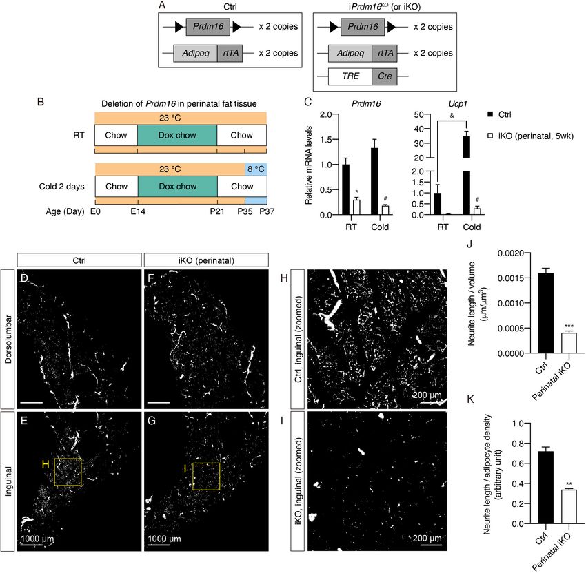
generated an inducible Prdm16 knockout mouse model (AdipoqrtTA; TRE-Cre; Prdm16lox/lox; hereaf-
ter noted as inducible Prdm16KO or iPrdm16KO mice), where Prdm16 can be deleted in adipocytes
in a doxycycline-dependent manner (Figure 4A). To test whether sympathetic parenchymal innerva-
tion may be developmentally determined during a defined time window, doxycycline was delivered
to mice from embryonic day (E) 14 until P21, the period of time when both beige adipocytes and
parenchymal innervation development become clearly apparent. Following doxycycline treatment,
iPrdm16KO and littermate control mice were switched back to chow diet for 2 weeks and subse-
quently exposed to either room temperature (RT) or 8˚C for 2 days (Figure 4B).
Chi et al. eLife 2021;10:e64693. DOI: https://doi.org/10.7554/eLife.64693 8 of 21Research article Developmental Biology Medicine Figure 3. Prdm16 regulates the emergence of early postnatal beige adipocytes and dense sympathetic parenchymal innervation. (A–D) Representative images of iWAT from (A–B) control and (C–D) cPrdm16KO mice at P14, immunolabeled with TH. MIPs from 50 mm z-stacks are shown. (E–H) Representative images of iWAT from (E–F) control and (G–H) cPrdm16KO mice at P21, immunolabeled with TH. MIPs from 50 mm z-stacks are shown. (A, C, E and G) Images of the dorsolumbar region. (B, D, F and H) Images of the inguinal region. Scale bars are indicated. Imaging was performed in at Figure 3 continued on next page Chi et al. eLife 2021;10:e64693. DOI: https://doi.org/10.7554/eLife.64693 9 of 21
Research article                                                                                     Developmental Biology Medicine

Figure 3 continued
least three independent animals per genotype, and representative images are shown. (I) Normalized gene expression of dorsolumbar vs. inguinal
regions in iWAT from control and cPrdm16KO mice at P6, P14, and P21, n = 2–3. Representative genes involved in the thermogenic program are shown.
Data are presented as mean ± SEM and analyzed by two-way ANOVA followed by Bonferroni’s multiple comparisons test. * denotes p
Research article                                                                                             Developmental Biology Medicine

Figure 4. Prdm16 deletion during early development causes decreased sympathetic parenchymal innervation. (A) Schematic representation of the
genetic components of the control and iPrdm16KO mice. iPrdm16KO mice carry floxed Prdm16 alleles (Prdm16lox/lox), two copies of AdipoqrtTA
transgene, and one copy of TRE-Cre transgene. Littermates carrying only Prdm16lox/lox and AdipoqrtTA (i.e. Cre-) were used as the control animals. (B)
Control (Cre-) and iPrdm16KO (Cre+) mice housed at RT (23˚C) were kept on a doxycycline-containing chow diet from E14 until P21 before being
switched to a regular chow diet for another 2 weeks. Control and iPrdm16KO mice were either maintained at RT (23˚C) or exposed to cold (8˚C) for 2
days at P35. (C) Normalized gene expression of inguinal regions from control and iPrdm16KO (perinatal, 5 weeks) mice exposed to RT or cold, n = 3–5.
Data are presented as mean + SEM and analyzed by two-way ANOVA followed by Bonferroni’s multiple comparisons test. * denotes p
Research article                                                                                        Developmental Biology Medicine

Figure 4 continued
sympathetic parenchymal innervation in inguinal regions with total neurite length normalized to (J) regional volume or (K) adipocyte density. N = 3
biological replicates per genotype were analyzed. Average neurite density from five to seven randomly selected tissue volumes (technical replicates)
contributes to neurite density measurement of one biological sample. Data are presented as mean + SEM and analyzed by Student’s t test. ** and ***
denote p
Research article                                                                                          Developmental Biology Medicine

Figure 5. Prdm16 is not required for maintaining sympathetic parenchymal innervation in mature iWAT. (A) Control (Cre-) and iPrdm16KO (Cre+) mice
housed at RT (23˚C) were kept on a regular chow diet until 8 weeks of age before being switched to a doxycycline-containing chow diet for 4 weeks.
Control and iPrdm16KO mice were either maintained at RT (23˚C) or exposed to cold (8˚C) for 2 days at the end of doxycycline treatment. (B)
Normalized gene expression of inguinal regions from control and iPrdm16KO (adult) mice exposed to RT or cold, n = 3–5. Data are normalized to
control RT group, presented as mean + SEM, and analyzed by two-way ANOVA followed by Bonferroni’s multiple comparisons test. * denotes p
Research article                                                                                 Developmental Biology Medicine

                                    Discussion
                                    Sympathetic innervation plays an important role in regulating two key aspects of adipose tissue func-
                                    tion: lipolysis and thermogenesis. Dense sympathetic parenchymal innervation is observed in both
                                    iBAT and the inguinal region of iWAT, where beige adipocytes are primarily located; however,
                                    sparse innervation is found in eWAT and the dorsolateral region of iWAT, an area of the tissue that
                                    is devoid of beige adipocytes even under long term cold stimulation (Chi et al., 2018b;
                                    Dichamp et al., 2019; Huesing et al., 2020; Murano et al., 2009; Zeng et al., 2019). The strong
                                    association between thermogenic adipocytes and dense sympathetic neurites as well as the associa-
                                    tion between white adipocytes and sparse innervation suggests that adipocyte type may determine
                                    the density of sympathetic parenchymal innervation. Indeed, when beige-to-white adipocyte identity
                                    change was achieved by adipocyte-specific deletion of Prdm16, the density of sympathetic paren-
                                    chymal innervation was significantly reduced in the inguinal region of iWAT (Chi et al., 2018b).
                                    Although many previous studies have attempted to define the relationships between sympathetic
                                    neurite density and beige adipocytes, when and how such relationships are established has remained
                                    unclear.
                                        Here, we delineated key stages of sympathetic nervous system development in iWAT using whole
                                    tissue imaging. Specifically, we observed that sympathetic parenchymal innervation in close apposi-
                                    tion to adipocytes is established between P6 and P28. Importantly, the appearance of UCP1+ beige
                                    adipocytes precedes the emergence of dense parenchymal neurites during early postnatal develop-
                                    ment. We further demonstrated that both early postnatal beige adipocytes and dense parenchymal
                                    neurites depend on Prdm16 expression in adipocytes. Using an inducible Prdm16 deletion model,
                                    we identified an early critical period during which beige adipocytes modulate sympathetic axon
                                    growth. However, Prdm16 deletion in adult mice did not alter the sympathetic structure.
                                        Assisted by whole-tissue images, our study carefully examined the growth of sympathetic axons
                                    in iWAT. We observed that sympathetic fibers travel in nerve fascicles to arrive at iWAT, and then
                                    depart from nerve fascicles to reach main blood vessels within iWAT. By P6, the sympathetic innerva-
                                    tion on blood vessels demonstrated a dense mesh-like structure, resembling that of mature iWAT.
                                    However, dense parenchymal innervation surrounding adipocytes, which several studies have charac-
                                    terized in mature iWAT (Chi et al., 2018b; Jiang et al., 2017), is not established at P6. Instead, we
                                    observed sparse discrete sympathetic fibers and small vessels in congruence within the tissue paren-
                                    chyma, a common phenomenon in sympathetic nerve fiber development where vasculature serves as
                                    a guide to direct developing fibers to reach their targets, such as the heart (Nam et al., 2013). Sub-
                                    sequently, from P10 until P28, dense parenchymal innervation becomes obvious where clusters of
                                    UCP1+ beige adipocytes are located.
                                        Although beige adipocytes emerge in white fat depots of adult mice following cold challenge, we
                                    observed strong beige adipogenesis during early postnatal development, similar to the ‘peri-wean-
                                    ing’ beige adipocytes reported in recent studies (Wu et al., 2020a; Wu et al., 2020b). Using high-
                                    resolution whole tissue imaging focusing on the inguinal region of iWAT, we detected scattered
                                    UCP1+ adipocytes at P6, subsequent emergence of small UCP1+ adipocyte clusters at P10, and an
                                    expansion to almost all lobules from P14 to P28. On the other hand, beige adipogenesis is delayed
                                    in the dorsolumbar region, where small clusters of beige adipocytes are not detected until P14. It
                                    will be of interest to investigate the mechanisms driving the preferential localization of early postna-
                                    tal beige adipocytes. Although beige adipocyte recruitment in adult mice requires sympathetic stim-
                                    ulation, early postnatal beige adipocytes develop normally in mice born and raised at
                                    thermoneutrality with minimal sympathetic activity as well as in mice with genetic sympathetic abla-
                                    tion (Wu et al., 2020b). Interestingly, we found several genes involved in the transcriptional control
                                    of beige adipocyte determination and function to have higher expression levels in the inguinal than
                                    the dorsolumbar region throughout early development, suggesting adipocytes or their progenitors
                                    in different regions may exhibit unique properties. Additionally, as early postnatal beige adipocytes
                                    first emerge close to the core of the inguinal region, other tissue structures such as blood or lymph
                                    vessels that are found in the same area (Figure 1B–C and Figure 5—figure supplement 2A–E) may
                                    play a role in promoting early beige adipogenesis.
                                        It is also worth noting that the whole-tissue UCP1+ patterns at P21 and P28 closely resemble that
                                    of adult mice following cold exposure. In addition, the thermogenic gene expression of early postna-
                                    tal beige adipocytes diminishes as mice mature. It is possible that early postnatal beige adipocytes

Chi et al. eLife 2021;10:e64693. DOI: https://doi.org/10.7554/eLife.64693                                                           14 of 21
Research article Developmental Biology Medicine
gradually become inactivated during maturation, and cold challenge re-activates the same cells in
adult mice. Follow-up studies will be needed to examine the fate of early postnatal beige adipo-
cytes. Of note, emerging studies have described additional beige adipocytes that rely on pathways
other than UCP1 to dissipate heat (Chen et al., 2019; Ikeda et al., 2017; Kazak et al., 2015). Thor-
ough characterization of these beige adipocytes using specific markers will be needed to delineate
their development and fate.
Using constitutive and inducible adipocyte-specific Prdm16 deletion models, we ablated early
postnatal beige adipocyte function and found that this dramatically reduced parenchymal sympa-
thetic neurites in iWAT. Our findings suggest that early postnatal beige adipocytes may express
PRDM16-dependent neurotrophic factors that stimulate sympathetic axon growth or downregulate
inhibition cues. Recent studies have unveiled important roles of brown adipocyte-derived factors,
such as S100B and TGFb1, in regulating sympathetic innervation in iBAT (Hu et al., 2020;
Zeng et al., 2019). As beige and brown adipocytes share similarities, it is possible that these factors
may also affect sympathetic innervation in iWAT. Interestingly, S100b, but not Tgfb1, showed a
regional pattern at the mRNA level during early iWAT development, with higher expression in the
inguinal region in a PRDM16-dependent manner (Figure 5—figure supplement 2F). This result sug-
gests that S100B may be one of the potential cues in regulating iWAT sympathetic development. It
is also likely that sympathetic neurites are indirectly regulated by additional beige adipocyte-associ-
ated cell types, such as immune or stromal cells. Follow-up studies will need to evaluate the role of
S100B and other potential factors in modulating sympathetic axon growth during early iWAT
morphogenesis.
Our studies using an inducible adipocyte-specific Prdm16 deletion model indicated a critical
developmental window for the interactions between beige adipocytes and sympathetic nerve termi-
nals. Restricted Prdm16 deletion during early tissue morphogenesis resulted in a lasting reduction in
parenchymal neurite density. However, Prdm16 deletion in fully mature mice failed to alter sympa-
thetic neurite density. These results indicate that sympathetic neurites in iWAT respond to signals
from beige adipocytes or associated cells during early development. However, when the innervation
pattern is fully established, such signals are no longer required for maintaining the innervation level
during adulthood. In line with this, we have previously shown that cold-induced beige adipocytes do
not promote sympathetic neurite outgrowth in adult iWAT when equivalent tissue regions are com-
pared. Taken together, our data suggest that sympathetic neurite density is regulated by local cues
from beige adipocytes during a specific developmental window and exhibits limited plasticity once
the pattern is established.
Of note, a recent study demonstrated leptin-mediated central regulation of sympathetic innerva-
tion in adipose tissue (Wang et al., 2020). Specifically, chronic leptin treatment acting on hypotha-
lamic neurons was found to rescue the defect in sympathetic innervation in iBAT and iWAT of adult
leptin-deficient mice. It is possible that the central and local regulation of sympathetic innervation in
adipose tissue acts with different timing and through distinct mechanisms. Interestingly, the sympa-
thetic axon growth period we observed (P10-P21) largely overlaps with a postnatal leptin surge (P8-
P20) reported previously (Ahima et al., 1998; Wu et al., 2020a). Future studies are needed to
uncouple central (leptin surge) and local (beige adipocyte-associated factors) effects to fully under-
stand how adipose sympathetic innervation is regulated by each mechanism. It is also worth noting
that current studies rely on neurite morphological changes such as length to characterize sympa-
thetic growth or remodeling. As adipocyte size dramatically changes in response to caloric excess or
deprivation, sympathetic neurite density may appear different even without active remodeling. A
better understanding of sympathetic neurite structural change will be assisted by identifying markers
specific to actively remodeling neurites.
Thermogenic adipocytes have been demonstrated to provide metabolic benefits that may com-
bat obesity and associated metabolic diseases. As thermogenic adipocytes are primarily induced by
sympathetic stimulation, many studies have turned to the sympathetic nervous system in search of
novel therapeutic targets for enhancing thermogenic adipocyte function. Our studies here demon-
strated a critical developmental window during which beige adipocytes regulate sympathetic neurite
density, providing fundamental knowledge about the development of the sympathetic nervous sys-
tem in mouse subcutaneous white fat and providing a framework for future attempts to target this
pathway for therapeutic benefit.
Chi et al. eLife 2021;10:e64693. DOI: https://doi.org/10.7554/eLife.64693 15 of 21Research article Developmental Biology Medicine
Materials and methods
Key resources table
Reagent type
(species) or Source or Additional
resource Designation reference Identifiers information
Strain, strain Adipoq-Cre Jackson Laboratory RRID:IMSR_JAX:028020
background
(Mus musculus)
Strain, strain Adipoq-rtTA PMID:22451920 RRID:IMSR_JAX:033448
background
(Mus musculus)
Strain, strain TRE-Cre Jackson Laboratory RRID:IMSR_JAX:006234
background
(Mus musculus)
Strain, strain Prdm16lox/lox PMID:24439384 RRID:IMSR_JAX:024992
background
(Mus musculus)
Antibody Anti-tyrosine hydroxylase (Rabbit polyclonal) Millipore Cat# AB152, IF(1:200)
RRID:AB_390204
Antibody Anti-tyrosine hydroxylase (Sheep polyclonal) Millipore Cat# AB1542, IF(1:200)
RRID:AB_90755
Antibody Anti-CD31/PECAM-1 (Goat polyclonal) R and D Systems Cat# AF3628, IF(1:200)
RRID:AB_2161028
Antibody Anti-UCP1 (Rabbit polyclonal) Abcam Cat# ab10983, IF(1:200)
RRID:AB_2241462
Antibody Anti-LYVE1 (Rabbit polyclonal) Abcam Cat# ab14917, IF(1:200)
RRID:AB_301509
Antibody Anti-PRDM16 (Sheep polyclonal) R and D Systems Cat# AF6295, WB(1:500)
RRID:AB_10717965
Antibody Anti-Lamin A/C (Mouse monoclonal) Santa Cruz Biotechnology Cat# sc-376248, WB(1:2000)
RRID:AB_10991536
Antibody Anti-Rabbit IgG (H+L), Alexa Fluor 568 (Donkey polyclonal) Thermo Fisher Scientific Cat# A10042, IF(1:200)
RRID:AB_2534017
Antibody Anti-Rabbit IgG (H+L), Alexa Fluor 647 (Donkey polyclonal) Thermo Fisher Scientific Cat# A32795, IF(1:200)
RRID:AB_2762835
Antibody Anti-Sheep IgG (H+L), Alexa Fluor 647 (Donkey polyclonal) Thermo Fisher Scientific Cat# A-21448, IF(1:200)
RRID:AB_2535865
Antibody Anti-Goat IgG (H+L), Alexa Fluor 568 (Donkey polyclonal) Thermo Fisher Scientific Cat# A-11057, IF(1:200)
RRID:AB_2534104
Software, algorithm Imaris Bitplane http://www.bitplane.
com/imaris/imaris;
RRID:SCR_007370
Software, algorithm FilamentTracer Bitplane http://www.bitplane.
com/imaris/
filamenttracer;
RRID:SCR_007366
Animals
Young wild-type mice of various ages were generated by crossing male and female mice from the
C57BL/6J background (C57BL/6J, JAX 000664) obtained from the Jackson Laboratories and main-
tained in our facilities. The constitutive Prdm16KO (cPrdm16KO) mice were generated as previously
described (Cohen et al., 2014) by crossing Adipoq-Cre mice (JAX 028020) with Prdm16lox/lox mice.
The inducible Prdm16KO (iPrdm16KO) mice were generated by crossing Adipoq-rtTA (provided by
Dr. Philipp E. Scherer) (Sun et al., 2012), TRE-Cre (B6.Cg-Tg(tetO-cre)1Jaw/J, JAX 006234), and
Prdm16lox/lox mice. All animals in this study were male mice on a pure C57BL/6J background.
All mice were maintained on a 12 hr light/dark cycle with free access to food and water. To gen-
erate mice born and raised at thermoneutrality, pregnant female mice were housed at 30˚C 14 days
Chi et al. eLife 2021;10:e64693. DOI: https://doi.org/10.7554/eLife.64693 16 of 21Research article Developmental Biology Medicine
after vaginal plug formation until the pups reach the indicated ages. All other mice were housed at
23˚C. For perinatal Prdm16 deletion, pregnant female mice were fed with a chow diet containing
600 mg/kg doxycycline (Bio-Serv, S4107) 14 days after vaginal plug formation until the pups reach
P21. For adult Prdm16 deletion, the inducible Prdm16KO (Cre+ and Cre-) mice were placed on a
doxycycline-containing chow diet for the indicated time. All other mice were fed with a standard
rodent chow diet. For cold exposure experiments, mice were placed at 8˚C for 48 hr with two mice
in each cage. Animal care and experimentation were performed according to procedures approved
by the Institutional Animal Care and Use Committee at the Rockefeller University.
iWAT regional dissection
Various regions of iWAT were dissected for qPCR or western blot analyses as illustrated in
Figure 1A and Figure 1—figure supplement 2K. After removal of the lymph node, the region
between the bottom two dotted lines, guided by the entry of the main blood vessel in the inguinal
portion and the upper boundary of the lymph node, was dissected as the inguinal region. The region
from the upper boundary of the lymph node to the back was considered as the dorsolumbar region.
When indicated, the dorsolumbar region was further divided into dorsomedial and dorsolateral
regions by making a cut alongside the blood vessel that travel through the dorsolumbar region.
Gene expression analysis
Total RNA was extracted from tissue using TRIzol (Invitrogen) along with RNeasy kits (QIAGEN). An
RNeasy mini kit was used for adult tissue samples, while an RNeasy micro kit was used for small tis-
sue samples from young mice. For qPCR analysis, RNA was reverse transcribed using the high-capac-
ity cDNA reverse transcription kit (Applied Biosystems). cDNA was used in qPCR reactions
containing SYBR-green fluorescent dye (Applied Biosystems). Relative mRNA expression was deter-
mined by normalization with Tbp (TATA-box binding protein) levels using the DDCt method. The
sequences of primers used in this study are listed in Supplementary file 1.
Nuclear extraction and immunoblotting
Frozen iWAT and iBAT were minced and homogenized in a hypotonic buffer (10 mM HEPES, 10 mM
KCl, 1.5 mM MgCl2, 0.5 mM DTT, and 1x protease inhibitor cocktail (cOmplete Mini, Roche)) by a
dounce homogenizer. Homogenate was incubated on ice for 10 min and then mixed with 1/20 vol of
10% IGEPAL CA-630 (Sigma-Aldrich, I8896). Samples were then filtered through a 100 mm cell
strainer and centrifuged at 1000 x g for 10 min. After centrifugation, lipid and cytoplasmic fractions
were removed and nuclear pellets were resuspended in lysis buffer (20 mM HEPES, 1.5 mM MgCl2,
0.42 M NaCl, 0.2 mM EDTA, 0.5 mM DTT, 1x protease inhibitor cocktail, and 20% Glycerol). Samples
were incubated on ice for 30 min and vortexed for 15 s every 10 min during the incubation. After
lysis, samples were centrifuged at 20,000 x g for 10 min and the supernatant was taken as the
nuclear extract. The following antibodies were used in immunoblotting: anti-PRDM16 (1:500, R and
D systems, AF6295), anti-Lamin A/C (1:2000, Santa Cruz, sc-376248).
Adipo-Clear
Adipo-Clear was performed following a previously published protocol (Chi et al., 2018a; Chi et al.,
2018b). In this study, primary antibodies including anti-UCP1 (1:200, abcam, ab10983), anti-TH
(1:200, Millipore, AB1542 and AB152), anti-CD31 (1:200, R and D systems, AF3628), and anti-LYVE1
(1:200, abcam, ab14917), as well as secondary antibodies conjugated with Alexa-568 and Alexa-647
(1:200, Thermo Fisher Scientific, A10042, A21448, A32795, A11057) were used.
Immunostaining and imaging with iBAT cryo-sections were dissected from mice perfused and
fixed with 1x PBS followed by 4% PFA. Harvested iBAT samples were post-fixed in 4% PFA at 4˚C
overnight and subsequently washed with 1x PBS for 1 hr at RT three times. Samples were then deli-
pidated and permeabilized as described in the Adipo-Clear protocol (Chi et al., 2018a). Fully delipi-
dated samples were incubated in 25% sucrose/PBS solution for 2 hr until sinking, and then frozen in
Tissue-Tek O.C.T Compound (Sakura Finetek USA, 4583). Frozen iBAT samples were sectioned into
40 mm slices using a Leica CM3050 S cryostat. Cryo-sections were blocked with PBS/0.1% Triton
X-100/0.05% Tween 20/2 mg/ml heparin (PtxwH buffer) containing 3% donkey serum for 1 hr at RT,
and then incubated with primary antibodies diluted in PtxwH buffer at RT overnight. Samples were
Chi et al. eLife 2021;10:e64693. DOI: https://doi.org/10.7554/eLife.64693 17 of 21Research article Developmental Biology Medicine
then rinsed in PtxwH buffer for 5 min, 10 min, and 30 min to remove unbound antibodies. Secondary
antibodies diluted in PtxwH buffer were then applied to samples at RT for 4 hr. Samples were next
rinsed with PtxwH buffer for 5 min, 10 min, and 30 min, followed by 1x PBS for 10 min twice. Finally,
samples were immersed in antifade mountant (ProLong Gold, ThermoFisher Scientific, P10144) and
sealed with a coverslip. Anti-TH (1:200, Millipore, AB152) and Alexa-647 conjugated anti-rabbit sec-
ondary (1:200, Invitrogen, A32795) antibodies were used for staining cryo-sections. Fluorescently
labeled samples were imaged on an inverted LSM 880 NLO laser scanning confocal and multiphoton
microscope (Zeiss) with a 20X lens (NA 0.8).
Light sheet microscopy
Whole-tissue iWAT samples were all imaged on a light sheet microscope (Ultramiscroscope II, LaVi-
sion Biotec) equipped with 1.3X and 4X objective lenses and an sCMOs camera (Andor Neo). Images
were acquired with the ImspectorPro software (LaVision BioTec). Samples were positioned in an
imaging chamber filled with benzyl ether and illuminated from one side by the laser light sheet with
488, 561, and 640 nm laser channels. Samples were scanned at a step-size of 4 mm for the 1.3x
objective and 3 mm for the 4x objective.
Image processing
All images and videos were generated using Imaris software (version 9.5.1, Bitplane). 3D tissue
reconstruction was generated using the ‘Volume’ function. Maximum intensity projections and opti-
cal slices were obtained using the ‘Ortho Slicer’ function. All images were captured using the ‘Snap-
shot’ tool, while all videos were made using the ‘Animation’ tool.
Neurite density quantification
Inguinal regions of iWAT from iKO and control iWAT samples were imaged with the 4X objective
lens on the light sheet microscope. Three animals from each group were imaged and analyzed. In
each 3D image, we randomly isolated small cuboidal volumes (4–8 volumes per sample) that were
completely contained within lobules using the ‘Surfaces’ tool followed by the mask channel option
of Imaris. Volumes of the isolated segments were automatically generated by ‘Surfaces’. To sample
parenchymal neurites, we avoided placing volumes in areas that contain nerve bundles or blood ves-
sel innervation. Using the ‘Filament’ tool, we computationally reconstructed parenchymal neurites by
automatically tracing the TH signal and calculated the total neurite length within each volume. We
presented the ratio of total neurite length (mm) by regional volume (mm3) as neurite density within a
volume. To adjust for adipocyte size/number, we manually counted adipocyte number as shown by
the tissue autofluorescence signal from multiple representative slices within each volume. The aver-
age adipocyte number per slice was then multiplied by the height (z depth) of that volume to gener-
ate a factor representing adipocyte density. The ratio of total neurite length (mm) by adipocyte
density (arbitrary unit) is presented (Figure 4—figure supplement 2).
Statistical analysis
All statistical analyses were performed using GraphPad Prism 8 (GraphPad Software, San Diego, CA,
USA). For gene expression analysis, neurite density quantification, and body weight measurement,
we estimated the approximate effect size based on independent preliminary studies. When indi-
cated, an unpaired two-tailed Student’s t test was used to analyze statistical differences. Two-way
ANOVA followed by Bonferroni’s multiple comparisons test was applied to determine the statistical
differences for the rest of the analyses. The statistical details for each experiment can be found in
the figure legends. p Values below 0.05 were considered significant throughout the study.
Acknowledgements
We thank Dr. Philipp Scherer for the Adipoq-rtTA mice line. We thank the Cohen lab for critical com-
ments. We also thank Christina Pyrgaki, Tao Tong, Katarzyna Cialowicz, and Alison North from the
Rockefeller Bioimaging Resource Center for assistance and support. JC is supported by the Center
for Basic and Translational Research on Disorders of the Digestive System through the generosity of
the Leona M and Harry B Helmsley Charitable Trust. This work was supported by the National
Chi et al. eLife 2021;10:e64693. DOI: https://doi.org/10.7554/eLife.64693 18 of 21Research article Developmental Biology Medicine
Institute of Diabetes and Digestive and Kidney Disease grant R01 DK120649 and by the American
Diabetes Association Pathway Program (Grant # 1–17-ACE-17).
Additional information
Funding
Funder Grant reference number Author
Leona M. and Harry B. Helms- Center for Basic and Jingyi Chi
ley Charitable Trust Translational Research on
Disorders of the Digestive
System Pilot Award
National Institute of Diabetes R01 DK120649 Paul Cohen
and Digestive and Kidney Dis-
eases
American Diabetes Association Grant # 1-17-ACE-17 Paul Cohen
The funders had no role in study design, data collection and interpretation, or the
decision to submit the work for publication.
Author contributions
Jingyi Chi, Conceptualization, Data curation, Formal analysis, Funding acquisition, Validation, Investi-
gation, Visualization, Methodology, Writing - original draft, Project administration, Writing - review
and editing; Zeran Lin, William Barr, Formal analysis, Investigation, Writing - review and editing;
Audrey Crane, Investigation, Writing - review and editing; Xiphias Ge Zhu, Investigation, Visualiza-
tion, Writing - review and editing; Paul Cohen, Conceptualization, Supervision, Funding acquisition,
Project administration, Writing - review and editing
Author ORCIDs
Jingyi Chi https://orcid.org/0000-0001-6013-8544
Zeran Lin https://orcid.org/0000-0003-4418-2443
William Barr https://orcid.org/0000-0001-6723-7684
Audrey Crane https://orcid.org/0000-0003-1573-6099
Paul Cohen https://orcid.org/0000-0002-2786-8585
Ethics
Animal experimentation: This study was performed in strict accordance with the recommendations
in the Guide for the Care and Use of Laboratory Animals of the National Institutes of Health. All of
the animals were handled according to approved institutional animal care and use committee
(IACUC) protocols (#18016-H) of The Rockefeller University.
Decision letter and Author response
Decision letter https://doi.org/10.7554/eLife.64693.sa1
Author response https://doi.org/10.7554/eLife.64693.sa2
Additional files
Supplementary files
. Supplementary file 1. qPCR primer sequences used in this study.
. Transparent reporting form
Data availability
All data generated or analyzed during this study are included in the manuscript and supporting files.
Chi et al. eLife 2021;10:e64693. DOI: https://doi.org/10.7554/eLife.64693 19 of 21You can also read


