Adhesive defence mucus secretions in the red triangle slug (Triboniophorus graeffei) can incapacitate adult frogs - bioRxiv
←
→
Page content transcription
If your browser does not render page correctly, please read the page content below
bioRxiv preprint first posted online Feb. 8, 2019; doi: http://dx.doi.org/10.1101/544775. The copyright holder for this preprint (which
       was not peer-reviewed) is the author/funder, who has granted bioRxiv a license to display the preprint in perpetuity.
                                     It is made available under a CC-BY 4.0 International license.

             Adhesive defence mucus secretions in the red triangle slug (Triboniophorus graeffei) can
                                                          incapacitate adult frogs

                                             John Gould1*, Jose W. Valdez2*, Rose Upton1
            1
            School of Environmental and Life Sciences, University of Newcastle, Callaghan, 2308 NSW,
            Australia
            2
             Department of Bioscience - Biodiversity and Conservation, Aarhus University, 8410 Rønde,
            Denmark

            *John Gould and Jose W. Valdez should be considered joint first author.

            Correspondence:

            John Gould, Conservation Biology Research Group, School of Environmental and Life Sciences,
            University of Newcastle, University Drive, Callaghan, NSW 2308, Australia
            email: john.gould@uon.edu.au

            Jose Valdez, Biodiversity and Conservation, Department of Bioscience, Grenåvej 14, Rønde
            8410, Denmark
            email: jose.valdez@bios.au.dk

            Abstract

                     Gastropods are known to secrete mucus for a variety of purposes, including locomotion,
            reproduction, adhesion to surfaces, and lubrication. A less commonly known function of mucus
            secretion in this group involves its use as a defence against predation. Among the terrestrial slugs,
            mucus that serves this particular purpose has been studied for only a handful of species under
            laboratory conditions, where it is thought to be produced for self-fouling or to make individuals
            difficult to consume. However, the mechanisms of how these defensive secretions operate and
            their effectiveness in deterring predation in the natural world have not be described in much
            detail. In this study, we provide evidence of adhesive mucus secretions in the red triangle slug
            (Triboniophorus graeffei) as an adaptation against predation. Field observations of a large red-
            eyed green tree frog (Litoria chloris) trapped in the mucus secretions of a nearby T. graeffei
            revealed that this mucus serves to incapacitate predators rather than just simply as an overall
            deterrence. Mechanical stimulation of T. graeffei under laboratory conditions revealed that
            adhesive secretions were produced from discrete sections of the dorsal surface when disturbed,
            leading to the production of a highly sticky and elastic mucus that was unlike the thin and
            slippery mucus used during locomotion. The adhesiveness of the defensive secretions was
            strengthened and reactivated when in contact with water. This appears to not only be the first
            description of defensive mucus production in this slug species but one of the first natural
            observations of the use of adhesive defence secretions to incapacitate a predator. The
            biomechanical properties of this mucus and its ability to maintain and strengthen its hold under
            wet conditions make it potentially useful in the development of new adhesive materials.

            Keywords: antipredator, adhesive gel, bioadhesive, Litoria chloris, mollusc, predator-prey
            interactions
bioRxiv preprint first posted online Feb. 8, 2019; doi: http://dx.doi.org/10.1101/544775. The copyright holder for this preprint (which
       was not peer-reviewed) is the author/funder, who has granted bioRxiv a license to display the preprint in perpetuity.
                                     It is made available under a CC-BY 4.0 International license.

            Introduction

                      Animals have evolved a diverse array of anti-predator traits, including those that are
            physical (camouflage, mimicry, and weaponry), behavioural (defensive displays, colouration),
            and chemical (venom, noxious chemicals). The production of mucus is a prime example of a
            chemical response to predation risk which has been recorded amongst velvet worms (Baer &
            Mayer, 2012), echinoderms (Flammang, Demeuldre, Hennebert, & Santos, 2016), fish (Schubert,
            Munday, Caley, Jones, & Llewellyn, 2003; Shephard, 1994), arthropods (Betz & Kölsch, 2004),
            lizards (Brau, Lanterbecq, Zghikh, Bels, & Damman, 2016), aquatic gastropods (Rice, 1985),
            terrestrial slugs (Barber et al., 2015; Deyrup-Olsen, Luchtel, & Martin, 1983), and amphibians
            (Arnold, 1982; Evans & Brodie, 1994; Graham, Glattauer, Li, Tyler, & Ramshaw, 2013). Such
            bioadhesives are typically secreted quickly and exhibit a rapid curing process, with some able to
            be exposed for weeks without losing their bonding capability (von Byern et al., 2017). These
            secretions are produced at the onset of an attack, most noticeably mechanical stimulation, acting
            as a protective barrier or overall deterrent that reduces the chances of the prey species from being
            consumed.

                       Gastropods are the archetypal animals known for their viscoelastic mucous secretions
            which aid in locomotion, reproduction, and adhesion to surface substrates while foraging (Smith,
            2010). Many gastropod species which have transitioned to a life on land also secrete a thin
            coating of mucosa in order to remain well lubricated, as their soft bodies and permeable
            epidermal linings mean they are particularly vulnerable to mechanical damage and desiccation
            (South, 2012; Verdugo, 1991). In addition to these well-known functions of mucus secretions
            amongst the gastropods, some species have also evolved supplementary secretions that serve to
            reduce the threat of predation (Rollo & Wellington, 1979; Triebskorn & Ebert, 1989). This has
            been recorded among the terrestrial slugs, which are very susceptible to predation due to their
            lack of a protective shell, soft bodies and slow pace. This defence-specific mucus is often distinct
            from the usually thin and slippery mucus produced for the purposes of lubrication and
            locomotion, acting as a protective barrier that prevents contact between the potential threat and
            the slug’s body (Deyrup-Olsen et al., 1983; Smith, 2006).

                      Although defensive mucus secretions in terrestrial slugs are thought to serve as methods
            of self-fouling that deters predation, recent findings have found that some species secrete mucus
            with adhesive properties as part of their anti-predation repertoire (Foltan, 2004; Landauer &
            Chapnick, 1981; Rice, 1985; Smith, 2010). For example, Arion subfuscus and Ariolimax
            columbianus have been shown to possess dorsal epithelial that secretes an adhesive defensive
bioRxiv preprint first posted online Feb. 8, 2019; doi: http://dx.doi.org/10.1101/544775. The copyright holder for this preprint (which
       was not peer-reviewed) is the author/funder, who has granted bioRxiv a license to display the preprint in perpetuity.
                                     It is made available under a CC-BY 4.0 International license.

            mucus when disturbed (Deyrup-Olsen et al., 1983; Mair & Port, 2002; Martin & Deyrup-Olsen,
            1986). In these species, the defensive mucus starts out as a viscous slime, which sets into a highly
            sticky and elastic mass which, although composed of 95% water, can sustain stressor over 100
            kPa due to gel-stiffening proteins which bind with metals (Smith, 2010; Wilks, Rabice, Garbacz,
            Harro, & Smith, 2015). Likewise, slugs in the genera Veronicella also produce their own form of
            sticky mucus in response to irritation (Cook, 1987). The mechanical properties of these adhesive
            secretions are so remarkable they have inspired the development of new surgical glues (Li et al.,
            2017), and are thought to confer an evolutionary advantage for terrestrial slugs by making them
            unpalatable or difficult to consume.

                      The use of adhesive mucus across multiple genera suggests that it may be a common
            defence mechanism among gastropods, particularly terrestrial slugs which lack a protective
            structure. However, studies on defensive secretions in gastropods have been conducted nearly
            exclusively on the terrestrial slugs A. subfuscus and A. columbianus (Smith & Callow, 2006).
            This presents an issue since there are likely to be many more species with defensive mucus
            secretions that have unique adhesive properties due to differences in the composition of their
            mucus secretions (Foltan, 2004). Moreover, many of these studies test the properties of adhesive
            secretions in a laboratory setting where their use as a form of antipredator defence is assumed. As
            such, the mechanism of how adhesive defence mucus operates in the natural world and their
            effectiveness in deterring predation has not been described in much detail in the literature to date.
            In this study, we show the first evidence of adhesive mucus secretions in the red triangle slug
            (Triboniophorus graeffei) as an adaptive response to predation threat. We also show that these
            secretions can act to incapacitate predators rather than simply methods of self-fouling that make
            individuals unpalatable or noxious to predators.

            Materials and Methods

                      Field observations occurred within the Watagans Mountain Range, New South Wales,
            Australia on October 27, 2017. Nightly fieldwork resulted in the chance discovery of an adult
            male red-eyed green tree frog (Litoria chloris) that appeared to be stuck to a fallen eucalyptus
            branch during a period of heavy rainfall. Since the frog was found in close proximity to a large T.
            graeffei individual, it was hypothesised that it had become stuck due to the secretions of the slug,
            either by misfortune or after a predation attempt. Observations were made in the field for a period
            of ten minutes without interference to examine any change in the situation and whether L. chloris
            would free itself. No changes occurred so the frog and the nearby slug were then taken back to
            the Conservation Biology laboratory at the University of Newcastle for further investigation.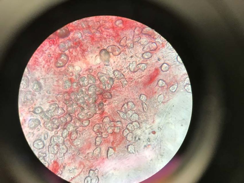
bioRxiv preprint first posted online Feb. 8, 2019; doi: http://dx.doi.org/10.1101/544775. The copyright holder for this preprint (which
       was not peer-reviewed) is the author/funder, who has granted bioRxiv a license to display the preprint in perpetuity.
                                     It is made available under a CC-BY 4.0 International license.

                      The adult frog, while still attached to the branch, was transferred to a 27 x 17 x 15 cm
            container will with 2-3 cm of aged tap water and regularly checked for recovery or signs of stress
            until it was no longer adhered to the branch. The T. graeffei individual was placed onto a petri
            dish, where the typical mucus properties exhibited during locomotion were examined. To
            determine whether it produced adhesive secretions that could explain the incapacitated state of
            the adult frog, mucus production was encouraged by disturbance via mechanical stimulation for a
            period of 60 seconds using a gloved finger. On November 24, 2018, an additional three T.
            graeffei individuals were collected from the Watagans Mountain Range to gain additional
            information regarding the potential adhesive properties of this defensive mucus. Specimens were
            housed together in a 27 x 17 x 15 cm container with leaf litter for seven days. Over this period,
            we evaluated the quantity, thickness, and adhesive quality of the mucus left behind during
            locomotion, as well as the mucus of the dorsal surface before and after 60 seconds of tactile
            stimulation.

            Results

                      Observations made in the field indicate that the ventral skin area of L. chloris was
            strongly adhered to the surface of the branch, including the lower throat, abdomen, and inner
            thighs of the hind legs (Fig. 1). Particularly noteworthy was the unusual positioning of the frog,
            with the body very close to the surface of the branch and the legs splayed out (Fig. 2).
            Additionally, the toe pads and webbing of the front legs were bonded to each other and partially
            to the branch, while the toe pads of the back legs were also stuck to the branch (Fig. 2). On
            multiple occasions during field observations, the individual attempted escape but was unable to
            remove itself from its unorthodox position. Attempts to physically remove the frog also failed,
            with the individual producing a distress call each time. Closer observations of the branch and
            surrounding leaf litter showed no signs that the frog was covered in fallen sap from nearby trees.
            Throughout this period of field observations, the T. graeffei individual did not move, remaining
            less than 1 cm away from the mouth of the frog (Fig. 1, Fig. 2).

                      Following its collection, the frog remained attached to the branch, with the skin of the
            abdomen, legs, and toes remaining covered in the sticky residue. Additional residue was also
            present on the section of the branch that was collected which, along with affected areas of skin,
            appeared to be mostly translucent with sections that were slightly red in colouration (Fig. 3).
            After more than 24 hours it was still unable to remove itself, so to prevent any further distress we
            assisted in its removal by carefully peeling away sections of skin that were adhered to the branch.
            Even after the frog was no longer stuck to the branch, its skin remained covered in mucus that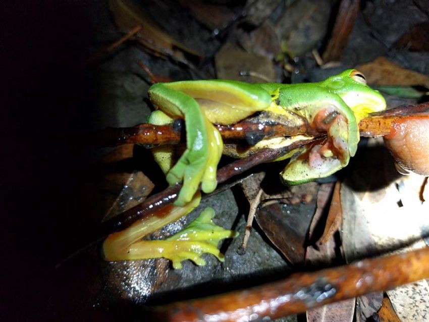
bioRxiv preprint first posted online Feb. 8, 2019; doi: http://dx.doi.org/10.1101/544775. The copyright holder for this preprint (which
was not peer-reviewed) is the author/funder, who has granted bioRxiv a license to display the preprint in perpetuity.
It is made available under a CC-BY 4.0 International license.
would often cause it to become adhered to the bottom of the container while it was immersed in
water.
When examining the T. graeffei individual, the mucus layer left behind during periods of
locomotion was found to be quite thin and lacking in adhesiveness (Fig. 4). Following the
examination of the additional specimens that were collected, it was discovered that the dorsal
surface was typically dry to the touch prior to disturbance but became wet with copious amounts
of extremely adhesive mucus during periods of mechanical stimulation. Only those portions of
dorsum that were stimulated showed an increased expression of mucus while surrounding regions
remained relatively dry. Often a single touch of the dorsum resulted in contractions in the area
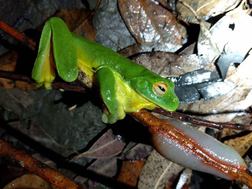
and the immediate secretion of mucus (Fig. 5), which was expelled onto the surface in the form of
tiny droplets that quickly spread over the surrounding surface. This mucus became adhesive
within a matter of seconds and often resulted in the fingers of gloved hands becoming stuck
together or to surrounding paper and plastic when handled. However, the mucus gradually lost its
adhesive quality over a few minutes, depending on quantity, as it began to desiccate, which was
only gained upon rehydration. Once expressed, this mucus became increasingly thick, sticky and
opaque with repeated stimulation. On some occasions, disturbance also resulted in the production
of red coloured mucus which became dispersed throughout the mostly clear mucus. This
particular mucus was only expressed during periods when mechanical stimulation was applied on
the red tissue located on the perimeter around the foot of the slug (Fig. 6) and was similar to the
residue present on the frog’s skin (Fig. 3).
Discussion
Our study indicates that T. graeffei secretes a highly adhesive mucus as an anti-predatory
response that is distinct from the mucus produced for lubrication and locomotion. This defensive
mucus has the ability to incapacitate comparatively large predators, such as frogs, for an extended
period of time, which may facilitate escape or reduce the chance of a predator mounting further
attacks while an escape is being made. The covering of L. chloris ventral surface in a sticky
residue and its proximity to the T. graeffei specimen suggests that the frog activated an anti-
predator response in the slug, resulting in it becoming covered in the adhesive mucus. This is also
supported by the similarity in colouration of the mucus present on the frog and the red mucus
often produced by T. graeffei when disturbed. The reddish colouration and the large quantity of
mucus present on the frog’s skin, as well as the underlying branch, are in contrast to the thin and
slippery locomotive mucus produced by the sole of the slug during locomotion, suggesting that
the frog wasn’t simply trapped behind the slug by chance. Instead, it is more likely that the frog
bioRxiv preprint first posted online Feb. 8, 2019; doi: http://dx.doi.org/10.1101/544775. The copyright holder for this preprint (which
       was not peer-reviewed) is the author/funder, who has granted bioRxiv a license to display the preprint in perpetuity.
                                     It is made available under a CC-BY 4.0 International license.

            mounted a predatory attack on the slug, leading to the secretion of a large quantity of defensive
            mucus that resulted in it becoming stuck to the branch shortly after.

                      The slug found in proximity to the frog was able to produce sufficient mucus to
            incapacitate its frog predator in a state that would have likely persisted for at least two days,
            making the frog vulnerable to desiccation and its own predation. In order to incapacitate such a
            large predator, T. graeffei would have had to produce a large supply of mucus that could become
            adhesive in a relatively short period of time. The presence of mucus across nearly the entire
            ventral surface of the L. chloris adult and its close proximity to the slug are highly suggestive of
            these properties and further supported by analysis of the mucus under laboratory conditions.
            These qualities correspond with the defensive secretions produced by other slugs, such as
            Ariolimax columbianus and Ario subfuscus, which can produce copious amounts of mucus (5.5%
            of total body weight) that becomes adhesive within a matter of seconds (Deyrup-Olsen et al.,
            1983; Mair & Port, 2002; Martin & Deyrup-Olsen, 1986). The observations made in this study
            also demonstrate that the adhesive nature of the T. graeffei defence mucus would be reactivated
            repeatedly upon rehydration. Since amphibians have mucus glands to keep their skin moist, is
            likely the frog’s own secretions facilitated the sustained adhesiveness of the mucus, as well as its
            transference over a large surface area of its skin, especially if it began to struggle or attempted to
            remove the secretions using its foot pads. This may also have been further exacerbated by rainfall
            during this period, which may have sustained the adhesive quality of the mucus, while also
            allowing it to become more easily spread across the surface. Another possibility is that L. chloris
            could have produced its own adhesive secretions during its distress, which may have increased
            the adhesive properties of the slug mucus even further. Amphibians are known to release
            antipredator skin secretions which can be toxic or noxious, but many species also secrete
            adhesive substances which can be five times stronger than rubber cement (Evans & Brodie,
            1994), and one experimental study on salamanders found that its adhesive mucus could
            incapacitate a snake predator for up to 48 hours (Arnold, 1982). Whether or not this was the case,
            other species also possess adhesive secretions for predation and the synergistic effect of such
            predator-prey adhesive secretions is not fully known and warrants further investigation.

                      Based on the experiments conducted in this study, it can be deduced that the cells
            responsible for secreting adhesive mucus in T. graeffei are located across the dorsal surface,
            which appear to be selectively activated in each discrete section of dorsum that becomes
            disturbed. Although many other regions have the capacity to produce mucus in this species,
            including the head, pneumostome, and sole, these do not seem to be involved in the production of
            defensive mucus. The exact mechanisms of mucus production in this species are yet to be
bioRxiv preprint first posted online Feb. 8, 2019; doi: http://dx.doi.org/10.1101/544775. The copyright holder for this preprint (which
was not peer-reviewed) is the author/funder, who has granted bioRxiv a license to display the preprint in perpetuity.
It is made available under a CC-BY 4.0 International license.
determined, but it seems to be similar to production in A. columbianus, which also secrete
adhesive mucus from glands located across the dorsal surface that is in contrast to the thin,
slippery mucus secreted by the pedal foot (Luchtel, Deyrup-Olsen, & Martin, 1991; Martin &
Deyrup-Olsen, 1986). In these species, defence adhesives typically gain their mechanical strength
from a network of proteins and polysaccharides that stiffen in the presence of substantial metal-
binding proteins by forming a network of cross-linked proteins (Braun, Menges, Opoku, &
Smith, 2013; Pawlicki et al., 2004; Smith, 2002; Smith, 2006; Werneke, Swann, Farquharson,
Hamilton, & Smith, 2007). These products are released from mucus glands in the dorsum as
microscopic packets that rupture by ATP or shear stress to form a uniform viscoelastic secretion
(Luchtel et al., 1991; Smith, 2010; Werneke et al., 2007). Without sheer, the mucus would not
possess adhesive properties and it would just flow off the slug (Deyrup-Olsen et al., 1983).
The ability of mucus packets to rupture in the presence of stress suggests that rubbing of
the dorsal surface may trigger the formation of mucus, which would account for the initiation of
secretion in T. graeffei upon tactile stimulation, as well as the specificity of mucus secretion to
the specific dorsal regions disturbed. It is also possible that stimulation of the dorsum leads to
contractions within the immediate area, which may be the primary mechanism used for the
expression of the mucus to the surface, though this process requires further investigation. Another
observation is that the red mucus expressed during periods of stimulation appears to be derived
from the red epidermis that skirts the perimeter of the slug’s foot, with cells from this section
possibly becoming dislodged into the translucent mucus when the epidermis in this region is
damaged. Although this particular mucus didn’t seem to have adhesive properties of its own,
further research may determine if it has other important properties such as making the slug
unpalatable or reacting with the adhesive secretions to increase its adhesiveness and
effectiveness. Nevertheless, this defence mechanism in T. graeffei is likely to differ from
previously studied species. As such, there is potential for comparative studies to be conducted,
since the biochemical makeup and secretory structures vary amongst gastropod species (Foltan,
2004; Smith, 2010).
A strong bioadhesive is a valuable defensive tool for a terrestrial slug that is slow and
often chooses to move when conditions are moist. However, this strategy is likely to come at a
considerable cost to the slug in terms of future survival, as mucus production requires an
investment of water that usually derives from the animal itself. Like many other biological
adhesives, the mucus of T. graeffei is able to adhere strongly and non-specifically which, along
with its ability to maintain or even strengthen its hold under wet conditions, makes it potentially
useful in the development of new generations of glues (Callow & Callow, 2006), such as thebioRxiv preprint first posted online Feb. 8, 2019; doi: http://dx.doi.org/10.1101/544775. The copyright holder for this preprint (which
was not peer-reviewed) is the author/funder, who has granted bioRxiv a license to display the preprint in perpetuity.
It is made available under a CC-BY 4.0 International license.
recent medical adhesives developed based on studies of A. subfuscus secretions. Furthermore,
investigating the mucus packet systems in this and other species may also help in understanding
their ability to quickly harden the mucus secretions, which could provide guidance for future
designs in fast-reacting artificial systems.
Adaptations that allow species to defend themselves from predatory attack, particularly
those species lacking in speed or camouflage, are evident throughout the animal kingdom.
Although not common, the use of adhesives to avoid predation has been found in many types of
animals, and typically assumed to make the prey unpalatable or difficult to consume. The ability
to use adhesives to immobilize predators has only been examined in the laboratory on amphibians
(Arnold, 1982; Evans & Brodie, 1994) and arthropods (Betz & Kölsch, 2004), and observations
made on hagfishes which use adhesive mucus to suffocate predators by clogging their gills
(Zintzen et al., 2011). However, this appears to not only be the first description of defensive
mucus production in this slug species but one of the first natural observations of the use of
adhesive defence secretions incapacitating a predator. As such, this finding is important from a
natural history perspective, especially since the evolution of defensive mucus secretions in most
terrestrial slugs remains unknown. Detailed research on the biochemical structure of such
bioadhesives may lead to significant advancements in the development of new materials that are
able to exploit their unique mechanical properties.
Acknowledgements
The authors thank William Legge for providing technical support during fieldwork. This work
was conducted under ethics number A-2011-138 approved by the University of Newcastle. All
experimental procedures were performed in accordance with the Australian code for the care and
use of animals for scientific purposes.
References
Arnold, S. J. (1982). A Quantitative Approach to Antipredator Performance: Salamander Defense
against Snake Attack. Copeia, 1982(2), 247-253. doi:10.2307/1444602
Baer, A., & Mayer, G. (2012). Comparative anatomy of slime glands in Onychophora (velvet worms).
Journal of morphology, 273(10), 1079-1088. doi:10.1002/jmor.20044
Barber, J. R., Leavell, B. C., Keener, A. L., Breinholt, J. W., Chadwell, B. A., McClure, C. J., . . .
Kawahara, A. Y. (2015). Moth tails divert bat attack: evolution of acoustic deflection.bioRxiv preprint first posted online Feb. 8, 2019; doi: http://dx.doi.org/10.1101/544775. The copyright holder for this preprint (which
was not peer-reviewed) is the author/funder, who has granted bioRxiv a license to display the preprint in perpetuity.
It is made available under a CC-BY 4.0 International license.
Proceedings of the National Academy of Sciences, 112(9), 2812-2816.
doi:10.1073/pnas.1421926112
Betz, O., & Kölsch, G. (2004). The role of adhesion in prey capture and predator defence in
arthropods. Arthropod Structure & Development, 33(1), 3-30.
doi:https://doi.org/10.1016/j.asd.2003.10.002
Brau, F., Lanterbecq, D., Zghikh, L.-N., Bels, V., & Damman, P. (2016). Dynamics of prey
prehension by chameleons through viscous adhesion. Nature Physics, 12, 931.
doi:10.1038/nphys3795
Braun, M., Menges, M., Opoku, F., & Smith, A. M. (2013). The relative contribution of calcium, zinc
and oxidation-based cross-links to the stiffness of Arion subfuscus glue. Journal of
experimental biology, 216(8), 1475-1483.
Callow, J. A., & Callow, M. E. (2006). The Ulva spore adhesive system. In Biological adhesives (pp.
63-78): Springer.
Cook, A. (1987). Functional aspects of the mucusproducing glands of the Systellommatophoran
slug, Veronicella floridana. Journal of Zoology, 211(2), 291-305.
Deyrup-Olsen, I., Luchtel, D., & Martin, A. (1983). Components of mucus of terrestrial slugs
(Gastropoda). American Journal of Physiology-Regulatory, Integrative and Comparative
Physiology, 245(3), R448-R452.
Evans, C. M., & Brodie, E. D. (1994). Adhesive Strength of Amphibian Skin Secretions. Journal of
Herpetology, 28(4), 499-502. doi:10.2307/1564965
Flammang, P., Demeuldre, M., Hennebert, E., & Santos, R. (2016). Adhesive secretions in
echinoderms: a review. In A. M. Smith (Ed.), Biological Adhesives (2nd Edition ed., pp. 193-
222). Switzerland: Springer.
Foltan, P. (2004). Influence of slug defence mechanisms on the prey preferences of the carabid
predator Pterostichus melanarius (Coleoptera: Carabidae). European Journal of Entomology,
101(3), 359-364.
Graham, L. D., Glattauer, V., Li, D., Tyler, M. J., & Ramshaw, J. A. (2013). The adhesive skin
exudate of Notaden bennetti frogs (Anura: Limnodynastidae) has similarities to the prey
capture glue of Euperipatoides sp. velvet worms (Onychophora: Peripatopsidae).bioRxiv preprint first posted online Feb. 8, 2019; doi: http://dx.doi.org/10.1101/544775. The copyright holder for this preprint (which
was not peer-reviewed) is the author/funder, who has granted bioRxiv a license to display the preprint in perpetuity.
It is made available under a CC-BY 4.0 International license.
Comparative Biochemistry and Physiology Part B: Biochemistry and Molecular Biology,
165(4), 250-259.
Landauer, M. R., & Chapnick, S. D. (1981). Responses of Terrestrial Slugs to Secretions of Stressed
Conspecifics. Psychological Reports, 49(2), 617-618. doi:10.2466/pr0.1981.49.2.617
Li, J., Celiz, A. D., Yang, J., Yang, Q., Wamala, I., Whyte, W., . . . Mooney, D. J. (2017). Tough
adhesives for diverse wet surfaces. Science, 357(6349), 378-381.
doi:10.1126/science.aah6362
Luchtel, D., Deyrup-Olsen, I., & Martin, A. (1991). Ultrastructure and lysis of mucin-containing
granules in epidermal secretions of the terrestrial slug Ariolimax columbianus (Mollusca:
Gastropoda: Pulmonata). Cell and tissue research, 266(2), 375-383.
Mair, J., & Port, G. R. (2002). The influence of mucus production by the slug, Deroceras reticulatum,
on predation by Pterostichus madidus and Nebria brevicollis (Coleoptera: Carabidae).
Biocontrol Science and Technology, 12(3), 325-335.
Martin, A. W., & Deyrup-Olsen, I. (1986). Function of the epithelial channel cells of the body wall of
a terrestrial slug, Ariolimax columbianus. Journal of experimental biology, 121(1), 301-314.
Pawlicki, J., Pease, L., Pierce, C., Startz, T., Zhang, Y., & Smith, A. (2004). The effect of molluscan
glue proteins on gel mechanics. Journal of experimental biology, 207(7), 1127-1135.
Rice, S. H. (1985). An anti-predator chemical defense of the marine pulmonate gastropod Trimusculus
reticulatus (Sowerby). Journal of experimental marine biology and ecology, 93(1-2), 83-89.
Rollo, C. D., & Wellington, W. G. (1979). Intra-and inter-specific agonistic behavior among
terrestrial slugs (Pulmonata: Stylommatophora). Canadian Journal of Zoology, 57(4), 846-
855.
Schubert, M., Munday, P. L., Caley, M. J., Jones, G. P., & Llewellyn, L. E. (2003). The toxicity of
skin secretions from coral-dwelling gobies and their potential role as a predator deterrent.
Environmental Biology of Fishes, 67(4), 359-367.
Shephard, K. L. (1994). Functions for fish mucus. Reviews in fish biology and fisheries, 4(4), 401-
429.
Smith, A. M. (2002). The structure and function of adhesive gels from invertebrates. Integrative and
Comparative Biology, 42(6), 1164-1171.bioRxiv preprint first posted online Feb. 8, 2019; doi: http://dx.doi.org/10.1101/544775. The copyright holder for this preprint (which
was not peer-reviewed) is the author/funder, who has granted bioRxiv a license to display the preprint in perpetuity.
It is made available under a CC-BY 4.0 International license.
Smith, A. M. (2006). The biochemistry and mechanics of gastropod adhesive gels. In S. A. M. & C. J.
A. (Eds.), Biological Adhesives (pp. 167-182). Berlin, Germany: Springer.
Smith, A. M. (2010). Gastropod Secretory Glands and Adhesive Gels. In J. von Byern & I. Grunwald
(Eds.), Biological Adhesive Systems: From Nature to Technical and Medical Application (pp.
41-51). Vienna: Springer Vienna.
Smith, A. M., & Callow, J. A. (2006). Biological adhesives. Berlin, Germany: Springer.
South, A. (2012). Terrestrial slugs: biology, ecology and control. London: Springer Science &
Business Media.
Triebskorn, R., & Ebert, D. (1989). The importance of mucus production in slugs' reaction to
molluscicides and the impact of molluscicides on the mucus producing system. University of
Tübingen, Heidelberg, Germany.
Verdugo, P. (1991). Mucin Exocytosis1-3. The American Review of Respiratory Disease, 144, S33-
S37.
von Byern, J., Müller, C., Voigtländer, K., Dorrer, V., Marchetti-Deschmann, M., Flammang, P., &
Mayer, G. (2017). Examples of Bioadhesives for Defence and Predation. In S. N. Gorb & E.
V. Gorb (Eds.), Functional Surfaces in Biology III: Diversity of the Physical Phenomena (pp.
141-191). Cham: Springer International Publishing.
Werneke, S., Swann, C., Farquharson, L., Hamilton, K., & Smith, A. (2007). The role of metals in
molluscan adhesive gels. Journal of experimental biology, 210(12), 2137-2145.
doi:10.1242/jeb.006098
Wilks, A. M., Rabice, S. R., Garbacz, H. S., Harro, C. C., & Smith, A. M. (2015). Double network
gels and the toughness of terrestrial slug glue. Journal of experimental biology, jeb. 128991.
Zintzen, V., Roberts, C. D., Anderson, M. J., Stewart, A. L., Struthers, C. D., & Harvey, E. S. (2011).
Hagfish predatory behaviour and slime defence mechanism. Scientific Reports, 1, 131.
doi:10.1038/srep00131bioRxiv preprint first posted online Feb. 8, 2019; doi: http://dx.doi.org/10.1101/544775. The copyright holder for this preprint (which
was not peer-reviewed) is the author/funder, who has granted bioRxiv a license to display the preprint in perpetuity.
It is made available under a CC-BY 4.0 International license.
Figures
Figure 1. Adult Litoria chloris adhered to a eucalyptus branch in close proximity to a
Triboniophorus graeffei individual. The frog is in an unusual position, with the skin of the throat
stretched.bioRxiv preprint first posted online Feb. 8, 2019; doi: http://dx.doi.org/10.1101/544775. The copyright holder for this preprint (which
was not peer-reviewed) is the author/funder, who has granted bioRxiv a license to display the preprint in perpetuity.
It is made available under a CC-BY 4.0 International license.
Figure 2. Adult Litoria chloris adhered to a eucalyptus branch with the front toes fixed to each
other around the branch.bioRxiv preprint first posted online Feb. 8, 2019; doi: http://dx.doi.org/10.1101/544775. The copyright holder for this preprint (which
was not peer-reviewed) is the author/funder, who has granted bioRxiv a license to display the preprint in perpetuity.
It is made available under a CC-BY 4.0 International license.
Figure 3. Underside of adult Litoria chloris covered with adhesive mucus and slight red
coloration.bioRxiv preprint first posted online Feb. 8, 2019; doi: http://dx.doi.org/10.1101/544775. The copyright holder for this preprint (which
was not peer-reviewed) is the author/funder, who has granted bioRxiv a license to display the preprint in perpetuity.
It is made available under a CC-BY 4.0 International license.
Figure 4. Analysis of mucus secretions used for locomotion from the base of the foot of
Triboniophorus graeffei.bioRxiv preprint first posted online Feb. 8, 2019; doi: http://dx.doi.org/10.1101/544775. The copyright holder for this preprint (which
was not peer-reviewed) is the author/funder, who has granted bioRxiv a license to display the preprint in perpetuity.
It is made available under a CC-BY 4.0 International license.
Figure 5. Microscopic photo of the mucus secretions from the dorsal surface of Triboniophorus
graeffei.bioRxiv preprint first posted online Feb. 8, 2019; doi: http://dx.doi.org/10.1101/544775. The copyright holder for this preprint (which
was not peer-reviewed) is the author/funder, who has granted bioRxiv a license to display the preprint in perpetuity.
It is made available under a CC-BY 4.0 International license.
Figure 6. Red tissue located around the perimeter of the foot of Triboniophorus graeffei which
produced a red coloured mucus which disturbed.You can also read