Atypical Clinical Presentation of Laryngopharyngeal Reflux: A 5-year Case-Series - Preprints.org
←
→
Page content transcription
If your browser does not render page correctly, please read the page content below
Preprints (www.preprints.org) | NOT PEER-REVIEWED | Posted: 15 April 2021                       doi:10.20944/preprints202104.0409.v1

                   Review
                   Atypical Clinical Presentation of Laryngopharyngeal
                   Reflux: A 5-year Case-Series
                   Jerome R. Lechien, MD, PhD, MS1-4, Stéphane Hans, MD, PhD, MS2,3, Francois Bobin, MD5,
                   Christian Calvo-Henriquez, MD6, Sven Saussez, MD, PhD1,4,7*, Petros D. Karkos, MD, PhD,
                   MPhil8*
                     1.  Department of Human Anatomy and Experimental Oncology, Mons School of Medicine, UMONS
                    Research Institute for Health Sciences and Technology, University of Mons (UMons), B7000 Mons, Belgium.
                     2.  Department of Otolaryngology-Head & Neck Surgery, Foch Hospital, School of Medicine, UFR Simone
                    Veil, Université Versailles Saint-Quentin-en-Yvelines (Paris Saclay University), 92150 Paris, France.
                     3.  Department of Otolaryngology-Head & Neck Surgery, Ambroise Paré Hospital, APHP, Paris Saclay
                    University, 92150 Paris, France.
                     4.  Department of Otolaryngology-Head & Neck Surgery, CHU Saint-Pierre, Faculty of Medicine, University
                    Libre de Bruxelles, 1000 Brussels, Belgium.
                     5.  Elsan polyclinic of Poitiers, Poitiers, France.
                     6.  Department of Otolaryngology, Hospital Complex of Santiago de Compostela, Santiago de Compostela,
                    Spain.
                     7.  Department of Otolaryngology-Head & Neck Surgery, EpiCURA Hospital, Mons, Belgium.
                     8.  Department of Otorhinolaryngology and Head and Neck Surgery, AHEPA University Hospital,
                    Thessaloniki Medical School, 54621 Thessaloniki, Greece.
                     *Dr Saussez & Dr Karkos equally contributed to the paper and have to be co-senior authors.

                     Correspondence to: Dr. Jerome R. Lechien, M.D., Ph.D., M.S.; Department of Otolaryngology-Head & Neck
                       Surgery, Foch Hospital, School of Medicine, UFR Simone Veil, Université Versailles
                       Saint-Quentin-en-Yvelines (Paris Saclay University), Paris, France. Email: Jerome.Lechien@umons.ac.be
                       Telephone: +32 65 37 35 84

                     Abstract:
                     Background: Laryngopharyngeal reflux (LPR) is a common disease in otolaryngology
                     characterized by an inflammatory reaction of the mucosa of the upper aerodigestive tract caused by
                     digestive refluxate enzymes. LPR has been identified as etiological or favoring factor of laryngeal,
                     oral, sinonasal or otological diseases. In this case-series, we reported atypical clinical presentation
                     of LPR in patients presenting in our clinic with reflux.
                     Methods: A retrospective medical chart review of 351 patients with LPR treated in the European
                     Reflux Clinic in Brussels, Poitiers and Paris was performed. In order to be included, patients had to
                     report atypical clinical presentation of LPR, consisting of symptoms or findings that are not
                     described in reflux symptom score and reflux sign assessment. The LPR diagnosis was confirmed
                     with 24-hour hypopharyngeal-esophageal impedance pH-study and patients were treated with a
                     combination of diet, proton pump inhibitors and alginates. The atypical symptoms or findings had
                     to be resolved from pre- to posttreatment
                     Results: From 2017 to 2021, 21 patients with atypical LPR were treated in our center. The clinical
                     presentation consisted of recurrent aphthosis or burning mouth (N=9), recurrent burps and
                     abdominal disorders (N=2), posterior nasal obstruction (N=2), recurrent acute suppurative otitis
                     media (N=2), severe vocal fold dysplasia (N=2), and recurrent acute rhinopharyngitis (N=1), tearing
                     (N=1), aspirations (N=1) or tracheobronchitis (N=1). Abnormal upper aerodigestive tract reflux
                     events were identified in all of these patients. Atypical clinical findings resolved and did not recur
                     after an adequate anti-reflux treatment.
                     Conclusion: LPR may present with various clinical presentations including mouth, eye,
                     tracheobronchial, nasal or laryngeal findings, which may all regress with an adequate treatment.

            © 2021 by the author(s). Distributed under a Creative Commons CC BY license.
Preprints (www.preprints.org) | NOT PEER-REVIEWED | Posted: 15 April 2021 doi:10.20944/preprints202104.0409.v1
2 of 13
Future studies are needed to better specify the relationship between LPR and these atypical
findings through analyses identifying gastroduodenal enzyme in the enflamed tissue.
Keywords: Reflux; Laryngopharyngeal; Clinical; Atypical; Nasal; Otological; Respiratory;
Management; Treatment; Diagnosis.
Introduction
Laryngopharyngeal reflux (LPR) may be defined as an inflammatory condition of the upper
aerodigestive tract tissues related to the direct and indirect effect of gastric or duodenal content
reflux, inducing morphological changes in the upper aerodigestive tract [1]. Many basic science
studies demonstrated that the mucosal lesions are mainly due to the extra- or intracell pepsin
activity into the upper aerodigestive tract mucosa [2,3]. Pepsin was found in nasal mucosa of
patients with resistant chronic rhinosinusitis and LPR [4]. Others identified LPR as a key condition
responsible of nasal symptoms in patients who do not report sinonasal infection [5]. In the same
way, pepsin and LPR were identified as important factors in the development of chronic media otitis
in children and adults [6], laryngeal disorders [7], or bronchial irritation in patients with asthma [8].
The involvement of LPR in many respiratory and digestive conditions may lead to atypical clinical
presentation of the disease, which may be difficult to detect in clinical practice.
This paper aims to present a case-series of patients with atypical clinical presentation of LPR
diagnosed in our reflux clinic.
Methods
Design, data collection, and setting.
A retrospective medical chart review of patients who were diagnosed with LPR from 2017 to 2021 at
the European Reflux Clinics (Brussels, Paris, Poitiers) [9] was performed. The LPR diagnosis was
made through 24-hour hypopharyngeal-esophageal multichannel intraluminal impedance-pH
monitoring (HEMII-pH) respecting predefined criteria in patient who initially reported LPR-related
symptoms, e.g. hoarseness, dysphagia, throat pain, throat clearing, halitosis or globus sensation [10].
The atypical LPR was defined as a clinical presentation with symptoms or findings that are not
reported in reflux symptom score (RSS) [11] or reflux sign assessment (RSA) [12]. RSS is 22-item
patient-reported outcome questionnaire that report the most prevalent otolaryngological, digestive
and respiratory symptoms associated with LPR. The score was developed after a systematic review
of the most prevalent LPR symptoms and signs reported in the literature [13] and may be considered
as a complete and reliable patient-reported outcome questionnaire [11]. The association between
atypical finding and LPR was confirmed if the LPR diagnosis was confirmed with HEMII-pH, if the
finding resolved posttreatment and if the atypical finding of patient was not explained by another
condition. Rigorous exclusion criteria were subsequently used to select well-matched samples, to
minimize bias and to eliminate confounding factors. Patients with other comorbidities different from
LPR or gastroesophageal reflux disease (GERD) such as smoking, drinking, or active allergy at the
time of the evaluations were excluded. Incomplete medical records were also excluded.
The epidemiological, medical and therapeutic data of each patient who consulted in our center were
all recorded, electronically available in our system and were easily extracted for the purpose of the
study using the following key word: “atypical”, “unusual”, “uncommon”, “nasal”, “respiratory”,
“bronchial”, “ear”, and “eye”.
24-hour HEMII-pH
The HEMII-pH catheter was composed of 8 impedance ring pairs and 2 pH electrodes (Versaflex Z®,
LPR ZNID22+8R FGS 9000-17; Digitrapper pH-Z testing System, Medtronic, Hauts-de-France,
France, supplementary file). The catheter model used was introduced transnasally and chosen basedPreprints (www.preprints.org) | NOT PEER-REVIEWED | Posted: 15 April 2021 doi:10.20944/preprints202104.0409.v1
3 of 13
on the esophageal length of patient. Six impedance segments were placed along the esophagus zones
(Z1 to Z6) below the upper esophagus sphincter (UES). Two additional impedance segments were
placed 1 and 2 cm above the UES in the hypopharyngeal cavity. The configuration of this catheter
enabled the recording of changes in intraluminal impedance at each point. The two pH electrodes
were placed 5 cm above LES and 1-2 cm above UES. The HEMII-pH probe was placed in the
morning before breakfast (8:00 AM). A hypopharyngeal reflux event (HRE) was defined as an
episode that reached two hypopharyngeal impedance sensors. A LPR diagnosis was given if there
was ≥1 acid or nonacid HRE [14]. Acid reflux was defined as an episode with pH≤4.0. Nonacid reflux
consisted of an episode with pH>4.0. The HEMII-pH tracing was electronically analyzed by the
software and the result was verified by two senior physicians. LPR was defined as acid when the
ratio of number of hypopharyngeal acid reflux episodes/number of nonacid reflux episodes was >2.
LPR was defined as nonacid when the ratio of number of acid reflux episodes/number of nonacid
reflux episodes 14.72 or a length of time >4.0% of the 24-hour recording spent below pH 4.0.
Finding evolution, treatment and management of atypical clinical presentations
The management of patients in our reflux clinics is summarized in Figure 1. In practice, after the
HEMII-pH diagnosis, the laryngologists started a treatment depending on the HEMII-pH features.
The treatment scheme included diet, behavioral changes, and use of proton pump inhibitors (PPIs),
alginate or magaldrate for 3 months. PPIs were taken once or twice daily before meals depending
on the pattern of reflux events (daytime, nighttime reflux events). Alginates were taken twice or
thrice daily after the main meals in case of weakly acid (mixed) or nonacid LPR. Diet
recommendations were based on a validated European diet scheme [15]. Treatment of patient was
tailored at 3- and 6-month regarding the evolution of RSS.
Nonresponders or those presenting with unusual clinical presentation benefited from additional
general and specific (related to the anatomical findings) examinations in order to identify differential
diagnosis or comorbidities associated with LPR (Figure 2).
Results
From the 351 patients who had a positive HEMII-pH diagnosis, 24 patients met our inclusion
criteria. Three patients were excluded because there were no posttreatment data in the medical
record. The atypical findings consisted of recurrent aphthosis or burning mouth (N=9), recurrent
burps and abdominal disorders (N=2), posterior nasal obstruction (N=2), recurrent acute
suppurative media otitis (N=2), severe vocal fold dysplasia (N=2), recurrent acute rhinopharyngitis
(N=1), chronic tearing (N=1), recurrent aspirations (N=1) and tracheobronchitis (N=1). The patient
features are reported in Tables 1 to 3.
Oral atypical manifestations.
Eleven patients reported oral atypical manifestations. From them, two patients had recurrent
aphthosis and burning mouth syndrome, while seven individuals had severe isolated burning
tongue/mouth. Before the realization of HEMII-pH, the patients benefited from a complete dental
and maxillofacial examinations, excluding the following lesions or conditions associated with
secondary burning mouth syndrome: atrophic glossitis, geographical tongue, other aphthosis
causes, dysplasia, lichen, mycosis, Sjogren, autoimmune disease, vitamin disorders, or
hypersensitivity to dental materials. After the exclusion of these causative factors, they benefited
from a reflux consultation and a 24-hour HEMII-pH. As exhibited in Table 1, LPR was identified in all
patients, consisting of acid (N=7), weakly acid (N=1) and alkaline (N=1) LPR. Regarding the
HEMII-pH features, patients received a personalized treatment, and the disorders/lesions regressed
after 3- to 6-month therapy. There was no recurrence of the disorder at the last follow-up time,
ranging from 6 months to 3 years. Note that the patient number 6 also developed fissured tongue
(Figure 3 (2)), which did not change after treatment.Preprints (www.preprints.org) | NOT PEER-REVIEWED | Posted: 15 April 2021 doi:10.20944/preprints202104.0409.v1
4 of 13
Patient number 3 was living abroad and was referred to our clinic with severe anorexia related to
burning mouth syndrome resistant to 3- to 6-month anti-reflux therapy (i.e. the use of PPIs, alginate,
antireflux diet). The patient lost 15 kg over the previous 6 months. The HEMII-pH revealed alkaline
LPR, and patient were treated with magaldrate (four times daily) for 6 months. Because the
symptoms did not improve, an additional check-up was proposed to patient and a histamine
intolerance was detected. The symptoms and findings disappeared after 2 months of a
histamine-free diet.
Among the patients with oral findings, two patients complained of recurrent burps, halitosis and
abdominal pain. Because they were resistant to PPI therapy, patients were addressed to our clinics.
The LPR diagnosis was confirmed with the HEMII-pH and the digestive work-up (biology and lactose
hydrogen breath test) revealed gluten (patient n10) and lactose (patient n11) intolerance. The
gluten-free and lactose-free diets were sufficient to significantly improve laryngopharyngeal and
digestive symptoms in these patients over the long-term follow-up (4 years).
Otological and nasal atypical manifestations.
Six patients had otological or nasal atypical LPR presentations (Table 2). Among them, three
individuals reported resistant chronic nasal obstruction, which was not related to nasal or
rhinopharyngeal tumor, polyposis, chronic rhinosinusitis, septal deviation, allergic rhinitis,
inflammatory nasal disease, cartilage hypotonia, infection, chemical or drug-induced rhinitis.
CT-scan of nose and sinuses was unremarkable. There was no history of nasal surgery and they did
not respond to a 3-month topical treatment including saline solution irrigation and two different
corticosteroids (mometasone furoate and budesonide). The patients benefited from acoustic
rhinomanometry to confirm the nasal obstruction, which was related to inferior turbinate
hypertrophy. In patient number 13, the turbinate edema was located in the back of the turbinate. The
RSS and the nasal obstruction of patients significantly improved after 3- to 6-month antireflux
therapy. Two patients were weaned from the antireflux medication and were clinically controlled
with the antireflux diet over the long-term. One patient was not weaned from the alginate-based
treatment because she continued to have laryngopharyngeal symptoms (LPR chronic course). At
baseline, this patient also had chronic tearing related to an inferior meatus edema. Although the
laryngopharyngeal symptoms persisted, the inferior meatus edema and the related tearing disorder
disappeared.
Three patients had otological clinical presentation associated with LPR (Table 2). Patient number 15
had 3 to 4 times yearly acute suppurative otitis throughout the last decade. During the last episode,
otolaryngologist observed bulging and erythema of both tympanic membranes and patients
benefited from antibiotic/anti-inflammatory treatment. The favoring factors of recurrent otitis media
were all excluded (e.g. immunological disorders, nasal disorder, rhinitis, chemical exposure). Patient
number 16 also reported otological disorder (retraction pocket) without history or favoring factors.
These two patients were addressed to the reflux clinics by general otolaryngologists who observed
LPR-related signs and erythema of the nasopharyngeal cavity (Figure 3 (3)). Acid gaseous upright
and daytime LPR was confirmed in both cases. The personalized treatments led to a complete
resolution of recurrent acute otitis media history and retraction pocket after treatment. There was no
recurrence at one year posttreatment. The last patient had a chronic course of rhinopharyngitis with
severe rhinorrhea, postnasal drip, nasal obstruction, face and ear pressure. Nasal fibreoptic
endosopy revealed important nasopharyngeal sticky mucus and erythema (Figure 3 (4)). CT-scan
and otological examinations (i.e. otoscopy, tympanometry and audiometry) were unremarkable.
There was also a dust allergy that was controlled by antihistamines. As for the other patients, the
HEMII-pH confirmed the diagnosis and the rhinopharyngitis-related symptoms disappeared after a
personalized treatment.Preprints (www.preprints.org) | NOT PEER-REVIEWED | Posted: 15 April 2021 doi:10.20944/preprints202104.0409.v1
5 of 13
Table 1: Data of patients with oral manifestations.
Baseline features Post-treatment features
PN G Age Atypical presentation HEMII-pH/RSS Treatment RSS/RSA Presentation evolution Long-term follow-up
1 M 36 Recurrent aphthosis & burning mouth Upright acid reflux Strict diet RSS: 13 Resolution of aphthosis & Long-term diet
Dental check-up: normal GI: normal PPIs RSA: 10 burning mouth No recurrence (3-y)
RSS: 48 - RSA: 26 Magaldrate
2 F 55 Recurrent aphthosis & burning mouth Upright acid reflux Strict diet RSS: 5 Resolution of aphthosis & Long-term diet
Dental check-up: normal GI: not performed PPIs RSA: NP burning mouth No recurrence (6-m)
RSS: 50 - RSA: 26 Alginate
3 M 31 Mouth burning & severe anorexia Upright nonacid reflux Histamine-free RSS: 64 Resolution of pain & Long-term histamine-free
GI/dental check-up: normal GI: normal Diet RSA: NP weight gain diet.
Nutritionist: Histamine intolerance RSS: 148 - RSA: 14 No recurrence (1-y)
4 M 38 Tongue burning Upright weakly acid reflux Strict diet RSS: 16 Resolution of tongue Long-term diet
Dental check-up: normal GI: normal PPIs RSA: 23 burning No recurrence (1-y)
RSS: 131 - RSA: 37 Alginate
5 M 55 Tongue burning Upright acid reflux Strict diet RSS: 48 Reduction of tongue Long-term diet
Dental check-up: normal GI: GERD, hiatal hernia PPIs RSA: 21 burning Long-term PPIs &
RSS: 76 - RSA: 24 Magaldrate Magaldrate (1-y)
6 F 53 Tongue burning & fissured tongue Upright acid reflux Strict diet RSS: 135 Reduction of tongue Long-term diet
Dental check-up: normal GI: GERD, esophagitis PPIs RSA: 22 Burning but no change Long-term intermittent
RSS: 247 - RSA: 28 Magaldrate of fissured tongue Magaldrate (9-m)
7 F 54 Tongue & mouth burning Upright acid reflux Strict diet RSS: 19 Resolution of tongue Long-term diet
Dental check-up: normal GI: GERD PPIs RSA: 13 burning No recurrence (3-y)
RSS: 88 - RSA: 26 Magaldrate
8 F 62 Tongue & mouth burning Upright acid reflux Strict diet RSS: 19 Resolution of tongue Long-term diet
Dental check-up: normal GI: GERD, esophagitis PPIs RSA: 32 burning One recurrence controlledPreprints (www.preprints.org) | NOT PEER-REVIEWED | Posted: 15 April 2021 doi:10.20944/preprints202104.0409.v1
6 of 13
RSS: 203 - RSA: 22 Alginate with alginate (3-y)
9 F 64 Tongue & mouth burning Upright acid reflux Strict alcaline RSS: 12 Resolution of tongue Long-term diet
Dental check-up: normal GI: normal Diet RSA: NP burning No recurrence (6-m)
RSS: 124 - RSA: 32
10 F 31 Recurrent burps & abdominal pain Upright acid reflux Gluten-free RSS: 40 Resolution of burps & Long-term gluten-free
GI check-up: gluten intolerance GI: bulbitis Diet RSA: 17 Abdominal pain diet.
RSS: 167 - RSA: 20 No recurrence (4-y)
11 F 36 Recurrent burps & abdominal pain Upright nonacid reflux Lactose-free RSS: 16 Resolution of burps & Long-term lactosis-free
GI check-up: lactose intolerance GI: normal Diet RSA: 21 Abdominal pain diet.
RSS: 111 - RSA: 31 No recurrence (4-y)
Table 1 footnotes: Abbreviations: G=gender; GERD=gastroesophageal reflux disease; GI=gastrointestinal; HEMII-pH=hypopharyngeal-esophageal multichannel intraluminal
impedance pH-monitoring; m=month; NP=not provided; PN=patient number; PPI=proton pump inhibitor; RSA=reflux sign assessment; RSS=reflux symptom score; y=year.Preprints (www.preprints.org) | NOT PEER-REVIEWED | Posted: 15 April 2021 doi:10.20944/preprints202104.0409.v1
7 of 13
Table 2: Data of patients with oral manifestations.
Baseline features Post-treatment features
PN G Age Atypical presentation HEMII-pH/RSS Treatment RSS/RSA Presentation evolution Long-term follow-up
12 F 64 Resistant chronic nasal obstruction* Upright weakly acid reflux Strict diet RSS: 67 Resolution of nasal Long-term diet
Nasosinusal check-up: hypertrophy of GI: normal PPIs RSA: NP obstruction No recurrence (6 months)
the posterior part of the inferior turbine RSS: 250 - RSA: 29 Alginate
13 F 50 Resistant chronic nasal obstruction* Upright acid reflux Strict diet RSS: 43 Resolution of nasal Long-term diet
Nasosinusal check-up: hypertrophy of GI: not performed PPIs RSA: NP obstruction No recurrence (6 months)
the posterior part of the inferior turbine RSS: 58 - RSA: 12 Alginate Septoplasty not required
14 F 66 Resistant chronic nasal obstruction Upright weakly acid reflux Strict alcaline RSS: 184 Resolution of nasal Long-term diet & alginate
and tearing GI: not performed Diet RSA: NP obstruction & tearing No recurrence (6-m) of
Nasosinusal check-up: hypertrophy of RSS: 210 - RSA: 32 Resolution of edema of tear & nasal symptoms
the posterior part of the inferior turbine inferior & middle meatus Chronic throat symptoms
15 F 35 Recurrent suppurative media otitis & Upright acid reflux Strict alcaline RSS: 2 Resolution of media Long-term diet
Ear pressure and pain GI: not performed Diet RSA: 7 otitis No recurrence (1-y) of
Nasosinusal check-up: normal RSS: 11 - RSA: 20 symptoms or tympanic
Otological check-up: retraction pocket membrane findings.
16 M 37 Chronic media otitis Upright acid reflux Strict alcaline RSS: 48 Improvement of nasal Long-term diet & short
Nasosinusal check-up: obstruction and GI: not performed Diet RSA: NP obstruction period of alginate
erythema of the Eustachian tube.** RSS: 73 - RSA: 36 No recurrence (1-y)
17 M 36 Recurrent rhinopharyngitis/otitis Upright weakly acid reflux Strict diet RSS: 52 Resolution of rhino- Long-term diet
Otological check-up: normal GI: not performed PPIs RSA: 20 pharyngitis posttreatment No recurrence (6 months)
Nasal check-up: controlled dust allergy RSS: 107 - RSA: 29 Alginate
Table 2 footnotes: *Resistant chronic nasal obstruction=resistant to two type of nasal sprays (mometasone furoate and budesonide). **Eustachian tube disorder highlighted with
abnormal tympanometry and audiometry reporting retrotympanic membrane liquid. Abbreviations: F/M=female/male; G=gender; GERD=gastroesophageal reflux disease;
GI=gastrointestinal; HEMII-pH=hypopharyngeal-esophageal multichannel intraluminal impedance pH-monitoring; m=month; NP=not provided; PN=patient number; PPI=proton
pump inhibitor; RSA=reflux sign assessment; RSS=reflux symptom score ; y=year.Preprints (www.preprints.org) | NOT PEER-REVIEWED | Posted: 15 April 2021 doi:10.20944/preprints202104.0409.v1
8 of 13
Broncho-laryngeal atypical manifestations.
The third patient group included two individuals with severe dysplasia of the vocal fold, one with
recurrent aspirations and related lung infections, and another with recurrent tracheobronchitis. No
patient smoked or had tobacco or chemical exposure history. Prior to the reflux consultation,
patients with vocal fold leukoplakia underwent direct laryngobronchoscopy with biopsy confirming
severe dysplasia (Figure 3 (1)). Patients with aspiration and tracheobronchitis benefited from
pulmonologist work-up, including lung spirometry, bronchoscopy and chest CT-scan, which were
normal. Patient with aspiration had no neurological disorder and videofluoroscopy and
bronchoscopy were unremarkable. HEMII-pH identified acid or weakly acid LPR in these patients.
The patient disorders disappeared with the personalized treatment and there was no recurrence
over the follow-up period.
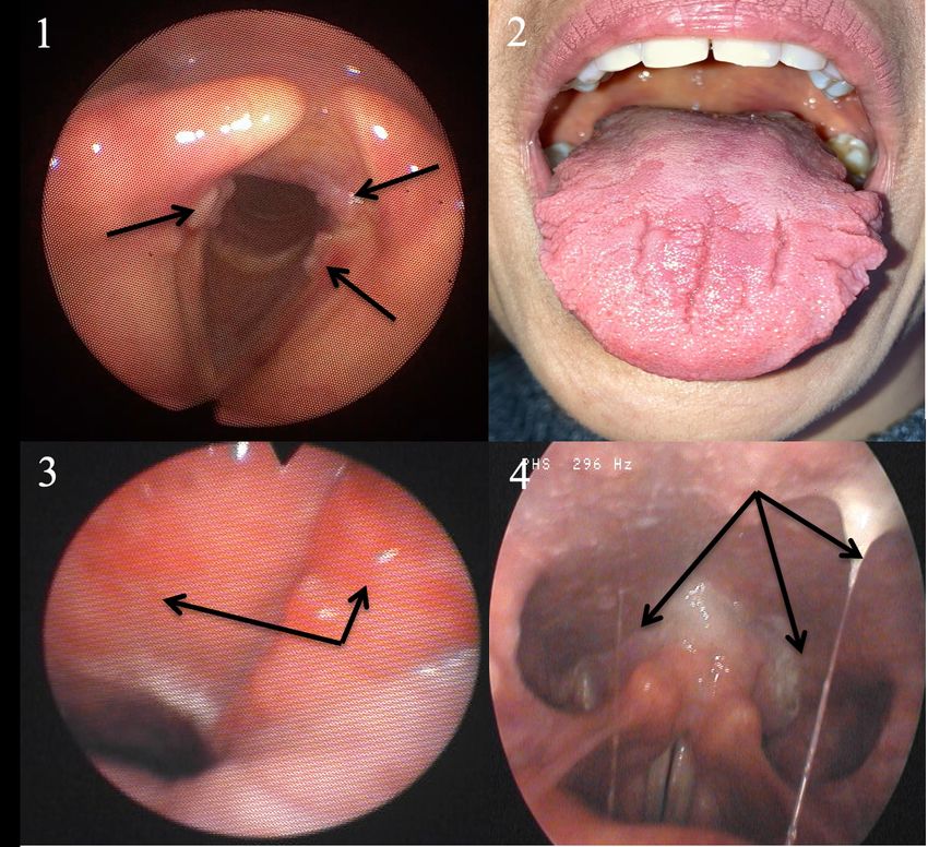
Figure 3: Some Atypical Findings associated with Reflux.
Figure 3 footnote: Leukoplakia (1), fissured tongue (2), erythema of the nasopharynx and
Eustachian meatus (3), and sticky mucus from nasopharynx to oropharynx (4).Preprints (www.preprints.org) | NOT PEER-REVIEWED | Posted: 15 April 2021 doi:10.20944/preprints202104.0409.v1
9 of 13
Table 3: Broncho-laryngeal manifestations of reflux.
Baseline features Post-treatment features
PN G Age Atypical presentation HEMII-pH/RSS Treatment RSS/RSA Presentation evolution Long-term follow-up
18 F 34 Severe idiopathic vocal fold dysplasia Upright acid reflux Strict alcaline RSS: 9 Resolution of dysplasia Long-term diet
Laryngeal check-up: normal GI: not performed Alginate RSA: 20 within 6 months No recurrence (6 months)
No tobacco/toxic exposition history RSS: 34 - RSA: 39 Diet
19 M 45 Severe idiopathic vocal fold dysplasia Upright acid reflux Strict alcaline RSS: 16 Resolution of dysplasia Long-term diet
Laryngeal check-up: normal GI: not performed Diet RSA: 18 within 6 months No recurrence (9 months)
No tobacco/toxic exposition history RSS: 73 - RSA: 23
20 M 38 Daily aspirations & pneumonia Upright acid reflux Strict alcaline RSS: 81 Resolution of dysplasia Long-term diet
Lung/Swallowing check-up: normal GI: esophagitis Diet RSA: 18 within 6 months No recurrence (6 months)
RSS: 156 - RSA: 27 Alginate
21 F 65 Recurrent tracheobronchitis Upright weakly acid reflux Strict diet RSS: 110 Resolution of tracheo- Long-term diet
Lung check-up: normal GI: LES insuffisiency Magaldrate RSA: 19 bronchitis within 6 months Magaldrate (sometimes)
No tobacco/asthma history RSS: 415 - RSA: 24 No recurrence (6 months)
Table 3 footnotes: Abbreviations: F/M=female/male; G=gender; GERD=gastroesophageal reflux disease; GI=gastrointestinal;
HEMII-pH=hypopharyngeal-esophageal multichannel intraluminal impedance pH-monitoring; LES=lower esophageal sphincter; m=month; NP=not provided;
PN=patient number; RSA=reflux sign assessment; RSS=reflux symptom score; y=year.Preprints (www.preprints.org) | NOT PEER-REVIEWED | Posted: 15 April 2021 doi:10.20944/preprints202104.0409.v1
10 of 13
Discussion
Laryngopharyngeal reflux is occasionally associated with nonspecific symptoms and findings,
which make diagnosis challenging for unaware physicians [16]. The involvement of LPR in the
development of several inflammatory conditions of the upper aerodigestive tract was increasingly
studied over the past decades, reporting potential involvement in rhinological, otological and
laryngological diseases [5-8]. In this study, our team shared some clinical observations where the
diagnosis and the treatment of LPR disease had a significant impact of the resolution of specific
conditions that are currently not or poorly known to be associated with reflux.
The involvement of LPR in the development of oral disorders was suspected for a long time, the first
reports dating from the seventies [17]. In the present study, we reported several patients with
primary burning mouth syndrome that was not attributed to any dental or general condition.
Interestingly, we observed that symptoms significantly improved or resolved with an adequate
treatment and a long-term antireflux diet. A few studies investigated the involvement of reflux in
dental lesions [18], or primary burning mouth syndrome [19-21] but authors reported conflicting
results, which may be related to methodological discrepancies across studies [22]. Indeed, the
majority of authors studied the association between burning mouth syndrome and reflux
considering GERD and not LPR diagnostic criteria [19-21]. To date, it has been demonstrated that
patients with LPR may not have GERD and vice versa [1]. The development of burning or pain mouth
may be related to mucosal injury related to pepsin, which may be easily detected in saliva samples
with peptest. Thus, the saliva pepsin detection could be useful to investigate the potential
involvement of LPR in primary burning mouth syndrome, ‘idiopathic’ aphtosis or fissured tongue
could require.
Several studies demonstrated that reflux events may reach nasopharyngeal and nasal regions
[23,24]. In this study, we identified patients who had nasal or otological findings associated with
LPR, i.e. nasal obstruction, excessive nasopharyngeal mucus or recurrent acute media otitis. The
pharyngeal reflux events are known to be mainly gaseous, occurring upright and daytime [25]. The
occurrence of rhinopharyngeal reflux episodes may easily support the development of a
reflux-related nasopharyngeal inflammation and the local production of sticky nasopharyngeal
mucus, the obstruction of the Eustachian tube and the development of otitis media disorders.
Furthermore, pepsin has been identified in secretion of otitis media in several studies [6,26,27].
According to nasal obstruction, two recent studies supported that LPR may lead to edema of the
nasal mucosa, including the posterior part of the inferior turbinate as observed in this study [28,29].
Interestingly, XX et al. found pepsin in the tears [30], which may support the occurrence of a
relationship between laryngopharyngeal reflux and tear disorders through the injury of the nasal
mucosa of the inferior meatus.
The pepsin-related mucosal injury was initially studied in vocal fold tissues [3,31]. Pepsin may induce
macroscopic and microscopic changes in the vocal fold mucosa, including epithelial cell dehiscence,
microtraumas, inflammatory infiltrates, Reinke space dryness, mucosal drying, and epithelial
thickening [32]. The development of severe dysplasia and its resolution after LPR treatment may
probably support the potential impact of LPR in the development of some vocal fold morphological
changes in non-smoker patients. Clinically, LPR may have an impact on the clinical presentation and
the therapeutic response of patients with asthma [8], which supports that the LPR-related
inflammation may reach the bronchi. The observation of patients with LPR and chronic bronchitis
that was not attributed to another disease supports the importance to keep in mind that LPR may
be an irritative factor of the lower airway. In the same way, pepsin was found in trachea and bronchi
of patients with idiopathic stenosis [33].Preprints (www.preprints.org) | NOT PEER-REVIEWED | Posted: 15 April 2021 doi:10.20944/preprints202104.0409.v1
11 of 13
In this case-series, the association between LPR and atypical findings is possible but not proven. The
detection of pepsin and other gastroduodenal enzymes in saliva, nasal or bronchial secretions may
form the basis for a future study and perhaps demonstrate the impact of LPR in the development of
many unusual conditions. Gastroduodenal enzymes may irritate the upper aerodigestive tract
mucosa but they may have an additional role on the local microbiota [34]. In the digestive area,
many researches demonstrated the importance of gut bacteria in the mucosa homeostasis,
protection, recovery, or renewal [35,36]. Similarly, the critical role of microbiota was reported in
respiratory tract diseases, such as tracheal stenosis or asthma [37,38]. Thus, it seems conceivable
that LPR may impact the upper aerodigestive tract microbiota leading to the development of some
disorders.
The primary limitation of the present clinical study is the lack of tissue-related demonstration of the
involvement of reflux in the development of the atypical disorders. However, the occurrence of LPR
at the HEMII-pH study and the complete resolution after treatment strongly support a clinical
association. The retrospective design, the low number of included patients and the short follow-up
time of some patients are additional limitation of the study.
Conclusion
LPR may present with various clinical presentations including mouth, eye, tracheobronchial, nasal or
laryngeal findings, which may all regress with an adequate treatment. Future studies are needed to
better specify the relationship between LPR and these atypical findings through analyses identifying
gastroduodenal enzyme in the enflamed tissue.
Author Contributions: Acknowledgments:
Author Contributions: Conceptualization, JRL, SH, PK and SS; Methodology/Organization, FB, CC; Writing –
Original Draft Preparation, JRL, PK; Writing – Review & Editing, PK, SS, SH; Supervision, SS, SH, PK.
Conflicts of interest: The authors declare no conflict of interest.
References:
1.Lechien JR, Akst LM, Hamdan AL, Schindler A, Karkos PD, Barillari MR, Calvo-Henriquez C, Crevier-Buchman L,
Finck C, Eun YG, Saussez S, Vaezi MF. Evaluation and Management of Laryngopharyngeal Reflux
Disease: State of the Art Review. Otolaryngol Head Neck Surg. 2019; 160(5):762-782. doi:
10.1177/0194599819827488.
2. Klimara MJ, Randall DR, Allen J, Figueredo E, Johnston N. Proximal reflux: biochemical mediators, markers,
therapeutic targets, and clinical correlations. Ann N Y Acad Sci. 2020; 1481(1):127-138. doi:
10.1111/nyas.14366.
3.Samuels TL, Johnston N. Pepsin as a marker of extraesophageal reflux. Ann Otol Rhinol Laryngol. 2010;
119(3):203-8. doi: 10.1177/000348941011900310.
4. Ren JJ, Zhao Y, Wang J, Ren X, Xu Y, Tang W, He Z. PepsinA as a Marker
of Laryngopharyngeal Reflux Detected in Chronic Rhinosinusitis Patients. Otolaryngol Head Neck Surg. 2017;
156(5):893-900. doi: 10.1177/0194599817697055.
5. Pawar S, Lim HJ, Gill M, Smith TL, Merati A, Toohill RJ, Loehrl TA. Treatment of postnasal drip with proton
pump inhibitors: a prospective, randomized, placebo-controlled study. Am J Rhinol. 2007; 21(6):695-701. doi:
10.2500/ajr.2007.21.3098.
6. Lechien JR, Hans S, Simon F, Horoi M, Calvo-Henriquez C, Chiesa-Estomba CM, Mayo-Yáñez M, Bartel R,
Piersiala K, Nguyen Y, Saussez S. Association Between Laryngopharyngeal Reflux and Media Otitis: A Systematic
Review. Otol Neurotol. 2021. doi: 10.1097/MAO.0000000000003123.
7. Lechien JR, Akst LM, Saussez S, Crevier-Buchman L, Hans S, Barillari MR, Calvo-Henriquez C, Bock JM, Carroll
TL. Involvement of Laryngopharyngeal Reflux in Select Nonfunctional Laryngeal Diseases: A Systematic Review.
Otolaryngol Head Neck Surg. 2020: 194599820933209. doi: 10.1177/0194599820933209.
8. Pearson JP, Parikh S, Orlando RC, Johnston N, Allen J, Tinling SP, et al. Review article: reflux and its
consequences--the laryngeal, pulmonary and oesophageal manifestations. Conference held in conjunction withPreprints (www.preprints.org) | NOT PEER-REVIEWED | Posted: 15 April 2021 doi:10.20944/preprints202104.0409.v1
12 of 13
the 9th International Symposium on Human Pepsin (ISHP) Kingston-upon-Hull, UK, 21-23 April 2010. Aliment
Pharmacol Ther. 2011; 33 Suppl 1:1-71. doi: 10.1111/j.1365-2036.2011.04581.x.
9. Lechien JR, Bobin F, Muls V, Saussez S, Remacle M, Hans S. Reflux clinic: proof-of-concept of a
Multidisciplinary European Clinic. Eur Arch Otorhinolaryngol. 2021. doi: 10.1007/s00405-021-06705-9.
10. Lechien JR, Bobin F, Muls V, Mouawad F, Dequanter D, Horoi M, Thill MP, Rodriguez Ruiz A, Saussez S. The
efficacy of a personalised treatment depending on the characteristics of reflux at multichannel intraluminal
impedance-pH monitoring in patients with acid, non-acid and mixed laryngopharyngeal reflux. Clin
Otolaryngol. 2021; 46(3):602-613. doi: 10.1111/coa.13722.
11. Lechien JR, Rodriguez Ruiz A, Dequanter D, Bobin F, Mouawad F, Muls V, Huet K, Harmegnies B, Remacle S,
Finck C, Saussez S. Validity and Reliability of the Reflux Sign Assessment. Ann Otol Rhinol Laryngol. 2020;
129(4):313-325. doi: 10.1177/0003489419888947.
12. Lechien JR, Rodriguez Ruiz A, Dequanter D, Bobin F, Mouawad F, Muls V, Huet K, Harmegnies B, Remacle S,
Finck C, Saussez S. Validity and Reliability of the Reflux Sign Assessment. Ann Otol Rhinol Laryngol. 2020;
129(4):313-325. doi: 10.1177/0003489419888947.
13. Lechien JR, Saussez S, Schindler A, Karkos PD, Hamdan AL, Harmegnies B, De Marrez LG, Finck C, Journe F,
Paesmans M, Vaezi MF. Clinical outcomes of laryngopharyngeal reflux treatment: A systematic review and
meta-analysis. Laryngoscope. 2019; 129(5):1174-1187. doi: 10.1002/lary.27591.
14. Hoppo T, Sanz AF, Nason KS, et al. How much pharyngeal exposure is “normal”? Normative data for
laryngopharyngeal reflux events using hypopharyngeal multichannel intraluminal impedance (HMII). J
Gastrointest Surg. 2012. 16:16–24.
15. Lechien JR, Bobin F, Muls V, Horoi M, Thill MP, Dequanter D, Rodriguez A, Saussez S. Patients with acid,
high-fat and low-protein diet have higher laryngopharyngeal reflux episodes at the impedance-pH monitoring.
Eur Arch Otorhinolaryngol. 2020; 277(2):511-520. doi: 10.1007/s00405-019-05711-2.
16. Lechien JR, Allen JE, Barillari MR, Karkos PD, Jia H, Ceccon FP, Imamura R, Metwaly O, Chiesa-Estomba CM,
Bock JM, Carroll TL, Saussez S, Akst LM. Management of Laryngopharyngeal Reflux Around the World: An
International Study. Laryngoscope. 2020. doi: 10.1002/lary.29270.
17. Howden GF. Erosion as the presenting symptom in hiatus hernia. A case report. Br Dent J. 1971;
131(10):455-6. doi: 10.1038/sj.bdj.4802772.
18. Lechien JR, Chiesa-Estomba CM, Calvo Henriquez C, Mouawad F, Ristagno C, Barillari MR, Schindler A, Nacci
A, Bouland C, Laino L, Saussez S. Laryngopharyngeal reflux, gastroesophageal reflux and dental disorders: A
systematic review. PLoS One. 2020; 15(8):e0237581. doi: 10.1371/journal.pone.0237581.
19. Preetha A, Sujatha D, Patil BA, Hegde S. Oral manifestations in gastroesophageal reflux disease. Gen Dent.
2015; 63(3):e27-31.
20. Hakeem A, Fitzpatrick SG, Bhattacharyya I, Islam MN, Cohen DM. Clinical characterization and treatment
outcome of patients with burning mouth syndrome. Gen Dent. 2018; 66(3):41-47.
21. Cheung D, Trudgill N. Managing a patient with burning mouth syndrome. Frontline Gastroenterol. 2015;
6(3):218-222. doi: 10.1136/flgastro-2014-100431.
22. Lechien JR, Hans S, De Marrez LG, et al. Prevalence and Features of Laryngopharyngeal Reflux in Patients
with Primary Burning Mouth Syndrome. Submitted Laryngoscope, 2021.
23. Brunworth JD, Mahboubi H, Garg R, Johnson B, Brandon B, Djalilian HR. Nasopharyngeal acid reflux and
Eustachian tube dysfunction in adults. Ann Otol Rhinol Laryngol. 2014; 123(6):415-9. doi:
10.1177/0003489414526689.
24. Brunworth JD, Garg R, Mahboubi H, Johnson B, Djalilian HR. Detecting nasopharyngeal reflux: a
novel pH probe technique. Ann Otol Rhinol Laryngol. 2012; 121(7):427-30. doi: 10.1177/000348941212100701.
25. Lechien JR, Bobin F, Dapri G, Eisendrath P, Salem C, Mouawad F, Horoi M, Thill MP, Dequanter D, Rodriguez
A, Muls V, Saussez S. Hypopharyngeal-Esophageal Impedance-pH Monitoring Profiles of
Laryngopharyngeal Reflux Patients. Laryngoscope. 2021; 131(2):268-276. doi: 10.1002/lary.28736.
26. Lei L, Yu Z, Yu R, Yang H, Zou J, Ren J, Zhang J, Zhong D. Correlation of pathogenic effects of
laryngopharyngeal reflux and bacterial infection in COME of children. Acta Otolaryngol. 2021: 1-5. doi:
10.1080/00016489.2021.1883732.
27. O'Reilly RC, Soundar S, Tonb D, Bolling L, Yoo E, Nadal T, Grindle C, Field E, He Z. The role of
gastric pepsin in the inflammatory cascade of pediatric otitis media. JAMA Otolaryngol Head Neck Surg. 2015;
141(4):350-7. doi: 10.1001/jamaoto.2014.3581.
28. Hamizan AW, Choo YY, Loh PV, Abd Talib NF, Mohd Ramli MF, Zahedi FD, Husain S. The association
between the reflux symptoms index and nasal symptoms among patients with non-allergic rhinitis. J Laryngol
Otol. 2021; 135(2):142-146. doi: 10.1017/S0022215120002492.
29. Ceylan SM, Kanmaz MA, Disikirik I, Karadeniz PG. Peak nasal inspiratory airflow measurements for assessing
laryngopharyngeal reflux treatment. Clin Otolaryngol. 2021. doi: 10.1111/coa.13737.Preprints (www.preprints.org) | NOT PEER-REVIEWED | Posted: 15 April 2021 doi:10.20944/preprints202104.0409.v1
13 of 13
30. Magliulo G, Pace A, Plateroti R, Plateroti AM, Cascella R, Solito C, Rossetti V, Iannella G.
Laryngopharyngeal reflux disease in adult patients: tears and pepsin. J Biol Regul Homeost Agents. 2020;
34(2):715-720. doi: 10.23812/19-437-L-26.
31. Samuels TL, Johnston N. Pepsin in gastroesophageal and extraesophageal reflux: molecular
pathophysiology and diagnostic utility. Curr Opin Otolaryngol Head Neck Surg. 2020; 28(6):401-409. doi:
10.1097/MOO.0000000000000664.
32. Lechien JR, Saussez S, Harmegnies B, Finck C, Burns JA. Laryngopharyngeal Reflux and Voice Disorders: A
Multifactorial Model of Etiology and Pathophysiology. J Voice. 2017; 31(6):733-752. doi:
10.1016/j.jvoice.2017.03.015.
33. Blumin JH, Johnston N. Evidence of extraesophageal reflux in idiopathic subglottic stenosis. Laryngoscope.
2011; 121(6):1266-73. doi: 10.1002/lary.21776.
34. Lechien JR, De Vos N, Everard A, Saussez S. Laryngopharyngeal reflux: The microbiota theory. Med
Hypotheses. 2021; 146:110460. doi: 10.1016/j.mehy.2020.110460.
35. Biagi E, Candela M, Turroni S, Garagnani P, Franceschi C, Brigidi P. Ageing and gut microbes: perspectives
for health maintenance and longevity. Pharmacol Res. 2013; 69(1):11-20. doi: 10.1016/j.phrs.2012.10.005.
36. Motta JP, Wallace JL, Buret AG, Deraison C, Vergnolle N. Gastrointestinal biofilms in health and disease. Nat
Rev Gastroenterol Hepatol. 2021. doi: 10.1038/s41575-020-00397-y.
37. Hillel AT, Tang SS, Carlos C, Skarlupka JH, Gowda M, Yin LX, Motz K, Currie CR, Suen G, Thibeault SL.
Laryngotracheal Microbiota in Adult Laryngotracheal Stenosis. mSphere. 2019; 4(3):e00211-19. doi:
10.1128/mSphereDirect.00211-19.
38. Tang HHF, Teo SM, Sly PD, Holt PG, Inouye M. The intersect of genetics, environment,
and microbiota in asthma-perspectives and challenges. J Allergy Clin Immunol. 2021; 147(3):781-793. doi:
10.1016/j.jaci.2020.08.026.You can also read