YTHDF1 alleviates sepsis by upregulating WWP1 to induce NLRP3 ubiquitination and inhibit caspase-1-dependent pyroptosis
←
→
Page content transcription
If your browser does not render page correctly, please read the page content below
www.nature.com/cddiscovery

                  ARTICLE                  OPEN

                  YTHDF1 alleviates sepsis by upregulating WWP1 to induce
                  NLRP3 ubiquitination and inhibit caspase-1-dependent
                  pyroptosis
                  Shuyao Zhang1,8, Xinmin Guan2,8, Wei Liu3,8, Zhe Zhu4,8, Hong Jin2,8, Youfeng Zhu2, Yun Chen1, Min Zhang4, Chengcheng Xu1,
                                                                    ✉             ✉                        ✉
                  Xu Tang1, Jing Wang1, Wang Cheng1, Weihua Lin5 , Xiaoke Ma6 and Jianliang Chen 7

                  © The Author(s) 2022

                      Pyroptosis is inflammation-associated caspase-1-dependent programmed cell death, which confers a crucial role in sepsis. The
                      present study intends to investigate the regulatory network and function of the microarray-predicted YTHDF1 in caspase-1-
                      dependent pyroptosis of sepsis. Peripheral blood of patients with sepsis was collected to determine WWP1 and YTHDF1 expression.
                      An in vitro sepsis cell model was induced in RAW264.7 cells using lipopolysaccharide (LPS) and ATP and an in vivo septic mouse
                      model by cecal ligation and perforation (CLP). After gain- and loss-of-function assays in vitro and in vivo, TNF-α and IL-1β levels and
                      the cleavage of gasdermin-D (GSDMD) were detected by ELISA and Western blot assay, followed by determination of lactate
1234567890();,:

                      dehydrogenase (LDH) activity. Immunoprecipitation and meRIP assay were performed to detect the ubiquitination of NLRP3 and
                      the m6A modification of WWP1 mRNA. The binding of WWP1 to YTHDF1 was explored using RIP-RT-qPCR and dual luciferase gene
                      reporter assay. It was noted that WWP1 and YTHDF1 were downregulated in clinical sepsis samples, LPS + ATP-treated RAW264.7
                      cells, and CLP-induced mice. The ubiquitination of NLRP3 was promoted after overexpression of WWP1. WWP1 translation could be
                      promoted by YTHDF1. Then, WWP1 or YTHDF1 overexpression diminished LDH activity, NLRP3 inflammasomes and caspase-1-
                      mediated cleavage of GSDMD in LPS + ATP-induced RAW264.7 cells. Overexpressed YTHDF1 restrained inflammatory response in
                      CLP-induced mice. Collectively, the alleviatory effect of m6A reader protein YTHDF1 may be achieved through promotion of NLRP3
                      ubiquitination and inhibition of caspase-1-dependent pyroptosis by upregulating WWP1.

                      Cell Death Discovery (2022)8:244 ; https://doi.org/10.1038/s41420-022-00872-2

                  INTRODUCTION                                                                               [7]. Interestingly, it has been reported that m6A RNA methylation is
                  Sepsis is a life-threatening disorder that results from a dysfunc-                         associated with the heterogeneity of sepsis [8]. Of note, the
                  tion in host response to infection [1]. Due to its rising incidence                        bioinformatics prediction in the current study found WW domain
                  and complexity, sepsis poses a major increasing global burden                              containing E3 ubiquitin protein ligase 1 (WWP1) to be a differential
                  and a big challenge to intensive care clinicians and researchers                           gene in sepsis which can be recognized by YTHDF1. To our
                  [2]. Cytokine storm plays an important part in the pathogenesis of                         knowledge, WWP1 is identified as a HECT-type ubiquitin E3 ligase
                  sepsis [3]. It has also been reported that the increase in peripheral                      involved in a series of pathologies [9]. WWP1 can play a pivotal role
                  blood mononuclear cells (PBMCs) is associated with the severity                            in ubiquitin-proteasome pathway and is associated with many
                  of sepsis [4]. Although the majority of surgical sepsis patients will                      diseases including infectious diseases [10]. NOD-like receptor
                  have a rapid recovery, many patients will have chronic critical                            family pyrin domain-containing 3 (NLRP3) inflammasomes can
                  illness and suffer from dismal long-term outcomes [5]. Unfortu-                            stimulate the inflammation and induce immune cell apoptosis to
                  nately, current therapy for sepsis is restricted to antimicrobial                          exacerbate the development of sepsis [11]. Moreover, inactivation
                  treatment and retaining the functions of degenerating organs [6].                          of NLRP3 inflammasomes could alleviate septic liver injury through
                  In this context, it is of significance to seek novel targets for                            autophagy-mediated degradation [12]. As previously reported,
                  treatment of sepsis.                                                                       pyroptosis induced by the NLRP3/caspase-1 pathway could affect
                      YTH N6-methyladenosine (m6A) RNA binding protein 1                                     cognitive deficits in mice with sepsis-related encephalopathy [12].
                  (YTHDF1) is a reader of m6A, a type of dynamic mRNA modification                            Additionally, NLRP3-dependent caspase-1/11-GSDMD pathway
                  regulating protein expression in multiple posttranscriptional levels                       was previously demonstrated to mediate pyroptosis in the

                  1
                   Department of Pharmacy, Guangzhou Red Cross Hospital of Jinan University, Guangzhou 510220, P.R. China. 2Department of Emergency Medicine, Guangzhou Red Cross
                  Hospital of Jinan University, Guangzhou 510220, P.R. China. 3Department of Endocrinology, Guangzhou Red Cross Hospital of Jinan University, Guangzhou 510220, P.R. China.
                  4
                   Department of Respiratory Medicine, Guangzhou Red Cross Hospital of Jinan University, Guangzhou 510220, P.R. China. 5Department of Burns, Guangzhou Red Cross Hospital of
                  Jinan University, Guangzhou 510220, P.R. China. 6Xidian University, School of Computer Science and Technology, Xi’an 710071, P.R. China. 7Clinical Laboratory, Cancer Hospital of
                  Shantou University Medical College, Shantou 515041, P.R. China. 8These authors contributed equally: Shuyao Zhang, Xinmin Guan, Wei Liu, Zhe Zhu, Hong Jin.
                  ✉email: doctorlin@163.com; xkma@xidian.edu.cn; cjlchenjianliang@stu.edu.cn

                  Received: 22 September 2021 Revised: 12 January 2022 Accepted: 25 January 2022

                  Official journal of CDDpress
S. Zhang et al.
2

    Fig. 1 WWP1 is downregulated in sepsis. A The thermal map of differentially expressed genes in GSE100159 (12 normal control samples and
    35 sepsis samples). B The intersection of UbiBrowser database and downregulated genes in GSE100159 dataset. C The expression of WWP1 in
    PBMCs collected from sepsis samples (n = 40) and healthy controls (n = 40) detected by RT-qPCR. D The survival rates of CLP-induced mice.
    E The pathological changes of lung, liver and kidney of CLP-induced mice observed by HE staining. F The expression of WWP1 in PBMCs of
    CLP-induced mice detected by RT-qPCR. There were ten mice in each group. **p < 0.01, ****p < 0.0001.

    hippocampus of a sepsis model [13]. Of note, the upregulation of        detected with RT-qPCR, was upregulated in response to oe-
    NLRP3, cleaved caspase-1 and cleaved GSDMD that are related to          WWP1 treatment yet decreased following LPS + ATP treatment;
    caspase-1-dependent pyroptosis was found in pneumonia-induced           in the presence of LPS + ATP, oe-WWP1 still notably increased
    sepsis [14]. Taking the above reports into consideration, we set out    WWP1 expression (Fig. 2A). As indicated by ELISA results, TNF-α
    to explore whether YTHDF1 affects sepsis development through            and IL-1β levels in culture medium of cells treated with LPS +
    interaction with WWP1/NLRP3/caspase-1 axis.                             ATP had notable increases, which could be inhibited by
                                                                            overexpression of WWP1 (Fig. 2B). The LDH level after treatment
                                                                            with LPS + ATP increased significantly, while overexpression of
    RESULTS                                                                 WWP1 significantly inhibited the release of LDH (Fig. 2C).
    WWP1 was downregulated in sepsis                                        According to Western blot assay, LPS + ATP markedly activated
    We predicted the E3 ubiquitin ligase of NLRP3 through                   the inflammasomes, while overexpression of WWP1 inhibited
    UbiBrowser database (http://ubibrowser.ncpsb.org/ubibrowser/            this effect (Fig. 2D). Moreover, immunofluorescence found that
    home/index) and intersected the results with the downregu-              WWP1 overexpression led to reduced caspase-1 expression
    lated gene in the sepsis-related microarray GSE100159, whereby          but showed no obvious influence on ASC-pro-caspase-1 spots
    WWP1 was the only gene identified (Fig. 1A, B). RT-qPCR results          (Fig. 2E). Western blot assay showed that LPS + ATP markedly
    confirmed that WWP1 was downregulated in patients with                   promoted GasD-N protein expression, attenuated GasD-FL
    sepsis (Fig. 1C).                                                       protein expression, which could be reversed by overexpression
       To further explore the role of WWP1 in the regulation of sepsis,     of WWP1 (Fig. 2F).
    we first established a sepsis mouse model through CLP, and                  Since WWP1 restoration resulted in suppressed activation of
    analyzed the survival rate of the mice. The results revealed that       the NLRP3 inflammasome yet unaffected NLRP3 protein expres-
    the survival rate of CLP-induced septic mice was strikingly lower       sion (Fig. 2D), we speculated that WWP1 may confer a role in the
    than that in sham-operated mice (Fig. 1D). From the results of HE       degeneration of NLRP3 protein. Thus, we treated WWP1 over-
    staining, the CLP-induced septic mice had seriously damaged             expression cells with protease inhibitor MG132. Intriguingly, in
    morphology of cells in lung, liver, and kidney relative to sham-        the presence of MG132, an upregulation of NLRP3 expression
    operated mice, accompanied by inflammatory reaction (Fig. 1E),           was observed in the oe-WWP1 group relative to the oe-NC
    suggesting that the CLP mouse model was successfully estab-             group (Fig. 2G). We further detected the level of NLRP3
    lished. RT-qPCR results documented that WWP1 expression in              ubiquitination with Co-IP. The results displayed that over-
    PBMCs of CLP-induced septic mice was lower than that in sham-           expression of WWP1 induced the polyubiquitination of NLRP3
    operated mice (Fig. 1F).                                                in macrophages (Fig. 2H).
       These results suggest that WWP1 is poorly expressed in sepsis.          Further to explore the types of NLRP3 polyubiquitin chain, the
                                                                            levels of K63 and K48 ubiquitination of NLRP3 were detected by
    Overexpression of WWP1 promoted NLRP3 ubiquitination to                 Co-IP. The results showed that overexpression of WWP1 induced
    inactivate NLRP3 inflammasomes and caspase-1 mediated                    K48-linked NLRP3 polyubiquitination in macrophages and had no
    cleavage of GSDMD                                                       significant effect on K63 ubiquitination of NLRP3 (Fig. 2I).
    In order to further explore the molecular mechanism of WWP1                Overall, overexpression of WWP1 is able to promote NLRP3
    in sepsis, we treated RAW264.7 cells with LPS and ATP to                ubiquitination while inhibiting the activation of NLRP3 inflamma-
    construct an in vitro cell model of sepsis. WWP1 expression, as         somes and caspase-1-mediated cleavage of GSDMD.

                                                                                                  Cell Death Discovery (2022)8:244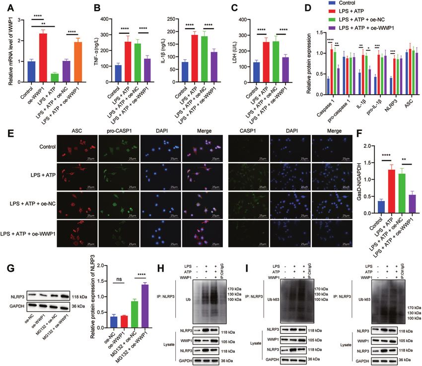
S. Zhang et al.
                                                                                                                                                3

Fig. 2 Overexpression of WWP1 promotes NLRP3 ubiquitination to inactivate NLRP3 inflammasomes and caspase-1-mediated cleavage
of GSDMD. A The expression of WWP1 detected by RT-qPCR. B The expression of TNF-α and IL-1βdetected by ELISA. C LDH level in cells with
different treatment. D The activation of NLRP3 inflammasomes detected by Western blot assay. E The co-localization of ASC with pro-caspase-
1 and caspase-1, respectively in macrophages detected by immunofluorescence. F Protein levels of GasD-N and GasD-FL detected by Western
blot assay. G The protein expression of NLRP3 in oe-WWP1-treated cells in response to MG132 treatment. H The ubiquitination of NLRP3
detected by Co-IP (WWP1: 105 kDa; NLRP3: 118 kDa). I K63 ubiquitination and K48 ubiquitination of NLRP3 detected by Co-IP (WWP1: 105 kDa;
NLRP3: 118 kDa). *p < 0.05; **p < 0.01; ***p < 0.001; ****p < 0.0001. ‘ns’ indicates no significant difference. Cell experiments were repeated
three times.

YTHDF1 promoted WWP1 expression in LPS + ATP-treated                    expression was markedly inhibited in RAW264.7 cells treated with
RAW264.7 cells                                                          LPS + ATP (Fig. 3D).
Furthermore, we found that there was an m6A modification site in            In order to further explore the effect of YTHDF1 on WWP1
WWP1 transcripts through the database of rmbase; therefore, we          expression, sh-YTHDF1 was transfected into RAW264.7 cells.
screened the m6A reader of WWP1 through the database of                 Based on the results from RT-qPCR and Western blot assay,
m6a2Target (http://m6a2target.canceromics.org/#/), which found          knockdown of YTHDF1 markedly downregulated the protein
that YTHDF1, IGF2BP1, IGF2BP2 and IGF2BP3 were potential                expression of WWP1, but had no significant effect on the RNA
factors to recognize the m6A modification of WWP1. Meanwhile,            level of WWP1 (Fig. 3E, F). meRIP assay was implemented to
GSE100159 dataset manifested that YTHDF1 was downregulated              assess the m6A modification status of WWP1 mRNA. The results
and IGF2BP3 was upregulated in sepsis (Fig. 3A). It has been            exhibited that compared with IgG antibody, the m6A antibody
reported that YTHDF1 can promote the translation of target              notably enriched WWP1 (Fig. 3G). RIP-RT-qPCR found that
transcripts [15], indicating that YTHDF1 may alleviate sepsis by        compared with IgG antibody, YTHDF1 antibody significantly
promoting WWP1 expression. We thus first used RT-qPCR to                 enriched WWP1 (Fig. 3H). Dual luciferase gene reporter assay
detect YTHDF1 expression in clinical samples, the results of which      revealed that oe-YTHDF1 resulted in a marked increase in the
revealed that YTHDF1 expression was remarkably low in patients          luciferase activity in cells after co-transfection with WWP1-WT
with sepsis (Fig. 3B). RT-qPCR also found that YTHDF1 expression        but no difference after co-transfection with WWP1-MUT (Fig. 3I).
in CLP-induced septic mice was noticeably lower than that in            These results suggest that YTHDF1 can upregulate WWP1 in the
sham-operated mice (Fig. 3C). RT-qPCR displayed that YTHDF1             form of m6A in sepsis.

Cell Death Discovery (2022)8:244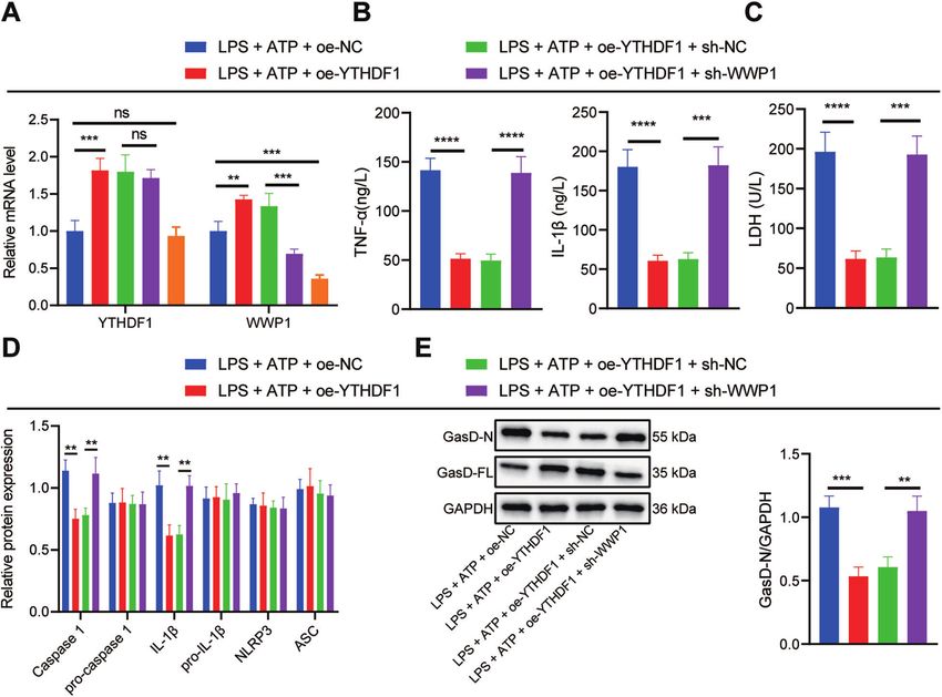
S. Zhang et al.
4

    Fig. 3 YTHDF1 facilitates WWP1 expression in LPS + ATP-treated RAW264.7 cells. A The expression of WWP1 and four m6A readers in
    sepsis-related microarray GSE00159 (measurement data were expressed as mean ± standard deviation, and paired samples t test was applied
    for comparison between the control group and the sepsis group). B RT-qPCR to detect the expression of YTHDF1 in PBMCs of sepsis samples
    (n = 40) and healthy controls (n = 40). C The expression of YTHDF1 in PBMCs of mice with different treatment detected by RT-qPCR. D The
    expression of YTHDF1 determined by RT-qPCR. E The expression of YTHDF1 and WWP1 in cells with different treatment assessed by RT-qPCR.
    F Western blot assay to measure the protein expression of YTHDF1 and WWP1 in cells with different treatment. G The m6A modification status
    of WWP1 mRNA evaluated by meRIP. H, RIP-RT-qPCR to assess the binding of WWP1 mRNA to YTHDF1 protein. I Dual luciferase gene reporter
    assay to evaluate the binding of WWP1 and YTHDF1. There were 10 mice in each group. *p < 0.05; **p < 0.01; ***p < 0.001. ****p < 0.0001. ‘ns’
    indicates no significant difference. Cell experiments were repeated three times.

    Upregulation of WWP1 by YTHDF1 inhibited activation of                   revealed that oe-YTHDF1 led to reduced inflammatory cell
    NLRP3 inflammasomes and caspase-1-mediated cleavage of                    infiltration in lung, liver, and kidney tissues of CLP-induced septic
    GSDMD in LPS + ATP-treated RAW264.7 cells                                mice, accompanied by restored tissue structure (Fig. 5C). In
    In order to further investigate the effect of YTHDF1/WWP1/NLRP3/         addition, oe-YTHDF1 contributed to markedly lowered LDH level
    caspase-1 axis on sepsis, RAW264.7 cells were transfected with oe-       in CLP-induced septic mice (Fig. 5D). ELISA demonstrated that the
    NC/oe-YTHDF1, or sh-NC/sh-WWP1. In the presence of LPS + ATP,            levels of TNF-α and IL-1β in CLP-induced septic mice treated with
    YTHDF1 and WWP1 expression in response to oe-YTHDF1 was                  oe-YTHDF1 were notably restrained (Fig. 5E). These results suggest
    substantially augmented, whereas WWP1 expression was decreased           that overexpression of YTHDF1 is capable of inhibiting the
    yet YTHDF1 expression remained unchanged in response to sh-              inflammatory response in CLP-induced septic mice.
    WWP1 treatment. In the presence of LPS + ATP + oe-YTHDF1, sh-
    WWP1 led to notably diminished WWP1 expression (Fig. 4A). ELISA
    revealed that oe-YTHDF1 brought about diminished levels of TNF-α         DISCUSSION
    and IL-1β in RAW264.7 cells induced by LPS + ATP, which was              This study clarified the regulatory role of YTHDF1 in sepsis with
    negated by sh-WWP1 (Fig. 4B).                                            the involvement of WWP1/NLRP3/caspase-1 axis and found that
       Moreover, LDH level in RAW264.7 cells induced by LPS + ATP            YTHDF1 could upregulate WWP1 to enhance NLRP3 ubiquitination
    was potently lowered by overexpressing YTHDF1, which was                 and restrict caspase-1-dependent pyroptosis in sepsis.
    reversed by silencing WWP1 (Fig. 4C). Based on the Western blot             In the first place, the present study revealed that WWP1 and
    assay results, oe-YTHDF1 significantly inhibited the activation of        YTHDF1 were downregulated in sepsis. It is known that YTHDF1 is
    NLRP3 inflammasomes in RAW264.7 cells induced by LPS + ATP,               the reader of m6A, which has been reported to participate in the
    which was counteracted by further sh-WWP1 treatment (Fig. 4D).           regulation of sepsis development. As previously reported, m6A
    As revealed by Western blot assay, oe-YTHDF1 markedly inhibited          RNA methylation was accountable for the heterogeneity of sepsis
    GasD-N protein expression and augmented GasD-FL expression in            [8]. Moreover, m6A modification of mRNA can exert important
    RAW264.7 cells induced by LPS + ATP, which was nullified by               functions in sepsis, with m6A-cis-eQTLs showing the most obvious
    additional sh-WWP1 treatment (Fig. 4E). Collectively, YTHDF1 can         role in individual variation in the progression of sepsis [16]. It was
    upregulate WWP1 to inhibit the activation of NLRP3 inflamma-              revealed that upregulation of YTHDF1 in cells could contribute to
    some and caspase-1-mediated cleavage of GSDMD.                           reduced HIV-1 infection mainly by diminishing HIV-1 reverse
                                                                             transcription [17]. Notably, the role of WWP1 in inflammation and
    Overexpression of YTHDF1 inhibited inflammation in CLP-                   infection has also been unfolded. As previously reported, WWP1 as
    induced septic mice                                                      an E3 ligase could negatively regulate TLR4-mediated release of
    To further explore the effect of overexpression of YTHDF1 on             TNF-α and IL-6 and induce K48-linked polyubiquitination under
    sepsis in vivo, we overexpressed YTHDF1 in CLP-induced septic            the stimulation of LPS to regulate TRAF6 proteasomal degradation
    mice. From RT-qPCR results, YTHDF1 expression in PBMCs of CLP-           [18]. Additionally, it was revealed that WWP1 ubiquitin ligases
    induced septic mice treated with oe-YTHDF1 was elevated                  could aid in inducing the release of HBV [19]. It is noteworthy that
    (Fig. 5A). Moreover, oe-YTHDF1 notably enhanced the survival             there is a paucity of reports regarding the relationship between
    rate of CLP-induced septic mice (Fig. 5B). The results of HE staining    YTHDF1 and WWP1. Interestingly, in the current study, it was

                                                                                                    Cell Death Discovery (2022)8:244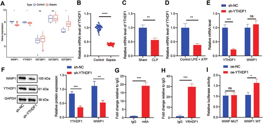
S. Zhang et al.
                                                                                                                                                 5

Fig. 4 Upregulation of WWP1 by YTHDF1 inhibits activation of NLRP3 inflammasomes and caspase-1-mediated cleavage of GSDMD.
A The expression of YTHDF1 and WWP1 in cells with different treatment assessed by RT-qPCR. B The expression of TNF-α and IL-1β
determined by ELISA. C LDH level in cells with different treatment. D The activation of NLRP3 inflammasomes measured by Western blot assay.
E Protein levels of GasD-N and GasD-FL detected by Western blot assay. *p < 0.05; **p < 0.01; ***p < 0.001; ****p < 0.0001. ‘ns’ indicates no
significant difference. Cell experiments were repeated 3 times.

found through rmbase-based analysis in combination with RT-             thereby alleviating the septic shock [26]. Intriguingly, the
qPCR and Western blot assay that YTHDF1 could promote the               regulation on NLRP3/caspase-1 by WWP1 has been rarely
expression of WWP1 in sepsis.                                           reported. However, a previous study revealed that downregulation
   Mechanistically, our study further demonstrated that WWP1            of WWP1 could activate caspase-3, thereby inhibiting growth and
suppressed caspase-1-dependent pyroptosis by promoting NLRP3            inducing apoptosis in hepatoma carcinoma cells [27]. Ubiquitina-
ubiquitination in sepsis. Strikingly, an increasing number of studies   tion is one of the main mechanisms regulating the activity of
have unfolded the involvement of NLRP3 inflammasomes in the              NLRP3 inflammasomes, the activation of which can be inhibited
development of sepsis and its related diseases. For instance, the       by E3 ubiquitin ligase through degradation of NLRP3 [28, 29]. In
inhibition of the NF-kB/NLRP3 inflammasome signaling pathway             the present study, we found that WWP1 could promote NLRP3
by Maf1 could lead to attenuation of sepsis-associated encephalo-       ubiquitination to repress the activation of NLRP3 inflammasomes.
pathy [20]. Moreover, the inactivated NLRP3 inflammasome due to          To conclude, the regulatory role of WWP1 in sepsis was achieved
melatonin could ameliorate sepsis-induced heart injury [21].            through its mediation of NLRP3 inflammasomes and caspase-1-
Downregulated activity of NLRP3 inflammasome in response to              dependent pyroptosis.
intravenous arginine administration alleviated acute kidney injury         Based on the results obtained in the current study, a conclusion
in a mouse model of polymicrobial sepsis [22]. Additionally,            is reached that YTHDF1 promotes NLRP3 ubiquitination by
repression of the NLRP3/IL-1β axis may exert protection against         upregulating WWP1, thereby inhibiting caspase-1-dependent
endothelial relaxation dysfunction induced by sepsis [23]. It is        pyroptosis, which contributes to attenuation of sepsis (Fig. 6).
known that proteolytic cleavage of GSDMD by caspase members             This finding may provide a novel direction for diagnosis and
including caspase-1 is a crucial step for executing pyroptosis in       treatment of sepsis. However, further studies are needed to
LPS-activated innate immune and endothelial cells and that              validate this finding and its clinical feasibility.
cleaved GSDMD can stimulate NLRP3-modulated caspase-1
activation of through an intrinsic pathway [24] Caspase-1-
dependent pyroptosis of PBMCs was unveiled to be able to                MATERIALS AND METHODS
predict the progression of sepsis in patients with severe trauma        Bioinformatics methods
[25]. It has been reported that inactivation of NLRP3 inflamma-          The sepsis-related microarray GSE100159 was downloaded from Gene
somes and inhibition of caspase-1-mediated cleavage of GSDMD            Expression Omnibus database, which contained 12 normal control samples
could prevent pyroptosis in LPS-induced sepsis in a mouse model,        and 35 sepsis samples. R language “limma” package was adopted to

Cell Death Discovery (2022)8:244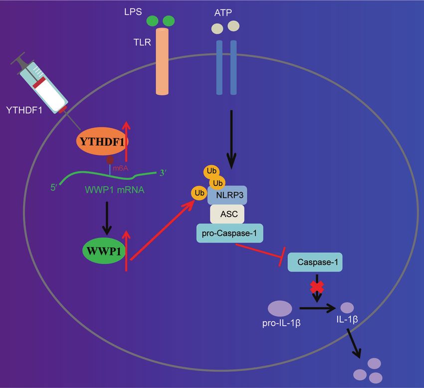
S. Zhang et al.
6
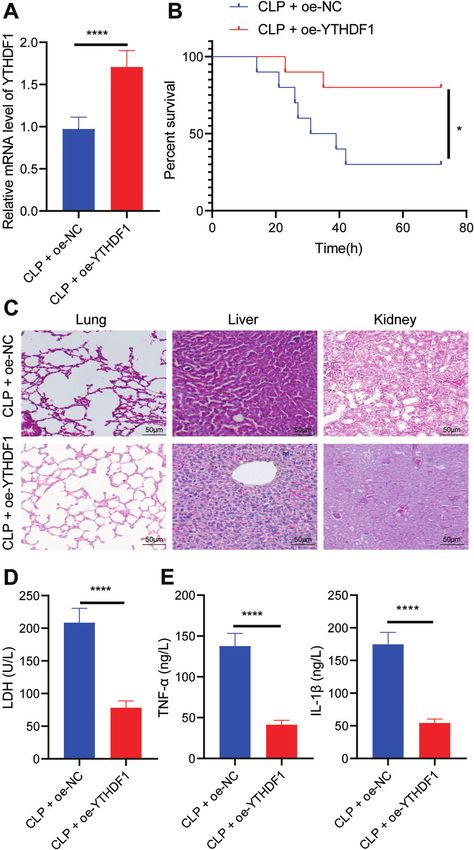
Fig. 5 Overexpression of YTHDF1 inhibits inflammation in CLP-induced septic mice. A RT-qPCR to detect the expression of YTHDF1 in
PBMCs of mice with different treatment. B The survival rates of mice with different treatment. C The pathological changes of lung, liver and
kidney tissues of mice with different treatment observed by HE staining. D LDH levels in serum of mice with different treatment. E The
expression of TNF-α and IL-1β detected by ELISA. There were 10 mice in each group. *p < 0.05; **p < 0.01; ***p < 0.001; ****p < 0.0001.
identify the differentially expressed genes (DEGs) in sepsis with |log fold Separation of PBMCs
change (FC) | > 1 and p value < 0.05 as the threshold. UbiBrowser database This study selected 40 patients with sepsis hospitalized in Cancer Hospital
was applied to predict E3 ubiquitin ligase of the protein, and m6A2Target of Shantou University Medical College from June 2018 to June 2020, and 40
database to predict mRNA m6A modifier. healthy individuals who underwent physical examination in the hospital at
Cell Death Discovery (2022)8:244S. Zhang et al.
7
Fig. 6 The molecular mechanism of m6A reader protein YTHDF1 in sepsis. YTHDF1 upregulates WWP1 to promote NLRP3 ubiquitination,
which inhibits caspase-1-dependent pyroptosis, thereby alleviating sepsis.
the same time period as controls. The peripheral blood samples of the was obtained via reverse transcription of the mRNA using the reverse
study subjects (30 mL) were collected. The PBMCs were isolated from transcription kits (RR047A, Takara, Kyoto, Japan). SYBR® Premix Ex TaqTM II
patients and healthy controls according to the instructions of kit using kits (DRR081, Takara) were used to prepare the reaction system. The
Ficoll-Paque-plus (GE Healthcare, Piscataway, NJ). The collected cells were samples were subjected to RT-qPCR in a real-time fluorescence qPCR
stored in a refrigerator at −80 °C for subsequent experimentation. instrument (ABI 7500, ABI, Foster City, CA). Primer design is shown in
Supplementary Table 1.
Cell culture
The mouse macrophage line RAW264.7 was purchased from Procell Life Western blot
Science&Technology (Wuhan, China) and cultured with Dulbecco’s Cells were detached in RIPA lysis buffer (P0013B, Beyotime, Shanghai,
modified Eagle medium (DMEM) (10569044, Gibco, Carlsbad, CA) contain- China) containing 1% protease inhibitor and phosphorylase inhibitor. The
ing 10% fetal bovine serum (10099141, Gibco) and 100 U/mL penicillin and total protein concentration was quantified using BCA assay kits (a53226,
100 μg/mL streptomycin at 37 °C with 5% CO2. Thermo Fisher Scientific, Rockford, IL). After sodium dodecyl sulfate-
polyacrylamide gel electrophoresis, the protein was transferred to a
polyvinylidene fluoride membrane (IPVH85R, Millipore Corp., Billerica, MA).
Cell transfection The membrane was blocked with 5% bovine serum albumin (BSA) for 1 h
The RAW264.7 macrophages (4 × 10⁵ cells/well) were seeded in a 6-well at room temperature, and then incubated overnight at 4 °C with primary
plate. Upon cell confluence reaching 70–80%, cells were grouped and antibodies against WWP1 (FNab09534, 1: 1000, Wuhan Fine Biotech,
transfected with WWP1 overexpression plasmid (oe-WWP1), oe-YTHDF1, oe- Wuhan, China), NLRP3 (ab263899, 1: 1000, Abcam, Cambridge, UK),
negative control (NC), plasmids carrying short hairpin RNA targeting WWP1 YTHDF1 (ab252346, 1: 1000, Abcam), pro-caspase-1/caspase-1 (ab179515,
(sh-WWP1), sh-YTHDF1, or sh-NC. The cell transfection was performed
1: 1000, Abcam), pro-IL-1β/IL-1β (Ab234437, 1: 1000, Abcam), ASC (sc-
following the protocols of lipofectamine 2000 reagent (11668-019, 514414, 1: 500, Santa Cruz Biotechnology, Santa Cruz, CA), GasD-FL
Invitrogen Inc., Carlsbad, CA). Briefly, 4 μg plasmid and 10 μL lipofectamine (ab219800, 1: 1000, Abcam), GasD-N (ab215203, 1: 1000, Abcam) and
2000 each diluted with 250 μL serum-free medium was mixed well and glyceraldehyde-3-phosphate dehydrogenase (GAPDH) (ab181602, 1:
added to the six-well plate after 20 min of standing. After 6-h incubation at 10000, Abcam). The membrane was then incubated with horseradish
37 °C with 5% CO2 under saturated moisture, the medium containing the peroxidase (HRP)-labeled secondary antibody against immunoglobulin G
transfection medium was then discarded and replaced with 10% FBS-
(IgG) (ab6721, 1: 5000, Abcam) for 2 h. Enhanced chemiluminescence
contained medium for 24–48 h for subsequent experiment. Sequences and
reagents were used to visualize the blots. ImageJ 1.48 u software (V1.48,
plasmids used were all provided by GenePharma (Shanghai, China). National Institutes of Health, Bethesda, MA) was used for protein
quantitative analysis through normalization of gray value of protein bands
Establishment of an in vitro sepsis cell model to the internal reference GAPDH.
RAW264.7 cells were treated with 500 ng/mL lipopolysaccharide (LPS) for 4 h,
and then with 5 mM ATP for 30 min to develop an in vitro sepsis cell model.
Coimmunoprecipitation (Co-IP)
RAW264.7 cells were lysed in protease-inhibitor cock tail (Roche
Reverse transcription-quantitative polymerase chain reaction Diagnostics GmbH, Mannheim, Germany)-containing cell lysis buffer
(RT-qPCR) (50 mM Tris HCl, pH 8.0, 150 mM NaCl, 5 mM ethylene diamine tetraacetic
The total RNA of tissue and cells was extracted using Trizol (16096020, acid, 1 mm phenylmethylsulfonyl fluoride, 0.1% sodium dodecyl sulfate
Thermo Fisher Scientific, Rockford, IL). The complementary DNA (cDNA) and 0.1% Triton X-100) (Sigma-Aldrich, St. Louis, MO), and the lysate was
Cell Death Discovery (2022)8:244S. Zhang et al.
8
centrifuged. The supernatant was incubated with protein A/G agarose The anesthetized mice were fixed in a supine (back down) position on
beads (Santa Cruz Biotechnology) at 4 °C for 30 min. The antibodies against the mouse plate. The midline incision was made on the abdominal wall of
NLRP3 (ab263899, 1: 30, Abcam), WWP1 (FNab09534, Wuhan Fine Biotech the mice to gently pull out the cecum. The feces of the upper end of the
Co., Ltd.), ubiquitin (ab209263, 1: 30, Abcam), ubiquitin K48 (ab140601, 1: cecum were squeezed to make the end full, and the mesenteric surface
1000, Abcam), ubiquitin K63 (ab179434, 1: 1000, Abcam), IgG (ab197767, 1: blood vessels were separated. The cecal wall was punctured with a 21-g
50, Abcam) were incubated with protein A/G agarose beads in PBS sterile needle at the midpoint between the ligation site and the top of
containing 0.1% Triton X-100 and protein inhibitor for 10 min. Subse- the cecum to cause perforation. A little content in the cecum was gently
quently, the supernatant was incubated with antibody protein A/G agarose squeezes out to ensure smooth perforation, and the extruded content
at 4 °C for 3 h. The precipitates were collected by centrifugation, washed, was wiped out. After laparotomy, the cecum in sham-operated mice was
resuspended in sodium dodecyl sulfate sample buffer and analyzed by gently pulled out without any treatment and then pushed back to the
Western blot assay. abdominal cavity, followed by closure of the abdominal cavity and
suturing layer by layer.
The mice were grouped into sham-operated and cecal ligation and
Radioinmunoprecipitacion (RIP) perforation (CLP)-induced septic mice (n = 10). The CLP-induced septic
RIP kits (#17-700, Millipore Corp.) were used to detect the binding between mice were subjected to CLP to establish a sepsis model, and the survival of
RNA and protein. Cells with different treatment were incubated with equal each was monitored three days after operation. CLP-induced septic mice
volume of RIPA lysate (P0013B, Beyotime) and then centrifuged at 4 °C at
were further treated with oe-NC or CLP + oe-YTHDF1 (n = 10). CLP-induced
14000 rpm to obtain the supernatant. One part of the cell extract was
septic mice treated with oe-NC were injected with 10 μg adenovirus-
taken out as input, and the other part was incubated with the antibody for mediated oe-NC (ad-oe-NC) and those treated with oe-YTHDF1 were
co-precipitation. The samples and input were detached with proteinase K injected with 10 μg adenovirus-mediated oe-YTHDF1 (ad-oe-YTHDF1) 24 h
to extract RNA for subsequent RT-qPCR detection of target RNA. The before operation. The survival of mice with different treatment was
dilution concentration of antibodies used in RIP was as follows: YTHDF1 monitored three days after operation. After the experiment, the mice were
(10 μL, FNab09572, Fine Biotech Co., Ltd.), IgG (10 μL, 10285-1-AP, euthanized.
Proteintech Group, Wuhan, China). The samples were mixed with the
antibodies at room temperature for 30 min. IgG was taken as NC.
Methylated RNA immunoprecipitation (MeRIP) m6A kits (Millipore Corp.) Hematoxylin and eosin (HE) staining
were applied for evaluation of the m6A level of WWP1. According to the After dewaxing and dehydration, sections of lung, liver and kidney tissues
instructions of the kit, MeRIP was performed, followed by RNA extraction of mice were soaked in Harris hematoxylin for 3–8 min, hydrolyzed with
and RT-qPCR determination on WWP1 expression. 1% hydrochloric acid alcohol, and treated with 0.6% ammonia to return to
blue. Sections were stained with eosin for 1–3 min, and then immersed in
95% alcohol I and II, anhydrous ethanol I and II each for 5 min, and in
Dual luciferase gene reporter assay xylene I and II each for 5 min for dehydration and cleaning purposes.
Following the instructions of Promega dual luciferase assay system, the Sections were sealed with neutral gum and examined under a microscope
cDNA containing full-length CDS of WWP1 was cloned into the pGL3-basic (Nikon, Tokyo, Japan, TE200). The images were collected to analyze lung,
luciferase reporter gene vector (Genecreate, Wuhan, China). RAW264.7 liver, and kidney pathology.
cells were seeded in a 24-well plate. After 24 h, oe-NC/oe-YTHDF1 and
pmirGLO-WWP1-wild type (WT)/pmirGLO-WWP1-mutant type (MUT) were
co-transfected into RAW264.7 cells using Lipofectamine 2000 (Invitrogen Enzyme-linked immunosorbent assay (ELISA)
Inc.). Then, 48 h after transfection, the cells were lysed and centrifuged at The expression of tumor necrosis factor-α (TNF-α) (Ab208348, Abcam) and
12000 g for, and the supernatant was collected. The dual luciferase interleukin-1β (IL-1β) (ab197742, Abcam) in serum and supernatant of
reporter gene assay system (E1910, Promega, Madison, WI). Each cell mouse was detected using ELISA kits. According to the instructions of the
sample was added with 100 μL firefly luciferase working solution to detect kits, 0.1 mL of cell supernatant was incubated in the reaction wells of the
firefly luciferase, and Renilla luciferase was detected using 100 μL Renilla 96-well plate at 37 °C for 1 h (blank well was made at the same time),
luciferase working solution. The ratio between firefly luciferase and Renilla followed by further reaction with 0.1 mL of freshly diluted enzyme labeled
kidney luciferase was used as the relative luciferase activity. antibody at 37 °C for 0.5–1 h. Subsequently, 0.1 mL of TMB substrate
solution was added to each reaction well for incubation at 37 °C for
10–30 min. A total of 0.05 mL of 2 M sulfuric acid was then added to each
Immunofluorescence reaction well to stop the reaction. On the Microplate Reader, the OD value
The cells were fixed in 95% absolute ethanol for 15 min, incubated with 5% of each well was measured at 450 nm and TNF-α, IL-1β and IL-6 levels
BSA to block non-specific staining, and then incubated with specific were measured
primary antibody against apoptosis-associated speck-like protein (ASC) (sc-
514414, 1: 100, Santa Cruz Biotechnology) and caspase-1 (sc-392736, 1:
100, Santa Cruz Biotechnology) at 4 °C overnight in darkness. Next, the cells Statistical analysis
were incubated with fluorescent secondary antibody against IgG H & L Measurement data were shown as the mean ± standard deviation from
(Life Technologies, Carlsbad, CA) at 37 °C for 2 h, followed by incubation at least three independent experiments performed in triplicate. All
with DAPI at room temperature for 15 min. Finally, the cells were observed statistical analyses were made using GraphPad Prism 5 software, with
under a confocal scanning microscope (LSM 700; Carl Zeiss MicroImaging, p < 0.05 as a level of statistical significance. The independent sample
Inc., Thornwood, NY). t test was used to analyze the data between two groups, and one-way
analysis of variance (ANOVA) was used for analysis among the multiple
groups. Pearson’s correlation analysis was performed to analyze the
Determination of lactate dehydrogenase (LDH) activity correlation between the observed indicators. p < 0.05 indicated that
The cell supernatant or mouse serum was collected, and then subjected to the difference was statistically significant.
detection according to the instructions of LDH assay kits (MAK066-1KT,
Sigma-Aldrich). The LDH activity was calculated in the samples. LDH
activity (U/L) = [(measured optical density (OD) value − control OD value/ DATA AVAILABILITY
standard OD value − blank OD value)] × concentration of the standard (0.2 The datasets generated/analyzed during the current study are available.
2 μmol/mL) × 1000.
Animal experiment REFERENCES
The animals used in this study were specific-pathogen-free male C57BL/6 J 1. Shen X, Cao K, Zhao Y, Du J. Targeting neutrophils in sepsis: from mechanism to
mice aged 8–12 weeks (purchased from Charles River, Beijing, China). The translation. Front Pharm. 2021;12:644270.
laboratory humidity was 60–65%, and the temperature was 22–25 °C. The 2. Perner A, Gordon AC, De Backer D, Dimopoulos G, Russell JA, Lipman J, et al.
mice were raised under 12-h light/dark cycles, with free access to food and Sepsis: frontiers in diagnosis, resuscitation and antibiotic therapy. Intensive Care
water. The experiment was started after one week of acclimatization. The Med. 2016;42:1958–69.
health status of mice was observed before the experiment. Ten mice were 3. Chousterman BG, Swirski FK, Weber GF. Cytokine storm and sepsis disease
randomly assigned to each group. pathogenesis. Semin Immunopathol. 2017;39:517–28.
Cell Death Discovery (2022)8:244S. Zhang et al.
9
4. Wang Y, Liu Y, Liu Q, Zheng Q, Dong X, Liu X, et al. Caspase-1-dependent pyr- 26. Xue Z, Xi Q, Liu H, Guo X, Zhang J, Zhang Z, et al. miR-21 promotes NLRP3
optosis of peripheral blood mononuclear cells is associated with the severity and inflammasome activation to mediate pyroptosis and endotoxic shock. Cell Death
mortality of septic patients. Biomed Res Int. 2020;2020:9152140. Dis. 2019;10:461.
5. Darden DB, Kelly LS, Fenner BP, Moldawer LL, Mohr AM, Efron PA. Dysregulated 27. Cheng Q, Cao X, Yuan F, Li G, Tong T. Knockdown of WWP1 inhibits growth and
immunity and immunotherapy after sepsis. J Clin Med. 2021;10:1742. induces apoptosis in hepatoma carcinoma cells through the activation of cas-
6. Wasyluk W, Zwolak A. PARP inhibitors: an innovative approach to the treatment of pase3 and p53. Biochem Biophys Res Commun. 2014;448:248–54.
inflammation and metabolic disorders in sepsis. J Inflamm Res. 2021;14:1827–44. 28. Tang J, Tu S, Lin G, Guo H, Yan C, Liu Q et al. Sequential ubiquitination of NLRP3
7. Zhuang M, Li X, Zhu J, Zhang J, Niu F, Liang F, et al. The m6A reader YTHDF1 by RNF125 and Cbl-b limits inflammasome activation and endotoxemia. J Exp
regulates axon guidance through translational control of Robo3.1 expression. Med. 2020;217:e20182091.
Nucleic Acids Res. 2019;47:4765–77. 29. Song H, Liu B, Huai W, Yu Z, Wang W, Zhao J, et al. The E3 ubiquitin ligase TRIM31
8. Zhang S, Liu F, Wu Z, Xie J, Yang Y, Qiu H. Contribution of m6A subtype classi- attenuates NLRP3 inflammasome activation by promoting proteasomal degra-
fication on heterogeneity of sepsis. Ann Transl Med. 2020;8:306. dation of NLRP3. Nat Commun. 2016;7:13727.
9. Kobayashi M, Hoshino S, Abe T, Okita N, Tagawa R, Nagai W, et al. Identification of
WWP1 as an obesity-associated E3 ubiquitin ligase with a protective role against
oxidative stress in adipocytes. Biochem Biophys Res Commun. 2019;508:117–22. AUTHOR CONTRIBUTIONS
10. Zhi X, Chen C. WWP1: a versatile ubiquitin E3 ligase in signaling and diseases. Cell SZ, XG and WL wrote the paper; JC, ZZ, HJ and YZ conceived the experiments; XM, YC
Mol Life Sci. 2012;69:1425–34. and MZ analyzed the data; CX, WL, XT, JW and WC collected and provided the sample
11. Zhao S, Chen F, Yin Q, Wang D, Han W, Zhang Y. Reactive oxygen species interact for this study. All authors have read and approved the final submitted manuscript.
with NLRP3 Inflammasomes and are involved in the inflammation of sepsis: from
mechanism to treatment of progression. Front Physiol. 2020;11:571810.
12. Hou N, Dai X, Lu W, Yang H, Yu H, Liu J, et al. Sophocarpine attenuates septic liver
injury through suppression of the NLRP3 inflammasome via autophagy-mediated COMPETING INTERESTS
degradation. Exp Ther Med. 2020;20:249. The authors declare no competing interests.
13. Chen H, Peng Y, Wang L, Wang X. Sevoflurane attenuates cognitive dysfunction
and NLRP3-dependent caspase-1/11-GSDMD pathway-mediated pyroptosis in ETHICS APPROVAL AND CONSENT TO PARTICIPATE
the hippocampus via upregulation of SIRT1 in a sepsis model. Arch Physiol The assays involving human beings were approved by the ethics committee of
Biochem. 2020;2020:1–8. Guangzhou Red Cross Hospital of Jinan University in line with Declaration of Helsinki,
14. Li LL, Dai B, Sun YH, Zhang TT. The activation of IL-17 signaling pathway pro- and all patients and their caregivers provided written informed consents. All
motes pyroptosis in pneumonia-induced sepsis. Ann Transl Med. 2020;8:674. procedures involving animals in this study were approved by the animal ethics
15. Li Z, Peng Y, Li J, Chen Z, Chen F, Tu J, et al. N(6)-methyladenosine regulates committee of Guangzhou Red Cross Hospital of Jinan University.
glycolysis of cancer cells through PDK4. Nat Commun. 2020;11:2578.
16. Sun X, Dai Y, Tan G, Liu Y, Li N. Integration analysis of m(6)A-SNPs and eQTLs
associated with sepsis reveals platelet degranulation and staphylococcus aureus ADDITIONAL INFORMATION
infection are mediated by m(6)A mRNA methylation. Front Genet. 2020;11:7.
Supplementary information The online version contains supplementary material
17. Tirumuru N, Zhao BS, Lu W, Lu Z, He C, Wu L. N(6)-methyladenosine of HIV-1 RNA
available at https://doi.org/10.1038/s41420-022-00872-2.
regulates viral infection and HIV-1 Gag protein expression. Elife. 2016;5:e15528.
18. Lin XW, Xu WC, Luo JG, Guo XJ, Sun T, Zhao XL, et al. WW domain containing E3
Correspondence and requests for materials should be addressed to Weihua Lin,
ubiquitin protein ligase 1 (WWP1) negatively regulates TLR4-mediated TNF-alpha
Xiaoke Ma or Jianliang Chen.
and IL-6 production by proteasomal degradation of TNF receptor associated
factor 6 (TRAF6). PLoS One. 2013;8:e67633.
Reprints and permission information is available at http://www.nature.com/
19. Garcia ML, Reynolds TD, Mothes W, Robek MD. Functional characterization of the
reprints
putative hepatitis B virus core protein late domain using retrovirus chimeras.
PLoS One. 2013;8:e72845.
Publisher’s note Springer Nature remains neutral with regard to jurisdictional claims
20. Chen S, Tang C, Ding H, Wang Z, Liu X, Chai Y, et al. Maf1 ameliorates sepsis-
in published maps and institutional affiliations.
associated encephalopathy by suppressing the NF-kB/NLRP3 inflammasome
signaling pathway. Front Immunol. 2020;11:594071.
21. Rahim I, Sayed RK, Fernandez-Ortiz M, Aranda-Martinez P, Guerra-Librero A,
Fernandez-Martinez J, et al. Melatonin alleviates sepsis-induced heart injury
through activating the Nrf2 pathway and inhibiting the NLRP3 inflammasome. Open Access This article is licensed under a Creative Commons
Naunyn Schmiedebergs Arch Pharm. 2021;394:261–77. Attribution 4.0 International License, which permits use, sharing,
22. Tanuseputero SA, Lin MT, Yeh SL, Yeh CL. Intravenous arginine administration adaptation, distribution and reproduction in any medium or format, as long as you give
downregulates NLRP3 inflammasome activity and attenuates acute kidney injury appropriate credit to the original author(s) and the source, provide a link to the Creative
in mice with polymicrobial sepsis. Mediators Inflamm. 2020;2020:3201635. Commons license, and indicate if changes were made. The images or other third party
23. Hu S, Pi Q, Luo M, Cheng Z, Liang X, Luo S, et al. Contribution of the NLRP3/IL-1beta material in this article are included in the article’s Creative Commons license, unless
axis to impaired vasodilation in sepsis through facilitation of eNOS proteolysis and indicated otherwise in a credit line to the material. If material is not included in the
the protective role of melatonin. Int Immunopharmacol. 2021;93:107388. article’s Creative Commons license and your intended use is not permitted by statutory
24. Gao YL, Zhai JH, Chai YF. Recent advances in the molecular mechanisms regulation or exceeds the permitted use, you will need to obtain permission directly
underlying pyroptosis in sepsis. Mediators Inflamm. 2018;2018:5823823. from the copyright holder. To view a copy of this license, visit http://creativecommons.
25. Wang YC, Liu QX, Liu T, Xu XE, Gao W, Bai XJ, et al. Caspase-1-dependent pyr- org/licenses/by/4.0/.
optosis of peripheral blood mononuclear cells predicts the development of
sepsis in severe trauma patients: a prospective observational study. Med (Baltim).
2018;97:e9859. © The Author(s) 2022
Cell Death Discovery (2022)8:244You can also read