Young adults with childhood-onset inflammatory bowel disease - aspects on bone mineral density, body composition and physical exercise
←
→
Page content transcription
If your browser does not render page correctly, please read the page content below
Young adults with childhood-onset
inflammatory bowel disease - aspects on
 bone mineral density, body composition
         and physical exercise

                      Vignir Sigurdsson

                   Department of Pediatrics
                 Institute of Clinical Sciences
     Sahlgrenska Academy at the University of Gothenburg

                       Gothenburg 2021

Cover illustration: Aron Ingi Vignisson
Illustrations in thesis: Ingibjörg Sigurðardóttir and Vignir Sigurðsson

Young adults with childhood-onset inflammatory bowel disease
- aspects on bone mineral density, body composition and physical exercise
© Vignir Sigurdsson 2021
vignir.sigurdsson@vgregion.se

ISBN: 978-91-8009-238-8 (Printed version)
ISBN: 978-91-8009-239-5 (Online version)
http://hdl.handle.net/2077/67345

Printed in Borås, Sweden 2021
Printed by Stema Specialtryck AB
Abstract
Background: Our research group has previously shown that low bone mineral
density (BMD) is common in children and adolescents with inflammatory bowel
disease (IBD). However, there is limited knowledge on the development of BMD
and body composition traits (skeletal muscle and body fat) in early adulthood in this
patient group.

Objective: The main objective of this thesis was to gain additional understanding of
BMD and body composition in young adults with childhood-onset IBD.

Method: We performed a follow-up in young adulthood in 74 patients with
childhood-onset IBD. Bone mineral density, skeletal muscle index (SMI), and
fat percentage (fat %) were measured with dual X-ray absorptiometry. Body
composition profiles were defined based on SMI and fat % Z-scores: i) normal,
ii) obese, iii) myopenic, iv) myopenic-obese. Bone geometry and microstructures
were estimated with high-resolution peripheral quantitative computed tomography.
Physical exercise during the previous year was registered. Results were compared to
normative control cohorts from nearby regions.

Results: Young adults, especially men with childhood-onset IBD, are at risk
for low areal BMD and those young men also show widespread deficits in bone
microstructures. Young adults with childhood-onset IBD have a risk for altered
body composition traits with an overrepresentation of abnormal body composition
profiles (myopenic, obese, and myopenic-obese) compared to controls. Young
men with Crohn’s disease have an especially high risk for myopenia. Despite the
detrimental effects of having childhood-onset IBD, we found that high levels of
regular physical exercise in young adulthood are associated with normal BMD and
body composition traits.

Conclusion: Young adult patients with childhood-onset IBD are at risk for
disturbances in BMD and body composition.

Keywords: IBD, BMD, Body composition, Physical exercise, HR-pQCT
Sammanfattning på svenska
Bakgrund: Vår forskningsgrupp har tidigare rapporterat att det är vanligt med låg
bentäthet hos barn och ungdomar med inflammatorisk tarmsjukdom (IBD). Det
finns dock begränsad kunskap om bentäthet samt kroppssammansättning (muskel
och fett) hos patientgruppen i ung vuxenålder.

Mål: Huvudmålet med denna avhandling var att öka kunskapen om bentäthet och
kroppssammansättning hos unga vuxna med pediatrisk IBD.

Metoder: Vi genomförde en uppföljning av 74 unga vuxna med pediatrisk IBD.
Bentäthet liksom skelettmuskel index (SMI) och fettprocent (fett %) mättes med
dual X-ray absorptiometri. Utifrån SMI och fett % Z-score definierade vi fyra
kroppssammansättnings profiler: i) normal, ii) obese (hög fett %), iii) myopenic
(lågt SMI), iv) myopenic-obese (både lågt SMI och hög fett %). Bengeometri
och benmikrostrukturer analyserades med hög-resolution peripheral quantitative
computed tomography. Patienternas fysiska träningsvanor senaste året registrerades.
Resultaten jämfördes med normativa kontroller från närliggande regioner.

Resultat: Unga vuxna patienter, speciellt män, med pediatrisk IBD löper risk för låg
bentäthet. Vidare har dessa unga vuxna utbredda förändringar i benmikrostrukturer.
Stor andel av de unga vuxna patienterna har även en störd kroppssammansättning
med överrepresentation av avvikande profiler (myopenic, obese och myopenic-
obese) jämfört med kontroller. Unga män med Crohns sjukdom löper störst risk
för myopeni. Regelbunden fysisk träning verkar dock kompensera för den IBD-
associerade risken för låg bentäthet och avvikelser i kroppssammansättning.

Slutsatser: Unga vuxna patienter med pediatrisk IBD har ökad risk för störningar i
bentäthet och kroppssammansättning.
List of papers
This thesis is based on the following studies, referenced in the text by their Roman
numerals.

I.      Sigurdsson GV, Schmidt S, Mellström D, Ohlsson C, Kindblom JM,
        Lorentzon M, Saalman R.
        Bone Mass Development from Childhood into Young Adulthood in Patients
        with Childhood-onset Inflammatory Bowel Disease.
        Inflamm Bowel Dis. 2017 Dec;23(12):2215-2226. PMID: 29064856.
II.     Sigurdsson GV, Schmidt S, Mellström D, Ohlsson C, Karlsson M,
        Lorentzon M, Saalman R.
        Altered body composition profiles in young adults with childhood-onset
        inflammatory bowel disease.
        Scand J Gastroenterol. 2020 Feb;55(2):169-177. PMID: 32008409.
III.    Sigurdsson GV, Schmidt S, Mellström D, Ohlsson C, Karlsson M,
        Lorentzon M, Saalman R.
        Physical exercise is associated with beneficial bone mineral density and
        body composition in young adults with childhood-onset inflammatory
        bowel disease.
        Manuscript submitted.
IV.     Sigurdsson GV, Schmidt S, Mellström D, Ohlsson C, Saalman R,
        Lorentzon M.
        A high proportion of young adult male patients with childhood-onset IBD
        have compromised cortical and trabecular bone microstructures.
        Manuscript.
Contents Abbreviations 12 1 Introduction 13 1.1 Inflammatory bowel disease......................................................................... 13 1.1.1 Clinical characteristics........................................................................................ 13 1.1.2 Diagnosis............................................................................................................. 14 1.1.3 Disease phenotype............................................................................................. 15 1.1.4 Epidemiology and etiology.................................................................................. 15 1.1.5 Treatment and disease activity........................................................................... 15 1.1.6 Complications of IBD........................................................................................... 16 1.1.7 Unique aspects of childhood-onset IBD............................................................. 16 1.2 Bone structure, physiology, and assessment of bone mineral density........ 17 1.2.1 Bone structure..................................................................................................... 17 1.2.2 Bone remodeling................................................................................................. 18 1.2.3 Regulating factors of bone turnover.................................................................... 18 1.2.4 Bone mineralization in childhood and peak bone mass..................................... 20 1.2.5 Bone mineral density measurements.................................................................. 21 1.3 Bone mineral density in patients with inflammatory bowel disease............. 21 1.4 Body composition......................................................................................... 22 1.4.1 Body composition in IBD..................................................................................... 23 1.5 Physical exercise in IBD............................................................................... 24 2 Aims 25 2.1 General aim.................................................................................................. 25 2.2 Specific aims................................................................................................ 25 3 Patients and methods 26 3.1 Patients......................................................................................................... 26
3.2 Patient cohorts.............................................................................................. 27
3.3 Control cohorts............................................................................................. 27
   3.3.1 Study I - Control cohorts – Bone mineral density data........................................ 27
   3.3.2 Study II - Control cohorts – Bone mineral density and body
         composition data ................................................................................................ 28
   3.3.3 Study III - Control cohorts – Bone mineral density, body
         composition, and physical exercise data............................................................ 28
   3.3.4 Study IV - Control cohort – HR-pQCT, body composition,
         and physical exercise data................................................................................. 28

3.4 Data collection.............................................................................................. 29
3.5 Bone mineral density.................................................................................... 29
3.6 HR-pQCT...................................................................................................... 30
3.7 Body composition......................................................................................... 31
3.8 Body composition profiles............................................................................ 31
3.9 Physical exercise.......................................................................................... 31
3.10 General statistics........................................................................................ 32
   3.10.1 Study I - Specific statistics................................................................................ 32
   3.10.2 Study II - Specific statistics............................................................................... 32
   3.10.3 Study III - Specific statistics.............................................................................. 33
   3.10.4 Study IV - Specific statistics.............................................................................. 33

4 Results                                                                                                                      34
4.1 Study I - BMD in young adults with childhood-onset IBD............................ 34
   4.1.1 Results................................................................................................................. 34
   4.1.2 Conclusion........................................................................................................... 35
4.2 Study II - Body composition in young adults with childhood-onset IBD...... 35
   4.2.1 Results................................................................................................................. 35
   4.2.2 Conclusion........................................................................................................... 36

4.3 Study III - Physical exercise in young adults with childhood-onset IBD...... 36
   4.3.1 Results................................................................................................................. 37
   4.3.2 Conclusion........................................................................................................... 38

4.4 Study IV - Young adult male patients with childhood-onset IBD
    had compromised cortical and trabecular bone microstructures................ 38
   4.4.1 Results................................................................................................................. 38
   4.4.2 Conclusion........................................................................................................... 39
5 Discussion 40 5.1 Bone health in young adults with childhood-onset IBD............................... 41 5.2 Body composition in young adults with childhood-onset IBD. .................... 42 5.3 Physical exercise.......................................................................................... 44 5.4 Gender differences ..................................................................................... 45 5.5 Crohn’s disease vs. ulcerative colitis........................................................... 46 5.6 Clinical implications...................................................................................... 47 6 Main conclusions 49 Acknowledgments 50 References 52
Young adults with childhood-onset inflammatory bowel disease

Abbreviations

  5-ASA             5-aminosalicylic acid
  aBMD              Areal bone mineral density
  BMD               Bone mineral density
  BMI               Body mass index
  CDAI              Crohn’s disease activity index
  DXA               Dual X-ray absorptiometry
  fat %             Fat percentage
  HR-pQCT           High-resolution peripheral quantitative computed tomography
  IBD               Inflammatory bowel disease
  IBD-U             Inflammatory bowel disease, unclassified
  IQR               Interquartile range
  OPG               Osteoprotegerin
  PBM               Peak bone mass
  PCDAI             Pediatric Crohn’s disease activity index
  PHV               Peak height velocity
  pQCT              Peripheral quantitative computed tomography
  PUCAI             Pediatric ulcerative colitis activity index
  RANK              Receptor activator of nuclear factor kappa-B
  RANKL             Receptor activator of nuclear factor kappa-B ligand
  SD                Standard deviation
  SMI               Skeletal muscle index
  TNF               Tumor necrosis factor
  vBMD              Volumetric bone mineral density

                                              12
Vignir Sigurdsson
1 Introduction
Inflammatory bowel disease (IBD) is a group of diseases that entails chronic
inflammation in different parts of the gastrointestinal tract. The prevalence of IBD
in Sweden is among the highest globally, affecting about 1/150 people1. Almost a
quarter of patients receive their diagnosis in childhood2, making IBD one of the
most common chronic childhood illnesses in the western world3. The prevalence
of childhood-onset IBD in Sweden is elevated as well, and the incidence rises both
in Sweden and globally4, 5. It is well known that children with IBD are at risk of
disturbances of growth due to multiple factors related to the disease6. During this
growth phase in childhood, the body composition changes and the majority of bone
development occurs7-9. Our research group has previously shown that children with
IBD run an increased risk to have low bone mineral density (BMD)10, 11. However,
there is limited knowledge on the development of bone mineralization and body
composition traits from adolescence to early adulthood in this patient group.
1.1 Inflammatory bowel disease
1.1.1 Clinical characteristics
Patients with IBD, both children and adults, suffer from chronic, relapsing
inflammation in different parts of the gastrointestinal channel, depending on the
disease subcategory. The main subcategories are ulcerative colitis and Crohn’s
disease, and a minority of patients with IBD fall into an unclassified category
(IBD-U)12. The disease often progresses slowly for weeks to months before a
diagnosis is established, but a more acute disease progression is also seen in some
patients. It is challenging to differentiate between ulcerative colitis and Crohn’s
disease on clinical symptoms only (Figure 1). Diarrhea and abdominal pain are
dominant symptoms in both disease subcategories. Bloody stools are more common
in ulcerative colitis, whereas in Crohn’s disease, low-grade fever, weight loss,
and perianal disease are more frequent12. Delayed puberty and growth retardation
are typical clinical features in pediatric patients with Crohn’s disease and may
be the only symptoms in some patients. The clinical picture of IBD is dependent
on various factors, such as subcategory, the extension of disease, and intensity of
inflammation12.
13Young adults with childhood-onset inflammatory bowel disease
Symptoms Ulcerative colitis Crohn´s disease
Abdominal pain
Diarrhea
Bloody stools
Weight loss
Low grade fever
Deylayed puberty
Growth retardation
Perianal disease
Symptom frequency +++ ++ +
Figure 1. Clinical symptoms and signs at diagnosis in patients with childhood-onset IBD.
1.1.2 Diagnosis
Diagnosis of IBD is based on clinical, endoscopic, histological, and radiological
findings and the exclusion of other differential diagnoses, mainly infectious colitis12.
In patients with ulcerative colitis, inflammation is most intense in the rectum
and can extend throughout the colon (Figure 2). The inflammation is continuous
and confined to the gastrointestinal wall’s superficial mucosal layer. In contrast,
patients with Crohn’s disease can have segmental inflammation in any part of the
gastrointestinal tract (Figure 2). In Crohn’s disease, all gastrointestinal wall layers
can be affected, leading to strictures or fistulae in some patients12.
14Vignir Sigurdsson
Ulcerative colitis Crohn´s disease
Figure 2. The extent of inflammation in patients with childhood-onset IBD.
1.1.3 Disease phenotype
In children with IBD, both ulcerative colitis and Crohn’s disease are classified
according to the phenotype with the Paris classification13, including age at diagnosis,
the extent of inflammation, growth restriction. Further, in Crohn’s disease, the
presence of perianal disease is included and the disease behavior is defined as
inflammatory, stricturing, or penetrating. The Paris classification is a modified
version of the Montreal classification14 of IBD used in the adult population.
1.1.4 Epidemiology and etiology
IBD can manifest in children of all ages, however, most cases debut usually after
ten years of age15. Sweden has one of the highest prevalence numbers of childhood-
onset IBD globally; 75/100.000 children were reported to have IBD in 2010, around
1500 children in total16. The prevalence of ulcerative colitis and Crohn’s disease in
children is similar in Sweden (30/100.000 and 29/100.000 respectively), with some
indication that the incidence of Crohn’s disease is on the rise5, whereas IBD-U is
less common (16/100.000)16.
The cause of IBD remains unknown, but current research on the pathogenesis
focuses on four main areas: the gut and systemic immune system, environmental
triggers, the microbiome, and genetics17.
1.1.5 Treatment and disease activity
Treatment of childhood-onset IBD in Sweden is based on national guidelines18,
which in turn follow European guidelines19.
Treatment options include nutritional, pharmaceutical, and surgical treatment. All
patients should also receive lifestyle advice (i.e., smoking cessation and physical
activity) and psychological support as needed (Figure 3). The treatment goal for
each patient is to achieve remission of both subjective (symptoms) and objective
(i.e., laboratory parameters and endoscopy) signs of the disease, without any
severe side effects or negative impact of therapies on quality of life18. In pediatric
patients, other important treatment goals are to achieve normal growth, pubertal
development, and optimal bone health19.
15Young adults with childhood-onset inflammatory bowel disease
Medical treatment
Third step
Biologics
Second step - Anti-TNF-alpha
- Ustekinumab
Thiopurines - Vedolizumab
First step Methotrexate*
5-ASA
Total enteral
nutrition*
Corticosteroids
Surgical treatment
Nutritional treatment and support
Psychological support
Lifestyle advice (i.e. physical activity support)
Figure 3. Treatment options in patients with childhood-onset IBD. 5-ASA, 5-aminosalicylic acid;
TNF, Tumor necrosis factor. *in patients with Crohn’s disease
Clinicians use validated disease activity indices to assess disease activity and the
need for acute medical or surgical intervention at a certain time point. In childhood,
the Pediatric Ulcerative Colitis Activity Index20 (PUCAI) and Pediatric Crohn’s
Disease Activity Index21 (PCDAI) are mainly used. In adulthood, several activity
indices exist22, i.e., the MAYO score23 for ulcerative colitis and Crohn’s Disease
Activity Index (CDAI)24. However, estimating inflammatory activity over a long
time, such as during a patient’s whole disease course, is challenging25. To date, there
is no consensus on how to estimate and describe disease course severity over time
reliably.
1.1.6 Complications of IBD
Patients with IBD have both long-standing chronic local enteric inflammation and
systemic inflammation. Disease-associated complications include, e.g., growth
retardation in pediatric patients26, iron deficiency, and decreased BMD11.
Patients with IBD can also develop several extraintestinal manifestations that
sometimes are unrelated to the degree of intestinal inflammation, for example, joint
pain and arthritis, eye disease (uveitis), skin disease (erythema nodosum), liver
diseases (primary sclerosing cholangitis, autoimmune hepatitis), inflammation in the
oral cavity (orofacial granulomatosis), and skeletal inflammation (chronic recurrent
multifocal osteomyelitis)27.
1.1.7 Unique aspects of childhood-onset IBD
The manifestation of IBD at a young age results in a long disease duration with
subsequent increased risk for complications and specifically in patients with
ulcerative colitis, an increased risk for cancer development18. The extent of
16Vignir Sigurdsson
inflammation at diagnosis is often greater in children with IBD than adults. Children
with ulcerative colitis have more often inflammation in the entire colon and in
patients with Crohn’s disease, a colonic engagement is more often reported in
children than adults28, 29.
When IBD affects children during puberty, a crucial period for the growth and
development of bones and body composition7-9, there are several risk factors for
BMD and body composition development disturbances. These include, i.e., lack of
physical exercise, chronic inflammation, nutrition problems, and pharmaceutical side
effects30, 31. There is, however, limited knowledge on how childhood-onset IBD per
se may influence BMD32 and body composition development33 into early adulthood, a
time when peak bone mass (PBM) should have been achieved.
1.2 Bone structure, physiology, and assessment of bone
mineral density
1.2.1 Bone structure
The skeleton divides into the axial and appendicular skeleton. The axial part (head,
vertebrae, and rib cage) defends some of the vital organs (brain, spinal cord, heart,
and lungs). In contrast, the appendicular part makes up the limbs and serves as
attachment sites for tendons and muscles, enabling body movement.
The skeleton comprises long bones (i.e., femur, tibia, and humerus) and flat bones
(skull, ileum, and scapula). Its primary function is mechanical, but it also stores
calcium and phosphate, has immunological and endocrine functions, and is home to
hematopoiesis.
Bones consist of an outer layer, periosteum, a thin membrane that overlies the
cortical bone, which surrounds the trabecular bone and the medullary cavity
(Figure 4). Cortical bone is mainly found in the shaft of long bones and the surface
of flat bones. The osteons in the cortical bone have a dense structure, concentrically
laid down around central canals known as Haversian canals (Figure 4). Around
the canal containing blood vessels, nerves, lymphatics, and connective tissue
are concentric layers of bone matrix (lamellae). There are tiny spaces (lacunae)
containing osteocytes between the lamellae layers (Figure 4).
Trabecular bone is less compact, lighter, and has an irregular structure. It is most
abundant in flat bones and the ends of long bones. The trabecular bone has a
honeycomb-like appearance (Figure 4) made from lamellae forming trabeculae
(plates and bars) aligned to support stress from external compression. Thicker and
more numerous trabeculae result in a more robust structure. The trabecular bone’s
resistance to compressive forces is why it is predominant in the vertebrae.
17Young adults with childhood-onset inflammatory bowel disease
Figure 4. Cortical and trabecular bone microstructures.
1.2.2 Bone remodeling
Bone tissue undergoes constant remodeling to maintain stability and integrity34.
Three cell types comprise the basic multicellular unit of the bone and act in a
coordinated manner: i) osteoblasts (4–6% of bone cells), are responsible for new
bone formation, ii) osteocytes (90–95% of bone cells), make up the majority of bone
structure and bone mass, and osteoclasts (1–2% of bone cells), have a role in bone
resorption35. The bone turnover process in adults is in equilibrium between bone
formation and bone resorption. The activation of this process results in endosteal
surface (outside the bone surface) absorption by osteoclasts, attracting osteoblasts
that form osteocytes and osteoid that calcifies to thicken and strengthen the bone
during an approximately three to six months process. The rate of bone turnover
is highest in sites where trabecular bone predominates, such as the vertebrae, and
lowest at sites with a high proportion of cortical bone, i.e., the hip36.
1.2.3 Regulating factors of bone turnover
The bone turnover process is regulated by both mechanical and biochemical factors,
including receptor activator of nuclear factor kappa-B ligand (RANKL), receptor
activator of nuclear factor kappa-B (RANK), and osteoprotegerin (OPG)37. The
RANKL-RANK-OPG system’s discovery changed the understanding of bone
homeostasis and osteoimmunity. The RANK receptor located on osteoclast precursor
cells and mature osteoclasts is activated by RANKL, inducing osteoclast proliferation
and bone resorption. Osteoblasts secrete RANKL to stimulate bone resorption and
OPG that inhibits RANKL from binding to RANK and, by that, decreases bone
resorption. The balance between RANKL and OPG regulates osteoclast activity and
18Vignir Sigurdsson
is influenced by multiple factors, i.e., mechanical loading, inflammatory cytokines,
and hormones37. By affecting this system, activated T and B cells, TNF-alpha and
corticosteroids can increase osteoclast activity and bone resorption36. Thus, patients
with IBD who suffer from local as well as systemic inflammation are especially at
risk.
Bone health can be defined as a state in which the skeleton serves its purpose, i.e.,
provides adequate mobility, protects against injury, stores minerals, and executes
hormonal as well as endocrinological functions38. Many factors influence the bone
mass accumulation and contribute to bone health. Of those, IBD is associated with
several of the modifiable factors that influence BMD directly as well as through their
effects on body composition traits (skeletal muscle and body fat) (Figure 5). These
are inflammation per se, physical exercise, nutrition, corticosteroids, and other/
unknown factors39-41. Childhood-onset IBD can affect height development, which can
influence BMD directly or through the body composition traits. Also, several non-
modifiable factors (genetics, age, and gender42, 43) influence BMD directly as well as
through their effect on height and body composition traits. Notably, genetic factors
are reported to account for 60–80% of the variation in BMD42, 44.
Figure 5. Inflammatory bowel disease is associated with several factors that possibly influence
BMD directly as well as through their effects on height and body composition traits (skeletal
muscle and body fat). Genetics, age, and gender are non-modifiable factors associated with BMD,
height, and body composition traits.
19Young adults with childhood-onset inflammatory bowel disease
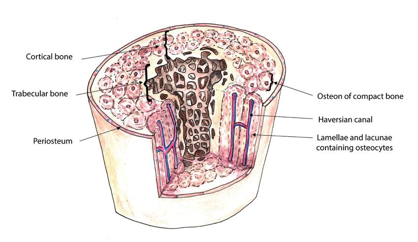
1.2.4 Bone mineralization in childhood and peak bone mass
In adulthood, the bone turnover process described above maintains the bones’
stability and strength. However, in childhood, bone formation dominates as the
child is growing and the majority of bone mass (around 40%) accumulates during
puberty7. Factors contributing to optimal bone mineralization such as regular
physical exercise41, adequate nutrition45, and absence of inflammation31 need to be
present during this period. They are crucial for bone development and achievement
of the genetically determined maximum PBM in young adulthood. According to
Baxter-Jones et al., PBM is achieved in the lumbar spine and total hip, regardless of
gender, about five years after the individual’s peak height velocity (PHV) and in the
total body about seven years after PHV7, 46. After attaining PBM in early adulthood,
BMD gradually declines with age47 (Figure 6). Peak bone mass is one of the most
important factors influencing fracture risk later in life48. The age-related decline
in BMD can lead to osteoporosis, especially in older women, as BMD decreases
more rapidly after menopause (Figure 6). Osteoporosis is defined by the WHO as
areal BMD (aBMD) value below -2.5 SD from the young adult mean (T-score)49.
However, the use of T-score is limited to adults over 50 years of age. Thus in children
and younger adults, aBMD measurements are often based on age- and gender-
matched controls and presented as aBMD Z-score50.
Figure 6. Development of BMD over time. The majority of BMD accumulates during childhood
and adolescence and reaches PBM in young adulthood. After that, age-related loss of BMD ensues.
This process accelerates in women after menopause.
20Vignir Sigurdsson
1.2.5 Bone mineral density measurements
The golden standard for measuring BMD in the clinical setting is dual x-ray
absorptiometry (DXA)51. This technique uses two x-ray beams of different energy
levels to differentiate between bone and soft tissue. The low-energy beams attenuate
more while passing through bone than through soft tissue, whereas the high-energy
beams are equally attenuated regardless of tissue type. Based on the differences in
attenuation, estimations of areal BMD (aBMD) will then be calculated in grams of
bone per cm2 for the whole body and at specific sites of the skeleton (i.e., lumbar
spine, hip).
The machine software provides values for aBMD, bone mineral content, and bone
area. Further, it calculates Z-scores of aBMD for children and adolescents compared
to age- and gender-appropriate normative references. DXA is mainly used in clinical
practice to diagnose bone fragility and estimate fracture risk in adult patients.
However, research interest in BMD development during childhood and adolescence
has increased, especially in patients with chronic inflammatory diseases, as low
BMD and even osteoporosis in early adulthood could have severe consequences in
late adulthood as bone health deteriorates with age52.
While DXA has advantages such as widespread availability, high precision, and
a low dose of radiation (5–10 µSv)53, one important limitation of DXA is that it
is only two-dimensional. Therefore, additional aspects of BMD such as three-
dimensional volumetric BMD (vBMD), bone geometry, and microstructures need
to be investigated with more advanced techniques. Bone geometry and vBMD can
be investigated with peripheral quantitative computed tomography (pQCT). In order
to assess bone microstructures and architecture, such as thickness and separation
of the trabeculae, high-resolution pQCT (HR-pQCT) is used. This microstructure
is the foundation of bone strength54. In contrast to the DXA method, these pQCT
techniques are primarily used in research and not in a clinical setting yet.
1.3 Bone mineral density in patients with inflammatory
bowel disease
Studies of BMD in patients with IBD are primarily conducted with DXA, resulting in
aBMD measurements. Several studies in children, but none in adulthood, have been
conducted using pQCT reporting vBMD measurements and bone geometry55-59. To
our knowledge, only two studies have been published using HR-pQCT60, 61, one in a
group of adolescent and adult patients and the other in middle-aged patients.
Children and adolescents with IBD have an increased risk of low aBMD10, 11, 62, 63.
Our research group and others have previously shown that almost 50% of children
and adolescents with IBD have aBMD Z-scores lower than -1 and 25% lower than
-2, without apparent gender differences10, 11, 62, 64, 65. Several studies have reported
lower aBMD in children with Crohn’s disease than those with ulcerative colitis64, 66,
whereas other studies found no difference11, 63.
21Young adults with childhood-onset inflammatory bowel disease
Adult patients with IBD have also an increased risk of low aBMD67, 68. Two recent
studies found lower aBMD in middle-aged male patients compared to female
patients69, 70. In most reports, no difference in aBMD is found between patients with
Crohn’s disease and those with ulcerative colitis67, 69, but a few studies have reported
lower aBMD in patients with Crohn’s disease68, 71.
Only one study has, to our knowledge, investigated the development of aBMD
prospectively from childhood to adulthood33. In this study from Laakso et al., adults
with childhood-onset IBD were reported to have low aBMD Z-scores at follow-up in
early adulthood without improving aBMD Z-scores since childhood. In accordance,
a retrospective study found that young adults with childhood-onset IBD who had
conducted a DXA scan in young adulthood showed aBMD Z-scores lower than -1 in
around 50% of cases32. Thus, despite limited data in young adulthood, low aBMD in
childhood appears to persist into early adulthood without substantial improvements.
However, Schmidt et al. from our research group reported that patients in late
adolescence (17 to 19 years of age) had improved their aBMD Z-scores in the lumbar
spine at first follow-up after two years11. Our research group concluded that there
might be evidence that patients potentially could improve their BMD by the time
they reached early adulthood. This finding raised the question of whether young
adults with childhood-onset IBD could continue to increase their aBMD beyond the
expected age for attaining PBM.
By using the three-dimensional measurement method pQCT, several studies have
found trabecular vBMD as well as cortical vBMD55, 58, 59 to be consistently low in
children with IBD55-59. Interestingly, Werkstetter and colleagues found cortical vBMD
to be high at diagnosis of IBD56 with normalization after 12 weeks of IBD treatment
and after reaching remission57.
Further examination of the bone microstructures, utilizing HR-pQCT in adolescents
and young adult patients with IBD (aged 12–33 years), some of them with
childhood-onset IBD, revealed deficits in trabecular thickness and larger separation
of trabeculae but no deficits in cortical thickness or density60. In contrast, in a group
of older adults with IBD (median age 44 years), both cortical thickness and density
were affected, but trabecular microstructures appeared to be intact61.
1.4 Body composition
Body composition describes the proportions of fat mass and fat-free mass of an
individual. Fat-free mass is all soft tissue except fat mass and is synonymous
with lean mass. As with bone mass development, the majority of both lean mass
and fat mass are acquired in adolescence72. The optimal analysis method of body
composition depends on localization. A two-component method using DXA is widely
used, measuring fat mass and lean mass. However, multicomponent models that
divide body composition into fat, water, protein, and mineral are more accurate for
measuring the composition of the total body73. The fat mass component is reliably
estimated with DXA and should be adjusted for body size either for height as a fat
mass index or for total body weight as percentage fat mass (fat %)73.
22Vignir Sigurdsson
Dual-X-ray absorptiometry can measure lean mass as a useful surrogate marker for
skeletal muscle74. However, lean mass is not only skeletal muscle but also other
soft tissue and water. Thus, skeletal muscle mass estimation is most reliable in the
appendices where the lean mass is primarily skeletal muscle mass, tendons, and
ligaments75. Further, for increased accuracy of skeletal muscle mass estimations with
DXA, Baumgartner et al.76 developed the appendicular skeletal muscle mass index
(SMI). It is attained by dividing appendicular lean mass by height squared (lean mass
in arms and legs [kg]/height [cm]2).
In healthy individuals, body composition components and BMD measurements
correlate with each other. The skeletal muscle affects bone tissue directly and there
is a strong correlation between SMI and BMD77. In contrast, fat mass and, thus,
increased weight are associated with higher BMD, although to a lesser extent than
SMI78. Further, Baumgartner et al.79 proposed that SMI and fat mass should be
interpreted simultaneously in the elderly by defining four specific body composition
profiles: normal profile, sarcopenic profile (low skeletal muscle mass), obese profile
(high fat mass), and sarcopenic-obese profile (a combination of both low skeletal
muscle mass and high fat mass). However, in recent years, a consensus has been
reached that sarcopenia is defined by both lack of skeletal muscle mass (myopenia)
and muscle function80. Thus, in this thesis, the term ´myopenia´ is used for lack of
skeletal muscle mass when there is no data on muscle function.
1.4.1 Body composition in IBD
Body composition has been studied only to a limited extent in children with IBD.
Total body lean mass deficits seem to be common81-83, but there are conflicting
reports on how fat mass is affected81-83. After the publication of study II in this thesis,
one study reported SMI in children with IBD, highlighting deficits of skeletal muscle
mass84.
Body composition has been more widely studied in adults with IBD, although with
divergent results. A recent review85 summarized seventeen studies that reported data
from adult patients with Crohn´s disease. Lean mass deficits were reported in 28% of
patients and fat mass was low in 31%. Eight studies reported data from adult patients
with ulcerative colitis; a reduction in lean mass was found in 13% of patients, fat
mass was reduced in 13%, and increased in 12% of patients compared to controls. It
appears that the male gender was more often found to be a risk factor for alterations
in body composition components85, especially in patients with Crohn’s disease86.
Two studies have focused on SMI and its association to BMD in adult patients with
IBD. One focused on patients with Crohn’s disease, finding a high prevalence of
myopenia associated with low aBMD87. The other found myopenia to be prevalent
(21%), regardless of disease subcategory or gender, and that low SMI predicted
low aBMD. A strong association has been found in pQCT studies between variables
indicating low skeletal muscle mass and deficits in trabecular and/or cortical
vBMD55-59.
23Young adults with childhood-onset inflammatory bowel disease
Body composition reflects the balance of physiological and, in chronic diseases such
as IBD, pathophysiological processes. Patients with IBD have several risk factors
that can affect this balance, such as nutritional problems88, inflammation per se89, 90,
lack of physical exercise91, and corticosteroid side effects92, and which potentially
manifest as altered body composition (Figure 5). These risk factors resemble those
known for low BMD.
In this context, it has to be noted that body mass index (BMI) as an estimation of
body composition in patients with IBD has proved to be unreliable85, 93. Thus, more
accurate methods are needed and DXA is the most widely available method.
1.5 Physical exercise in IBD
The terms ’physical exercise’ and ’physical activity’ are often used interchangeably.
However, physical activity is considered any movement by skeletal muscles resulting
in energy expenditure measured in kilocalories. It can be categorized into different
activities such as sports, occupational, household, and other activities. Physical
exercise is a physical activity category. It is a planned, structured, and regular activity
to improve or maintain physical fitness94.
Children with IBD are reported to be less physically active than age-matched healthy
controls95. As regular physical exercise in childhood promotes healthy exercise habits
in early adulthood, this could lead to less physical exercise in adulthood96. However,
to the best of our knowledge, there are no data available regarding this in young
adults with childhood-onset IBD.
Physical exercise habits in adults with IBD have also been studied. Tew et al.
reported that less than one-fifth of adults with IBD were engaged in a high amount of
regular physical exercise and one-third was more or less sedentary97. In comparison,
a study on physical exercise habits in healthy adults from Sweden reported higher
physical activity levels, where two-fifths of adults were heavily engaged in physical
exercise98.
Physical exercise plays a vital role in developing and maintaining bone health and
body composition. The importance of skeletal muscle activity is well-documented
in healthy individuals99-101. The relationships between physical exercise, BMD, lean
mass, and fat mass in patients with IBD have only been studied to a limited extent;
no data are available in young adults with childhood-onset IBD. Of the studies in
both children and adult patients with IBD102, 103, physical exercise appears to be
positively associated with higher aBMD and lean leg mass and lower fat mass.
24Vignir Sigurdsson
2 Aims
2.1 General aim
The main objective of this thesis was to gain additional understanding of bone
health and body composition in young adults with childhood-onset IBD.
2.2 Specific aims
I. This study aimed to investigate whether young adults with childhood-onset
IBD have compromised bone mineralization and to correlate their BMD
data to anthropological measures and disease subcategories. A secondary
aim was to examine whether patients with childhood-onset IBD have the
potential to improve their BMD into young adulthood beyond the expected
age for attaining PBM, as our previous data have indicated.
II. This study aimed to investigate body composition with the focus on SMI and
fat % in young adults with childhood-onset IBD. A secondary aim was to
evaluate to which extent BMD and body composition traits relate to each
other.
III. This study aimed to investigate the amount of physical exercise undertaken
by young adults with childhood-onset IBD and its associations to BMD,
SMI, and fat %. A secondary aim was to evaluate whether there is a link,
at the individual level, between physical exercise habits in adolescence and
later in early adulthood.
IV. This study aimed to investigate the extent of microstructural alterations
in young adult males with IBD and the association between these changes
and the patient’s SMI and the amount of physical exercise.
25Young adults with childhood-onset inflammatory bowel disease
3 Patients and methods
3.1 Patients
This thesis is a part of a longitudinal study conducted by our research group. The
current thesis is primarily based on the data from the second follow-up in young
adulthood. A total of 166 patients were initially identified from the only two centers
in the catchment area responsible for the diagnosis, treatment, and follow-up of
childhood-onset IBD and were invited to participate in our longitudinal study. As
a result, the recruited patient population included individuals with mild, moderate,
and severe disease, representing the entire clinical spectrum of childhood-onset
IBD. These two centers are The Queen Silvia Children’s Hospital at Sahlgrenska
University Hospital, Gothenburg, and the Department of Pediatrics at Södra
Älvsborgs Hospital, Borås.
In total, 144 of the 166 initially eligible patients participated in the baseline
measurement conducted between 2003 and 2005. These baseline aBMD results have
previously been published by Schmidt et al.10 Of those 144 patients, 126 participated
in the first follow-up measurement two years later, between 2005 and 2007, of which
the results were also previously published by Schmidt et al.11
In this current second follow-up, DXA measurements for BMD and the body
composition of 74 young adult patients with IBD were carried out between 2012 and
2015. Thus, a total of 52 out of 126 patients at the first follow-up did not participate
in the second follow-up. Patients who did not participate had relocated out of the
area, declined to participate, or we failed to establish contact. These non-participants
(n=52) did not differ significantly regarding clinical characteristics from the
participants included in the second follow-up (Table 1).
26Vignir Sigurdsson
Table 1. Characteristics of participants and non-participants at the first follow-up, the last
common study visit
Patient characteristics Participants (n=74) Non-participants (n=52) p value
Age (years) 16.7±2.9 17±3.1 0.650
Weight (kg) 60.7±15.6 59.4±16.1 0.643
Height (cm) 168±13.3 170.7±14.5 0.296
Female gender, n (%) 25 (34%) 19 (37%) 0.752
Crohn's disease, n (%) 25 (34%) 13 (25%) 0.287
Age at diagnosis (years) 11.1±3.3 11.2±3.4 0.924
Disease duration (years) 3.4±2.7 3.7±2.9 0.597
Current corticosteroid treatment 7 (10%) 5 (11%) 0.557
Any azathioprine treatment 37 (54%) 22 (46%) 0.235
Total body aBMD Z-score 0.1±1.2 0.2±1.3 0.649
Lumbar spine aBMD Z-score -0.8±1.4 -0.7±1.5 0.658
aBMD, Areal bone mineral density. Values are displayed as mean±SD or n (%). Difference
between groups tested with Student's t -test or Fisher's exact test.
3.2 Patient cohorts
Study I included all patients that participated in the described second follow-up.
Study II included all patients that had reached 18 years of age at any study visit and
not only the second follow-up. Study III included all patients that participated in
the second follow-up and answered the physical exercise questionnaire. Study IV
included all male patients at the second follow-up (Table 2).
3.3 Control cohorts
We used several control cohorts in this project regarding BMD and body composition
(Table 2).
3.3.1 Study I - Control cohorts – Bone mineral density data
The GOOD study: This cohort included 1,068 population-based young adult men
aged 18–25 years from the greater Gothenburg area, Sweden (the GOOD study46, 104).
LUNAR standard references: The normative healthy reference database, which
uses sex, weight, ethnicity- and age-specific reference data, is provided by the DXA
manufacturer LUNAR® (GE Medical Systems Lunar).
We used the current Swedish national standards for growth monitoring and
evaluation as the references for height, weight, and PHV9, 105.
27Young adults with childhood-onset inflammatory bowel disease
3.3.2 Study II - Control cohorts – Bone mineral density and body
composition data
The GOOD study: This cohort included 1,068 population-based young adult men
aged 18–25 years from the greater Gothenburg area, Sweden (the GOOD study46, 104).
Normative data, Malmö: This cohort entailed normative data collected from 221
young adults (113 men and 108 women) in the age range of 18–30 years from the
greater Malmö area, Sweden106.
3.3.3 Study III - Control cohorts – Bone mineral density, body
composition, and physical exercise data
The GOOD study: This cohort included 1,068 population-based young adult men
aged 18–25 years from the greater Gothenburg area, Sweden (the GOOD study46, 104).
Normative data, Malmö: This cohort entailed normative data collected from 221
young adults (113 men and 108 women) in the age range of 18–30 years from the
greater Malmö area, Sweden106. Of those, 86 had available physical exercise data and
were included in study III.
Pediatric osteoporosis prevention (POP) study: This cohort consisted of 187 young
adults (97 men and 90 women) aged 18–25 years from the Malmö region107.
3.3.4 Study IV - Control cohort – HR-pQCT, body composition,
and physical exercise data
The GOOD study: Each patient was matched by age and height with five controls
(n=245) from the GOOD study46, 104.
Table 2. Overview of study participants and control cohorts by study I-IV.
Study I Study II Study III Study IV
Patients 74 94 72 49
All patients at the
Study visit Any study visit Second follow-up Second follow-up
second follow-up
Age 17.6-27.7 ≥18 ≥18 ≥18
GOOD study GOOD study GOOD study GOOD study (n=245, age-
Controls (n=1068) (n=1068) (n=1068) and height-matched)
Standard Malmö normative Malmö normative
references LUNAR data (n=221) data (n=86)
POP study (n=187)
Controls in total 1068 1289 1341 245
28Vignir Sigurdsson
3.4 Data collection
We registered the following clinical data: age, gender, height, weight, disease
subcategory, disease duration, and age at disease-onset. Furthermore, data from
medical records were obtained regarding pharmacologic treatments received at or
before the study visit. These included corticosteroids, 5-aminosalicylic acid (5-
ASA), azathioprine, methotrexate, and biological therapy [anti-tumor necrosis factor
(TNF)-alpha]. No biological therapies other than anti-TNF-alpha were used in these
patients. Intestinal surgery due to IBD and fistula surgical procedures data were also
registered.
Height was measured to the nearest 0.5 cm using a wall-mounted stadiometer.
Weight was measured to the nearest 0.1 kg using a calibrated standard scale with the
participants in light clothes. The hand’s maximal grip strength (kg) was measured
at the second follow-up in the majority of patients (n=70) using the Jamar hydraulic
hand dynamometer (5030J1, Jackson, MI, USA) with an adjustable handgrip.
Detailed growth and weight charts from birth until 18 years of age were available
for 43 patients, with age at PHV being derived from each growth curve for
which there was sufficient information on all three growth phases and fitting the
Infancy-Childhood-Puberty model by minimizing the sum of the squares using a
modification of the Levenberg-Marquardt algorithm108. For PHV estimation, two or
more measurements during the critical “peri-PHV” period were needed, not more
than two years from PHV and not more than three years from each other. PHV is
generally believed to be reached within two years of pubertal onset105, 108. According
to Greulich and Pyle’s method, bone age was estimated at baseline by a radiograph of
the left wrist, as previously published by our group10.
3.5 Bone mineral density
A total of 74 patients participated in all three study visits (baseline, first follow-up,
and second follow-up), during which they underwent DXA scans for estimation
of aBMD (g/cm2), bone mineral content (g), and bone area (cm2) in the total body,
lumbar spine (L1–L4), and total hip. All DXA measurements were performed at
the Sahlgrenska University Hospital in Gothenburg (Sweden). We used a Lunar
densitometer (DPX-IQ version 4.7e; GE Medical Systems Lunar, Madison, WI), with
Lunar software ver. 4.7, for the baseline and first follow-up measurements. The data
for these measurements have previously been published10, 11. At the second follow-
up, we used the Lunar Prodigy DXA (GE Medical Systems Lunar). The GOOD
cohort controls were measured with the same Lunar Prodigy DXA apparatus46
as the patients. The control populations from the Malmö region were measured
using a Lunar DPX-L (version 1.3z; GE Medical Systems Lunar) apparatus106.
The correlation of aBMD measurements between the DPX-IQ and Prodigy DXA
machines is very strong at different measurement sites (R=0.98–0.99)109. The aBMD,
bone mineral content, and bone area values were expressed as absolute values. Areal
BMD was also expressed as age- and gender-adjusted Z-score, based on the control
29Young adults with childhood-onset inflammatory bowel disease
population and in the first study, the DXA manufacturer standard reference. The
aBMD Z-score for total hip could not be calculated at the first follow-up due to the
manufacturer’s lack of reference data.
3.6 HR-pQCT
The bone microstructure was measured non-invasively with HR-pQCT at the
ultra-distal tibia. The leg ipsilateral to the non-dominant arm of the participant was
fixated in anatomically formed carbon shells and placed in the HR-pQCT machine
(XtremeCT; Scanco Medical AG, Brüttisellen, Switzerland). The operator placed a
reference line at the tibia’s articular plateau, which was identified with an ordinary
X-ray. Images were taken at a fixed distance (22.5 mm) from the reference line.
With an isotropic resolution of 82 µm, the device captured 104 parallel images and
depicted a 9.02 mm 3D representation of the bone (Figure 7).
Figure 7. Representative image from our HR-pQCT scan at the tibia, showing cortical and
trabecular microstructures.
Images were obtained in approximately three minutes, and the effective dose
generated was 3 µSv per measurement. Measurements were repeated until quality
was sufficient. A contour was automatically placed around the bone to separate the
periosteal surface from the surrounding extra-osseous soft tissue. This contour could
be adjusted by the operator when needed. The standard analysis was performed
according to an earlier described protocol110. From the measurements the following
parameters were obtained: volumetric density (mg/cm3), cortical cross-sectional
area (mm2), cortical volumetric BMD (mg/cm3), cortical thickness (mm), periosteal
circumference (mm), trabecular cross-sectional area (mm2), trabecular bone volume
fraction (%), trabecular number (mm-1), trabecular thickness (mm), and trabecular
separation (mm). The two same operators performed all measurements and graded
image quality using recommendations from the manufacturer.
30Vignir Sigurdsson
3.7 Body composition
Lean soft tissue mass of the total body, arms, and legs (kg), as well as total body fat
mass (kg), were measured with DXA using a Lunar densitometer (DPX-IQ version
4.7e; GE Medical Systems Lunar, Madison, WI). The GOOD cohort controls were
measured with the same Lunar Prodigy DXA apparatus46 as the patients, while the
control populations from the Malmö region were measured using a Lunar DPX-L
(version 1.3z; GE Medical Systems Lunar) apparatus106. The correlation of body
composition measurements using different Lunar machines is strong (R=0.99 for
lean mass and R=0.99 for fat %)109. Therefore, no adjustments were made to the
raw measurement data. We calculated SMI as the sum of the arms and legs lean
soft tissue mass divided by the height squared (kg/m2). Skeletal muscle index is a
reasonable estimate of the total body skeletal muscle mass77. Fat mass was calculated
as the fat mass percentage (fat %) of the total body weight73.
3.8 Body composition profiles
By taking each patient’s SMI and fat % into account, we modified the theoretical
model proposed by Baumgartner79, defining four body composition profiles. In our
modified model, we use the term myopenia for an SMI Z-score -1 and fat % Z-score -1 and fat % Z-score >1;
iii) myopenic: SMI Z-score 27% for males. The cut-offs
for myopenia in the present study were higher than in the original publication of
Baumgartner79, where myopenia was defined as SMI Z-score lower than -2, cut-offs
were SMI of 5.5 kg/m2 for females and 7.26 kg/m2 for males. Cut-offs for fat % were
similar to Baumgartner79, >38% for females and >27% for males.
3.9 Physical exercise
At the second follow-up in early adulthood, we used a standardized physical
exercise questionnaire to gather physical exercise habits in the last 12 months.
The questionnaire also included questions regarding participation in sports during
childhood and adolescence. Physical exercise was defined as regular training,
whereas activities such as bicycling to work were not considered. Physical exercise
was registered as the time in hours per week (h/w) spent on training. Seasonality was
taken into account and the weekly amount of training was averaged for the entire
year. In this thesis, we use the term “amount of physical exercise” to describe the
31Young adults with childhood-onset inflammatory bowel disease
average amount of training over the past year in hours per week. Corresponding data
on physical exercise in the control group (n=1,341) were also registered in hours per
week (h/w) and averaged for the past year.
3.10 General statistics
The thesis author performed statistical analyses with SPSS ver. 26 software (IBM
Corp., Armonk, NY). Continuous variables were presented as median [range or
interquartile range (IQR)] or mean (SD). Categorical variables were presented
as n (%). Differences between three or more groups were tested with analysis of
variance (ANOVA), the Kruskal-Wallis one-way ANOVA, or Extended Fisher’s
exact test based on variable type and distribution. The differences between any two
groups were tested with the Mann-Whitney U-test, student’s t-test, or Fisher’s exact
test based on variable type and distribution. All tests were 2-tailed and conducted,
assuming a significance level of 0.05.
3.10.1 Study I - Specific statistics
A single sample t-test was used to compare the patient’s standard reference aBMD
Z-score to the standard reference population mean Z-score of zero. The regression
models were constructed with a stepwise approach with the total body aBMD
and lumbar spine aBMD Z-scores as dependent variables. The following primary
predictors were selected: aBMD Z-score at baseline; gender; weight at second
follow-up; and the average parental aBMD Z-score (previously published by our
group44). The following secondary predictors were used for the selection process:
height Z-score at second follow-up; age at second follow-up; disease subcategory;
age at diagnosis; been treated with biological agents, azathioprine, 5-ASA or
corticosteroids; and the difference between bone age and chronologic age (ΔBA-CA)
at baseline measurement.
The final model included all four primary predictors and three secondary predictors:
age at diagnosis, previous treatment with biologics, and corticosteroids. The other
secondary predictors were not significant confounders or predictors and did not
improve the model.
3.10.2 Study II - Specific statistics
We used measurements in our combined control cohort for SMI, fat %, and aBMD
in total body, spine, and femoral neck to calculate age- and gender-specific Z-scores.
Using linear regressions in females and males separately, we calculated age-specific
expected mean values for SMI, fat %, and aBMD. Using these expected values and
the measured values for each study participant, we calculated an individual Z-score
for SMI, fat %, and aBMD, using the following formula: the measured value of the
participant, minus the expected mean value of controls for the participant’s age,
divided by the root mean square error of the regression model.
Differences in continuous variables between groups of body composition profiles
32You can also read