Xq27.1 palindrome mediated interchromosomal insertion likely causes familial congenital bilateral laryngeal abductor paralysis (Plott syndrome)
←
→
Page content transcription
If your browser does not render page correctly, please read the page content below
www.nature.com/jhg

                  ARTICLE                  OPEN

                  Xq27.1 palindrome mediated interchromosomal insertion likely
                  causes familial congenital bilateral laryngeal abductor paralysis
                  (Plott syndrome)
                                         1✉
                  Felix Boschann             , Daniel Acero Moreno2, Martin A. Mensah1,3, Henrike L. Sczakiel1, Karolina Skipalova1, Manuel Holtgrewe4,
                                          1,5                                ✉
                  Stefan Mundlos               and Björn Fischer-Zirnsak 1,5

                  © The Author(s) 2022

                      Bilateral laryngeal abductor paralysis is a rare entity and the second most common cause of stridor in newborns. So far, no
                      conclusive genetic or chromosomal aberration has been reported for X-linked isolated bilateral vocal cord paralysis, also referred to
                      as Plott syndrome. Via whole genome sequencing (WGS), we identified a complex interchromosomal insertion in a large family with
                      seven affected males. The 404 kb inserted fragment originates from chromosome 10q21.3, contains no genes and is inserted
                      inversionally into the intergenic chromosomal region Xq27.1, 82 kb centromeric to the nearest gene SOX3. The patterns found at
                      the breakpoint junctions resemble typical characteristics that arise in replication-based mechanisms with long-distance template
1234567890();,:

                      switching. Non protein-coding insertions into the same genomic region have been described to result in different phenotypes,
                      indicating that the phenotypic outcome likely depends on the introduction of regulatory elements. In conclusion, our data adds
                      Plott syndrome as another entity, likely caused by the insertion of non-coding DNA into the intergenic chromosomal region Xq27.1.
                      In this regard, we demonstrate the importance of WGS as a powerful diagnostic test in unsolved genetic diseases, as this genomic
                      rearrangement has not been detected by current first-line diagnostic tests, i.e., exome sequencing and chromosomal microarray
                      analysis.

                      Journal of Human Genetics; https://doi.org/10.1038/s10038-022-01018-z

                  INTRODUCTION                                                                               genomic diversity [10–13]. Rare, disease-causing SVs can vary
                  Vocal cord paralysis is the second most common cause of stridor                            widely in size and include copy number alterations (CNVs:
                  in newborns, which can lead to respiratory distress and often                              deletions and duplications) and copy-number neutral changes
                  requires intubation or tracheostomy [1, 2]. Frequent causes of                             such as inversions and translocations [14]. The “upstream”
                  idiopathic and acquired cases are primary neurological defects,                            mechanisms leading to the formation of SVs can be categorized
                  birth trauma, infections or surgical procedures [3]. Familial cases of                     into recombination-based (e.g., non-allelic homologous recombi-
                  bilateral congenital vocal cord paralysis are rare and mostly                              nation, NAHR) and replication-based mechanisms (RBM) [15]. The
                  associated with other syndromic malformations [4]. X-linked                                “downstream” mechanisms leading to phenotypes are manifold
                  isolated bilateral vocal cord paralysis, also referred to as Plott                         [16, 17]. Aside alterations of gene dosage or disruptions of coding
                  syndrome (MIM: 308850) has been described in three different                               sequence, SVs affecting the non-coding part of the genome can
                  families [5–7]. Since the paralysis can improve over time, it was                          alter 3D chromatin architecture, which can result in misregulated
                  assumed that the cause was the immaturity of the chemorecep-                               gene expression [18, 19].
                  tive pathway between the nucleus ambiguus, the carotid body                                   Herein we report a new family with Plott syndrome and the
                  and the posterior cricoarytenoid muscle [4]. So far, no conclusive                         identification of a novel interchromosomal insertion via whole
                  genetic or chromosomal aberration has been reported [5–7].                                 genome sequencing (WGS) as the likely cause of X-linked isolated
                  Likewise, up to 60% of assumed monogenetic diseases remain                                 congenital bilateral vocal cord paralysis.
                  unsolved using current first-tier diagnostic tests (e.g., chromoso-
                  mal microarray analysis (CMA) and exome sequencing (ES)) [8, 9].
                  A shortcoming of these methods is the inaccurate detection of                              MATERIAL AND METHODS
                  structural variants (SVs, i.e., DNA rearrangements comprising more                         Ethics statement
                  than 50 nucleotides). On average more than 25k SVs are present in                          Consent of each participating individual or their legal guardian was
                  every human genome and thereby represent the largest source of                             obtained for all clinical and molecular studies of this report and for the

                  1
                   Charité – Universitätsmedizin Berlin, corporate member of Freie Universität Berlin and Humboldt-Universität zu Berlin, Institute of Medical Genetics and Human Genetics, Berlin,
                  Germany. 2Division of Pediatric Critical Care Medicine, Kliniken der Stadt Köln gGmbH, Cologne, Germany. 3Clinician Scientist Program, Berlin Institute of Health at Charité –
                  Universitätsmedizin Berlin, BIH Academy, Berlin, Germany. 4Core Facility Bioinformatics, Berlin Institute of Health Charité – Universitätsmedizin Berlin, Berlin, Germany. 5RG
                  Development & Disease, Max Planck Institute for Molecular Genetics, Berlin, Germany. ✉email: felix.boschann@charite.de; ; bjoern.fischer@charite.de

                  Received: 3 November 2021 Revised: 12 January 2022 Accepted: 13 January 2022
F. Boschann et al.
2
publication of clinical photographs. All studies and investigations were of 15 months showed a clear mobility of the right vocal fold and a
performed according to the declaration of Helsinki principles of medical still uncoordinated lateralization and fasciculation on the left side.
research involving human subjects. At the most recent physical examination at the age of 24 month,
he is still fitted with a tracheostoma. His psychomotor develop-
Molecular genetic analysis ment is regular. CMA and ES did not show conclusive findings.
Short-reads WGS. Genomic DNA from affected individual V-3 and his Family history revealed that seven male neonates had acute
unaffected brother (V-2) was isolated from peripheral blood for subsequent inspiratory stridor and died within the first days of life due to
genome sequencing on an Illumina platform using the TruSeq DNA PCR- respiratory distress. No autopsy or genetic testing had been
free protocol (Macrogen). Reads were aligned to human genome build performed on any of them.
GRCh37/hg19 using BWA-MEM 0.7.17 (arXiv:1303.3997v2). Structural
variants were called using Delly v0.8.1 (PMID: 22962449) [20] and analyzed
according to an in-house standard operating procedure using the VarFish
Molecular genetic findings
platform [21]. The BAM files were manually inspected for variants of interest More than 115 Gb of sequences were generated for each individual
in the Integrative Genome Viewer (IGV, http://software.broadinstitute.org/ (V-2 and V-3). More than 97% of sequenced bases had a Phred
software/igv/). Exact determination of breakpoint junction at nucleotide quality score of over 20 and >92.6% of over 30. WGS data evaluation
resolution was possible via alignment of split reads and further examination revealed no convincing coding variant, however, identified a ~10 kb
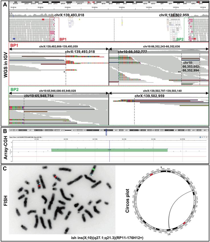
via the UCSC blat tool (https://genome.ucsc.edu/cgi-bin/hgBlat). spanning deletion in an intergenic region on chromosome Xq27.1
[NC_000023.10:g.139493018_139502959] (Fig. 1A) Split reads were
Breakpoint junction PCR. Genomic DNA was amplified using primer pairs visible at both breakpoints, with the non-aligned reads each mapping
spanning both breakpoint junctions (primer sequences are listed in to two genomic regions on chromosome 10q21.3 [NC_000010.10:g.
Supplementary Table 1). Fragments were sequenced directly after A66353052 and NC_000010.10:g.65948754] (Fig. 1A). HGVS: NC_
enzymatic purification by shrimp alkaline phosphatase and exonuclease
1 using BigDye® Terminator v3.1 (Applied Biosystems) and run on ABI 3730 000023.10:g.139493018_139502959ins [NC_000010.10:g.66353052_
DNA Analyzer (Applied Biosystems). 65948754inv]. These genomic locations represent the boundaries of
a 404 kb large fragment. Depth of coverage data and the previously
Quantitative real-time polymerase chain reaction (qPCR). We performed performed CMA confirmed that this region of chromosome 10q21.3
qPCR using genomic DNA of the index subject and family members to is duplicated (Fig. 1B). FISH analysis showed the presence of an
confirm the duplication of the 10q21.3 region and show segregation with additional aqua signal (representing the 10q21.3 region) on
the phenotype. The relative copy number (RCN) of the target sequence chromosome X (green), thereby confirming the interchromosomal
was measured using EVAgreen (Solis BioDyne) and quantified using the insertion event (Fig. 1C). ISCN nomenclature: ish der(X)ins(X;10)(q27.1;
comparative ΔΔCt method on a QuantStudio 3 Real-Time-PCR System q21.3)(RP11-176H12+).
(ThermoFisher Scientific) in comparison to Albumin. The primer sequences For accurate breakpoint determination, we performed breakpoint-
are given in Supplementary Table 1.
spanning PCRs. For the proximal breakpoint junction (BP1), the exact
Chromosomal microarray. CMA for individual V-3 was carried out using a location of the end sequence from chromosome X and start position
whole-genome 1 M oligonucleotide array (Agilent; Santa Clara, CA). Data of the 10q21.3 inserted sequence (joint point 1 *) could not be
were analyzed as previously reported [22]. unambiguously defined due to a 3-bp overlap/microhomology
(“TGC”) in the sequence (Fig. 2A and Supplementary Fig. 1). After the
FISH. Fluorescent in situ hybridization (FISH) analyses were performed on insertion of 59 bp [NC_000010.10:66352994-66353052], a new break
metaphase spreads prepared from 72 h PHA stimulated peripheral blood occurs (joint point 2 **). The following sequence also originates from
lymphocyte culture from the index patient (V-3) according to standard chromosome 10q21.3 and is located 217 bp centromeric
procedures following respective manufacturer’s protocol. The BAC clone (NC_000010.10:66352777). Between these parts 7 bp of unknown
RP11-176H12 (Empire Genomics, Buffalo, NY) in color aqua was selected to origin are inserted. The distal breakpoint junction (BP2) is formed by
cover the duplicated region of 10q21.3. Vysis CEP 10 SpectrumOrange the end of the large duplicated fragment (NC_000010.10:65948754)
Probe (Abbott) and Vysis CEP X (DXZ1) SpectrumGreen Probe (Abbott)
were used to label centromeres of chromosome 10 and chromosome X.
and chromosome X (NC_000023.10:139502959). At this junction we
found a 2 bp insertion (“AG”) (Fig. 2A and Supplementary Fig. 1).
Segregation analysis was performed using breakpoint specific
RESULTS PCRs and qPCR. By this approach we confirmed the derivative
Clinical report X-chromosome in the affected individual V-3. In addition, we
The affected boy (V-3) is the second child of non-consanguineous found this genomic rearrangement in the female individuals IV-5,
parents. He was born at 40 weeks of gestation with the following III-4, III-5, III-6 and II-6 (Fig. 2B, Supplementary Fig. 2).
birth measurements: weight 3200 g (P42, −0.96 SD), length 50 cm
(P54, −1.09 SD), head circumference 35 cm (P65, −0.46 SD). There DISCUSSION
were no congenital malformations or comorbidities. Shortly after Bilateral vocal cord paralysis is a rare entity with an incidence less
birth, stridor was noticed and due to respiratory distress, than 1/100.000 [1]. In 1964 Plott and colleagues first described
mechanical ventilation was necessary. Multiple extubation bilateral familial laryngeal abductor paralysis in four male siblings
attempts were frustrated by recurrence of stridor. A tracheo- and assumed X-linked inheritance [2]. Since then, two other
bronchoscopy showed significant ulceration of the mucosa at the families with suspected Plott syndrome (MIM: 308850) have been
level of the arytenoid cartilage on the left, corresponding to reported, but no genetic tests had been performed [6, 7].
damage due to long-term intubation. Submucosal injection of Furthermore, it has not been elucidated whether the laryngeal
Betamethasone in the affected area was applied. The subglottic abductor paralysis is caused by a central lesion at the level of the
space was unremarkable. After a new extubation attempt, the ventral division of the nucleus ambiguus or by a peripheral
child showed recurrence of stridor with progressive respiratory myogenic process. Via WGS, we identified a complex interchro-
failure necessitating reintubation. The follow-up tracheobroncho- mosomal insertion as the likely cause of X-linked congenital
scopy showed vocal folds in an intermediate position. Since no bilateral laryngeal paralysis, i.e., Plott syndrome, in an affected boy
vocal fold abduction during inspiration was achieved, a congenital with a conspicuous family history. Testing of the other six affected
bilateral vocal cord paresis was suspected. boys was not possible because they died several decades ago.
An MRI of the neck and brain showed no abnormalities. A However, segregation analysis revealed that each mother of an
tracheostomy was performed without complications and the affected child carries the genomic rearrangement, whereas it was
patient was subsequently discharged. A tracheoscopy at the age not detectable in any unaffected male family member.
Journal of Human GeneticsF. Boschann et al.
3
Fig. 1 Molecular genetic findings. A Split-reads and discordant pairs retrieved from WGS and visualized on IGV are shown in different colors
matching for their pairs. Non-split-reads that map to the genome of reference are shown in gray. Dashed vertical lines represent the
breakpoints. WGS identified a ~10 kb spanning deletion in the intergenic region of Xq27.1 [NC_000023.10:g.139493018_139502959]. The
proximal breakpoint junction (BP1) comprises two joint points (indicated by asterisks). The first joint-point * connects chromosome X to
chromosome 10q21.3. After the insertion of 59 bp another break occurs (second joint point **). Subsequently, the sequence of the large 404
kb fragment continues, which is located 219 bp centromeric. The distal breakpoint junction (BP2) connects the large duplicated fragment of
chromosome 10q21.3 back to chromosome X. The junctions are shown at the basepair level in Supplementary Fig. 1. B Array CGH shows a
404 kb spanning region of chromosome 10q21.3 to be duplicated. C FISH signals on metaphase chromosomes showing the interchromosomal
insertion. BAC probes are RP11-176H12 (10q21.3 - aqua), Vysis CEP 10 SpectrumOrange Probe (10p11.1-q11.1,- orange) and Vysis CEP X (DXZ1)
SpectrumGreen Probe (Xp11.1-q211.1 - green). ISCN: ish der(X)ins(X;10)(q27.1;q21.3)(RP11-176H12+). Schematic representation of the
interchromosomal insertion as circos plot retrieved from WGS data.
Journal of Human GeneticsF. Boschann et al.
4
Fig. 2 Schemativ overview of the complex chromosomal rearrangement and Segregation analysis. A The patterns found at the two
breakpoint junctions (i.e. microhomology, small insertions, large template insertion, deletion at insertion site) resemble typical characteristics
that arise due to replication-based repair mechanisms like MMBIR/FoSTes. The proximal breakpoint junction (BP1) comprises two joint points
(indicated by asterisks). Location of primers for break point spanning PCR and qPCR. B Pedigree showing our proband (V-3) and his five-
generation family with seven affected male neonates. Schematic view of the co-segregation analysis: blue dots indicate presence of the wild
type chromosome X, red dots show the presence of the derivative chromosome X. PCR amplification of the proximal breakpoint junction
(BP1) (exemplary). CL control locus PCR, gray filled = not consented for testing.
Interchromosomal insertions are complex genomic rearrange- breakpoint junctions is possible. In our case, the proximal
ments (CGR), characterized by chromosomal segments inserted breakpoint junction (BP1) comprises two joint points. At the first
into an interstitial region of non-homologous chromosome joint point we found a microhomology of 3-bp. After the insertion
generated by more than three DNA breakage and joining events of 59 bp, another break occurs, followed by the insertion of 7 bp
[23]. Approximately 2% of non-recurrent copy number gains are of unclear origin. Subsequently, the sequence of the large 404 kb
caused by inter- or intrachromosomal insertions [24]. The initial fragment continues, which is located 219 bp centromeric. This
trigger of the herein reported interchromosomal insertion is most discontinuous transition indicates that the polymerase has
likely the human specific AT-rich palindrome located in the 485 kb reduced processivity and that a stable replisome is established
spanning intergenic chromosomal region Xq27.1, 82 kb centro- only after iterative template switches. The distal breakpoint
meric to the nearest gene SOX3 [25]. Distinct phenotypes, such as displays features of a simple breakpoint junction. Taken together
congenital hypertrichosis, congenital ptosis, Charcot–Marie–Tooth the patterns found at the two breakpoint junctions (i.e.,
neuropathy and sex reversal have been reported with different microhomologies, small insertions, large template insertion,
inter- and one intrachromosomal insertions into the above- deletion at insertion site) resemble typical characteristics that
mentioned non-coding region [25–31]. arise in replication-based mechanisms with long-distance tem-
Since palindrome sequences can form hairpin loops, they are plate switching (i.e., MMBIR/and Fork Stalling and Template
prone to DNA double-strand breaks and thus pose a risk for the switching (FoSTes)) [33]. Notably, both ends of the sequences that
initiation of chromosomal rearrangements [32, 33]. As in our case form the proximal junction are located within LINE1-Elements,
(distal breakpoint junction BP2), at least one breakpoint of the demonstrating that repetitive elements further stimulate the
other reported cases was located near the center of this formation of complex insertions by providing sequences with
palindromic sequence [28]. The reestablishment of a broken microhomologies.
replication fork depends on stretches of microhomologies which The “downstream” mechanisms of genomic rearrangements
are used by error prone DNA polymerases for priming synthesis leading to phenotypic consequences can be caused by various
on a template strand, a mechanism called Microhomology- mechanisms. In addition to direct disruption of the coding region
Mediated Break-Induced Replication (MMBIR) [33, 34]. Such of genes, insertions affecting intergenic regions can modify
replication-based repair mechanisms are assumed to have the higher-order chromatin organization such as topologically asso-
largest contribution in the formation of nonrecurrent structural ciating domains (TADs) [19].
rearrangements [33, 35]. These “upstream” mechanisms can often In this case, the inserted 404 kb large fragment contains no
be identified by specific signatures at the breakpoint sequence. genes. Other cases featuring non-coding insertions at this
This is a major advantage of genome sequencing, since, in position resulted in varying other phenotype, indicating that the
contrast to CMA or ES, accurate nucleotide resolution of phenotypic outcome might result from “enhancer-adoption” (i.e.,
Journal of Human GeneticsF. Boschann et al.
5
the introduction of tissue specific enhancers resulting in ectopic 8. Yang Y, Muzny DM, Xia F, Niu Z, Person R, Ding Y, et al. Molecular findings among
expression of SOX3). Unfortunately, due to absent SOX3 expression patients referred for clinical whole-exome sequencing. JAMA.2014;312:1870–9.
in tissues accessible from patients, we were unable to perform 9. Dharmadhikari AV, Ghosh R, Yuan B, Liu P, Dai H, Al Masri S, et al. Copy number
precise gene expression analyses, a problem that has already been variant and runs of homozygosity detection by microarrays enabled more pre-
cise molecular diagnoses in 11,020 clinical exome cases. Genome Med.
addressed by Si and colleagues [26]. SOX3, together with SOX1
2019;11:30.
and SOX2, forms the SOXB1 subgroup and is an early marker of 10. Chaisson MJP, Sanders AD, Zhao X, Malhotra A, Porubsky D, Rausch T, et al. Multi-
vertebrate neurogenesis [36]. SOX3 is mainly expressed in platform discovery of haplotype-resolved structural variation in human genomes.
telencephalic neuronal precursor cells (NPC) throughout neuroaxis Nat Commun. 2019;10:1784.
and binds to neuronal genes, thereby priming them for 11. Sudmant PH, Rausch T, Gardner EJ, Handsaker RE, Abyzov A, Huddleston J, et al.
subsequent activation by SOX11 in differentiating neurons. SOX3 An integrated map of structural variation in 2504 human genomes.
regulation and expression appears to be tissue specific. During Nature.2015;526:75–81.
neuronal differentiation, SOX3 is generally downregulated but 12. Collins RL, Brand H, Karczewski KJ, Zhao X, Alfoldi J, Francioli LC, et al. A structural
during hypothalamic development SOX3 remains expressed in a variation reference for medical and population genetics. Nature.2020;581:444–51.
13. Pauper M, Kucuk E, Wenger AM, Chakraborty S, Baybayan P, Kwint M, et al. Long-
subset of differentiated hypothalamic cells in the adult brain
read trio sequencing of individuals with unsolved intellectual disability. Eur J
[37, 38]. Notably, SOX3 is crucial for early segmentation of Hum Genet. 2021;29:637–48.
pharyngeal pouches and proper neural crest cell migration into 14. Lupski JR. Genomic disorders: structural features of the genome can lead to DNA
pharyngeal arches and thereby for cranial nerve and epibranchial rearrangements and human disease traits. Trends Genet. 1998;14:417–22.
placode formation [39, 40]. Thus, SOX3 might also be an important 15. Carvalho CM, Lupski JR. Mechanisms underlying structural variant formation in
regulator for the chemoreceptive pathway between the nucleus genomic disorders. Nat Rev Genet. 2016;17:224–38.
ambiguus, the carotid body and the posterior cricoarytenoid 16. Hurles ME, Dermitzakis ET, Tyler-Smith C. The functional impact of structural
muscle, the perturbation of which is thought to be the cause of variation in humans. Trends Genet. 2008;24:238–45.
Plott syndrome [4]. We therefore hypothesize that altered 17. Stankiewicz P, Lupski JR. Structural variation in the human genome and its role in
disease. Annu Rev Med. 2010;61:437–55.
regulation and ectopic expression of SOX3 caused by the
18. Lupianez DG, Kraft K, Heinrich V, Krawitz P, Brancati F, Klopocki E, et al. Disrup-
identified complex interchromosomal insertion is the cause of tions of topological chromatin domains cause pathogenic rewiring of gene-
X-linked congenital bilateral laryngeal abductor paralysis (Plott enhancer interactions. Cell.2015;161:1012–25.
syndrome). 19. Spielmann M, Lupianez DG, Mundlos S. Structural variation in the 3D genome.
Further studies investigating the effect on SOX3 expression in Nat Rev Genet. 2018;19:453–67.
neuronal tissue (iPS-induced) and molecular characterization of 20. Rausch T, Zichner T, Schlattl A, Stütz AM, Benes V, Korbel JO. DELLY: structural
additional individuals with Plott syndrome would strengthen our variant discovery by integrated paired-end and split-read analysis. Bioinformatics.
assumption. 2012;28:i333–i339.
This study emphasizes that distinct diseases can result from 21. Holtgrewe M, Stolpe O, Nieminen M, Mundlos S, Knaus A, Kornak U, et al. VarFish:
comprehensive DNA variant analysis for diagnostics and research. Nucleic Acids
insertions into the same non-coding genomic region and that the
Res. 2020;48:W162–W169.
phenotypic outcome likely depends on the introduction of 22. Flottmann R, Kragesteen BK, Geuer S, Socha M, Allou L, Sowinska-Seidler A, et al.
regulatory elements. In this regard, we demonstrate the impor- Noncoding copy-number variations are associated with congenital limb mal-
tance of WGS as a powerful diagnostic test in unsolved rare cases, formation. Genet Med. 2018;20:599–607.
even though detection of structural variations with short-read 23. Zhang F, Carvalho CM, Lupski JR. Complex human chromosomal and genomic
technology has its imitations [5, 32]. Even if accurate detection of rearrangements. Trends Genet. 2009;25:298–307.
complex structural rearrangements and their breakpoint junctions 24. Gu S, Szafranski P, Akdemir ZC, Yuan B, Cooper ML, Magrina MA, et al.
by long read sequencing approaches becomes feasible, the Mechanisms for complex chromosomal insertions. PLoS Genet. 2016;12:
interpretation of non-coding genomic rearrangements remains e1006446.
25. Zhu H, Shang D, Sun M, Choi S, Liu Q, Hao J, et al. X-linked congenital hyper-
challenging [41, 42]. This requires the integration of multimodal
trichosis syndrome is associated with interchromosomal insertions mediated by a
layered biological data (gene expression, epigenetics, 3D struc- human-specific palindrome near SOX3. Am J Hum Genet. 2011;88:819–26.
ture) to functionally interpret their effect within multiple 26. Si N, Meng X, Zhao Z, Xia W, Zhang X. A 105 kb interstitial insertion in the Xq27.1
molecular contexts. palindrome from pseudoautosomal region PAR1 causes a novel X-linked reces-
sive compound phenotype. J Transl Med. 2019;17:138.
27. Bowl MR, Nesbit MA, Harding B, Levy E, Jefferson A, Volpi E, et al. An interstitial
DATA AVAILABILITY deletion-insertion involving chromosomes 2p25.3 and Xq27.1, near SOX3, causes
All data relevant to the study are included in the article or the Supplementary X-linked recessive hypoparathyroidism. J Clin Invest. 2005;115:2822–31.
Information. For further information, contact the corresponding author. 28. Brewer MH, Chaudhry R, Qi J, Kidambi A, Drew AP, Menezes MP, et al. Whole
genome sequencing identifies a 78 kb insertion from chromosome 8 as the cause
of Charcot-Marie-Tooth Neuropathy CMTX3. PLoS Genet. 2016;12:e1006177.
REFERENCES 29. DeStefano GM, Fantauzzo KA, Petukhova L, Kurban M, Tadin-Strapps M, Levy B,
et al. Position effect on FGF13 associated with X-linked congenital generalized
1. Cohen SR, Eavey RD, Desmond MS, May BC. Endoscopy and tracheotomy in the
hypertrichosis. Proc Natl Acad Sci USA. 2013;110:7790–5.
neonatal period: a 10-year review, 1967–1976. Ann Otol Rhinol Laryngol.
30. Haines B, Hughes J, Corbett M, Shaw M, Innes J, Patel L, et al. Interchromosomal
1977;86:577–83.
insertional translocation at Xq26.3 alters SOX3 expression in an individual with
2. King EF, Blumin JH. Vocal cord paralysis in children. Curr Opin Otolaryngol Head
XX male sex reversal. J Clin Endocrinol Metab. 2015;100:E815–20.
Neck Surg. 2009;17:483–7.
31. David J, Bunyan DOR, Anthony GTyers, Huang Shuwen, Vivienne KMaloney, et al.
3. Lesnik M, Thierry B, Blanchard M, Glynn F, Denoyelle F, Couloigner V, et al.
McMullan X-Linked dominant congenital ptosis cosegregating with an interstitial
Idiopathic bilateral vocal cord paralysis in infants: case series and literature
insertion of a chromosome 1p21.3 fragment into a quasipalindromic sequence in
review. Laryngoscope.2015;125:1724–8.
Xq27.1. Open J Genet. 2014;4:415–25.
4. Vanessa Abdelhalim J-PV. Isolated idiopathic bilateral vocal cord paralysis in two
32. Walsh E, Wang X, Lee MY, Eckert KA. Mechanism of replicative DNA polymerase
sisters: case report and review of familial vocal cord paralysis. Int J Pediatr
delta pausing and a potential role for DNA polymerase kappa in common fragile
Otorhinolaryngol Extra. 2011;6:368–72.
site replication. J Mol Biol. 2013;425:232–43.
5. Plott D. Congenital laryngeal-abductor paralysis due to nucleus ambiguus dys-
33. Hastings PJ, Ira G, Lupski JR. A microhomology-mediated break-induced repli-
genesis in three brothers. N Engl J Med. 1964;271:593–7.
cation model for the origin of human copy number variation. PLoS Genet. 2009;5:
6. Watters GV, Fitch N. Familial laryngeal abductor paralysis and psychomotor
e1000327.
retardation. Clin Genet. 1973;4:429–33.
34. Sakofsky CJ, Ayyar S, Deem AK, Chung WH, Ira G, Malkova A. Translesion poly-
7. Shatla ES, Prashanth GP, Aguiar R, Shivalingam G, Al, Haq AA. Neonatal stridor in
merases drive microhomology-mediated break-induced replication leading to
familial congenital laryngeal paralysis (Plott Syndrome): a case study in an Omani
complex chromosomal rearrangements. Mol Cell. 2015;60:860–72.
Family. Oman Med J. 2017;32:515–7.
Journal of Human GeneticsF. Boschann et al.
6
35. Bahrambeigi V, Song X, Sperle K, Beck CR, Hijazi H, Grochowski CM, et al. Distinct COMPETING INTERESTS
patterns of complex rearrangements and a mutational signature of micro- The authors declare no competing interests.
homeology are frequently observed in PLP1 copy number gain structural var-
iants. Genome Med. 2019;11:80.
36. Bylund M, Andersson E, Novitch BG, Muhr J. Vertebrate neurogenesis is coun- ETHICS APPROVAL AND CONSENT TO PARTICIPATE
teracted by Sox1-3 activity. Nat Neurosci. 2003;6:1162–8. All investigations were performed according to the declaration of Helsinki principles
37. Rogers N, Cheah PS, Szarek E, Banerjee K, Schwartz J, Thomas P. Expression of the of medical research involving human subjects. Participants or their legal guardians
murine transcription factor SOX3 during embryonic and adult neurogenesis. provided written informed consent for participation and publication in this study.
Gene Expr Patterns. 2013;13:240–8. Consents of all participants were archived.
38. Bergsland M, Ramskold D, Zaouter C, Klum S, Sandberg R, Muhr J. Sequentially
acting Sox transcription factors in neural lineage development. Genes Dev.
2011;25:2453–64. ADDITIONAL INFORMATION
39. Wang L, Xie J, Zhang H, Tsang LH, Tsang SL, Braune EB, et al. Notch signalling
Supplementary information The online version contains supplementary material
regulates epibranchial placode patterning and segregation. Development.
available at https://doi.org/10.1038/s10038-022-01018-z.
2020;147:dev183665.
40. Rizzoti K, Lovell-Badge R. SOX3 activity during pharyngeal segmentation is
Correspondence and requests for materials should be addressed to Felix Boschann;
required for craniofacial morphogenesis. Development.2007;134:3437–48.
or Björn Fischer-Zirnsak.
41. Ho SS, Urban AE, Mills RE. Structural variation in the sequencing era. Nat Rev
Genet. 2020;21:171–89.
Reprints and permission information is available at http://www.nature.com/
42. Logsdon GA, Vollger MR, Eichler EE. Long-read human genome sequencing and
reprints
its applications. Nat Rev Genet. 2020;21:597–614.
Publisher’s note Springer Nature remains neutral with regard to jurisdictional claims
in published maps and institutional affiliations.
ACKNOWLEDGEMENTS
We would like to thank the family for their collaboration and contribution to this
project. We thank Valerie Johnston and Anja Lekaj for excellent technical assistance.
Open Access This article is licensed under a Creative Commons
Attribution 4.0 International License, which permits use, sharing,
adaptation, distribution and reproduction in any medium or format, as long as you give
AUTHOR CONTRIBUTIONS appropriate credit to the original author(s) and the source, provide a link to the Creative
Conceptualization: FB, BFZ, SM. Methodology: MH, HS, BFZ. Formal analysis and Commons license, and indicate if changes were made. The images or other third party
investigation: FB, DAM, MAM, KS. Writing - original draft preparation: FB. Writing - material in this article are included in the article’s Creative Commons license, unless
review and editing: BFZ, MAM, HS, DAM. Resources: SM, BFZ. indicated otherwise in a credit line to the material. If material is not included in the
article’s Creative Commons license and your intended use is not permitted by statutory
regulation or exceeds the permitted use, you will need to obtain permission directly
from the copyright holder. To view a copy of this license, visit http://creativecommons.
FUNDING org/licenses/by/4.0/.
MAM is a participant in the BIH Charité Digital Clinician Scientist Program founded by the
late Prof. Duska Dragun and funded by the Charité-Universitätsmedizin Berlin and the
Berlin Institute of Health. Open Access funding enabled and organized by Projekt DEAL. © The Author(s) 2022
Journal of Human GeneticsYou can also read