Wnt Pathway Extracellular Components and Their Essential Roles in Bone Homeostasis
←
→
Page content transcription
If your browser does not render page correctly, please read the page content below
G C A T
T A C G
G C A T
               genes
Review
Wnt Pathway Extracellular Components and Their Essential
Roles in Bone Homeostasis
Núria Martínez-Gil * , Nerea Ugartondo                       , Daniel Grinberg          and Susanna Balcells

                                          Department of Genetics, Microbiology and Statistics, Faculty of Biology, Universitat de Barcelona, CIBERER,
                                          IBUB, IRSJD, 08028 Barcelona, Spain; nereaugartondo@gmail.com (N.U.); dgrinberg@ub.edu (D.G.);
                                          sbalcells@ub.edu (S.B.)
                                          * Correspondence: nuria.martinez.gil.91@gmail.com

                                          Abstract: The Wnt pathway is involved in several processes essential for bone development and
                                          homeostasis. For proper functioning, the Wnt pathway is tightly regulated by numerous extracellular
                                          elements that act by both activating and inhibiting the pathway at different moments. This review
                                          aims to describe, summarize and update the findings regarding the extracellular modulators of the
                                          Wnt pathway, including co-receptors, ligands and inhibitors, in relation to bone homeostasis, with
                                          an emphasis on the animal models generated, the diseases associated with each gene and the bone
                                          processes in which each member is involved. The precise knowledge of all these elements will help us
                                          to identify possible targets that can be used as a therapeutic target for the treatment of bone diseases
                                          such as osteoporosis.

                                          Keywords: Wnt pathway; inhibitors; ligands; co-receptors; bone


                                   1. Introduction
Citation: Martínez-Gil, N.;                     The Wnt pathway (wingless-type mouse mammary tumor virus integration site) is a
Ugartondo, N.; Grinberg, D.; Balcells,
                                          ubiquitous pathway involved in a wide variety of cellular processes, and highly conserved
S. Wnt Pathway Extracellular
                                          across species. It was first described for its role in carcinogenesis, and later demonstrated
Components and Their Essential
                                          to be essential for embryonic patterning and for maintaining adult tissues [1–3]. In the
Roles in Bone Homeostasis. Genes
                                          skeletal tissue, the Wnt pathway is involved in the differentiation of osteoblasts, osteoclasts
2022, 13, 138. https://doi.org/
                                          and chondrocytes. Therefore, it has a very important role in both skeletal development and
10.3390/genes13010138
                                          bone remodeling, and an essential function in mechanical communication.
Academic Editor: Zhousheng Xiao                 Historically, the Wnt pathway has been categorized into β-catenin-dependent (canoni-
Received: 10 December 2021
                                          cal pathway) and β-catenin independent (non-canonical) pathways (Figure 1). Although
Accepted: 11 January 2022
                                          some studies postulate that different Wnts activate either the canonical or the non-canonical
Published: 13 January 2022
                                          pathways, it has also been reported that some Wnt ligands (e.g., WNT5A) can act on both
                                          pathways [4,5]. In all cases, the Wnt ligands bind to one of the 10 frizzled family receptors
Publisher’s Note: MDPI stays neutral
                                          (FZD) and optionally to a co-receptor that can be from the low-density lipoprotein receptor-
with regard to jurisdictional claims in
                                          related protein (LRP) family (LRP5/6) or a transmembrane tyrosine kinase (ROR1/2 or
published maps and institutional affil-
                                          RYK) [6–8]. The FZDs are seven-pass transmembrane G-protein receptors that can transmit
iations.
                                          signals through dishevelled proteins or through heterotrimeric G proteins [9,10]. Although
                                          the canonical pathway has received much attention, there is increasing evidence on the role
                                          of the non-canonical pathways in bone and the crosstalk between the pathways [11–13].
Copyright: © 2022 by the authors.
                                          The canonical Wnt pathway begins with the formation of a heterotrimeric complex con-
Licensee MDPI, Basel, Switzerland.        sisting of an FZD, a Wnt ligand and an LRP co-receptor (Figure 1). This binding results
This article is an open access article    in the stabilization and translocation of β-catenin into the nucleus, where it activates the
distributed under the terms and           transcription of important target genes. When the pathway is not active, the ‘destructive
conditions of the Creative Commons        complex’ phosphorylates and sequesters β-catenin in the cytoplasm for its subsequent
Attribution (CC BY) license (https://     degradation in the proteasome. The canonical Wnt pathway is regulated by a series of extra-
creativecommons.org/licenses/by/          cellular inhibitors common to all the Wnt pathways (e.g., secreted frizzled-related proteins
4.0/).                                    (SFRP) and Wnt Inhibitory factor 1 (WIF-1)) but also by specific canonical Wnt pathway

Genes 2022, 13, 138. https://doi.org/10.3390/genes13010138                                                      https://www.mdpi.com/journal/genes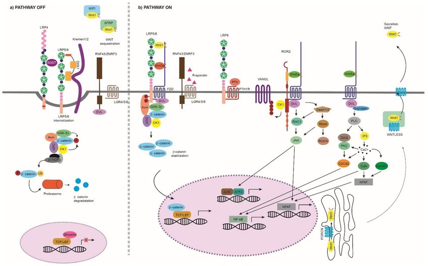
Genes 2022, 13, 138 2 of 52
inhibitors that block the LRP5/6 binding (e.g., sclerostin and DKK1) [12,13]. In addition to
the canonical pathway, Wnt ligands can activate multiple signaling cascades independent
of β-catenin. Within this category, the most widely studied are the WNT-planar cell polarity
(WNT-PCP) and the WNT-calcium (WNT-Ca2+ ) pathways (Figure 1) [14–17], which can
branch out into the JNK, PI3K-RAC1 and mTORC1/2 pathways, among others [18–22]. The
WNT-PCP pathway begins with the binding of a Wnt ligand, such as WNT5A, to FZD and
to ROR1/2 or RYK co-receptors. This binding activates small G proteins, such as Rac and
Rho, which are implicated in the establishment of cell polarity and cell migration [23]. In
addition, the activation of these pathways may inhibit the canonical Wnt pathway [24,25].
The WNT-Ca2+ pathway regulates calcium release from the endoplasmic reticulum in order
to control intracellular calcium levels. It begins with the binding of a Wnt ligand, such
as WNT5A, to FZD, which in turn activates the phospholipase C (PLC). Cell adhesion,
migration and embryonic development are regulated through this pathway [26].
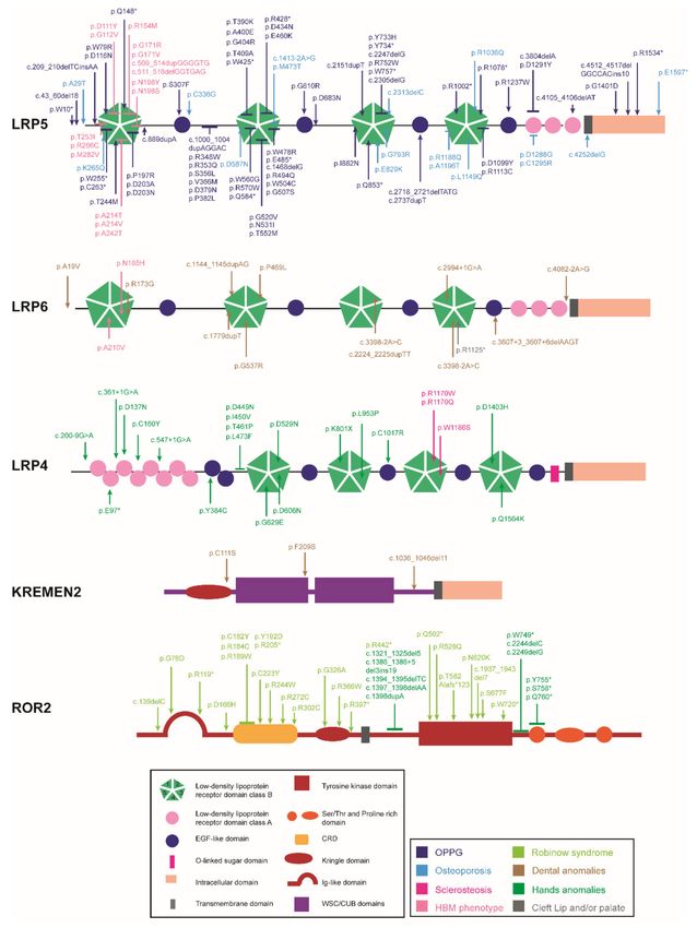
Figure 1. Wnt pathway. (a) Wnt pathway OFF: The Wnt pathway is inactive in the absence of Wnt
ligands or by the effect of extracellular inhibitors that prevent the activation of the pathway. We
can group the extracellular inhibitors into two categories depending on the action they perform:
(i) binding to the Wnt ligands and preventing their binding to the membrane receptors, such as
SFRP1–5 and WIF-1, and (ii) interfering with the LRP binding to FZD, such as DKK1 and sclerostin.
Due to their mechanism of action, the first type of inhibitors can act on both canonical and non-
canonical pathways, while the second type is specific to the canonical Wnt pathway. The inhibition
performed by DKK1 and sclerostin is mediated or enhanced by receptors such as Kremen proteins
and LRP4 [27,28]. When the canonical pathway is not activated, β-catenin is phosphorylated at
three critical residues and sequestered in the cytoplasm by the destruction complex. The destruction
complex is formed by scaffold proteins APC and AXIN1; Ser/Thr kinases, CK1α, ε and δ and GSK3β;
and transcriptional regulators YAP/TAZ of the Hippo pathway [29]. This phosphorylated β-catenin
is polyubiquitinated for its degradation in the proteasome by the complex containing, among others,
β-TrCP protein. Thus, it is not available in the nucleus and, in its absence, a complex is formed by
TCF/LEF and TLE/Groucho, inhibiting the transcription of target genes. (b) Wnt pathway ON: The
Genes 2022, 13, 138                                                                                                    3 of 52

                      binding of a Wnt ligand to FZD/LRP or ROR1/2/RYK/FZD activates the Wnt pathway that will
                      result in a different transcriptional regulation. Canonical Wnt Pathway ON: The pathway begins at
                      the cell surface with the formation of a heterotrimeric complex consisting of a Wnt ligand (19 different
                      Wnts), a LRP transmembrane co-receptor (LRP5/6) and an FZD receptor (10 different FZDs). The
                      formation of the LRP-FZD-Wnt complex results in phosphorylation of the LRP5/6 co-receptor by
                      CK1α and GSK3β. Then, the DVL (also called DSH) polymerizes and is activated, inhibiting the
                      destruction complex. This produces stabilization and accumulation in the cytoplasm of β-catenin,
                      which will translocate to the nucleus. Once there, it displaces the TLE/Groucho repressors, forming
                      an active complex with TCF/LEF proteins, which results in the recruitment of coactivators and
                      activation of transcription of genes important for the differentiation and formation of bone, such
                      as WISP1 and RUNX2 [12,29]. Non-canonical Wnt Pathway ON: (i) WNT/PCP: The WNT-PCP
                      pathway begins with the binding of a Wnt ligand, such as WNT5A, to FZD and the co-receptors
                      ROR1/2 or RYK. Then, DVL is recruited and activated, resulting in the activation of the scaffold
                      protein VANGL. DVL forms a complex with DAMM1, which activates the small GTPases RHOA
                      and RAC1, which in turn activate ROCK and JNK. This leads to rearrangements of the cytoskeleton
                      and/or the induction of transcription through ATF2 and/or NFAT. The activation of the WNT5A-
                      ROR1/2 pathway inhibits the canonical Wnt signaling [24,25]. (ii) WNT/Ca2+ : The binding of the
                      ligand to an FZD receptor results in the recruitment and activation of DVL. Then, DVL binds to
                      the small GTPases, which activate PLC. This activation leads to the breakage of PIP2 into DAG and
                      IP3. When IP3 binds to its receptor on the endoplasmic reticulum, calcium release occurs. When
                      the calcium concentration is increased, DAG activates PKC, which in turn can activate CDC42.
                      Increased intracellular calcium can also activate calcineurin and CaMKII, which in turn can induce
                      the activation of the NFAT transcription factor or NF-kB [26,30]. WNT signaling modulators: The
                      binding of RSPO to LGR and to RNF43/ZNRF3 maintains the Wnt signal ON by preventing the
                      polyubiquitination and endocytosis of FZD performed by RNF43/ZNF3 [31]. WNT production: Wnt
                      precursors undergo post-translational modifications such as porcupine-mediated palmitoylation,
                      other lipid modifications and glycosylation in the ER. Then, the transmembrane protein Wntless (Wls)
                      transports the functional Wnt ligands to the plasma membrane via the golgi apparatus. Wnt ligands
                      are secreted from the cell by solubilization, exosome formation or on lipid protein particles [32–34].
                      See next figures for the information on the different protein domains. APC: adenomatous polyposis
                      coli; AXIN1: axis inhibition protein 1; β-TrCP: β-Transducin repeat-containing protein; CAMKII:
                      calcium/calmodulin-dependent protein kinase II; CDC42: cell division control protein 42; CK1:
                      casein kinase 1; DAAM1: DVL-associated activator of morphogenesis; DVL: disheveled: FZD:
                      frizzled; GSK3β: glycogen synthase kinase 3β; IP3, inositol 1,4,5 triphosphate, JNK: JUN kinase;
                      LGR: leucine-rich repeat-containing G-protein-coupled receptor; NFAT: nuclear factor of activated
                      T cells; NF-kB: nuclear factor kappa B; PKC: protein kinase C; PLC: phospholipase C; RAC: Ras-
                      related C3 botulinum substrate; RHOA: Ras homolog gene family member A; ROCK: Rho kinase;
                      ROR1/2: bind tyrosine kinase-like orphan receptor 1 or 2; RYK: receptor-like tyrosine kinase; PORCN:
                      porcupine; RNF43/ZNRF3: ring finger protein 43/zinc and ring finger 3; RSPO; R-spondin ligand
                      family members; SFRP: secreted frizzled-related proteins; TCF/LEF: T-cell factor/lymphoid enhancer
                      factor; TLE: Transducin-Like Enhancer of Split Proteins; VANGL: Van Gogh-like; YAP/TAZ: Yes-
                      associated protein/transcriptional co-activator with a PDZ-binding domain; WIF-1: Wnt inhibitory
                      factor 1.

                           Despite the fact that the Wnt pathways are some of the most studied signaling routes,
                      their complexity means that there are still many aspects to discover. To precisely regulate
                      the transcription of Wnt pathway target genes, complex and tight regulation is carried out
                      by extracellular components including ligands (19 Wnt ligands), inhibitors (12 different:
                      SFRPs, Dickkopf (DKKs), sclerostin, among others), co-receptors (LRP, ROR, among others)
                      and receptors (10 FZD). A current challenge is determining the tissue-specific and transient
                      modulators of this pathway—for instance, in bone tissue—which could be useful as new
                      therapeutic targets. Importantly, many rare and complex diseases are associated with genes
                      from members of the Wnt pathway (Table 1). Since treatments for bone density diseases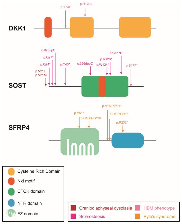
Genes 2022, 13, 138                                                                                                       4 of 52

                                 target genes that cause rare monogenic bone diseases, a comprehensive understanding of
                                 the causal variants and genes is fundamental.
                                      This review aims to describe, summarize and update the findings regarding all these
                                 extracellular modulators in relation to bone homeostasis, with an emphasis on the animal
                                 models generated, the diseases associated with each gene and the bone processes in which
                                 each member is involved. The review is structured into three main sections—co-receptors,
                                 ligands and inhibitors—within which we will review the main members of the Wnt pathway
                                 that have an important role in bone homeostasis.

                                 Table 1. Diseases or traits caused by mutations in Wnt pathway genes.

    Disease/Trait      OMIM                        Phenotype                      Gene      Inh.         Comment       Ref.
    Osteogenesis                       -   Low bone mass
                        615220                                                   WNT1      AR/AD            LoF       [35–37]
   imperfecta XV                       -   Increased bone fragility
   Osteoporosis-
                                       -   Congenital blindness
   pseudoglioma
                        259770         -   Severe juvenile-onset osteoporosis     LRP5       AR             LoF       [38,39]
     syndrome
                                       -   Spontaneous fractures
      (OPPG)
                        615221                                                   WNT1        AD             LoF        [35]
    Osteoporosis                   -       Low bone mass
                        166710                                                    LRP5       AD             LoF       [40–42]
                                       -   Massive generalized hyperostosis
 Craniodiaphyseal
                        122860         -   Sclerosis, especially involving the    SOST       AD           LoF/DN       [43]
    dysplasia
                                           skull and facial bones
                        269500     -       Progressive skeletal overgrowth        SOST       AR             LOF       [44–46]
    Sclerosteosis                  -       Syndactyly
                        614305                                                    LRP4     AD/AR            LOF       [47,48]
                                       -   Hyperostosis of the skull,
                                           mandible, clavicles, ribs and                                   * 52 kb
    van Buchem
                        239100             diaphyses of the long bones, and      SOST *      AR          downstream    [49]
      disease
                                           tubular bones of the hands                                      deletion
                                           and feet.
                                                                                  LRP5       AD             GoF       [50–52]
                        144750     -       High bone mass, affecting
       HBM                                 especially the skull and               LRP6       AD             GoF        [53]
    phenotype 1         607634
                        607636             tubular bones                          SOST       AR             LoF        [54]
                                   -       Low fracture risk                      DKK1       AD             LoF       [55,56]
                                   -       Dysmorphic facial features
                        268310                                                    ROR2       AR             LoF       [57–59]
                                   -       Hypertelorism
       Robinow
                                   -       Short-limbed dwarfism
      syndrome
                        180700     -       Vertebral segmentation                WNT5A       AD             LoF        [60]
                                   -       Short stature, genital hypoplasia
                                       -   Broadening of the long bones with
                                           wide and expanded trabecular
                                           metaphyses
                                       -   Cortical thinning
  Pyle’s syndrome       265900                                                   SFRP4       AR             LoF        [61]
                                       -   Bone fragility and fractures
                                       -   Genu valgum
                                       -   Widening of the ribs and clavicles
                                       -   Platyspondyly
                        616724                                                    LRP6       AD             LoF       [62,63]
   Selective tooth
  agenesis, types 7,    150400     -       Absence of one or more teeth          WNT10A    AD/AR            LoF       [64,65]
       4 and 8          617073                                                   WNT10B      AD             LoF        [66]
 Hand/foot anomalies:
    Cenani–Lenz                        -   Complex syndactyly of the hands
     syndactyly         212780         -   Bone malformations in the              LRP4       AR             LoF        [67]
     syndrome                              forearm and lower extremities
Genes 2022, 13, 138 5 of 52
Table 1. Cont.
Disease/Trait OMIM Phenotype Gene Inh. Comment Ref.
Isolated bilateral
- Fusion of two or more fingers LRP4 AR LoF [68,69]
syndactyly
Brachydactyly
113000 - Disproportionately short fingers ROR2 AD GoF [70,71]
type B1
- Syndactyly
Split hand/foot - Median clefts of the hands
malformation, and feet
225300 WNT10B AR LoF [72]
isolated form, - Aplasia and/or hypoplasia of the
type 6 phalanges, metacarpals
and metatarsals
Ectodermal dysplasia:
Ectodermal - Severe oligodontia
617392 KRM1 AR [73,74]
dysplasia 13 - Anomalies of hair and skin
Al-Awadi-Raas-
- Severe limbs malformations
Rothschild
276820 - Severely hypoplastic pelvis WNT7A AR complete LOF [75]
syndrome
- Abnormal genitalia
(AARRS)
- Bowing of the femurs
Furhman - Hypoplasia of the fibula, pelvis,
228930 WNT7A AR partial LOF [75]
syndrome fingers and fingernails
- Cleft lip and palate
- Fibular agenesis or hypoplasia
- Clubfeet with oligodactyly
- Acromial dimples
Santos syndrome 613005 - Motion limitations of the forearms WNT7A LoF [76]
and/or hands
- Severe nail hypoplasia
or anonychia
- Multiple eyelid apocrine
hidrocystomas
Schöpf–Schulz–
- Palmoplantar keratoderma
Passarge 224750 WNT10A AR LoF [77]
- Hypotrichosis
syndrome
- Hypodontia
- Nail dystrophy
- Hyperkeratosis and hyperhidrosis
of the palms and soles
Odonto-onycho-
- Atrophic malar patches
dermal dysplasia 257980 WNT10A AD/AR LoF [78]
- Hypodontia and conical teeth
(OODD)
- Onychodysplasia
- Dry sparse hair
Skeletal dysplasias:
- Osteopenia
- Bilateral coxa valga deformity
Other skeletal - Mild left radial and ulnar bowing
WNT3A AR [79]
dysplasias - Broadening of metaphyses
- Bilateral shortening of the great
toes and thumbs
Inh: Inheritance; Ref: References; AR: Autosomal recessive; AD: Autosomal dominant; DN: Dominant neg-
ative; LoF: Loss-of-function; GoF: Gain-of-function; 1 also known as: Osteosclerosis; Endosteal hyperostosis;
Osteopetrosis type 1, van Buchem disease type 2. *: A 52 kb deletion at 35 kb downstream of the SOST gene.
2. Co-Receptors
We define a co-receptor as a cell surface receptor that binds to both a signaling molecule
and to a primary receptor, thereby facilitating ligand recognition and initiation of down-
stream biological processes. Different co-receptors play essential roles in Wnt signaling,Genes 2022, 13, 138 6 of 52
both in the canonical and non-canonical pathways. This section includes a subsection
entitled LRPs, with information related to LRP4/5/6; a second subsection entitled Kremen,
which includes information on Kremen1/2; a third subsection, ROR, which includes infor-
mation related to ROR1/2, and a final subsection, LGR, which includes information related
to LGR4/5/6. We review the role of these co-receptors in bone remodeling, with a special
emphasis on mutations causing disease (Figure 2) and on the animal models available
(Table 2).
Figure 2. Domain structure of the co-receptors and localization of the mutations causing human
skeletal diseases according to human gene mutation database (HGMD). ↓ indicates a point mutation,
⊥ encompasses more than one aminoacid position (a cluster of point mutations). *: STOP codon.Genes 2022, 13, 138 7 of 52
Table 2. Mouse models of Wnt pathway co-receptor mutations.
Tissue Phenotype
Gene Model Expression Comments Refs.
BM BF BR
KO Total KO ↓ ↓ ↓/= I I ↓ in [80]; =in [81,82] [80–86]
cKO-Dermo1-Cre EM = = - [86]
cKO-Ocn-Cre mature OB ↓ ↓ = [87]
cKO-α1(I)-Col-Cre OB = = - [82]
cKO Rat 3.6Col1a1-Cre OB ↓ - - [84]
cKO-Dmp1-Cre OCy ↓ - - [88]
cKO-Vil1-Cre ISC ↓/= II ↓/= II - II ↓ in [82]; =in [88] [82,88]
CD and OB
cKO-Col21-Cre ↓ - - [89]
progenitors
KI-A214V or G171V Total KI ↑ ↑ = [88]
cKI-Dmp1-Cre A214V or
Lrp5 OCy ↑ ↑ = [88]
G171V
cKI-Prrx1-Cre A214V or
MSC ↑ ↑ = [88]
G171V
cKI-Vil1-Cre A214V ISC = = = [88]
cKI-Vil1-Cre G171V ISC ↑/= III ↑/= III = III ↑ in [82]; =in [88] [82,88]
cKI-1(I)-Col-Cre G171V OB = = = [82]
Tg Rat3.6Col1a1G171V OB ↑ [90]
Offtargets in Ocy
cKI-Ctsk-Cre A214V or IV line
mature OC ↑ ↑ ↓ IV Only in female [91]
G171V
mice
KO Total KO - - - NV [83,86,92]
cKO-Dermo1-Cre EM = = = [86]
Ringelschwanz ↓ = ↑ p.R868W mutation [93,94]
Lrp6
cKO-Ocn-Cre mature OB ↓ ↓ = [87]
CD and OB
cKO-Col2a1-Cre ↓ - - [89]
progenitors
KO-Lrp5 and Het Severe limb
Total KO ↓ - - [83]
KO-Lrp6 abnormalities
cKO-Dermo-Cre EM ↓ ↓ - NV [86]
Lrp5 and cKO-Ocn-Cre mature OB ↓ ↓ = NV [87]
Lrp6 CD and OB
cKO-Col2a1-Cre ↓ - - NV [89]
progenitors
cKO-Rank-Cre OC precursors ↓ ↓ ↓ [95]
cKO-Ctsk-Cre mature OC = = = [95]
KO Total KO NV [96–98]
ECD ↓ ↑ ↑ Only ECD domain [99]
cKO-OCN-Cre mature OB ↑ ↑ ↓ [96,100]
Lrp4 cKO-Dermo1-Cre OCy ↑ ↑ ↓ [100]
cKO-LysM-Cre Myeloid cells = = = [96]
KI-Lrp4-R1170W Total KI ↑ ↑ = [101]
KI-Lrp4-R1170Q Total KI ↑ ↑ = [102]
KO Total KO - - - NV [8,103–105]
Het KO Het KO ↑ = ↓ [103]
Ror2 cKO-Rank-Cre OC ↑ = ↓ [103]
Only in trabecular [103,106,
cKO-Ctsk-Cre mature OC ↑ = ↓
bone 107]
Krm1 KO Total KO = = - [108]
KO Total KO = = - In young mice [108]
Krm2 KO Total KO ↑ ↑ - In adult mice [109]
Tg Col1a1-Krm2 OB ↓ ↓ ↑ [109]
Krm1 and
KO Total KO ↑ ↑ = [108]
Krm2Genes 2022, 13, 138 8 of 52
Table 2. Cont.
Tissue Phenotype
Gene Model Expression Comments Refs.
BM BF BR
KO Total KO ↓ ↓/= V ↑ V ↓ [110,111]; = [112] [110–113]
Lgr4
cKO-LysM Myeloid cells ↓ = ↑ [112]
Lgr5 KO Total KO NV [114]
Lgr6 KO Total KO = = = [115,116]
Ref: references; BM: bone mass; BF: bone formation BR: bone resorption; KO: knock-out; cKO: conditional KO;
het KO: heterozygous KO; KI: knock-in; cKI: conditional KI; Tg: Transgenic; EM: embryonic mesenchyme; OB:
osteoblast; OC: osteoclast; Ocy: osteocyte; CD: chondrocyte; ISC: intestinal stem cells; NV: not viable. Roman
numerals: references to specific traits. ↑ increase; ↓ decrease; = not affected; - not available.
2.1. LRPs
The LRP proteins belong to the low-density lipoprotein receptor (LDLR) family of
highly conserved cell surface receptors [117], whose functions include endocytosis (e.g.,
lipoproteins or proteases) and direct signal transduction (e.g., bone morphogenic protein
(BMP) or Wnt pathways) [118,119]. These transmembrane proteins have a large extracel-
lular domain (ECD) and a small intracellular domain (ICD) separated by a single-pass
transmembrane domain. The ECD allows interaction with extracellular proteins (e.g., Wnt
ligands or extracellular inhibitors such as DKK1 or sclerostin), and the ICD is in charge of
the downstream transmission of signaling events [120].
The most studied LRP family members in relation to bone are LRP5 and LRP6, which
share 71% homology at the protein level [121]. Their ECDs consist of four β-propeller
domains (class B repeats or YWTD domains) alternating with four epithelial growth factor
(EGF)-like domains followed by three LDLa domains (class A repeats). Their ICDs include
various highly conserved PPPS/TP motifs containing serines and threonines that are
phosphorylated upon receptor activation (Figure 2). These phosphorylated sites bind Axin,
thereby preventing the phosphorylation of β-catenin [7,122,123]. LRP4, also known as
MEGF7, has a structure similar to that of LRP5/6 but with some differences. The ECD
features eight LDLa domains, four β-propeller domains alternating with six EGF-like
repeats and a domain for O-linked oligosaccharide modification. The ICD features two
domains: the endocytosis signal NPxY domain responsible for protein internalization, and
the PZD-interacting motif at the C-terminal end (Figure 2) [124–126].
The importance of these transmembrane co-receptors in bone was revealed by the
description of mutations in LRP5 that caused two diametrically opposed bone phenotypes:
osteoporosis pseudoglioma syndrome (OPPG), and the high bone mass (HBM) pheno-
type [38,50,51] (Table 1 and Figure 2). The difference between the two types of mutations
is that OPPG results from loss-of-function (LoF) mutations causing LRP5 to be unable to
activate the canonical Wnt pathway. In contrast, the mutations responsible for the HBM
phenotype are gain-of-function (GoF) and cause a loss of affinity for the extracellular in-
hibitors DKK1 and sclerostin. In this case, LRP5 can no longer be internalized and remains
available in the membrane for Wnt pathway activation [39,127–132]. In addition, the LRP5
locus has been associated with bone mineral density (BMD) and risk of fracture in a plethora
of genome-wide association studies (GWAS) [133–143].
To understand the effect of LRP5 on bone and the signaling pathways by which
it acts, genetically modified mouse models have been generated (Table 2). Lrp5 total
knock-out (Lrp5-KO) mouse models and conditional KOs (cKOs) in bone-related cells
do not display bone alterations at birth but acquire a decreased BMD during postnatal
development due to reduced bone formation [80,81,83–89] (Table 2). On the contrary,
global or conditional LRP5 transgenic mice carrying the human LRP5 HBM p.Gly171Val or
p.Ala214Val mutations reproduce the HBM phenotype with high rates of bone formation
and better bone quality [88,90] (Table 2). Furthermore, conditional knock-in (cKI) female
mice with the HBM mutations present only in osteoclasts show a reduction in bone re-
sorption, demonstrating that the in vivo effect of the HBM mutations is not only due toGenes 2022, 13, 138 9 of 52
osteoblast stimulation but also to osteoclast inhibition [91] (Table 2). Relative to skeletal
mechano-responsiveness, Lrp5-KO and Lrp5-cKO mice have been reported to show se-
vere impairment in later stages of the mechano-transduction signaling cascade, whereas
HBM mice show a higher osteogenic response to mechanical loading in vivo than WT
mice [84,144,145]. In 2008, Yadav et al. proposed that the effect of LRP5 on bone formation
and BMD was due to its inhibitory effect on tryptophan hydroxylase 1 and, therefore, on
serotonin synthesis in the duodenum, which would otherwise block bone formation by
signaling in osteoblasts [82,146,147]. However, these hypotheses have not been reproduced,
and there is much evidence that LRP5 exerts its effect directly on osteoblastic cells through
the Wnt canonical pathway [86–88,148,149].
Similar to LRP5, there is much evidence of a role of LRP6 in bone. Missense mutations
in LRP6 have been described in humans as the cause of tooth agenesis and oligodontia and,
recently, HBM [53,62,63] (Table 1 and Figure 2). Interestingly, the mutations causing the
HBM phenotype are located exclusively in the first β-propeller domain, and the phenotype
of these patients is identical to that of the patients with LRP5-HBM. Moreover, LRP6 has
been associated with heel BMD in three different studies [140–142].
The Lrp6-KO mouse model is embryonic lethal with loss of distal limb structures
and truncation of the axial skeleton, indicating that Lrp6 plays a fundamental role during
embryogenesis, during which it is widely expressed [83,86,92] (Table 2). Confirmation
that Lrp6 is also involved in postnatal bone metabolism was obtained using cKO mouse
models and models with hypomorphic mutations [87,89,93,94] (Table 2). Specifically, the
Ringelshwanz (rs) mouse with the p.R886W hypomorphic mutation in the third β-propeller
domain showed developmental malformations and delayed ossification, in addition to
an increase in bone resorption and a decrease in BMD and in the activity of the canonical
Wnt pathway in osteoblasts [93,94] (Table 2). Through different in vitro models, it was
determined that this effect was a consequence of the impaired interaction of mutant Lrp6
with the chaperone Mesd and its defective trafficking to the plasma membrane [93,94].
Lrp6-cKO mice in the early osteoblast lineage showed reduced BMD due to decreased bone
formation [87,89]. In contrast, there was no obvious bone phenotype for Lrp6-cKO in the
embryonic mesenchyme [86] (Table 2).
The double KO and cKO for both co-receptors (Lrp5 + Lrp6-dKO and Lrp5 + Lrp6-
cdKO) show a more severe phenotype than the single KOs and cKOs regarding BMD and
skeletal development during embryogenesis (Table 2). Many authors agree that there is
some redundancy in the functions of these co-receptors, but that they may regulate the
Wnt pathway in different time windows (Lrp6 in early stages and Lrp5 in the late stages
of osteoblast differentiation) and in different skeletal compartments (Lrp6 in trabecular
bone and Lrp5 in cortical bone) to promote the proper acquisition of BMD. Therefore, the
expression of the two co-receptors in osteoblasts would be necessary for normal skeletal
homeostasis [83,86,87,89,150] (Table 2). More research is needed to better define the tissue
distribution and the time at which these two co-receptors act. In this context, a study of
Lrp5 + Lrp6-cKO at early and late stages of osteoclast differentiation showed that there was
a low BMD with reduced osteoblast and osteoclast numbers in the early osteoclast model,
while the late osteoclast model did not have a bone phenotype [95] (Table 2).
The characterization of all these animal models indicates that Lrp5 and Lrp6 exert their
function in bone homeostasis mostly through the regulation of bone formation, controlling
the differentiation and proliferation of osteoblasts, bone matrix deposition in differentiated
osteoblasts and apoptosis of osteocytes. Furthermore, these effects are mainly due to
Wnt signaling through the canonical pathway. It has been determined that LRP6 acts
not only through the formation of the heterotrimeric complex LRP6-FZD-Wnt, but also
through the formation of a complex involving LRP6, parathyroid hormone (PTH) and PTH
receptor 1, which promotes the stabilization of β-catenin and thus activates the canonical
Wnt pathway [151].
LRP4 has been extensively studied in relation to its role in the neuromuscular junc-
tion, where it serves as a co-receptor for motor neuron-derived agrin/Wnt and facilitatesGenes 2022, 13, 138 10 of 52
acetylcholine receptor clustering on the myocyte [152]. In addition to this function, LRP4
has a very important role in bone homeostasis, where it enhances the inhibitory function of
sclerostin in the canonical Wnt pathway [47,96,99,100,153]. LRP4 can also enhance other
inhibitors of the Wnt pathway, such as DKK1 and SOSTdc1 [99,154].
Missense and splicing mutations in LRP4 have been implicated in several muscu-
loskeletal human diseases depending on the position of the mutation (Table 1 and Figure 2).
Mutations in the LDLa, EGF-like domain or in the first and second β-propeller domains
cause Cenani–Lenz Syndrome (CLS) [67]; mutations in the central cavity of the third β-
propeller domain cause sclerosteosis-2 [47,48]; and those in the fourth β-propeller domain
or in the EGF-like domain cause isolated bilateral syndactyly [68,69]. Thanks to in vitro
functional studies, it has been proven that all these mutations cause a significant decrease
in the inhibition of the canonical Wnt pathway by LRP4, this effect being more severe in
mutations associated with sclerosteosis-2. CLS-causing mutations decrease the levels of
LRP4 available in the membrane, while this is not observed for the sclerosteosis-causing
mutations. Instead, impaired binding between sclerostin and LRP4 has been verified in the
mutations causing sclerosteosis, which would explain the severity of the effect [48,67,68].
Interestingly, mutations in the edge of the third β-propeller domain have been found to
cause myasthenia gravis, the most common neuromuscular junction pathology. These
mutations cause impairment of the MuSK signaling pathway, whereas sclerosteosis-2 mu-
tations have no effect on this pathway, demonstrating the highly position-specific effect of
the different LRP4 mutations [155]. Furthermore, as with LRP5/6, LRP4 has been associated
with BMD in different GWAS studies [136,137,140–142].
Lrp4-KO or null mutant mouse models are not viable due to the absence of neuromus-
cular junctions and therefore the inability to breathe [96–98] (Table 2). In contrast, Lrp4-cKO
mouse models in the osteo lineage are viable. When the deletion occurs specifically in
osteoblasts or in osteocytes, a high BMD is observed, with high levels of bone formation
and reduced bone resorption but without polysyndactyly [96,100] (Table 2). The phenotype
is more severe in early osteoblasts, where Lrp4 is more highly expressed, than in osteo-
cytes [100]. In contrast, no bone phenotype is observed in osteoclast-specific KO mice [96]
(Table 2). The presence of Lrp4 has been shown to be essential in these animal models for
the functioning of sclerostin [96]. In 2017, Xiong et al. [156] proposed that reduced bone
resorption in Lrp4-cKO in the osteoblast lineage stabilizes the prorenin receptor and, as a
result, increases the production and secretion of the ATP derivative adenosine. Elevated
adenosine-A2aR signaling in osteoclast precursors reduces receptor activator of nuclear
factor kB (RANK)-mediated osteoclastogenesis. Furthermore, circulating sclerostin levels
are higher in Lrp4-cKO in the osteoblast lineage with unchanged expression of SOST
per cell, but it is possible that increasing the number of osteocytes with increasing bone
formation will also increase the total level of SOST. In addition to these models, LRP4-KI
mouse models with the p.R1170W or p.R1170Q mutations, which cause sclerosteosis in
humans, recapitulate the human HBM phenotype associated with this mutation without
the presence of syndactyly (Table 2). In addition to high BMD, these mice exhibit high levels
of bone formation with unchanged bone resorption and are protected against the effects of
anti-sclerostin antibodies and of SOST transgenic overexpression [101,102]. Polysyndactyly
and other limb deformity problems have been reported in different murine and bovine
hypomorphic models of Lrp4 [98,99,153,154,157–159]. Specifically, the Lrp4 ECD mouse
model that does not have the transmembrane or intracellular domains has polysyndactyly
and decreased BMD [99] (Table 2).
All these studies on animal models, together with many in vitro studies, seem to
indicate that Lrp4 exerts its function on the differentiation/function of osteoblasts through
the inhibition of the Wnt-induced activation of the canonical pathway.
2.2. ROR
ROR1/2 belong to the evolutionarily conserved Ror family of receptor tyrosine kinases,
which are type I transmembrane protein tyrosine kinases. These receptors contain differentGenes 2022, 13, 138 11 of 52
motifs, including an immunoglobulin-like domain, a frizzled-like cysteine rich domain
(CRD) and a kringle domain (KD) in the extracellular region, a transmembrane section and
an intracellular region with a tyrosine kinase domain, two serine/threonine-rich domains,
a proline-rich domain and a short C-terminus tail [160–162] (Figure 2). There is some
controversy about the activity of these receptors, regarding whether they are true kinases
or pseudokinases [163]. ROR1 and ROR2 are structurally very similar, with 58% amino acid
identity and high domain similarity. Both bind WNT5A through their CRD domains, and,
by acting as receptors or co-receptors along with FZD, they activate the non-canonical Wnt
signaling pathway [8,163]. The Ror1 + Ror2-dKO mouse model revealed that these two co-
receptors possess certain redundant functions in skeletal and cardiac development [164,165].
Ror1/2 are widely expressed in various tissues and organs, such as the limbs, heart, lungs,
gut, muscles and skeletal and nervous systems, during embryonic development, while their
expression decreases markedly in adult tissues [163]. While Ror2 is strongly expressed in
mature osteoclasts, Ror1 expression has not been detected in these cells [107]. In addition,
Ror2 has been reported to regulate the length of various organs, including limbs, through
interaction with both Wnt5a and Wnt9a [163,166].
In humans, recessive LoF mutations in ROR2 are associated with autosomal recessive
Robinow syndrome, and dominant GoF mutations are associated with brachydactyly type
B [57,58,70,107,160] (Table 1 and Figure 2). Moreover, ROR2 has been associated with hip
BMD in a GWAS study [164].
Ror2-KO mouse models show early lethality and abnormalities characteristic of Robi-
now patients. The Ror2-KO mice exhibit dwarfism, facial abnormalities, short and de-
formed limbs and tails, dysplasia of lungs and genitals, ventricular septal defects and
severe cyanosis [8,103–105] (Table 2). In these mice, the bones formed through endochon-
dral ossification are affected, but not those formed by intramembranous ossification. The
heterozygous Ror2-KO mouse and the Ror2-cKO in the osteoclast lineage exhibited a
HBM phenotype due to impaired bone resorption only in trabecular bone [103,106,107]
(Table 2). The impaired bone resorbing activities of osteoclasts can be restored by the con-
stitutively active form of RhoA, showing that this impaired bone resorption is an effect of
the non-canonical Ror2-Wnt5a signaling pathway [103,106,107,167]. Thus, Ror-2-mediated
signaling regulates the differentiation and bone resorbing activity of osteoclasts, thereby
maintaining bone mass.
2.3. KREMEN
The Kremen family of receptors (KRM1/2) are single-pass transmembrane proteins
that show high affinity for DKK1 and DKK2 [168,169]. KRM presents an ECD that contains
a KD, a carbohydrate binding (WSC) and a CUB domain that are necessary for binding
to DKK [168,170] (Figure 2). KRM1/2 can bind to the CRD2 domain of DKK1 and form
the DKK1-KRM-LRP5/6 ternary complex, which is internalized, thus removing LRP5/6
from the cell surface [171,172] (Figure 1). Similar to LRP4, which enhances sclerostin
inhibitory activity, Kremen acts by enhancing DKK1 function, but unlike LRP4, Kremen is
not essential for DKK1 to perform its function [168]. While Krm1 has a more widespread
expression pattern, Krm2 is expressed predominantly in bone [109]. In addition to this
effect, Krm1/2 can activate the Wnt pathway by binding directly to Lrp6 in the absence of
Dkk1 [109,173,174].
In humans, mutations in KRM1 affect different protein domains and have been associ-
ated with ectodermal dysplasia and oligodontia [73,74] (Table 1 and Figure 2).
Krm1-KO mice and young Krm2-KO mice (less than 24 weeks old) are viable with-
out abnormalities and show normal bone formation and bone mass [108], while adult
(24 weeks) Krm2-KO mice display higher bone formation and bone mass [108,109] (Table 2).
Krm1 + Krm2-dKO are also viable, but show ectopic postaxial forelimb and expanded
apical ectodermal ridges and increased bone formation, bone mass and canonical Wnt
signaling [108] (Table 2). The dKO mouse phenotype shows similar defects in the limb to
those described in the heterozygous Dkk1-KO mutants (see Section 4). In addition, theGenes 2022, 13, 138 12 of 52
ectopic growth of digits present in the Krm1 + Krm2-dKO mice is further enhanced by
the deletion of a single Dkk1 allele, but without any additional increase in BMD [108]. In
contrast, mice with osteoblast-specific Krm2 overexpression display a severe osteoporotic
phenotype caused by decreased bone formation and increased bone resorption with im-
paired fracture healing [109,175] (Table 2). Taking all these models together, it seems that
Krm1 and Krm2 show a certain degree of redundancy in skeletal growth and development
and that Krm2 is important for bone formation during skeletal maintenance/remodeling.
2.4. LGR
The leucine-rich repeat-containing G-protein-coupled receptors (LGRs) 4, 5 and 6 con-
stitute a subfamily of receptors belonging to the G-protein-coupled receptor (GPCR) super-
family [176]. Structurally, these transmembrane GPCRs belong to the class A rhodopsin-like
family [177], which have a characteristic leucine-rich domain (LRR) in the extracellular N-
terminal region that is responsible for ligand interaction [177–180]. All three receptors show
some homology, but while LGR4/5 have 17 LRR repeats, LGR6 only has 13 [179]. These re-
ceptors bind to R-spondins (RSPOs), causing the formation of the LGR-RSPO-ZNRF/RNF43
complex. This leads to the sequestration of ZNF/RNF43, which causes an increase in
membrane-available FDZs, since the transmembrane ubiquitin ligases ZNF/RNF43 are
responsible for degrading them [31]. In addition to this agonistic effect on the Wnt pathway,
LGR is a target gene of the Wnt pathway, which will produce a positive feedback loop [31].
It has also been reported that LGR proteins mark different adult stem cells and play a
very important role in defining progenitor and stem cell behavior [31,181,182]. Specifically,
it has been reported that LGR5 is a marker of adult stem cells in the stomach [183], hair
follicles [184] and small intestine and colon [181], among others, and LGR6 in the taste
buds, lungs and skin [115,181,183,185].
LGR4, also known as GPR48, is widely expressed in multiple tissues from early
embryogenesis to adulthood [186], being abundantly expressed in skeletal, adipose and
muscular tissue [110,113,178,187]. Specifically, expression has been observed in osteoblast
precursors in in vitro osteogenesis, and in osteoclasts in response to RANK ligand (RANKL)-
induced osteoclast differentiation. In addition to the Wnt pathway enhancement, LGR reg-
ulates a multitude of pathways through its classical G-protein signaling potential [110,188].
In one of these pathways, LGR4 acts as a secondary receptor for RANKL, competing with
RANK. Furthermore, this LGR4-RANKL binding is inhibited by both osteoprotegerin
(OPG) and RSPO1. Through in vitro and in vivo studies, LGR4 has been shown to inhibit
RANKL-induced osteoclast differentiation, survival and function [112].
No rare human diseases associated with LGR4 have been found, but a rare nonsense
variant has been linked to osteoporosis in two different association studies [189,190] and
common variants are associated with heel BMD [140,142]. Total Lgr4-KO mice are at risk of
perinatal death, with approximately 60% of newborn mice dying before day 1. These mice
exhibit decreased body weight, size and bone length [110–113] (Table 2). Lgr4-KO mice
have a low BMD due to both impaired bone formation and bone resorption. Regarding
bone formation, these mice present a dramatic delay in osteoblast differentiation and
mineralization during embryonic bone formation, and, in postnatal bone remodeling, there
is a decrease in the kinetic indices of bone formation rate and osteoid formation [110,111].
During bone resorption, they present osteoclast hyperactivation with an increase in the
number of osteoclasts, surface area, size and bone erosion [110,112] (Table 2), a phenotype
that is also observed in Lgr4-cKO mice in monocytes [112] (Table 2). Interestingly, the
injection of a soluble form of the Lgr4 extracellular domain abrogated RANKL-induced
bone loss in three mouse models of osteoporosis [112]. Thanks to in vitro and in vivo
studies, it has been verified that RSPO signaling has no effect on osteoclast differentiation
and that the Lgr4 effect in this cell type is determined by its signaling via RANKL [112,191].
In addition to the evidence from in vivo studies, many in vitro studies have been carried
out demonstrating the positive effect of LGR4 on osteogenic differentiation [111,192,193],
adipocyte and myocyte differentiation [111]. Although LGR4 is expressed within bone-Genes 2022, 13, 138 13 of 52
marrow-derived mesenchymal stem cells (BMSCs) undergoing osteogenic differentiation,
its expression is not correlated with any specific osteogenic marker but is maintained
throughout the process, suggesting that LGR4 may be necessary to support osteogenic
differentiation [194].
LGR5 is also known as GPR49, HG38 and FEX, and is expressed in osteoblast precur-
sors in osteogenesis in vitro. Lgr5-KO mice show neonatal lethality (during the first 24 h of
life) associated with ankyloglossia, characterized by the fusion of the tongue to the floor of
the mouth, leading to the inability to nurse and subsequent neonatal mortality. Perinatal
lethality is due partly to respiratory failure as the result of pressure against the diaphragm
from the markedly distended abdomen [114] (Table 2). This phenotype suggests the in-
volvement of LGR5 in craniofacial development. Regarding LGR6, as with LGR4, a rare
variant associated with the risk of postmenopausal osteoporosis has been described [189],
along with common variants associated with heel BMD in a GWAS study [140]. It is ex-
pressed in mouse primary calvarial cells, bone marrow cells and BMSCs [194]. In vitro
studies have highlighted the importance of LGR6 in promoting osteogenic differentiation,
correlating its expression with typical markers of osteogenic differentiation, such as os-
terix [194,195]. These results point to LGR6 as a novel marker of osteoprogenitor cells in
bone marrow [194]. A recent study shows that the enhancement of the Wnt pathway by
LGR6 occurs downstream of the BMP signaling pathway [196]. Despite this, Lgr6-KO mice
develop normally and no skeletal phenotype is evident, although Lgr6 is necessary for the
regeneration of the digit tip bone following amputation in mice [115,116] (Table 2).
3. WNT Ligands
Wnt ligands are highly conserved, secreted glycoproteins with a molecular weight
around 40 KDa. They are formed by 350–400 amino acids, 23–24 of which are conserved
cysteine residues. For secretion of an active Wnt ligand, Porcn-mediated lipidation by
palmitic acid is necessary and it occurs on a conserved serine residue (Figure 3). Following
lipidation, secretion into the extracellular matrix requires Wls binding [32,33,197]. However,
some authors have questioned the essential nature of this step [33,198,199]. Although the
degree of sequence identity between some Wnt family members is only 18%, it is thought
that all Wnt proteins form a similar three-dimensional structure, which is shaped similarly
to a fist with a cleft inside and thumb and index finger protrusions [197,198,200]. There
are four main regions of concentrated amino acid conservation that map onto the Wnt
structure: the tips of the thumb and index finger loops, the core of the N-terminal α-helical
domain (NTD) and a large continuous patch in between, facing the exterior. The two
main contact points between the Wnt ligand and the FZD receptor are the tips of the
thumb and index loops [197,198]. The cysteine-rich domain (CRD) at the second point
of contact with the receptor is similar to that of SFRP and ROR1/2, among others [201].
On the NTD, the lipidation of the conserved serine creates hydrophobic forces with the
receptor that, combined with shape complementarity, facilitate binding [198]. The other
conserved regions of the Wnt proteins, located opposite the FZD binding region, bind
to co-receptors such as LRP5/6/RYK [197,198,202]. The amino acid differences between
Wnt ligands enable their differential binding to receptors. For example, WNT1 binds to
the first β-propeller domain of LPR5/6, whereas WNT3A binds to the third β-propeller
domain [203]. It has been suggested that the activation of one or another Wnt pathway
relies on the available receptors [204,205], while other studies have suggested that the
selection of the pathway depends on the amount of Wnt ligand available [5,206]. A total
of 19 Wnt ligands have been identified in humans. Of these, WNT1, WNT3A, WNT4,
WNT5A, WNT5B, WNT7A, WNT7B, WNT9A, WNT10A, WNT10B and WNT16 have been
extensively studied in relation to their role in bone, both in its function, in those mutations
associated with human diseases (Figure 3), and in the animal models generated (Table 3).Genes 2022, 13, 138 14 of 52
Figure 3. Schematic protein representation of the Wnt ligands with the mutations causing human
skeletal diseases according to HGMD. ↓ indicates a point mutation, ⊥ encompasses more than one
aminoacid position (a cluster of point mutations). Numbers below the structure show the amino acid
position in the peptide before post-translational modification. *: STOP codon.Genes 2022, 13, 138 15 of 52
Table 3. Mouse models of Wnt pathway ligand mutations.
Tissue Phenotype
Gene Model Expression Comments Refs.
BM BF BR
KO Total KO - - - NV [207,208]
Het KO Total Het KO ↓ = = Mild osteopenia [207]
Sway Hypomorphic ↓ ↓ = G565del [209,210]
cKO-Prrx1-Cre MSC ↓ ↓ ↑ [207]
cKO-Runx2-Cre OB ↓ - - [211]
Wnt1 cKO-Dmp1-Cre late OB and Ocy ↓ ↓ - [212]
Monocytic
cKO-Lyz2-Cre lineage (OC = - - [211]
included)
Tg-Col1a1-tTA OB, inducible ↑ ↑ = [211]
Ov. in late OB
Dmp1-Cre-R26 het Wnt1 ↑ ↑ = HBM phenotype [212]
and Ocy
OB-targeted
Wnt1 and Tg-Wnt1; inducible Ov. of Similar phenotype to
↑ ↑ = [211]
Lrp5 Lrp5-KO Wnt1 in LRP5+/+
LRP5-KO
Wnt3a KO Total KO - - - NV [213,214]
KO Total KO - - - NV [215]
Wnt4 cKO-Prrx1-Cre MSC ↑ I - - I femoral, in females [216]
Tg-Col2.3kb-Wnt4 OB ↑ ↑ ↓ [217]
Wnt4 and Ectopic cartilage
dKO Total dKO ↓ - - [218]
Wnt9a formation
KO Total KO - - - NV [219,220]
Wnt5a Increased
Het Wnt5a Total Het KO ↓ ↓ ↓ [103,221]
adipogenesis
cKO-Osx-Cre OB ↓ ↓ ↓ [103]
Wnt5b KO Total KO - - - [222]
Abnormal limb
Wnt7a KO Total KO - - - [223]
development
KO Total KO - - - NV [224]
Skeletal
cKO-Dermo1-Cre ↓ - - [18]
progenitors
Little bone marrow
Tg-OSX-Cre;R26 Ov. in preOB ↑ ↑ = [225]
space
Wnt7b Little bone marrow
Tg-Col1-Cre;R26 Ov. in mature OB ↑ ↑ = [225]
space
Ov. in OB Little bone marrow
Col2-Cre; R26 ↑ ↑ - [210]
and CD space
Inducible
Tg-Runx2-Wnt7b ↑ ↑ = [225]
total Ov.
Inducible Ov.
Osx-rtTA; tetO-Cre; R26 ↑ ↑ ↑ II II less than formation [226]
in OB
NV, shortened long
Wnt9a KO Total KO ↓ - - [218]
bones
Total Wnt9a KO, Deletion of Ror1 3rd
Wnt9a Ror1hyp/hyp;
hypomorphic ↓ - - exon, containing [166]
and Ror1 Wnt9a-KO
Ror1 Ig-like domain
Wnt9a Shortened long
dKO Total dKO ↓ - - [166,218]
and Ror2 bonesGenes 2022, 13, 138 16 of 52
Table 3. Cont.
Tissue Phenotype
Gene Model Expression Comments Refs.
BM BF BR
Impaired dental
Wnt10a KO Total KO ↓ ↓ - [227–229]
formation
Wnt10b-KO Total KO ↓ ↓ - Early age dependent [230–232]
Resistant to obesity
Wnt10b Tg-FABP4-Wnt10b Ov. in MSC ↑ - - and age/hormone- [232]
related bone loss
Tg-Oc-Wnt10b Ov. in OB ↑ ↑ = [231]
Tg-Oc-Wnt10b Ov. in mature OB ↑ ↑ ↑ [230,231]
in cortical bone
KO Total KO ↓ =/↓ III ↑ II I ↓ [233]; ↑ = [234] [233–236]
cKO-Runx2-Cre OB ↓ - - In cortical bone [234]
cKO-Dmp1-Cre Ocy = - - [234]
Wnt16 Tamoxifen-
CAG-Cre-ER ↓ ↓ ↑ In cortical bone [237]
inducible
Tg-2.3Kb IV = [238]; ↑ [239]
OB ↑ =/↑ IV =/↓ V V [238]
rat-Col1a1-Wnt16 = [238]; ↓ [239]
Tg-Dmp1-Wnt16 OCy ↑ ↑ = [240]
Ref: references; BM: bone mass; BF: bone formation; BR: bone resorption; KO: knock-out; cKO: conditional KO;
het KO: heterozygous KO; Tg: Transgenic; Ov: overexpression; OB: osteoblast; OC: osteoclast; Ocy: osteocyte; CD:
chondrocytes; NV: not viable. Roman numerals: references to specific traits.
3.1. WNT1
WNT1, also known as INT1, plays an important role in osteoblastogenesis and can
also inhibit osteoclastogenesis and chondrogenesis. WNT1 is expressed in mesenchymal
stem cells (MSCs), osteoblasts, monocytes and osteocytes, in which it can activate the
canonical Wnt pathway in an autocrine or paracrine way, and also the mTORC1 pathway
in osteoblasts and the PCP-JNK pathway in osteoclasts [211,212,230,241]. Activation of
the canonical pathway by WNT1 inhibits MSC differentiation into adipocytes, promoting
osteoblastogenesis [207,230]. At a later stage, WNT1 activation of mTORC1 in osteoblasts
promotes their differentiation and mineralization [212]. Activation of JNK-dependent
non-canonical signaling by WNT1 seems to contribute to the suppression of osteoclast
differentiation [207]. In adult bone, WNT1 expression can be induced by mechanical
loading or physical activity to activate osteoblastogenesis [242–244]. Importantly, the
extent of WNT1 induction declines with aging and may underlie the muted bone anabolic
response to mechanical loading in aged mice [244].
WNT1 has been linked to bone physiology in humans, as LoF mutations can cause
early-onset osteoporosis or osteogenesis imperfecta (OI) [35,36,40] (Table 1 and Figure 3).
The differential diagnosis between early-onset osteoporosis and OI has turned out to
be a difficult task, given their relatedness and the genotypic and phenotypic variability
within each clinical entity. Lately, they have been considered to be part of a spectrum
rather than two delimited disorders. Moreover, WNT1 is associated with total-BMD and
heel-BMD [136,140–142,235].
Due to the importance of Wnt1 in the early stages of embryonic development of
the nervous system, total Wnt1-KO mouse models resulted in perinatal death [208,241]
(Table 3). The heterozygous Wnt1-KO models suffered mild osteopenia but no significantly
impaired bone resorption or formation, although osteoblast differentiation and activity
were impaired in vitro [207]. The hypomorphic Wnt1 Swaying mouse model has a better
neonatal survival rate than the total Wnt1-KO and it recapitulates several phenotypes of
OI patients, such as spontaneous fractures and severe osteopenia, caused by a decrease in
osteoblast activity (Table 3) [209]. Similarly, the Wnt1-cKO in MSCs [212] and the Wnt1-cKOGenes 2022, 13, 138 17 of 52
in late osteoblasts and osteocytes [212] also recapitulate the OI phenotype, with impaired
osteoblast activity, decreased mineralization and bone formation rate and spontaneous
fractures, the latter displaying a milder phenotype, with conserved osteoblast and osteoclast
numbers [207,212] (Table 3). In contrast, when Wnt1 is overexpressed in osteoblasts or
osteocytes, a HBM phenotype is obtained [212] (Table 3). The osteoblast number and
mineral apposition rate are increased while the osteoclast number per bone surface remains
similar to controls. Inhibition of mTORC1 in the Wnt1 GoF model was associated with
reduced mineralization and bone formation [212]. Likewise, the OI phenotype of the
hypomorphic Wnt1 Swaying mice was also corrected by the mTORC1 activation provoked
by Tsc1-cKO in late osteoblasts and osteocytes [212]. In a recent study, a comparison
between Wnt1-cKO in osteoblasts and cKO in monocytes showed the development of
general osteoporosis with high bone fragility only in the osteoblast cKO. The effect was
gender- and age-independent and independent of LRP5 [211] (Table 3).
3.2. WNT3A
WNT3A can signal through both the canonical and non-canonical Wnt pathways [18–22].
Although WNT3A is not expressed in osteoblasts, it can activate the canonical pathway
in them to promote their differentiation (in vitro) [245,246]. Because of this feature, Wnt3a
is, together with Wnt1, the most widely used Wnt ligand for the stimulation of canonical
Wnt signaling, which induces cell proliferation and survival in osteoblasts [247,248]. In
uncommitted osteoblast precursors and differentiated osteoblasts, Wnt3a also prevents
apoptosis [225,245,249]. Wnt3a treatment does trigger a negative feedback mechanism, as
demonstrated by the increase in Wnt antagonists (axin2, DKK2, SFRP2) and the decrease
in WNT2B.
In vitro, WNT3A plays an important role in the differentiation of MSCs into osteoblasts
by the activation of the canonical pathway and the activation of PKC δ through the non-
canonical pathway [18,250]. The expression of alkaline phosphatase (ALP), bone sialopro-
tein, osteocalcin and osterix is increased by WNT3A [18,248]. It is likely that, in vivo, the
WNT3A concentration determines the fate of MSCs towards an adipogenic or osteogenic
lineage through the activation of the expression of ALP and other bone markers. These
results contradict those of Boland et al. that stated that WNT3A promoted the proliferation
of osteoblast precursors in their undifferentiated state by the inhibition of ALP and bone
sialoprotein [245,251]. Liu et al. demonstrated that, although WNT3A can inhibit osteogenic
differentiation, inhibition towards the adipogenic lineage will prevail over the inhibition of
osteogenic differentiation under lower WNT3A concentrations, thus promoting osteogenic
differentiation [252]. WNT3A also inhibits MSC differentiation into chondrocytes [21].
A WNT3A mutation has been described in homozygosis in one patient with a skeletal
dysplasia consisting of osteopenia, bilateral coxa valga deformity, mild left radial and
ulnar bowing, broadening of metaphases and bilateral shortening of the great toes and
thumbs [79] (Table 1, Figure 3). Wnt3a-KO mice were not viable due to several defects in
embryonic development [213,214] (Table 3).
3.3. WNT4
WNT4 plays a role in the differentiation of osteoblasts through the non-canonical
pathways. While the expression levels of Wnt4 decrease during the proliferative expan-
sion period of MSCs, they significantly increase during the osteoblastic differentiation
stage [253]. In addition, BMP-induced osteogenic differentiation is enhanced in vitro in
human and murine MSCs by WNT4 overexpression, which activates the non-canonical p38
MAPK-mediated signaling pathway without activating either the canonical or JNK path-
way [254]. WNT4 also inhibits bone resorption and bone-loss-related inflammation [217].
WNT4 has also been reported to play a role in blood vessel development, suggesting that,
in vivo, WNT4 may promote bone regeneration by promoting MSCs to form a suitable
microenvironment to generate an entire bone/bone marrow structure [254].Genes 2022, 13, 138 18 of 52
In humans, the region containing WNT4 and ZBTB40 has been repeatedly reported
to be associated with bone mass in different genome-wide association studies or SNP
microarrays [137,141,255]. In vitro studies clearly showed that the knock-down of ZBTB40
in osteoblasts displayed disrupted osteoblast differentiation and mineralization while the
WNT4 knock-down did not [216]. In vivo, Zbtb40-KO mice showed a reduction in lumbar
spine areal BMD and bone volume, but not the decrease in femoral BMD observed in
Wnt4-cKOs [216] (see below). Therefore, although a role of WNT4 in bone cannot be ruled
out, ZBTB40 may also be responsible for the association of this region with bone mass.
Wnt4-KO mice die soon after birth and do not display any skeletal phenotype, al-
though a delay in chondrocyte maturation has been observed [215,218] (Table 3). Wnt4-cKO
in MSC showed decreased femoral BMD in females [216] (Table 3). The difference in results
between the in vivo and in vitro KO models might reside in the paracrine effect of Wnt4
affecting bone resorption, which would not be observable in vitro [216]. On the other
hand, overexpression of Wnt4 in differentiated osteoblasts leads to higher BMD and higher
osteoblast number and activity [217] (Table 3). Estrogen-deficiency-induced and age-related
bone loss were also lower in these Wnt4 overexpressing mice, with higher bone formation
and lower bone resorption. Proinflammatory cytokines typical in estrogen deficiency bone
loss were also inhibited under Wnt4 overexpression through the inhibition of the NFkB in
macrophages and osteoclast precursors. Overexpression of Wnt4 partly mitigated the TNF
inflammation-caused bone erosion phenotype in TNF + Wnt4 overexpressing mice [217].
Wnt4 suppresses RANKL-induced osteoclast differentiation by inhibiting the binding of
the activated RANK to the TNF receptor-associated factor 6 (Traf6) in the osteoclast [217].
Mouse models have demonstrated that although the absence of Wnt4 can be compen-
sated for in bone, its presence provides protection against bone resorption and strengthens
bone formation.
3.4. WNT5A
Although WNT5A has been historically classified as a non-canonical Wnt ligand, it has
recently been shown that it can influence multiple Wnt pathways depending on the avail-
able FZD receptors [256]. Furthermore, recent studies have demonstrated that WNT5A can
both activate and inhibit the canonical Wnt signaling pathway [205,247,256] and that it takes
part in the differentiation of both osteoblasts and osteoclasts [103,221,257,258]. WNT5A is
expressed in MSCs, osteoblasts and osteoclasts [257]. In vitro studies showed that in the
absence of FZD4 and LRP5, cells responded to WNT5A by inhibiting the WNT3A-mediated
canonical pathway [205]. WNT5A can induce β-catenin degradation independently of
GSK3 phosphorylation [220] and through the non-canonical WNT/Ca2+ pathway, thus
inhibiting TCF-mediated transcription downstream of β-catenin stabilization [256,259].
In the presence of FZD4 and LRP5 receptors, however, WNT5A can activate canonical
Wnt signaling [205]. It has been suggested that sphingosine-1-phosphate (S1P) promotes
the expression of WNT5A and LRP5 in osteoblasts [260], and that WNT5A promotes
osteoblast differentiation via LRP5/6 expression in an osterix-dependent manner [221].
Similar to WNT10B, WNT5A inhibits stem cell differentiation into adipocytes, thus pro-
moting osteoblastogenesis through the non-canonical pathways [261]. Wnt5a treatment
stimulated bone nodule formation and ALP expression in hMSCs [257,262]. WNT5A
plays an important role in osteoclastogenesis, especially during embryonic development.
Osteoblast-lineage cells and osteoclast precursors express WNT5A and ROR2, respectively,
and ROR2 signaling in osteoclast precursors enhances RANKL-induced osteoclastogenesis
through the JNK/cJun/Sp1 pathway [103,258].
In humans, LoF mutations in WNT5A result in the dominant Robinow syndrome [60,247]
(Table 1 and Figure 3). On the other hand, WNT5A is highly expressed in synovial tissues
in rheumatoid arthritis (RA) patients. Excess WNT5A-ROR2 signaling may contribute to
bone loss in this disease, as shown in rheumatoid arthritis models, where the inhibition of
Ror2 signaling suppressed bone loss [103,258].You can also read