Veterinary Integrative Sciences - ThaiJO
←
→
Page content transcription
If your browser does not render page correctly, please read the page content below
Veterinary Integrative Sciences

                                                           Veterinary Integrative Sciences 2021; 19(3): 305-315.

                                              Vet Integr Sci
                                   Veterinary Integrative Sciences
                                                                 ISSN; 2629-9968 (online)
                                                              Website; www.vet.cmu.ac.th/cmvj

  Research article

       Histopathological observation and health status of the zebra-snout
          seahorse Hippocampus barbouri Jordan & Richardson, 1908
                                  in captivity
                     Thatpon Kamnurdnin1, Sinlapachai Senarat2,*, Jes Kettratad1,3, Supanut Pairohakul1,*,
                                     Wannee Jiraungkoorskul4, Ratree Suksuwan5,
                                   Woranop Sukparangsi6 and Chanyut Sudtongkong2

                        1
                         Department of Marine Science, Faculty of Science, Chulalongkorn University, Bangkok 10330, Thailand
                             2
                               Department of Marine Science and Environment, Faculty of Science and Fisheries Technology,
                                        Rajamangala University of Technology Srivijaya, Trang 92150, Thailand
                        3
                         Marine Ecology and Marine Resources Utilization Research Unit, Aquatic Resources Research Institute,
                                                   Chulalongkorn University, Bangkok 10330, Thailand
                            4
                              Department of Pathobiology, Faculty of Science, Mahidol University, Bangkok 10400, Thailand
    5
      Marine and Coastal Resources Research Center, Lower Gulf of Thailand. Khao Rup Chang, Mueang Songkhla District, Songkhla 90000, Thailand
                               6
                                 Department of Biology, Faculty of Science, Burapha University, Chon Buri 20131, Thailand

  Abstract
  The health status of the zebra-snout seahorse, Hippocampus barbouri in captivity has been required for approval for
  aquaculture. In this study, we investigated the histopathological appearance of three vital organs including gill, kidney and
  liver in captive H. barbouri during its juvenile and adult stages, by using histological techniques. In juveniles from stage
  14-days (100% prevalence) towards stage 30-days adults (100% prevalence), the gills exhibited intraepithelial edema and
  necrosis while hepatic tissue showed evidence of intracytoplasmic vacuoles. In addition, histological alteration to renal
  tissues was observed the degeneration of renal tubules, the presence of melanomacrophage, and the infection of trematode
  parasites. The parasites were found in stage 30-days adult fish in the kidney (33.3 % prevalence). Taken together, this
  study highlights the issue of health in captive rearing of H. barbouri, in particular histopathological alterations in gill,
  liver and kidney tissues, suggesting that aquaculture of this seahorse species requires improved methods and protocols for
  maintenance and preventing infection.

  Keywords: Captive zebra-snout seahorse, Histopathology, Kidney, Liver, Gill

  Corresponding author:
  1. Sinlapachai Senarat, Department of Marine Science and Environment, Faculty of Science and Fisheries Technology, Rajamangala University of
     Technology Srivijaya, Trang 92150, Thailand. E-mail: Sinlapachai.s@rmutsv.ac.th
  2. Supanut Pairohakul, Department of Marine Science, Faculty of Science, Chulalongkorn University, Bangkok 10330, Thailand. E-mail: Supanut.P@
     chula.ac.th

  Article history;     received manuscript: 1 April 2021,
  		                   revised manuscript: 19 April 2021,
  		                   accepted manuscript: 10 May 2021,
  		                   published online: 17 May 2021
  Academic editor;     Korakot Nganvongpanit

Vet Integr Sci              Kamnurdnin et al. Vet Integr Sci. 2021; 19(3): 305 - 315                                                         305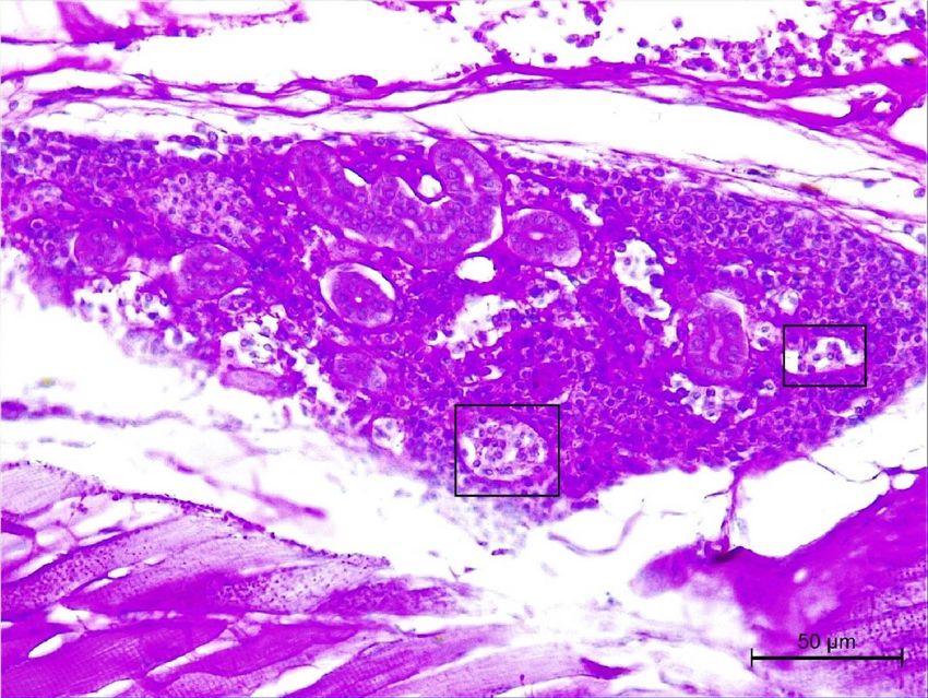
Veterinary Integrative Sciences

                                INTRODUCTION
                                         The zebra-snout Seahorse, Hippocampus barbouri Jordan & Richardson,
                                1908, belongs to the family Syngnathidae, is one of the most important fishery
                                resources of Thailand. This seahorse species can be found along tropical and
                                temperate shallow shelter reefs, and in algae and seaglass beds (Foster and
                                Vincent, 2004). In recent years, overexploitation, particularly for traditional
                                Chinese medicine (TCM), overfishing and habitat deterioration have been the
                                main causes of dramatic declines in H. barbouri (Vincent et al., 2011). Further
                                indicative of its decline, H. barbouri is also listed in the Appendix II of the
                                Convention on International Trade in Endangered Species of Wild Fauna and
                                Flora (CITES) (Hutton and Dickson, 2000).
                                         Due to population declines in H. barbour, initial and ongoing work
                                by the Phuket Marine Biological Center (PMBC), Thailand under its Culture
                                Project has developed a strategy involving research and development of captive
                                breeding and aquaculture of this species in Thailand toward stock recovery and
                                conservation. Numerous feeding studies involving live feed for aquaculture
                                of H. barbour have been performed, with results showing increased growth
                                rates (Kamnurdnin, 2017; Wung et al., 2020; Wilson and Vincent, 2000; Planas
                                et al., 2008; Sheng et al., 2006); however, there are no known studies on the
                                health status of this species of seahorse in captivity through microanatomical
                                methods. This information is essential for understanding environmental
                                factors influencing its reproductive capacity, growth rate, and survival under
                                aquaculture. Hence, in the present study, we assessed the health of captive
                                juvenile to adult H. barbour using histopathological methods.

                                MATERIALS and METHODS

                                Seahorse collection
                                        Captive broodstock of Hippocampus barbouri were reared in a standard
                                culture system from the Phuket Marine Biological Center (PMBC), Thailand.
                                Juvenile stages (at 1-day, 5-days, 7-days, 14-days and 20-days) and adult stage (at
                                30-days) (Table 1) were received as voucher specimens from Kamnurdnin (2017).
                                Previous observations showed that the adult stage of H. barbouri in captivity is
                                considered as individuals over 30-days of age, from which secondary growth
                                occurs (Kamnurdnin, 2017). The experimental protocol was approved by the
                                Animal Care and Use Committee of Faculty of Science in accordance with the
                                guide for the care and use of laboratory animal prepared by Chulalongkorn
                                University (Protocol Review No. 1623004).

Vet Integr Sci   Kamnurdnin et al. Vet Integr Sci. 2021; 19(3): 305 - 315                                   306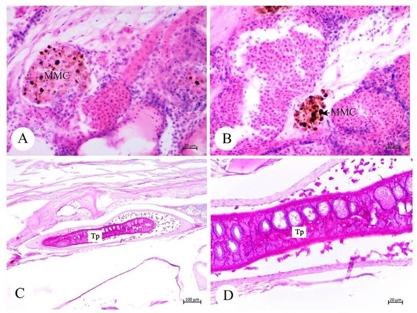
Veterinary Integrative Sciences

Table 1 Summary of size and number of samples of Hippocampus barbouri in captivity.
     Seahorse stages                           Days                             Numbers        Total length (cm)
                                                  1                                3                15.60±0.78
                                                  5                                3                21.70±3.94
         Juveniles                                7                                3                29.00±3.22
                                                 14                                3                36̀.00±2.75
                                                 20                                3                42.90±2.96
          Adult                                  30                                3                57.30±3.75

                                    Histological techniques
                                              All seahorse samples were quickly euthanized by a rapid cooling
                                    method (Wilson et al., 2009). The external anatomy was visually evaluated
                                    under stereomicroscope, then the whole fish were transferred to Davidson's
                                    fixative. Cross sections of all samples were dissected approximately 1 cm from
                                    head to tail and were then processed using standard histological protocols
                                    (Presnell and Schreibman, 1997; Senarat et al., 2020a; Senarat et al., 2020b).
                                    The paraffin blocks were sectioned at a thickness of 4 µm and stained with
                                    Harris’s hematoxylin and eosin (H&E), Masson's Trichrome (MT) and periodic
                                    acid-Schiff (PAS) (Presnell and Schreibman, 1997; Suvarna et al., 2013).
                                    Histopathological abnormalities of the fish were examined and photographed
                                    under a light microscope (Leica digital 750). Each histopathological abnormality
                                    in the respective organs was calculated and presented as a percent prevalence
                                    (%). The occurrence of melanomacrophage centers and infection of trematode
                                    parasites was also documented from the stained tissue sections.

                                    RESULTS
                                             We observed histopathological conditions in several vital organs (gill,
                                    liver, kidney) and the mesentary through histological techniques, as shown in
                                    Figures 1–3. The prevalence of histopathological lesions is also provided in
                                    Table 2.

Table 2 Prevalence alteration (%) of histopathological observations in Hippocampus barbouri in captivity.
       Tissues                                                                                Percent
                           Days                             Lesions
       (n = 3)                                                                              prevalence
 Gills                      14         Lamellar epithelium lifting and intraepithelial         100.0
                                       edema of secondary gills
                            20                                                                  33.3
Livers                           14             Lipidosis and cellular degeneration of hepatocyte          100.0
                                 20                                                                        100.0
Kidneys                          14             Renal degeneration                                         100.0
                                 20                                                                        100.0
                                 30             MMC and trematode parasites                                33.3

Vet Integr Sci       Kamnurdnin et al. Vet Integr Sci. 2021; 19(3): 305 - 315                                      307
Veterinary Integrative Sciences
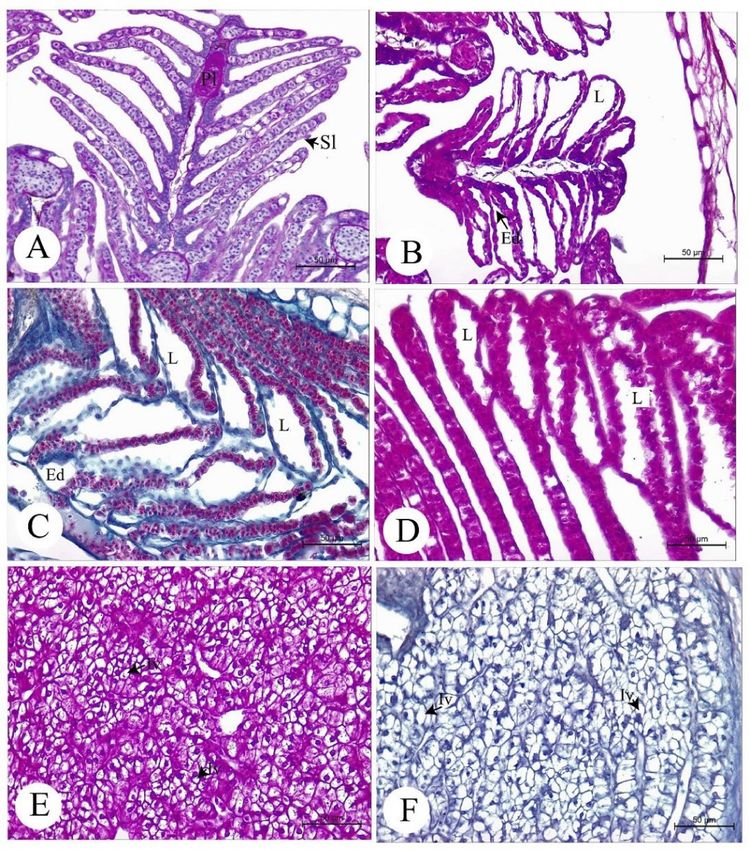
Histology and histopathology of gills
Histology of the H. barbouri gill in healthy individuals is shown in
Figure 1A. The structure of the four-gill filament (or respiratory filament) arises
from the gill arch in a perpendicular manner, where they are parallel to each
other. Each gill filament is histologically observed as double rows, in which
each filament is divided into the primary and secondary lamellae [respiratory
lamellae (Figure 1A)]. Only an intense lifting of the lamellar epithelium and
intraepithelial edema of the secondary gills clearly showed alterations in
juveniles at 14-days (100% prevalence, Figure 1B), 20-days (33% prevalence)
and adults at 30-days (100% prevalence, Figures 1C-1D).
Figure 1 Light photomicrograph of gill histology (A) consisting of primary
lamella (PI) and secondary lamella (SI) of Hippocampus barbouri in captivity.
Lamellar epithelium lifting (L) and edema (Ed) of the secondary lamellar gills
at 14 days (B-D), and intracytoplasmic vacuoles (Iv) were observed in the
liver. Note: periodic acid-Schiff (A); Harris's hematoxylin and eosin; (B, D, E);
Masson's Trichrome (C).
Vet Integr Sci Kamnurdnin et al. Vet Integr Sci. 2021; 19(3): 305 - 315 308Veterinary Integrative Sciences
Histopathology of livers
Necrosis and intracytoplasmic vacuoles, also called “hepatocellular
lipidosis”, in juvenile livers occurred at 14-days juvenile stage and at 30-days
adult stage (100 % prevalence) (Figures 1E-1F).
Histopathology of kidneys
Both 14 and 30-days stages exhibited histological changes in the renal
degeneration (100% prevalence) throughout the melanomacrophage aggregates
(100% prevalence, Figures 2A-2B).
Melanomacrophage centers (MMCs) and infection of trema-
tode parasites
We observed MMCs in the mesentery in 30-days adult stage (33.3 %
prevalence) (Figures 2A-2B). Interestingly, trematode parasites were found
only in those at stage 30-days. Also, the location of the parasites was observed
in kidney tissue at 30-days stage (33.3 % prevalence, Figures 2C-2D), and that
the degeneration of renal tubules in the kidney was also found near the site of
trematode infection (Figure 3).
Figure 2 Light photomicrograph of melanomacrophage centers (MMCs) and
trematode parasites of Hippocampus barbouri in captivity. A-B: MMCs at
30-days. C-D: Trematode parasites (Tp) at 30-days in kidney tissue. Note: A-F:
Harris's hematoxylin and eosin.
Vet Integr Sci Kamnurdnin et al. Vet Integr Sci. 2021; 19(3): 305 - 315 309Veterinary Integrative Sciences
Figure 3 Light photomicrograph of the degeneration of the renal tubule (square)
of Hippocampus barbouri in captivity; representative figure at 30-days. Note:
periodic acid-Schiff.
DISCUSSION
Due to a lack of information on H. barbouri health under captive
conditions, one requirement was to identify histopathological alterations of
captive broodstock H. barbouri for documentation and help in applying to
aquaculture development. This method has been routinely recommended and
used to assess the general health status of fishes under both laboratory and field
conditions (Blazer, 2002; OECD, 2009; Dietrich and Krieger, 2009; Senarat et
al., 2015; Senarat et al., 2018a; Senarat et al., 2018b; Senarat et al., 2019; Senarat
et al., 2020c).
Intense lamellar epithelium lifting and intraepithelial edema of the
secondary gills in H. barbouri were observed. This indicates that the gill may
be a more specific and sensitive organ in H. barbouri. It was reported that
these lesions serve as a mechanism of defense by increasing the structural
distance between the epithelium and pollution or low-quality environmental
condition. This notion was supported by several studies on the gills of teleost fish
(Bury et al., 1998; Sola et al., 1995), for example, Cyprinus carpino after
exposed to 10 mg/L Thiamethoxam (Georgieva et al., 2014), Acanthopagrus
latus after treated to HgCl2 concentrations (Hassaninezhad et al., 2014) and
Poecilia reticulate exposed to chlorpyrifos (de Silva and Samayawardhena,
2002). Additionally, some reports have shown that histopathological lesions
Vet Integr Sci Kamnurdnin et al. Vet Integr Sci. 2021; 19(3): 305 - 315 310Veterinary Integrative Sciences
on the gills could develop due to sensitization of the mucosal membrane
lining the lamellae, with the activity of mucous and chloride cells towards the
water supplied to the fishes (Boyd et al., 1980), which needs to be observed
of H. barbouri in further study. Although here we found a few lesions in the
gill, the overall histopathological conditions indicate impaired gills in captive
H. barbouri which reflect a reduced physiological function (the respiration,
excretory and osmoregulatory functions).
Hepatocellular lipidosis in juvenile livers was found at 14 and 30-days
(100 % prevalence) (Figures 1E-1F). The occurrence of the hepatocellular
lipidosis and cellular degeneration of hepatocytes was known to be related to
liver damage, as well as a consequent loss of liver function (Greenfield et al., 2008).
A similar mechanism was previously suggested by Hinton and Laure´n (1990),
who postulated that a derangement in lipid and protein metabolism (lipidosis)
might play a negative role in the abnormal accumulation of triglycerides in the
hepatocytes. It was reported that the appearance of the hepatocellular lipidosis
is caused by several factors, for example, chlorinated hydrocarbons and other
contaminants (Hendricks et al., 1984; Hinton et al., 1992; Robertson and Bradley
1992; Schrank et al., 1997), including PCBs (Anderson et al., 2003; Teh et al.,
1997). However, liver histopathology is also related to old age, poor nutritional
value of food sources, and/or other environmental conditions (Hinton et al.,
1992; Robertson and Bradley, 1992). These ideas were in accordance with liver
histopathological observations from Sparus aurata fed on the different dietary
lipid contents (Caballero et al., 1999; Zilberg and Munday, 2002). Therefore,
based on the hepatic histopathological condition of H. barbouri under captivity,
contamination by pollutant(s) and other factors as above (Hinton and Laure´n
1990) may play some role in deteriorating health of the seahorse in aquaculture.
The kidney is one of the major organs affected from contaminated
aquatic environments, which can be visualized through histological observation.
The renal degeneration of H. barbouri exhibited both 14 and 30 days, and it was
similar to histological damage of teleostean renal tissues exposed to carbofuran
(Yenchum, 2010), ammonia (Aysel et al., 2008), and methyl mercury exposure
(Melaa et al., 2007). This underscores the importance of the kidney in the
excretion of metal and hydrophilic pollutants as shown by Pritchard and Bend
(1984); Meinelt et al. (1997); Takashima and Hibya (1995). In addition, renal
tissues may be used as a histopathological biomarker to observe pollutants in
captivity. Further investigations are needed to accurately measure contaminants
such as metals and other chemical pollutants in captive water conditions, and
the relation to histological damage to renal tissue and their functions.
Melanomacrophage centers (MMCs), accumulation of pigmented
phagocytes functioning in phagocytosis of erythrocytes and infectious
materials, play central roles in several vertebrates’ immune system
(Takashima and Hibya, 1995; Steinel and Bolnick, 2017). In teleost fishes,
the immune responses to parasites, pollutants, as well as stress, can trigger
the emergence of MMCs, which can be used as a histological indicator for
pathogen infection (Alvarez-Pellitero et al., 2007; Robert, 2012; Sitja-Bobadilla,
2008). The appearance of MMCs together with trematode parasites was found,
supporting the notion that MMCs occurred when fish hosts were infected with
parasites. In addition, we observed that the location of MMC accumulation was
not in close proximity to the parasites, which were protected by thick cyst wall.
Vet Integr Sci Kamnurdnin et al. Vet Integr Sci. 2021; 19(3): 305 - 315 311Veterinary Integrative Sciences
It is possible that the presence of MMCs was a good indication of general stress
in this seahorse. This was in agreement with previous observations, which can
help in better understanding the dynamics of fish health either in the wild or
captivity (Steinel & Bolnick, 2017; Tsujii & Seno, 1990).
CONCLUSION
We provided evidence of various histopathological observations
in several vital organs from the zebra-snout seahorse, H. barbourin under
captivity, including edema of the gills, hepatocellular lipidosis and kidney
degeneration, and infection of parasites. In addition, different life history stages
of seahorse exhibited a distinct prevalence of disease. From our findings, we
propose that the presence of histopathological lesions here reflect an unhealthy
status of juvenile to adult stages of the seahorse under captive culture. As a
conservation and fisheries management tool, successful rearing and aquaculture
of healthy stocks are needed to offset overharvest and habitat degradation. In
captive and other aquaculture settings, the regular monitoring of water quality
and providing optimal nutrition for H. barbouri is required and should be
continuously assessed by the Phuket Biological Center (PMBC), Thailand in
order to reach successful captive breeding of this species.
ACKNOWLEDGEMENTS
Special thanks to all staff of the Phuket Marine Biological Center
(PMBC), Thailand and members of the Fish Biology and Aquatic Health
Assessment Laboratory (FBA-LAB), Department of Marine Science,
Chulalongkorn University, Aquatic Toxicopathology Unit, Department of
Pathobiology, Faculty of Science, Mahidol University, Thailand for their help
in the laboratory. We also specially thank Dr. Todd W. Miller for reading the
grammatical structure in this manuscript.
CONFLICT OF INTEREST
The authors declare that no conflict of interest.
REFERENCES
Alvarez-Pellitero, P., Palenzuela, O., Sitjà-Bobadilla, A., 2007. Histopathology and cellular
response in Enteromyxum leei (Myxozoa) infections of Diplodus puntazzo
(Teleostei). Parasitol. Int. 57, 110–120.
Anderson, M.J., Cacela, D., Beltman, D., The, S.J., Okihiro, M.S., Hinton, D.E., et al. 2003.
Biochemical and toxicopathic biomarkers assessed in smallmouth bass recovered
from a polychlorinated biphenyl-contaminated river. Biomark. 8, 371–393.
Aysel, C.K.B., Gulten, K., Ayhan, O., 2008. Sublethal ammonia exposure of nile tilapia
(Oreochromis niloticus L.): Effects on gill, liver and kidney histology. Chemosphere.
72, 1355–1358.
Blazer, V.S., 2002. Histopathological assessment of gonadal tissue in wild fishes. Fish Physiol.
Biochem. 26, 85–101.
Vet Integr Sci Kamnurdnin et al. Vet Integr Sci. 2021; 19(3): 305 - 315 312Veterinary Integrative Sciences
Boyd, R.B., De Vries, A.L., Eastman, J.T., Pietra, G.G., 1980. The secondary lamellae of the
gills of cold water (high latitude) teleosts. A comparative light and electron
microscopic study. Cell Tissue Res. 213, 361–367.
Bury, N.R., Li, J., Flik, G., Lock, R.A.C., Wendelaar-Bonga, S.E., 1998. Cortisol protects
against copper induced necrosis and promotes apoptosis in fish gill chloride cells
in vitro. Aquat. Toxicol. 40, 193–202.
Caballero, M.J., Lopez-Calero, G., Socorro, J., Roo, F.J., lzquierdo, M.S., Fernandez, A.J.,
1999. Combined effect of lipid level and fish meal quality on liver histology of
gilthead seabream (Sparus aurata). Aquaculture. 179, 277–290.
de Silva, P.M.C.S., Samayawardhena, L.A., 2002. Effects of chlorpyrifos on reproductive
performances of guppy (Poecilia reticulata). Chemoshere. 58, 1293–1299.
Dietrich, D.R., Krieger, H.O., 2009. Histological analysis of endocrine disruptive effects in
small laboratory fish. John Wiley and Sons, New Jersey, U.S.A.
Foster, S.J., Vincent, A.C., 2004. Life history and ecology of seahorses: implications for
conservation and management. J. Fish Biol. 65, 1–61.
Georgieva, E., Stoyanova, S., Velcheva, I., Yancheva, V., 2014. Histopathological alterations in
common carp (Cyprinus carpio L.) gills caused by thiamethoxam. Braz. Arch. Biol.
Technol. 57, 991–996.
Greenfield, B.K., The, S.J., Ross, J.R., Hunt, J., Zhang, G., Davis, J.A. et al., 2008.
Contaminant concentrations and histopathological effects in Sacramento splittail
(Pogonichthys macrolepidotus). Arch. Environ. Contam. Toxicol. 55, 270–281.
Hassaninezhad, L., Safahieh, A., Salamat, N., Savari, A., Majd, N.E., 2014. Assessment of gill
pathological responses in the tropical fish yellowfin seabream of Persian Gulf under
mercury exposure. Toxicol. Rep. 1, 621–628.
Hendricks, J.D., Meyers, T.R., Shelton, D.W., 1984. Histological progression of hepatic
neoplasia in rainbow trout (Salmo gairdneri). J. Natl. Cancer Inst. 65, 321–336.
Hinton, D.E., Laure´n, D.J., 1990. Liver structural alterations accompanying chronic toxicity
in fishes: potential biomarkers of exposure. In: McCarthy, J.F., Shugart L.R. (Eds.),
Biomarkers of Environmental Contamination, pp. 17–57.
Hinton, D.E., Baumann, P.C., Gardner, G.R., Hawkins, W.E., Hendricks, J.D., Murchelano,
R.A. et al., 1992. Histopathologic Biomarkers. Biochemical, Physiological, and
Histological Markers of Anthropogenic Stress. Biomark. 155–209.
Hutton, J., Dickson, B., 2000. Endangered species, threatened convention: the past, present
and future of CITES, the Convention on International Trade in Endangered Species of
Wild Fauna and Flora, Earthscan London.
Kamnurdnin, T., 2017. Effects of food on growth and gonadal development of Seahorse,
Hippocampus sp (Master degree, Chulalongkorn University.
Meinelt, T., Krüger, R., Pietrock, M., Osten, R., Steinberg, C., 1997. Mercury pollution and
macrophage centres in pike (Esox lucius) tissues. Environ. Sci. Pollut. Res. Int. 4,
32–36
Melaa, M., Randi, M.A.F., Ventura, D.F., Carvalho, C.E.V., Pelletier, E., Oliveira, C., Ribeiro,
C.A.. 2007. Effects of dietary methyl mercury on liver and kidney histology in the
neotropical fish Hoplias malabaricus. Ecotoxicol. Environ. Saf. 68, 426–435.
Organisation for Economic Co-operation and Development (OECD). 2009. OECD guidance
document for the diagnosis of endocrine-related histopathology of fish gonads [cited
2018, September 21]. Available from: http://www.oecd.org/dataoecd/33/27/42140701.pdf
Planas, M., Chamorro, A., Quintas P., Vilar. A., 2008. Establishment and maintenance of
threatened long-snouted seahorse, Hippocampus guttulatus, broodstock in captivity.
Aquaculture 283, 19–28.
Presnell, J.K., Schreibman, M.P., 1997. Humason’s Animal Tissue Techniques. 5th ed. US,
Johns Hopkins University Press.
Pritchard, J.B., Bend, J.R., 1984. Mechanisms controlling the renal excretion of xenobiotics in
fish: effects of chemical structure. Drug Metab. Rev. 15, 655-671.
Robertson, J.C., Bradley, T.M., 1992. Liver ultrastructure of juvenile Atlantic salmon (Salmo
salar). J. Morphol. 211, 41–54.
Robert, R.J., 2012. Fish Pathology. Blackwell Publishing Ltd.
Schrank, C.S., Cormier, S.M., Blazer, V.S., 1997. Contaminant exposure, biochemical, and
histopathological biomarkers in white suckers from contaminated and reference sites
in the Sheboygan River, Wisconsin. J. Great Lakes Res. 23, 119–30.
Vet Integr Sci Kamnurdnin et al. Vet Integr Sci. 2021; 19(3): 305 - 315 313Veterinary Integrative Sciences
Senarat, S., Poolprasert, P., Kettratad, J., Boonyoung, B., Jiraungkoorskul, W., Huang,
S., Pengsakul, T., Kosiyachinda, P., Sudtongkong, C., 2020a. Histological observation
of digestive system of malayan halfbeak, Dermogenys pusilla (Kuhl & van Hasselt,
1823) during juvenile stage from Thailand. Vet. Integr. Sci. 18, 33-41.
Senarat, S., Boonyoung, P., Kettratad, J., Jiraungkoorskul, W., Poolprasert, P., Huang,
S., Pengsakul, T., Mongkolchaichana, E., Para, C. 2020b. The identification and
distribution of the mucous secreting cells in the integument of the Schaap’s dragonet,
Callionymus schaapii, Bleeker, 1852. Vet. Integr. Sci. 18(1), 23-32.
Senarat, S., Kettratad, J., Siriwong, W., Bunsomboonsakul, S., Kenthao, A., Kaneko, G., Sopon,
A., Sudtongkong, C., Jiraungkoorskul, W., 2020c. Oogenesis and ovarian health
problems in economically important fishes from different habitats potentially affected
by pollution in Thailand. Asian Fish. Sci. 33, 274–286.
Senarat, S., Kettratad, J., Gerald Plumley, F., Wangkulangkul, S., Jiraungkoorskul,
W., Boonyoung, P., Poolprasert, P., 2019. Pathological microscopy in liver
parenchyma of gray-eel catfish, Plotosus canius, from Ang-Sila area, Chonburi
Prov-ince, Thailand. Vet. Integr. Sci. 17, 255-261
Senarat, S., Kettretad, J., Poolprasert, P., Tipdomrongpong, S., Plumley, F.G., Jiraungkoorskul,
W. 2018a. Health status in wild and captive Rastrelliger brachysoma from Thailand:
Histopathology. Songklanakarin J. Sci. Technol. 40, 1090–1097.
Senarat, S., Kettratad, J., Tipdomrongpong, S., Pengsakul, T., Jiraungkoorskul, W., Boonyoung,
P., Huang, S., 2018b. Histopathology of kidney and liver in the captive broodstock
(Rastrelliger brachysoma) during its juvenile stage. Vet. Integr. Sci. 16, 87-93.
Senarat, S., Kettratad, J., Poolprasert, P., Jiraungkoorskul, W., Yenchum, W., 2015.
Histopathological findings of liver and kidney tissues of the yellow mystus,
Hemibagrus filamentus (Fang and Chaux, 1949), from the Tapee River, Thailand.
Songklanakarin J. Sci. Technol. 37, 1–5.
Sheng, J., Lin, Q., Chen, Q., Gao, Y., Shen L., Lu. J., 2006. Effects of food, temperature and
light intensity on the feeding behavior of three-spot juveniles seahorses, Hippocampus
trimaculatus Leach. Aquaculture. 256, 596–607.
Sitja-Bobadilla, A. 2008. Fish immune response to myxozoan parasites. Parasitology. 15,
420–425.
Sola, F., Isaia, J., Masoni, A., 1995. Effects of copper on gill structure and transport function in
the rainbow trout, Oncorhynchus mykiss. J. Appl. Toxicol. 15, 391–359.
Steinel, N.C., Bolnick, D.I., 2017. Melanomacrophage centers as a histological indicator of
immune function in fish and other poikilotherms. Front Immuno. 8, 827.
Suvarna, K.S., Layton, C., Bancroft, J.D., 2013. Bancroft’s Theory and Practice of Histological
Techniques. 7th ed. Canada, Elsevier.
Takashima, F., Hibiya, T., 1995. An Atlas of Fish Histology, Normal and Pathological Features,
2nd edition. Kodansha, Tokyo.
The, S.J., Adams, S.M., Hinton, D.E., 1997. Histopathologic biomarkers in feral freshwater
fish populations exposed to different types of contaminant stress. Aquatic Toxicol. 37,
51–70.
Tsujii, T., Seno, S., 1990. Melano-macrophage centers in the aglomerular kidney of the sea
horse (teleosts): morphologic studies on its formation and possible function. Anat.
Rec. 226, 460–470.
Vincent, A.C.J., Foster, S.J., Koldewey H.J., 2011. Conservation and management of seahorses
and other Syngnathidae. J. Fish Biol. 78, 1681–1724.
Wung, L.Y., Christianus, A., Zakaria, M.H., Min, C.C., Worachananant, S. 2020. Effect of
cultured artemia on growth and survival of juvenile Hippocampus barbouri. JFE. 44,
40-49.
Wilson, J.M., Bunte, R.M., Carty, A.J., 2009. Evaluation of rapid cooling and tricaine
methanesulfonate (MS 222) as methods of euthanasia in zebrafish (Danio rerio).
J. Am. Assoc. Lab. Anim. Sci. 48, 785–789.
Wilson, M.J., Vincent, A.C.J., 2000. Preliminary success in closing the life cycle of exploited
seahorse species, Hippocampus spp., in captivity. Aquarium Sci. and Conserv. 2,
179–196.
Yenchum, W., 2010. Histological effects of carbofuran on guppy Poecilia reticulata Peters
(Doctoral thesis, Chulalongkorn University).
Vet Integr Sci Kamnurdnin et al. Vet Integr Sci. 2021; 19(3): 305 - 315 314Veterinary Integrative Sciences
Zilberg, D., Munday, B.L., 2002. Pathology of experimental amoebic gill disease in Atlantic
salmon, Salmo salar L., and the effect of pre-maintenance of fish in sea water on the
infection. J. Fish Dis. 23, 401–407.
How to cite this article;
Thatpon Kamnurdnin, Sinlapachai Senarat, Jes Kettratad, Supanut Pairohakul, Wannee
Jiraungkoorskul, Ratree Sooksuwan, Woranop Sukparangsi and Chanyut Sudtongkong.
Histopathological observation and health status of the zebra-snout seahorse Hippocampus
barbouri Jordan & Richardson, 1908 in captivity. 2021; 19(3): 305-315.
Vet Integr Sci Kamnurdnin et al. Vet Integr Sci. 2021; 19(3): 305 - 315 315You can also read