UNIT Qualitative Evaluation of Inclusions In Moderna, AstraZeneca and Pfizer Covid-19 vaccines. by - Project CUNIT-2-112Y6580
←
→
Page content transcription
If your browser does not render page correctly, please read the page content below
Global Humanitarian Crisis Prevention and Response Unit

                Project CUNIT-2-112Y6580

   Qualitative Evaluation of Inclusions
                                  In
Moderna, AstraZeneca and Pfizer Covid-19
               vaccines.

                                  by
                            UNIT

                                                                    Page 1 of 48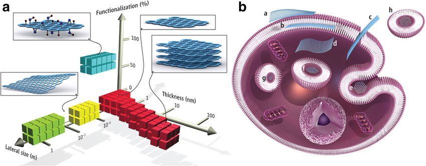
Global Humanitarian Crisis Prevention and Response Unit

                                       Executive Summary

UNIT was commissioned by EbMCsquared CiC under project UNITC-112980 to investigate

the contents of four injection vials (Moderna 01, Moderna 02, AstraZeneca, Pfizer) for any

undeclared ingredients that may cause bodily harm.

This report is the submission of initial findings that confirm the presence of graphene

compounds in each of the injection vials. Though a quantitative estimate has not be established

for the concentration of graphene in the samples, its occurrence is on a high frequency range

on an average 2cm transect when counts were conducted on a higher magnification (40x).

                                                                                      Page 2 of 48
Global Humanitarian Crisis Prevention and Response Unit

                                                   TABLE OF CONTENTS
1.      Introduction ...................................................................................................................... 5
     1.1.     Background ................................................................................................................ 5
     1.2.     Descriptions of Vials.................................................................................................. 5
     1.3.     Aims and Objectives of the study ............................................................................ 6
     1.4.     Report Structure and Outline .................................................................................. 6
2.      Methodology ..................................................................................................................... 8
     2.1.     Vial Descriptions ....................................................................................................... 8
     2.2.     Process of Sampling and Slide Preparation ............................................................ 8
     2.3.     Raman Spectroscopy ............................................................................................... 10
3.Results .................................................................................................................................. 11
     3.1. Description of Inclusions ............................................................................................ 11
        3.1.1.       Graphene Composites in the form of Nano Ribbons ......................................... 11
        3.1.2.       Graphene Composite form 1 .............................................................................. 12
        3.1.3.       Graphene Composite Form 2 ............................................................................. 12
        3.1.4.       Calcite ................................................................................................................ 13
        3.1.5.       Graphene Nano Forms ....................................................................................... 13
        3.1.6.       Crystalised forms of the solution ....................................................................... 14
     3.2.     Moderna 01 .............................................................................................................. 15
        3.2.1.       Microscopy ........................................................................................................ 15
        3.2.2.       Raman Spectroscopic Investigation ................................................................... 18
        3.2.3.       Counts ................................................................................................................ 20
     3.3.     Moderna 02 .............................................................................................................. 21
        3.3.1.       Microscopy ........................................................................................................ 21
        3.3.2.       Raman Spectroscopic Investigation ................................................................... 25
        3.2.3. Counts .................................................................................................................... 26
     3.4.     AstraZeneca ............................................................................................................. 28
        3.4.1.       Microscopy ........................................................................................................ 28
        3.4.2.       Raman Spectroscopic Investigation ................................................................... 32
        3.4.3.       Counts ................................................................................................................ 34
     3.5.     Pfizer ......................................................................................................................... 35
        3.5.1.       Microscopy ........................................................................................................ 35
        3.5.2.       Raman Spectroscopic Investigation ................................................................... 38
        3.5.3.       Counts ................................................................................................................ 39
4.      Interpretation, Discussions and Conclusion ................................................................ 41
5.      Bibliography ................................................................................................................... 45

                                                                                                                             Page 3 of 48
Global Humanitarian Crisis Prevention and Response Unit

1. Introduction
The following report is a product of a joint cooperation between EbMCsquared CiC and UNIT
in an attempt to identify the undeclared contents of the current vaccines that are being
administered to the UK public causing high numbers of adverse reactions and deaths.
   1.1. Background
UNIT was commissioned by EbMCsquared under UNITC-112980 to 91 to analyse contents of
four vaccine vials and identify if any of the following components were present in these vials
 graphene, graphene oxide, parasites, biological filaments.
The four vaccines that form the subject of this first investigation by UNIT belonged to
Moderna, Pfizer, and AstraZeneca.
The entire process of collection and delivery is reported in ANNEXE 1A.
   1.2. Descriptions of Vials
       1.2.1. Moderna 01 Labelling

The manufacturing label had the following information on the vial:
COVID-19 Vaccine Moderna. 020mg/mL Dispersion for injection. COVID-19 mRNA Vaccine
(nucleoside modified) Multidose vial doses of .5mL.
Lot-3004737 Exp. 24/01/2022.
The liquid contained in the Moderna bottle was cloudy to naked eye against the sunlight.

       1.2.2. Moderna 02 Labelling

The manufacturing label had the following information on the vial:
COVID-19 Vaccine Moderna. 020mg/mL Dispersion for injection. COVID-19 mRNA Vaccine
(nucleoside modified) Multidose vial doses of .5mL.
Lot- Lot-3004737 Exp. 24/01/2022.
The bottle weight prior to breaking of the seal was 18.842gm.
The liquid contained in the Moderna bottle was cloudy to naked eye against the sunlight.

       1.2.3. AstraZeneca Labelling

The manufacturing label had the following information on the vial:

                                                                                      Page 5 of 48
Global Humanitarian Crisis Prevention and Response Unit

Covid-19        Vaccine    4        ml,         Astrazeneca              Injection.      Covid-19      Vaccine:
(ChAd0x1-S (recombinant)), Intramuscular use. Multidose vial (8x0.5ml doses). 108439/2 Lot-
PW40167 Expiry- 01-22. (Figure 2.1)
The bottle weight prior to the breaking of the seal was 12.184g and after the breaking of the
seal was 11.803g.
The liquid contained in the AstraZeneca bottle was transparent to naked eye.

       1.2.4. Pfizer Labelling

The manufacturing label had the following information on the vial as in figure 1.1:

                                      Figure 1.1. Pfizer vaccine vial
Lot-FC9001 Exp. 09/01/2022.
The liquid contained in the Pfizer bottle was cloudy to naked eye against the sunlight

       1.2.5.
   1.3. Aims and Objectives of the study
The aim of the study was to identify any solid inclusions in the vials as were undeclared by the
manufacturers.
The study was to verify the findings of graphene related compounds such as graphene oxide,
graphene hydroxide by Campara (2021) and report any other biological inclusions that may be
interpreted as toxic to human body.
   1.4. Report Structure and Outline
The vials went through evaluation of contents at four different laboratory sites that are
identified in ANNEXE 1B on request and the methodology adopted is presented in Chapter
two.

                                                                                                    Page 6 of 48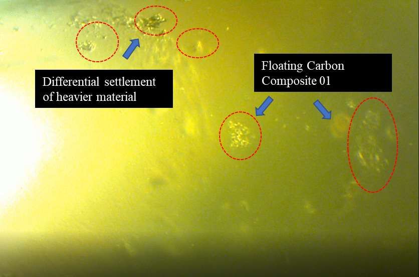
Global Humanitarian Crisis Prevention and Response Unit
      The report presents an in depth analysis of the findings of this project and is divided into
      five chapters. Chapter one is the introduction, the second chapter is a description and
analysis of the methods used for the evaluation. Chapter three presents the results of the
analysis, Chapter four is the interpretation, discussions and conclusions of the study. The
Bibliography forms the last chapter of the report.
The entire project involved the inputs of experts in individual fields and their names, affiliations
and expertise are identified for reference purposes in Annex 2.

                                                                                        Page 7 of 48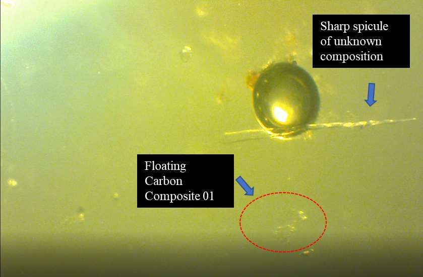
Global Humanitarian Crisis Prevention and Response Unit

    2. Methodology
    2.1.    Vial Descriptions
The secured vials were made of glass and the vaccines contained within the vials were in a
liquid form. The different parts of the vial are shown in figure 2.1. Each of these vials were
stored at 4°C in the sample storage rooms until the evaluation of their contents took place.
Each of the four vials undertook the same process for evaluation.

Figure 2-1 Different parts of the sealing process of the vial. The vial shown here is the AstraZeneca vial.
The same three seal components were present for the Moderna and Pfizer vials.

    2.2.    Process of Sampling and Slide Preparation

Stage 1 - Opening of the vial

Stage 1 involved the opening of the vial while the process was recorded on the camera for
posterity. Each of the vials were sealed with a plastic seal on the top (figure 2.1). This seal had
to be broken off before one could access the rubber top of the injection vial. The rubber top
was held securely in place with a metal ring.
There were two options of accessing the liquids from within the vials. One, was to use an
injection syringe and draw out the required amount of liquid. However, knowing that the
objective of the study was to identify and report any nano graphene particles within the vials,
this method of sample acquisition was not thought to be appropriate. The reasoning being, that
during the penetration of the vial, the syringe would carry nanoscopic amounts of rubber seal
material, which is a carbon product, each time, it would access the liquid. Therefore, the second
option of material access was adopted, which meant, breaking off the metal ring and simply
removing the rubber seal on top each time the sample material was required.

                                                                                            Page 8 of 48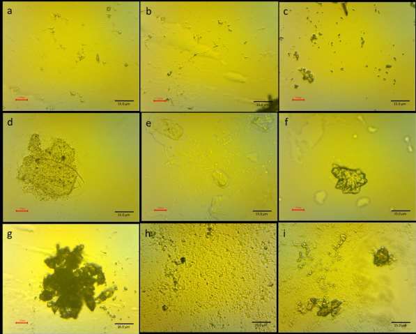
Global Humanitarian Crisis Prevention and Response Unit
Stage 2   Subsampling for evaluation of experimental steps required

Stage 2 had the simple purpose of the first evaluation of the sample. As there was no prior
study available at hand to determine the chemistry and the physical properties of the material
at hand, a small subsample was used from the vial to be evaluated under a polarising
microscope to identify the gross composition of the material of the vaccine and to have a brief
understanding of any inclusions if they were present.
Each of the vaccine s active ingredients seemed to have been preserved in a sucrose-salt rich
medium. The optical and physical properties of the preserving medium posed a challenge to
the identification of the actual inclusions. In addition to being of nearly the same refractive
index as the medium, the micro and nano crystalline nature of the expected inclusions rendered
an additional impediment to the task of their isolation.
During, wet microscopic evaluation at the dry laboratories, it was noted from the onset, that
the denser material settled at the bottom of the slide and got impregnated and encrusted with
the medium solution, while the slide was still in the process of wet examination. Additionally,
the deposition of the inclusions occurred at different depths within the medium.
Cummulatively, such characteristics, posed a grave challenge to the central objective of the
study, in that, isolation of the objects of interest was deemed impossible. The medium masked
all signatures of the inclusions by impregnation and encrustation in a manner that the inclusions
were completely camouflaged into the background.
These challenges were attempted to be overridden by vacuum filteration. The first attempt at
vacuum filteration failed due to equipment failure. This resulted in the loss of the test material.
As, at the time of this experiment, the material at hand was quite limited, any further attempts
for both vacuum filteration processes were paused.
(It should be noted, that the ongoing continuation of this project will be using checmical
filteration to isolate the inclusions from the onset as the current project has given a clear insight
into the chemistry of the product.)

Stage 3- Partitioning of sample for clean slide preparation

This stage included the partitioning of sample into two. The clean portions of the sample were
stored in a pyrex glass vial which was cleaned using hydrogen per oxide, hydrochloric baths,
then rinsed with distilled water. The vials were then sterilised in the sterilisation chamber prior
to the transfer of the sample into them.

                                                                                        Page 9 of 48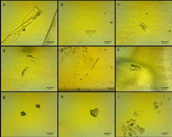
Global Humanitarian Crisis Prevention and Response Unit
0.0125ml of sample was then transferred onto clean slides within fume cupboard and left there
to air dry at 20°C under a glass chamber.
The dried slides were mounted using glass coverslips and used for microscopic evaluation. It
should be noted that none of the prepared clean slides dried out completely even after a few
days.
Stage 3- Optical and Petrological Microscopy
Prepped slides were examined under a reflection and transmission light microscope for organic
content and petrological microscope for mineral contents.
Microscopic stage involved preparation of several test slides to identify the typical nature of
the content.
On representative slides, counts were done along a specific transect. These were then
represented on psimpol to get density options on a typical 0.006ml of sample.
Stage 4   Raman sampling and preparation
Subsamples from original vials were obtained for Raman spectroscopy. These were transferred
onto standard slides using sterilised glass pipettes. The slides were left to dry under glass
chambers inside a fan heating oven before being taken for examination to the Raman
Laboratories.
   2.3.    Raman Spectroscopy
As most of the observed inclusions belonged categorically to carbon compounds, Raman
Spectroscopy was the chosen method for initial identification of the inclusions. All Raman
spectra were recorded in air and at room temperature in back scattering geometry using
Renishaw in Via Raman spectrometer. A tunable Ar ion laser was used as an excitation source
of 488 nm. The laser beam was tightly focused on the sample surface through a Leica 50x
LWD microscope objective with a numerical aperture equal to .5, leading to a laser beam
diameter of about 2µm. Spectral resolution was about 2cm-1.

                                                                                      Page 10 of 48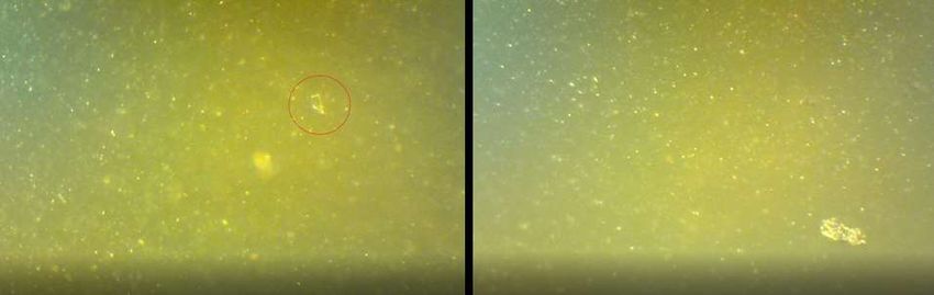
Global Humanitarian Crisis Prevention and Response Unit

    3.Results
3.1. Description of Inclusions
The analysis of all four vial contents identified objects that are similar. For ease of
nomenclature and related descriptions per vaccine, these inclusions are illustrated and defined
individually below-

The identified inclusions were-
    1. Graphene nano ribbons coated with Polyethylene Glycol
    2. Graphene Composite Form 1
    3. Graphene Composite Form 2
    4. Microcrystalline Calcite with Carbonaceous inclusions
    5. Graphene Nano Forms with and without fluorescence
            a. Graphene nano objects
            b. Graphene nano scrolls

    3.1.1. Graphene Composites in the form of Nano Ribbons

Figure. 3.1. Carbon Composite Ribbon Forms from a sample in Moderna 01. The first picture is of a wet
sample and at a low magnification. The second picture is that of the same sample when it is dry and
embedded in solution at a high magnification.

This micro inclusion form appears dark grey in wet slides, resembling filamentous ribbons. As
the material dries, the form becomes nearly transparent. On high magnifications of 40x,
lamellar structures can be identified as sheets. Raman spectrum on these sheets shows a carbon
oxygen bond with added polyethylene glycol imprints (figure 3.9). A longer range of the
Raman requires to be shot for a better grasp on the distribution of defects and other
characteristics of this form.

                                                                                          Page 11 of 48
Global Humanitarian Crisis Prevention and Response Unit
3.1.2. Graphene Composite form 1
GC1 appears in a translucent folded form of about 10-15microns across. The form is
transparent to translucent in transmitted light and shows light structure within it.
Figure 3.2. An assemblage of different forms with some embedded in the solution appearing translucent
and those on the top show a good relief.
GC1 is isotropic under crossed polars. Raman spectroscopic results for this form shows
dominant dual peaks of calcite at 1100 cm-1 and of some form of iron oxide nearly 500 cm-1 .
The spectrum beyond 1300 cm-1 is quite noisy due to the presence of some fluorescence.
3.1.3. Graphene Composite Form 2
These forms are visibly more intricate and give a rather complex Raman signature. The
deciphered components were graphene with iron oxide and calcite. The forms are quite
distinct in their lamellar, structure.
The signature for carbon-carbon bond is quite distinct at 1600 cm-1 , 1100 cm-1 fpeak is
picked up for calcite.
Figure 3.3. Carbon composite Form 2
Page 12 of 48Global Humanitarian Crisis Prevention and Response Unit
3.1.4. Calcite
Figure 3.4. Microcrystalline Calcite with graphene inclusions.
Microcrystalline Calcite is another inclusion which is present in the samples. The form can be
described as lumpy with inclusions of graphene nano forms. The form gives a very clear Raman
signal for Calcite. Calcite is also present in GC1 and GC2 as identified through Raman spectra.
3.1.5. Graphene Nano Forms
Nano forms of graphene were identified in all the samples that were evaluated (figure 3.5).
Upon shooting of the Raman when focused on some of these nano objects (in different
vaccines), the obtained signals were found to be markedly masked with fluorescence.
Identification of Graphene nano forms was therefore conducted on the basis of microscopic
morphological correlation with the forms where, the signal was clear and undebatable (figure
3.24).
Nano forms of Graphene dominated the counts in all the samples. They were found to be
present both in roundish shapes and long spiculate shapes. The rounded forms were almost
entirely found in association with nano particles.
Figure 3.5. (a) Nano forms of Graphene. (b) Nano scrolls.
Page 13 of 48Global Humanitarian Crisis Prevention and Response Unit
Quite conspicuously the spiculate forms were noticeably strewn in random orientations at the
bottom of all the samples that were examined (figure 3.5).
These nano Graphene spicules were impossible to be evaluated by Raman, as their radius was
measurably smaller than the resolution of the laser.
Given, the sheer number of the Graphene scrolls that were noted to be present in the samples,
a separate project with the central focus on these scrolls is underway, where, high resolution
Raman and invasive FTIR investigations are being undertaken to establish the structures of
these scrolls and to get a quantitative estimate of their concentrations.
3.1.6. Crystalised forms of the solution
All the four vaccines are sugar based, and on the edges of the coverslip, as the material dried it
crystallised into sugar crystals form in varied shapes. Figure 3.6. shows some varied forms of
these crystals under polarised light.
Figure 3.6. Four different forms of sugar base crystals identified in a dried Moderna slide. The crystals
display third order interference colours under polarised light with an angular extinction in most cases.
The crystals stem from distinctive nucleii.
Page 14 of 48Global Humanitarian Crisis Prevention and Response Unit
3.2. Moderna 01
3.2.1. Microscopy
Moderna 01 was the first sample that was evaluated. Under wet microscopy, the sample showed
several filamentous forms (figure 3.7). These forms seemed to shed off some of their filaments
in form of small flakes (figure 3.7).
As the slide dried, these filamentous forms became incorporated in the solution medium and
were optically difficult to distinguish from the background (figure 3.8).
Figure 3.7. Filament forms as observed under wet microscopy.
In the dried state, various forms of particulate inclusions were identified. The settlement of
these inclusions took place at multiple levels within the solidified medium. Lighter material
came to rest and settle on top and denser material was found at the bottom of the slide.
The crystalised solution seemed to be quite viscous, and it solidified in a thick multi-layered
pattern which is quite obvious in form of large sheets with perforations (figure 3.9). On the
edges of the slide, the material crystallised into pleochroic crystals stemming from a seed
nucleus (figure 3.6). The optical properties of these structures displayed third order interference
colours along with the distinct presence of growth nucleus clearly identifying these crystals to
be formed of the host solution which includes sucrose.
Page 15 of 48Global Humanitarian Crisis Prevention and Response Unit
The ribbon like structures were seen in two states of deposition. One, where partial structure
of the inclusion was visible above the solidified medium and part of it was almost transparent
and embedded into the medium and the other state was, where the ribbon was incorporated in
the medium and was barely distinguishable. The embedded portions seemed to create a
quadrilateral geometry with a faint visual signal that seemed like a folded ribbon (figure 3.6).
Figure 3.8. Ribbon shaped forms half embedded in the medium. Square and quadrilateral crystals in the
background.
Figure 3.9. Flat, perforated several layered settlements of the medium.
Figure 3.10 exhibits various representative forms that were found in the vaccine. These forms
ranged from transparent or translucent sheet like forms, to dark almost opaque amorphous
carbon like materials of different sizes (figure 3.10j,k).
Page 16 of 48Global Humanitarian Crisis Prevention and Response Unit
Figure 3.10. Various inclusions found within Moderna 01.
Page 17 of 48Global Humanitarian Crisis Prevention and Response Unit
3.2.2. Raman Spectroscopic Investigation
Representative inclusions from Moderna 01 were examined by Raman spectroscopy. The
investigation clearly showed that all the inclusions have a strong carbon signal with confirmed
graphene compositions of some representative forms.
Page 18 of 48Global Humanitarian Crisis Prevention and Response Unit
Figure 3.11. Raman spectrum of representative inclusions in Moderna01.
The two clear Raman signals were obtained from two objects. The flat ribbon like inclusions
exhibited clear graphene spectra integrated with the spectrum of polyethylene glycol and other
minor compounds. The other clear signal was obtained from a calcite microcrystalline form
with a distinct strong peak at 1100cm-1.
The carbon composite forms had a highly complicated signal with clear graphene peak at
1600cm-1, but other peaks at 1100 cm-1 making the signal quite difficult to separate. Further
analysis is currently underway to isolate these signals and identify the other components of this
form of carbon. Some nano amorphous carbon forms showed a clear Graphene signal however,
these forms also exhibited fluorescence which masked the Graphene peak.
These same forms were identified in other vaccine vials as well allowing for the establishment
of their composition consistency across the spectrum of various samples with confidence.
Page 19 of 48Global Humanitarian Crisis Prevention and Response Unit
The compositional identities derived of the inclusions through Raman spectroscopy was used
to quantify the comparative occurrence of these inclusions along a 2D track. This abundance
is presented as counts in the next section.
3.2.3. Counts
Figure 3.12. Count sheets at low and high resolutions along two tracks each for Moderna 01.
The inclusions were counted along two transects. Each transect was 2cm long. The counts were
performed at both: low and high magnifications.
The count distribution is dominated at the low resolution by GComposite 01. At low resolution,
GComposite 2 is present in very low quantities, but the abundance reverses at higher
magnifications. The counts are overall dominated by Graphene nano objects (figure 3.11).
It should be noted, that Graphene nano scrolls were omitted in the counts. This step was
necessary because though, these nano scrolls form a significant percentage of the total counts,
a confirmation of their composition could not be attained within the limitations of this project.
As mentioned above in Section 3.1.5, thorough investigation of these forms now constitutes the
subject of enhanced second investigation project following this report.
Page 20 of 48Global Humanitarian Crisis Prevention and Response Unit
3.3. Moderna 02
3.3.1. Microscopy
The sample material from Moderna 2, was translucent on slide with granular particulate
material in suspension. On observation of the slide material under wet microscopy, several
floating bits of transparent sheet like objects were observed (figure 3.12). Heavier material
sank below as traction bedload and salted across the slide while the medium evaporated under
the microscopic lamp light.
Figure 3.13. Floating sheets of translucent material in a wet sample as observed under an optical
microscope. (b) The settled detrital material at the bottom of the slide. The shadows of the lighter floating
material can be seen as dark hazy figures.
In regions where the solution column was of a significant thickness, bubbles were observed as
flowing with the convection undercurrent towards the edge of the extent of the material. The
locus of these particles was clearly striking, even under the lowest magnification.
The graphs obtained by tracking the movement for these particles were typical of self-
assemblage systems composed of particles coming together under the influence of various
intermolecular forces (figure 3.14).
An interpretation is here drawn in light of the knowledge, that these particles carry the required
m-RNA load and under the designated conditions, exhibit the self assemblage characteristics
using a combination of non-covalent intermolecular interactions such as electrostatic,
hydrophobism, vander Waals and pi effects (figure 3.15).
The self assembly processes seem to be driven by a constantly changing competitive
environment which is driven by the kinematic and thermodynamically driven cursours
following a typical LaMer model. The seeding of the process seems to be around the nucleic
acid form of molecules and Graphene nano objects. According to Kulkarni et al. (2018), the
growth of the particle relies on the pH neutralisation and migration of neutral, unbound
Page 21 of 48Global Humanitarian Crisis Prevention and Response Unit
ionisable lipid towards the LNP core regardless of the payload (mRNA, minicircular DNA or
pDNA).
These particles were observed in ubiquity across the sample preparations and each of these
structures began with the formation of a small seed like particle to which the surrounding
particles aggregated, based on hydrophobic interactions.
What seems to be obvious through observation is that hydrophobic interactions appear to be
the dominant driving force of the LNP growth and electrostatic interactions guide the seed
formation and stability of the final assembly.
Figure 3.14. Motion graph of a characteristic self-assemblage particle. Accelerations in both X and Y
directions show typical staggered forms that typify hydrophobic/philic jumps and movements.
Figure 3.15. Self-Assembled Nano Particles with Payload of mRNA.
On drying of the sample, the solution settled into a thick layer, which retained some form of
moisture. The relatively denser material settled to the bottom, while, the lighter material was
on the top of the solidified solution with an additional detrital layer in the middle of the
medium.
General overview of the dried slide at low resolution exhibited a translucent medium with
forms akin to fibres and transparent sheets on the top surface (figure 3.16).
Page 22 of 48Global Humanitarian Crisis Prevention and Response Unit
Figure 3.16. Moderna 02 Low Magnification overview of the slide.
Page 23 of 48Global Humanitarian Crisis Prevention and Response Unit
On higher magnification, the slide material seemed to abound in carbon-related forms. Figure
3.17 shows different shapes and forms that were noted along various transects across the slide.
It is noteworthy, that the noted deposits are on three separate planes, with a significant
difference in the depth of focus.
Figure 3.17. Representative inclusions across Moderna 02 at high magnification.
Page 24 of 48Global Humanitarian Crisis Prevention and Response Unit
3.3.2. Raman Spectroscopic Investigation
Page 25 of 48Global Humanitarian Crisis Prevention and Response Unit
Figure 3.18. Raman spectra of various inclusions in Moderna 02.
Raman spectroscopy was used on the inclusions of Moderna 2 to identify the main
representations of the sample (figure 3.18). With the exception of calcite composite samples,
the rest showed a highly interfered spectrum. The carbon peaks at around 1600 cm-1 and 1350
cm-1 were only vaguely discernable in the Graphene nano objects. With the fluorescent
background, it was extremely difficult to interpret the spectrum for any other component except
carbon.
A reshooting and further processing of the data is highly recommended for these Moderna
inclusions to be reasonably identified with some confidence.
In the absence of confident Raman signtures, for analysis of these inclusions, a comparative
similarity was used in the presence of the known chemistries of the other inclusions. These
chemistries were used to do the counts which are present in figure.
3.2.3. Counts
Counting of inclusions along four tracks of two at low and two at high magnifications showed
results similar to Moderna 01 (figure 3.19). Graphene Composite 01 were prominently present
Page 26 of 48Global Humanitarian Crisis Prevention and Response Unit
at lower resolution and at higher resolutions, Graphene Nano objects are present in great
abundance.
It is clear from the counts, that the nanoscopic structures far exceed the density counts than
microscopic structures.
It should be noted, that Graphene nano scrolls were omitted in the counts. This step was
necessary because though, these nano scrolls form a significant percentage of the total counts,
a confirmation of their composition could not be attained within the limitations of this project.
As mentioned above in Section 3.1.5, thorough investigation of these forms now constitutes the
subject of enhanced second investigation project following this report.
Figure 3.18. Count sheets at low and high resolutions along two tracks each for Moderna 02.
Page 27 of 48Global Humanitarian Crisis Prevention and Response Unit
3.4. AstraZeneca
3.4.1. Microscopy
AstraZeneca was the third vaccine that was evaluated for its inclusions. Several fresh runs of
the wet samples were seen under the microscope. The AstraZeneca vaccine is almost
transparent when seen through the microscope, making the spotting of any inclusions with
inherent colours slightly easier.
The wet microscopic observation was that the fresh solution exhibited instantaneous
movement of nanoscopic particulate material (figure 3.20) which when observed closely,
evolved from being driven by the convection current, to being quite random. As the solution
dried up, the particles exhibited traction movement.
Figure 3.20. Nano Particles in motion when depth of the solution is greater than the height of the particle.
The particles coalesce to form bigger particles and the dominating influence on the direction of the
movement is through the currents within the solution.
These nanoscopic particles were quite noticeable as white specs in the beginning, as moving in
a swarm like motion in the same general direction. With time, these evolved into bigger
droplets with more random vectors following the principles of self assembly.
As the solution dried, from the heat of the lamp of the microscope from the top, the
sedimentation process began with sheet like forms being deposited on the top of the medium
which begins to crystallise early while the liquid below is still able to flow partially. This
difference in the crystallisation or solidification pace was clearly discernible from a few slide
shots off a video, as are presented in figure 3.21. In the figure, the microscopic form is clearly
visible as it is lying on top of the solid film while the nanoparticles are still in motion in the
background as can be seen by the shifting position of the shadow. A clear output of this
mechanism was that as the medium solidified, it becames more difficult for the nanoparticles
to navigate through the viscous material. The core of the nanoparticles was seeded with carbon
nanotubes and carbon nano objects: formed of some form of detrital carbon particles. These
were dragged along with the eddie currents at the bottom of the medium and as the material
Page 28 of 48Global Humanitarian Crisis Prevention and Response Unit
settled, the flute casts were left behind which were identified easily in the dry microscopic
sections.
Figure 3.21. Lamellar convection columns with solidified top resulting in deposition of the flaky particle.
The arrow points to the position of the nanoparticle in the deeper column of cooler material.
Even, in a wet slide under the microscope, several forms of inclusions were observed. These
are described in detail below.
Nano particle movement and self assembly process was noted in AstraZeneca, similar to what
was noted in the Moderna vaccines. Here too, the nano particles self assembled through typical
signatory movements attributed to the inter-particle forces. While the solution dried up, the
heavier carbon material that had sunk to the bottom of the slide was released from the nucleus
of the nanoparticles and these now began to exhibit traction movement. Figure 3.22, shows the
relative movement vectors of some of these solid materials. When these inclusions settled
down, they sank to the lower strata. A later, high resolution examination of the lower strata
shows a dump of graphene nano objects (figure 3.23) including graphene nano scrolls (figure
3.24).
Page 29 of 48Global Humanitarian Crisis Prevention and Response Unit
Figure 3.22. Distance covered by the object through combined movement vectors
Figure 3.23. Graphene based structures settled down on drying. These structures demonstrated self-
movement against the growing viscosity of the drying solution. When the solution dried, they were found
to have settled at the bottom of the slide.
Figure 3.24. Graphene scrolls or spicules dumped at the bottom of the slide.
Page 30 of 48Global Humanitarian Crisis Prevention and Response Unit
Figure 3.25. Representative inclusions found within AstraZeneca vaccine.
The inclusions identified within AstraZeneca were of the same category as described in section
3.1. These included Graphene composites of two variations, Graphene ribbons impregnated
with polyethylene glycol, Graphene Nano Objects including a vast number of nano scrolls and
a high count of amorphous carbon. Calcite was also distinctly present in the vaccine in a
microcrystalline carbon composite form.
The type inclusions were targeted for Raman identification.
Page 31 of 48Global Humanitarian Crisis Prevention and Response Unit
3.4.2. Raman Spectroscopic Investigation
Page 32 of 48Global Humanitarian Crisis Prevention and Response Unit
Figure 3.26. Raman spectra of various inclusions in AstraZeneca.
Raman spectroscopy results on inclusions within AstraZeneca yielded direct confirmation of
the presence of Graphene in all the identified representative forms.
The carbon composites are of two forms as they are in the Moderna vaccines. These two forms
showed distinct graphene signatures through the characteristic Graphene peaks at 1600 cm-1
and 1350 cm-1. Besides Graphene, the spectrum is dominated by iron oxide and some other
forms of carbon associations.
Further work is ongoing to isolate the signals and help to identify individual compounds. With
the exception of one sample of graphene composite, the effect of fluorescence was found to be
minimal in AstraZeneca .
Calcium composites with carbon also showed the same signature as the ones present in the two
Moderna vaccines.
The inclusions in the calcite composites contained pristine graphene nano particles. These
particles were evaluated to show a clear signal for graphene. Though, the Graphene nano
objects were present in clear association with the Graphene nano scrolls, Raman investigations
were not carried out on scrolls due to the limitation of the laser size and the microscope
Page 33 of 48Global Humanitarian Crisis Prevention and Response Unit
magnification. In any future work on AstraZeneca, shooting Raman on the scrolls would be
one of the main targets in quantifying the concentration of Graphene.
The identification of the inclusions assisted in accurate counting process in the following stage.
Figure 3.25. Count sheets at low and high resolutions along two tracks each for AstraZeneca.
3.4.3. Counts
AstraZeneca counts do not pick up Graphene composite 2 in the lower magnifications however
these are present in significant numbers at higher magnifications. It is clear from the graph that
graphene nano objects dominate the count at higher resolutions.
It should be noted, that Graphene nano scrolls were omitted in the counts. This step was
necessary because though, these nano scrolls form a significant percentage of the total counts,
a confirmation of their composition could not be attained within the limitations of this project.
As mentioned above in Section 3.1.5, thorough investigation of these forms now constitutes the
subject of enhanced second investigation project following this report.
Page 34 of 48Global Humanitarian Crisis Prevention and Response Unit
3.5. Pfizer
3.5.1. Microscopy
Pfizer was the fourth vaccine vial that was evaluated for its inclusions. The vaccine was noted
to be of the same offwhite colour as Moderna. 0.006 µL of the sample was transferred on the
slide for wet evaluation and an equal amount was left in a slightly inclined pippette to allow
for examination in a closed environment in 3dimensions.
The pippete specimen showed some extremely interesting inclusions which were not found
when the slide dried. As the material was sucked into the pipette, distinct translucent to
transparent sheets were seen floating about (figure 3.28). These were recognized from previous
observations in AstraZeneca and Moderna to be the Graphene Composite 1.
Active sedimentation of the denser material on the bottom curve of the cylinder was noted
immediately afterwards (figure 3.29).
Figure 3.28. Floating lighter material. In the background, the golden sparkly particles are the future self-
assembly nano particles that will encapsulate the mRNA.
The two objects of interest that were clearly noted to be floating about but could not be located
once the slide had dried, were: (1) an extremely pointy transparent spicule like object (figure
3.30) and the other was a thin translucent perforated sheet (figure 3.31).
Where, both the objects are of interest to this study, the nature of the spicule remains vital for
future work to identify.
As the solution was poured onto a slide for observation, the mixture exhibited the same
nanoparticulate self assembly mechanism as was observed in both Moderna and AstraZeneca
vaccines. As the material dried out, the inclusions settled at various depths depending upon
their relative densities.
Figure 3.32 shows an assembly of various forms of inclusion that were identified within Pfizer.
These fall in the same category as mentioned in section 3.1.
Page 35 of 48Global Humanitarian Crisis Prevention and Response Unit
Figure 3.29. Sedimentation of the heavier load on the curved bottom of the pipette and the floating
transparent composite 01.
Figure 3.30. Sharp transparent spicule floating in the liquid.
Page 36 of 48Global Humanitarian Crisis Prevention and Response Unit
Figure 3.31. Floating perforated membrane.
Ribbon forms of nearly transparent microforms are found in fair number in the slide. These are
often half embedded in the solution with one end projecting outside the material.
The carbon composites of both form 1 and 2 also are present in great numbers. Form 1 settles
on top of the material while form 2 is found at mid levels of the solidified medium.
Graphene nano forms are present in fair numbers within the slide material along with some
scrolls.
Figure 3.32. Representative inclusions found within Pfizer vaccine.
Page 37 of 48Global Humanitarian Crisis Prevention and Response Unit
3.5.2. Raman Spectroscopic Investigation
Figure 3.33. Raman spectra of various inclusions in AstraZeneca.
Raman spectroscopy was conducted on four selected representative samples for Pfizer (figure
3.32). Three of these samples showed carbon composite signatures with possible graphene in
Page 38 of 48Global Humanitarian Crisis Prevention and Response Unit
them. The signals of amorphous carbon like materials were extremely complex with carbon
along with iron oxide and several other compounds in them. The graphene complex 1 is
graphene with polyethylene glycol signal forming the bulk of the spectrum. Though, for initial
assessments, this study can confirm the presence of graphene in Pfizer, however, the complex
with which it is associated still requires to be established through further work.
One of the sample that was shot, displayed a fair influence of fluorescence. Reshooting this
sample at longer exposures is important to separate the signal from the article of interest with
the signal of the background.
In summary, Raman helped identify the inclusions within Pfizer and these identifications were
classed in the counts.
3.5.3. Counts
Figure 3.34. Count sheets at low and high resolutions along two tracks each for AstraZeneca.
The counts for Pfizer were conducted along four tracks of two each for Low and High
magnifications (figure 3.34). The relative numbers of carbon composite 2 is quite high in one
of the tracks whereas it is completely absent in the first track. At higher magnifications the
counts are dominated by the sheer number of nano graphene objects.
Page 39 of 48Global Humanitarian Crisis Prevention and Response Unit
It should be noted, that Graphene nano scrolls were omitted in the counts. This step was
necessary because though, these nano scrolls form a significant percentage of the total counts,
a confirmation of their composition could not be attained within the limitations of this project.
As mentioned above in Section 3.1.5, thorough investigation of these forms now constitutes the
subject of enhanced second investigation project following this report.
Page 40 of 48Global Humanitarian Crisis Prevention and Response Unit
4. Interpretation, Discussions and Conclusion
Moderna
Active Ingredients Table 1
mRNA List of declared ingredients for
Vehicles the vaccines.
SM102
Polyethylene Glycol
2000 dimyristoyl glycerol (DMG)
Cholesterol
1,2-distearoyl-sn-glycero-3phosphocholine (DSPC)
Inactive Ingredients
Tromethmine
Tromethamine hydrochloride
Acetic Acid
Sodium Acetate
Sucrose
AstraZeneca
Active Ingredients
Adenovirus
Vehicles
L-histidine
L-histidine hydrochloride monohydrate
Inactive Ingredients
Magnesium Chloride Hexahydrate
Polysorbate 80
Ethanol
Sucrose
Sodium Chloride
Disodium edetate dihydrate (EDTA)
Water
Pfizer
Active Ingredients
mRNA
Vehicles
4-hydroxybutyl azanediyl bis hexane -6.1-diyl bis 2 hexyldecanoate
Polyethylene Glycol
N,N di tetra decylacetamide
1,2 di stearoyl sn glycerol 3phosphocholine
Cholesterol
Inactive Ingredients
Potassium Chloride
Monobasic Potassium Phosphate
Sodium Chloride
Dibasic sodium, phosphate dehydrate
Sucrose.
Page 41 of 48Global Humanitarian Crisis Prevention and Response Unit
The study subjects were four vaccine samples (2 of Moderna, 1 of AstraZeneca and 1 of Pfizer).
The objective of this study was to examine these four vaccine vials and document any
undeclared ingredients in the composition of the vaccines, with special focus on graphene and
related products as well as any biological forms.
It should be noted, that all three vaccines have very different chemical ingredients as declared
by the manufacturers (Table 1). Despite, this genetic difference between them, upon
examination, the results reveal common undeclared inclusions in the four vials.
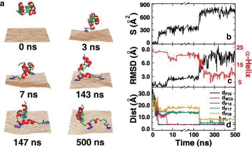
These inclusions are primarily with a focus on graphene and carbon related nano structures in
form of carbon or graphene composites, graphene in association with polyethylene glycol,
graphene oxide, iron oxide compounds, and calcite. The variation in shapes of the logged
inclusions, points to their varied purposes in the wider arena of drug delivery and biosensory
fields.
The shapes identified during the course of this project can be classified into five different
categories-
1. Ribbon Forms (G-PEG)
2. Sheet forms (GC-1)
3. Tubular forms (GC-2)
4. Nano Dots
5. Nano Scrolls
Where, the roles of the Ribbons and sheets remain unclear due to their microscopic sizes, the
tubular forms along with nano dots and scrolls seem to be aimed at enhancing cell acceptance
for the drug.
All three vaccines commonly employ the self-assembling lipid nano particles as drug delivery
mechanisms. Where, the central find of this project has been the confirmation of the presence
of graphene in all four samples, it is important to evaluate this find in the context of the subject
itself.
As mentioned above, self-assembly processes are the core delivery mechanisms for the three
types of vaccines. As has been reported under observations in the Chapter 3, the process in
itself, is primarily dependent on weak intermolecular interactions (Mendes et al. 2013) which,
in the case of lipid nanoparticles is driven and determined by the kinetics and the
thermodynamic environment. However, based on the observations within this study, to
kickstart the nucleation or seeding of these particles in the first state, Graphene nanoparticles
Page 42 of 48Global Humanitarian Crisis Prevention and Response Unit
seem to play an important role. This interpretation is drawn from the observation that each of
the nanoparticles seem to have a graphene core made of nano particles that float in the
beginning and then saltate prior to settling down. This process creates a defect in the solution
medium thereby, facilitating the formation of the nano particle seed. Given, the high
concentration of graphene nano objects in all four vaccines, as the nanoparticles move through
the medium, they must be incorporating not only graphene nanodots, but also nano scrolls,
thereby increasing their binding efficiency and their ability to carry their pay load.
As is already well known, the structure of the lipid nano particle plays a crucial role in
determining its efficiency to carry the nucleic acid payload. However, it is the payload
structure that defines the geometry of the lipid nanoparticles (Hajj et al. 2019, Kulkarni et al.
2018).
Though, these structures can be varied, depending upon the payload itself, the core
responsibilities of the nano lipid particles are the same; viz (1) protection of nucleic acids from
nucleases, (2) controlled release of nucleic acid, (3) cell and tissue selectivity, (4) high delivery
yield, (5) minimal toxicity, and (6) stability especially in long-term storage. These required
characteristics for a vector can been enhanced significantly by use of carbon nanotubes,
graphene and graphene oxide. This process was observed in action, when the experiments were
being conducted during the course of this study and as reported in detail within in Chapter 3.
Nano tubes, both single and multiwalled, along with graphene forms are increasingly, paving
their way into targeted drug delivery (Sattari et al. 2021, Wu et al. 2018, Wierzbicki 2017,
Eatemadi et al. 2014). The samples evaluated during the course of this project have identified
graphene and other carbon composites as forming a striking percent portion of the ingredients
of these vaccines. Given the context of the increasing use of graphene in drug delivery, the
findings of this project seem to fit in the reference frame of attempts towards enhanced and
tailored drug delivery.
The work carried out so far has been essentially a qualitative assessment of the contents. There
are several forms within these vaccines that require quantitative assessment and determination.
A major hurdle in achieving a good quantitative result was the failure to isolate the solid
faction. The method used in this project was the traditional method of slide preparation. It is
Page 43 of 48Global Humanitarian Crisis Prevention and Response Unit
hoped, that if similar work were to be performed on additional samples, vacuum filtering would
be adopted as a mechanism of attaining cleaner samples for both Raman and SEM imaging.
Further, it is quite important to mention, that the source of fluorescence within the samples was
unknown while Raman investigations were underway. Due to extremely tight timescales, it
was not possible to complete the Raman re-runs where the effects of fluorescence would have
been removed through targeted emittance and processing of the data. Despite this, the signals
were interpreted using similar data from various catalogues. It is hoped that further work will
continue on the Raman spectroscopic investigations to attain cleaner spectra and to isolate the
individual spectrums of the current specimens.
In conclusion, it can be stated that the four samples of vaccines (Moderna 1, Modern 2,
AstraZeneca, Pfizer) all contain significant amount of carbon composites, graphene
compounds and iron oxide. These ingredients were undeclared by the manufacturers and are
absent from the list of ingredients for the vaccines.
Page 44 of 48Global Humanitarian Crisis Prevention and Response Unit
5. Bibliography
Bonneville, S., Delpomdor, F., Preat, A., Chevaller,C., Arkaki, T., Kazemlan, M.,
Steele, A., Schrelber, A., Wirth, R., Benning, L.G. (2020). Molecular identification of
fungi microfossils in a Neoproterozoic shale rock. Palentology, 6, 1-11.
Buzgar, N. and Apopei, A. (2009). The Raman study of certain carbonates. Analele
stiintifice ale univesrsitatii, al. i. cuza iasi. Geologie. Tomul LV, 2.
Brennan, T.V., Lin, L., Huang, X. and Yang, Y. (2018). Generation of Luciferase-
expressing Tumor Cell Lines. Bioprotocol, 8(8), 1-1
Childres, I., Jauregui, L.A., Park, W., Cao, H. and Chen, Y.P. Raman spectroscopy of
Graphene and related materials. Chapter 19.
Crystal, R.G. (2014). Adenovirus: The First Effective In Vivo Gene Delivery Vector.
Human Gene Therapy 25,3-11.
Das, A., Chakraborty, B. and Sood, A.K. (2008). Raman Spectroscopy of graphene on
different subsrates and influence of defects. Bulletin of Material Sciences, 31, 3, 579-
584.
Doerfler, W. (2021). Adenoviral Vector DNA- and SARS-CoV-2 mRNA-Based Covid-
19 Vaccines: Possible Integration into the Human Genome Are Adenoviral Genes
Expressed in Vector -based Vaccines.
Dychalska, A., Popielarski, P., Frankow, W., Fabisiak, K., Paprocki, K. and Szybowicz.
(2015). Study of CVD diamond layers with amorphous carbon admixture by Raman
scattering spectroscopy. Materials Science-Poland.
Ghosh, S., Calizo, I., Teweldebrhan, D., Pokatilov, E.P., Nika, D.L., Balandin, A.A.,
Bao, W., Miao, F. and Lau, C.N. (2008). Extremely high thermal conductivity of
graphene: Prospects for thermal management applications in nanoelectronic circuits.
Applied Physics Letters, 92, 151911-1-3.
Griffith, W.P. (1969). Raman spectroscopy of Minerals. Nature, 244, 264-266.
Guo, W., Chen, Z., Feng, X., Shen, G., Huang, H., Liang, Y., Zhao, B., Lo, G. and Hu,
Y. (2021). Graphene oxide (GO)- based nanosheets with combined
chemo/photothermal/photodynamic therapy to overcome gastric cancer (GC) paclitaxel
resistance by reducing mitochondria-derived adenonsine-triphosphate (ATP).
Haskin, L.A., Wang, A., Rockow, K.M., Jolliff, B.L., Korotev, R.L. and Viskupic, K.M.
(1997). Raman spectroscopy for mineral identification and quantification for insitu
Page 45 of 48Global Humanitarian Crisis Prevention and Response Unit
planetary surface analysis: A point count method. Journal of Geophysical Research,
102, 8. 19293-19306.
Hajj, A., Ball, R.L., Deluty, S.B., Singh, S.R., Strelkova, D., Knapp, C.M., Whitehead,
K. (2019) Branched-tail lipid nanoparticles potently deliver mRNA in vivo due to
enhanced ionization at endosomal pH. Small, 15,1 7.
Jiao, L., Zhang, L., Wang, X., Diankov, G. and Dai, H. (2009). Narrow graphene
nanoribbons from carbon nanotubes. Nature Letter, 458, 877-880.
Jiang, W., Liang, F., Wang, J., Su, L., Wu, Y and Wang, L. (2014). Enhanced
electrochemical performances of FeO graphene nanocomposite as anode materials for
alakaline nickel-iron batteries. RSC Adv., 2014,4, 15394-15399.
Kato, M., Guan, S. and Zhao, X. (2021). In-situ observation of graphene using an
optical microscope. Applied Surface Science Advances, 6,
Kim, J., Eygeris, Y., Gupta, M. and Sahay, G. (2021). Self-assembled mRNA vaccines.
Advanced Drug Delviery Review, 170, 83-112.
Kulkarni, A., Darjuan, M.M., Mercer, J.E., Chen, S., van der Meel, R., Thewalt, J.L.,
Tam, Y.Y.C., and Cullis, P.R. (2018). On the formation and morphology of lipid
nanoparticles containing ionizable cationic lipids and siRNA. ACS Nano, 12, 4787
4795.
Kuzmin, V.V., Novikov, V.S., Ustynyuk, L.Y., Prokhorov, K.A., Sagitova, E.A. and
Nikolaeva, G.Y. (2020). Raman Spectra of polyethylene glycols: Comparative
experimental and DFT study. Journal of Molecular Structure, 1217, 5,
Liu, J., Cui, L. and Losic, D. (2013). Graphene and Graphene oxide as new nanocarriers
for drug delivery applications. Acta Biomaterialia, 9(22).
Malard, L.M., Pimenta, M.A., Desselhaus, G., Dresselhaus, M.S. (2009). Raman
spectroscopy in graphene. Physics Reports, 473, 51-87.
Mendes, A.C., Baran, E.T., Reis, R.L. and Azevedo, H.S. (2013). Self-assembly in
nature: Using the principles of nature to create complex nanobiomaterials. Wiley
Interdiscip Rev. Nanomed. Nanobiotechnol 5, 582-612.
Osvath, Z., Zambo, D., Sulyok, A., Palinkas, A. and Deak, A. 2021. Tuning the
nanoparticle rippling of graphene with PEGylated gold nanoparticles and ion radiation.
Carbon Trends, 5, 1-5.
Paillard, V. (2001). On the origin of the 1100cm-1 Raman band in amorphous and
nanocrystalline sp3 carbon. Europhysics Letters, 54(2), 194-198.
Page 46 of 48Global Humanitarian Crisis Prevention and Response Unit
Pierna, J.A.F., Abbas, O., Dardenne,P. and Baeten, V. (2011). Discrimination of
Corsican honey by FT-Raman spectroscopy and chemometrics. Biotechnological
Agron. Society Environment, 15,1, 75-84.
Pop, E., Varshney, V., Roy, A. (2012). Thermal properties of graphene: Fundamentals
and applications. MRS Bulletin 37, 1273-1281
Prawer,S and Nemanich, R.J. (2004). Raman Spectroscopy of diamond and doped
diamond. Philosophical Transactions of the Royal Society of London. Series A.
Mathematical, Physical and Engineering Sciences. 362, 2537- 2565.
Romann, J., Valmalette, J.C., Chevallier, V. and Merlen, A. (2010). Surface
interactions between moleules and nanocrystals in copper oxalate nanostructures.
Journal of Physical Chemistry, C. 114, 10677-19682.
Roy, S. and Jaiswal, A. (2017). Graphene-based nanomaterials for theranostic
applications. Reports in Advances of Physical Sciences, 1,4, 53pp.
Sagitova, E.A., Prokhorov, K.A., Nikolaeva, G.Y., Baimova, A.V., Pashinin, P.P.,
Yarysheva, A.Y. and Mendeleev, D.I. (2018). Raman analysis of polyethylene glycols
and polyethylene oxides. Journal of Physics Conference Series. 999 012002, 1-10.
Sharma, S.K., Misra, A.K., Ismail, S. and Singh,U.N. (2006). Remote Raman
Specroscopy of Various Mixed and Composite Mineral Phases at 7.2m Distance.
https://www.researchgate.net/publication/252521525.
Tallant, D.R., Friedmann, T.A., Missert, N.A., Siegal, M.P. and Sullivan, J.P. (1998).
Raman spectroscopy of amorphous carbon. Material Research Society Symposium
Proceedings, 498, 37-47.
Zhao, X., Liu, L., Li, X., Zeng, J., Jia, X. and Liu P. (2014). Biocompatible graphene
oxide nanoparticle-based drug delivery platform for tumor microenvironment-
responsive triggered release of doxorubicin. Langmuir, 30(34),
Wierzbicki, M., Jaworski, S., Kutwin, M., Grodzik, M., Strojny, B., Kurantowicz, N.,
Zdunek, K., Chodun, R., Chwalibog, A., Sawosz, E. (2017). Diamond, graphite and
graphene oxide nanoparticles decrease migration and invasiveness in glioblastoma
celllines by impairing extracellular adhesion. International Journal of Nanomedicine,
12, 7241-7254.
Wu, J., Phillips, J.A., Liu, H. and Tan, Y.W.(2008). Carbon nanotubes protect DNA
strnds during cellular delivery. ACS Nano.2 (2008) 2023-2028.
Page 47 of 48You can also read