All Roads Lead to Rome: Different Molecular Players Converge to Common Toxic Pathways in Neurodegeneration
←
→
Page content transcription
If your browser does not render page correctly, please read the page content below
cells
Review
All Roads Lead to Rome: Different Molecular Players Converge
to Common Toxic Pathways in Neurodegeneration
Shirel Argueti-Ostrovsky † , Leenor Alfahel † , Joy Kahn and Adrian Israelson *

                                           Department of Physiology and Cell Biology, Faculty of Health Sciences and The Zlotowski Center for
                                           Neuroscience, Ben-Gurion University of the Negev, P.O. Box 653, Beer Sheva 84105, Israel;
                                           shirela@post.bgu.ac.il (S.A.-O.); leenor@post.bgu.ac.il (L.A.); kahnjo@bgu.ac.il (J.K.)
                                           * Correspondence: adriani@bgu.ac.il
                                           † Theses authors contributed equally to this study.

                                           Abstract: Multiple neurodegenerative diseases (NDDs) such as Alzheimer’s disease (AD), Parkin-
                                           son’s disease (PD), amyotrophic lateral sclerosis (ALS) and Huntington’s disease (HD) are being
                                           suggested to have common cellular and molecular pathological mechanisms, characterized mainly
                                           by protein misfolding and aggregation. These large inclusions, most likely, represent an end stage of
                                           a molecular cascade; however, the soluble misfolded proteins, which take part in earlier steps of this
                                           cascade, are the more toxic players. These pathological proteins, which characterize each specific
                                           disease, lead to the selective vulnerability of different neurons, likely resulting from a combination
                                           of different intracellular mechanisms, including mitochondrial dysfunction, ER stress, proteasome
                                           inhibition, excitotoxicity, oxidative damage, defects in nucleocytoplasmic transport, defective axonal
                                           transport and neuroinflammation. Damage within these neurons is enhanced by damage from the

                                    nonneuronal cells, via inflammatory processes that accelerate the progression of these diseases. In
Citation: Argueti-Ostrovsky, S.;
                                           this review, while acknowledging the hallmark proteins which characterize the most common NDDs;
Alfahel, L.; Kahn, J.; Israelson, A. All   we place specific focus on the common overlapping mechanisms leading to disease pathology despite
Roads Lead to Rome: Different              these different molecular players and discuss how this convergence may occur, with the ultimate
Molecular Players Converge to              hope that therapies effective in one disease may successfully translate to another.
Common Toxic Pathways in
Neurodegeneration. Cells 2021, 10,         Keywords: neurodegenerative diseases; proteostasis; misfolded proteins; ALS; Parkinson’s diseases;
2438. https://doi.org/10.3390/             Alzheimer’s diseases; Huntington’s disease
cells10092438

Academic Editor: Naweed I. Syed

                                           1. Introduction
Received: 25 August 2021
Accepted: 14 September 2021
                                                Neurodegenerative diseases (NDDs) are becoming increasingly prevalent in an age-
Published: 16 September 2021
                                           dependent manner, partly because life expectancy has increased in recent years due to
                                           our advanced medical knowledge [1]. The most prevalent of NDDs includes Alzheimer’s
Publisher’s Note: MDPI stays neutral
                                           disease (AD), Parkinson’s disease (PD), Huntington’s disease (HD), amyotrophic lateral
with regard to jurisdictional claims in
                                           sclerosis (ALS), frontotemporal dementia (FTD), spinocerebellar ataxias (SCA), prion dis-
published maps and institutional affil-    eases (PrD) and others. Although diverse in their clinical manifestation, with some causing
iations.                                   memory and cognitive impairments while others affect movement, speech, and breath-
                                           ing [2–5], a large number of NDDs have many common features, including their chronic
                                           and progressive nature, increasing prevalence with age and degeneration of neurons in
                                           specific areas in the central nervous system (CNS) [6]. Interestingly, although NDDs are
                                           typically defined by specific protein accumulations and anatomic vulnerability, they share
Copyright: © 2021 by the authors.
Licensee MDPI, Basel, Switzerland.
                                           many pathophysiological processes.
This article is an open access article
                                                Common underlying mechanisms in neurodegeneration.
distributed under the terms and
                                           1.1. Mitochondrial Dysfunction and Oxidative Stress
conditions of the Creative Commons
Attribution (CC BY) license (https://           The greatest risk factor for NDDs is aging. Mitochondria have been thought to con-
creativecommons.org/licenses/by/           tribute to aging through the accumulation of mitochondrial DNA (mtDNA) mutations [7]
4.0/).                                     and net production of reactive oxygen species (ROS) which then leads to oxidative stress, a

Cells 2021, 10, 2438. https://doi.org/10.3390/cells10092438                                                     https://www.mdpi.com/journal/cells
Cells 2021, 10, x FOR PEER REVIEW 2 of 47
Cells 2021, 10, 2438 2 of 46
and net production of reactive oxygen species (ROS) which then leads to oxidative stress,
aprominent
prominent common
common mechanism
mechanism in in NDDs
NDDs [8].[8]. Oxidative
Oxidative stress
stress is aiscondition
a condition caused
caused by
by the
the imbalance between oxidants and antioxidants in a biological
imbalance between oxidants and antioxidants in a biological system. The imbalance occurs system. The imbalance
occurs as aof
as a result result of the level
the excess excessoflevel
ROS,ofreactive
ROS, reactive
nitrogen nitrogen
species,species, or improper
or improper func-
functioning
tioning of the antioxidant
of the antioxidant system.system. ROS significantly
ROS significantly contribute
contribute to thetodegeneration
the degeneration of neu-
of neuronal
ronal
cells bycells by modulating
modulating the function
the function of biomolecules
of biomolecules (DNA, (DNA,
RNA,RNA, lipidslipids and proteins)
and proteins) and
and processes
processes (nucleic
(nucleic acidacid oxidation,
oxidation, lipid
lipid peroxidation)
peroxidation) in inthethe cell[9].
cell [9].ItItisis well
well known
that
that the brain has a higher oxygen demand and thus consumes 20% more oxygen than
other parts
other parts of the body, making it somewhat of a ‘ROS factory’ as well as the ‘hotspot’ of
neurodegeneration [10].
neurodegeneration [10]. Accordingly,
Accordingly, therethere isis extensive
extensive literature
literature supporting
supporting aa role for
mitochondrial dysfunction
mitochondrial dysfunction and
and oxidative
oxidative damage
damage in in the
the pathogenesis
pathogenesis of of different
different NDDs,
NDDs,
including AD,
including AD, PD,PD, HD,
HD, ALS,
ALS, etc. [8] and Figure 1. In this context, studies related to AD
indicate a relationship between Aβ-induced
indicate oxidative imbalance
Aβ-induced oxidative imbalance and elevated levels of
byproducts of lipid peroxidation, protein oxidation
byproducts of lipid peroxidation, protein oxidation and DNA/RNA and DNA/RNA oxidation
oxidation [11].[11]. In
In PD,
PD, inhibition of mitochondrial complex I and a subsequent increase
inhibition of mitochondrial complex I and a subsequent increase in the production of ROS in the production
of aROS
is is a leading
leading cause for cause for the
the loss loss of dopaminergic
of dopaminergic neuronsneurons
[12,13].[12,13].
In ALS,Inmutated
ALS, mutated
Cu/Zn
Cu/Zn superoxide dismutase (SOD1), the first protein identified
superoxide dismutase (SOD1), the first protein identified to cause familial ALS, to cause familial ALS,
was found
was
to foundinteract
directly to directly
withinteract with voltage-dependent
voltage-dependent anion channelanion (VDAC1) channeland(VDAC1) and to
to subsequently
subsequently disrupt proper mitochondrial function [14,15]. Furthermore,
disrupt proper mitochondrial function [14,15]. Furthermore, a correlation was found be- a correlation
was found
tween between mitochondrial
mitochondrial association and association
ALS disease and ALS disease
progression forprogression
multiple SOD1 for multiple
mutants
SOD1
[16,17]. mutants [16,17].
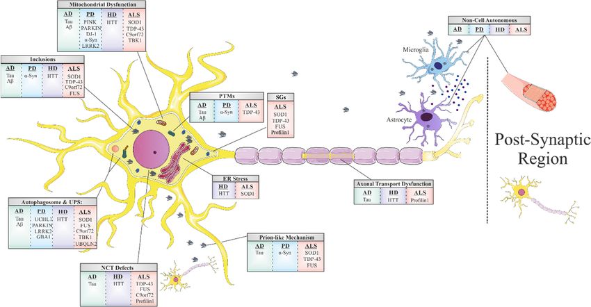
Figure 1.
Figure 1. Shared
Shared mechanisms
mechanisms of toxicity in
of toxicity in the
the most
most common
common neurodegenerative
neurodegenerative diseases.
diseases. Mitochondrial
Mitochondrial dysfunction,
dysfunction,
ER stress, autophagosome and proteasome inhibition, nucleocytoplasmic transport defects, axonal transport dysfunction
ER stress, autophagosome and proteasome inhibition, nucleocytoplasmic transport defects, axonal transport dysfunction
and prion-like propagation are induced by protein misfolding and abnormal interactions. Post translational modifications
and prion-like propagation are induced by protein misfolding and abnormal interactions. Post translational modifications
and non-cell autonomous toxic mechanisms are also common features among the different neurodegenerative diseases.
and non-cell autonomous toxic mechanisms are also common features among the different neurodegenerative diseases.
1.2.
1.2. Stress
Stress Granules
Granules
Another
Another common
common feature
feature underlying
underlying neurodegeneration
neurodegeneration is is the
the formation
formation of stress
of stress
granules (SGs). SGs are subtypes of RNA granules that assemble from the interaction
granules (SGs). SGs are subtypes of RNA granules that assemble from the interaction of of
RNA-binding proteins (RBPs) with untranslated messenger ribonucleoproteins
RNA-binding proteins (RBPs) with untranslated messenger ribonucleoproteins (mRNPs). (mRNPs).
These
These mRNPs
mRNPs areare formed
formed from
from mRNAs
mRNAs halted
halted in
in translation
translation initiation
initiation due
due to
to stress
stress re-
re-
sponse or drugs [18]. SG components that do not bind RNA are presumably recruited
sponse or drugs [18]. SG components that do not bind RNA are presumably recruited to SGs to
SGs through protein-protein interactions. These RNA–protein or protein–protein
through protein-protein interactions. These RNA–protein or protein–protein complexes com-
plexes form membraneless
form membraneless organellesorganelles thatoccur
that usually usually
viaoccur via liquid–liquid
liquid–liquid phase separa-
phase separation (LLPS).
The formation of these membraneless organelles is a strategy of cellular compartmentaliza-
tion that plays a role in several fundamental physiological processes. Interestingly, manyCells 2021, 10, 2438 3 of 46
RBPs contain “low complexity domains” (LCDs), also referred to as intrinsically disordered
protein regions (IDPRs), which consist of different residues that facilitate binding affinity
between SG components. The binding between SG components may result in undesired
amyloid aggregation formation [19,20] and subsequent neurodegenerative responses [4].
The proteins best characterized in this regard include hnRNPA1, hnRNPA2, Fused in
Sarcoma (FUS), TAR DNA-binding protein 43 (TDP43) and tau [21]. The nuclear pore
complex (NPC) is another target in the cell that interfaces the formation of pathological SGs.
Under stress, nuclear pore components, 30 types of proteins called nucleoporins (NUPs),
and other proteins of the nucleocytoplasmic transport (NCT) system translocate to the
cytoplasm and are sequestered to existing SG [22].
1.3. Disruption of Nucleocytoplasmic Transport (NCT)
Impairment of the NPC, in general, and NCT, in particular, has recently emerged as
a central disease mechanism in different NDDs (Figure 1). Proteins that are smaller than
40 kDa can diffuse freely across the nuclear membrane; however, proteins above 40 kDa
require active transport to cross the nuclear membrane. Thus, NCT refers to the active
import and export of large molecules from the cell nucleus via the NPC. This process
facilitates the transition of a protein possessing either a nuclear localization sequence (NLS)
and/or nuclear export signal (NES) which are recognized by specific auxiliary carrier
proteins, importins, and/or exportins, respectively. Dating back to 2015, a connection was
revealed between the disruption of NPC components as well as the nuclear import–export
machinery and different models of the chromosome 9 open reading frame 72 (C9ORF72)
ALS-linked mutation [23]. Following this publication, extensive research progressed in this
regard, with consistent results substantiating the disruption of intact NCT in ALS, caused
by different mutations: TDP43 [24], FUS [25], PFN1 [26], and in other NDDs, including
AD [27] and HD [28,29].
1.4. Prion-Like Propagation
The majority of adult NDDs are characterized by intra- or extracellular aggregation
of misfolded proteins, a subject that will be further discussed later in this review. These
misfolded proteins, frequently, possess self-propagation properties as a mechanism of
spreading in an individual organism, another common feature of NDDs often referred to as
a ‘prion-like’ mechanism (Figure 1). In the self-propagation mechanism, the normal form
of the prion protein (PrPC) undergoes a conformational change into a misfolded protein,
by interacting with pathogenic prion protein (PrPSc). The ability of misfolded proteins
to ‘seed’, that is, to recruit physiological proteins of the same kind and to induce their
conversion into a pathological form, and propagate from cell-to-cell, with the continuous
conversion of the normal protein into its misfolded form, is eventually the cause of the
formation of amyloid aggregates [30]. In vitro and in vivo studies indicate that amyloid
beta (Aβ) and tau in AD, alpha-synuclein (α-syn) in PD [31], and TDP43 and SOD1 in
ALS [32] have similar “prion-like” characteristics. Interestingly, prion-like diseases arise
not only through inherited mutations in the prion protein but also sporadically from the
wild-type form of the protein [33].
1.5. Non-Cell-Autonomous Toxicity
In most of the NDDs, it remains an enigma as to whether the toxicity that arises
from these misfolded prion-like proteins is cell-autonomous. More specifically, whether
the source of neurodegeneration evolves from mutant protein expression and toxicity
exclusively in the vulnerable neuronal population, or whether mutant damage accumu-
lated within other cell types interacting with the affected neurons also contributes to their
degeneration. This question has led to extensive research providing evidence for a non-cell-
autonomous mechanism in which neurodegeneration is strongly influenced by toxicity or
mutant protein expression in both neuronal and non-neuronal cells in their surrounding en-
vironment, notably glial cells in the CNS: astrocytes, oligodendrocytes, and microglia, eachCells 2021, 10, 2438 4 of 46
of which has intimate interaction with neurons (Figure 1). Dysfunctional astrocytes do not
provide sufficient nutrients and antioxidants to the neurons, while dysfunctional microglia
cannot efficiently clear pathogens and cell debris from extracellular space, thus resulting in
chronic inflammatory processes in the brain. In ALS, for instance, there is clear evidence
of damage caused by mutated proteins within microglia and astrocytes as a contributor
to disease progression [34–36]. Furthermore, in PD, α-syn was found to cause damage in
axon-unsheathing oligodendrocytes, thereby inducing secondary neurodegeneration of the
associated neurons [37]. Additionally in HD, mutant huntingtin accumulates in astroglial
nuclei which increases neuronal vulnerability to excitotoxicity [38]. Therefore, research into
targeting glial metabolism and autoimmunity in the CNS can improve the survival and
function of neurons and thus provide a basis for future neuroprotective treatments [39].
1.6. Disruption of Axonal Transport
Another common hallmark of NDDs is the disruption in axonal transport along neu-
ronal cells (Figure 1). Owning their unique morphological structure, neurons are extremely
polarized cell types in comparison to others. This feature makes the regulation of intracel-
lular cargo trafficking crucial to maintain neuronal homeostasis and survival. Anterograde
transport, from the soma to the terminal of an axon, delivers substances such as RNAs,
proteins, and organelles. In the opposite direction, retrograde transport is essential for
processes such as neurotrophic factor signaling, the autophagy–lysosomal pathway (ALP),
and the response to nerve injury. The exception to this is the mitochondria, which moves
bidirectionally in contrast to other cargos [40]. Given that, it is not surprising that mutations
in the axonal transport machinery are associated with neurological diseases [41,42]. For
instance, when axonal transport is disrupted, cargos start to accumulate in an abnormal
fashion, causing the axon to swell. This phenomenon appears also in neurons from post-
mortem studies of patients that suffered from diverse NDDs [40]. Brains from early-stage
AD patients display swellings in basal forebrain axons before amyloid formation [43],
motor axons in ALS patients accumulate phosphorylated neurofilament proteins and or-
ganelles causing swelling [44], and axonal accumulation of synaptic vesicles and α-syn
have been found in hippocampal neurons of patients with PD [45].
1.7. Protein Misfolding
The last and most remarkable common mechanism of NDDs is the misfolding event
of key proteins that accumulate and form toxic aggregates which disrupt the proper
functioning of the cell’s different processes ([2] and Table 1). This well-known feature is
the foundation of all other mechanisms we have mentioned before in this review, and it
has a large impact on our understanding of these mechanisms. Fascinating research from
genetic, neuropathological, cellular, and biochemical studies, as well as from experiments
with in vivo models [46] and postmortem tissues [47], confirm that protein misfolding,
oligomerization, and accumulation in the CNS are the main events triggering pathological
abnormalities that are responsible for NDDs. These proteins undergo misfolding from their
native states to form β -sheet-rich structures, ranging from small oligomers to large fibrillar
aggregates. For each NDD, there is one or more prototype protein which is known to
misfold, accumulate, and form aggregates. These proteins include Aβ and tau in AD; α-syn
in PD, TDP43, SOD1, C9Orf72, and FUS in ALS; TDP43 and tau in FTD; and Huntingtin
(HTT) in HD. In healthy cells, misfolded proteins are either degraded or refolded correctly
by chaperones (proteins that are involved in protein folding). However, if the misfolded
protein accumulates into an amyloid aggregate with a fibril structure, then it has a higher
resistance to degradation due to its extremely stable thermodynamic state. This state
enables it to convert more native proteins into an amyloid form (as mentioned in the ‘prion-
like’ mechanism), thereby escalating the pathology of the cell, leading to the progression of
the disease [48].Cells 2021, 10, 2438 5 of 46
Table 1. Neurodegenerative diseases: principal proteins, mechanism of toxicity, related chaperones and current treatments.
Associated/Pathogenic Related
Disease Associated Genes Normal Function Toxicity Mechanism Current Treatments
Protein Chaperones
MAPT Tau Polymerizes tubulin into microtubules Gain and loss of function
There are evidence for its precursor AChE inhibitors:
participation in: HSP90
HSP104 • Donepezil
AD APP - Neuronal growth • Rivastigmine
HSP70
PSEN1 Amyloid β - Synaptogenesis Gain of toxic function • Galantamine
HSP60
PSEN2 - Neuronal protein trafficking • NMDA antagonist:
HSC70
- Signal transduction • Memantine
- Cell adhesion
- Calcium metabolism
- Regulation of synaptic plasticity
- Synaptic vesicle recycling
SNCA α-Synuclein Gain of toxic function
- Dopaminergic neurotransmission
- Cell differentiation Precursor of dopamine
UCHL1 UCHL1 Neuron-specific deubiquitinating enzyme Gain of toxic function • Levodopa
- E3 ubiquitin ligase Dopaminergic agonists:
PARK2 Parkin Loss of function
- Mitophagy • Ropinirole
function • Pramipexole
of the mitochondria and mitophagy • Transdermal rotigotine,
HSP70
function • Apomorphine
PD PINK1 PINK1 Loss of function HSP40
of the mitochondria and mitophagy • Safinamide
HSP90
- Mitochondrial function NMDA antagonist:
- Mitophagy • Amantadine
- ROS scavenging Anticholinergic drugsBenztropine
- Metal ion binding
PARK7 DJ-1 Loss of function • MAOs and COMTs inhibitors
- Chaperone activity
- Transcriptional regulation
LRRK2 LRRK2 Mitochondrial clearance Gain of toxic function
GBA GCase Lysosomal enzyme Gain and loss of functionCells 2021, 10, 2438 6 of 46
Table 1. Cont.
Associated/Pathogenic Related
Disease Associated Genes Normal Function Toxicity Mechanism Current Treatments
Protein Chaperones
HSP70
- Embryonic development Only supportive treatments are
HD HTT HTT Gain of toxic function HSP40
- Formation of the CNS available
HSP90
Catalyzes the conversion of superoxide
SOD1 SOD1 Gain of toxic function
radical into peroxide and oxygen
TARDBP TDP-43 RNA metabolism Gain and loss of function CCS
C9orf72 C9orf72 Yet to be established Gain and loss of function HSP70
HSP40 Glutamate antagonist:
ALS - Regulates DNA damage
FUS FUS Gain and loss of function HSP110
- RNA metabolism • RiluzoleFree radical scavenger
HSPB8
UBQLN2 Ubiquilin2 Degradation of ubiquitinated proteins Yet to be established • Edaravone
HSP27
- Autophagy Mitophagy MIF
TBK1 TBK1 - Innate immunity Neuroinflammation Loss of function 4-PBA
Apoptosis
PFN1 Profilin1 Actin polymerization Gain and loss of functionCells 2021, 10, 2438 7 of 46
2. Proteostasis
Beyond the role of proteins and their conformational changes in the process of neu-
rodegeneration, they are an essential component in the biological system as a whole,
facilitating almost every process, and thus, their correct formation and regulation is crucial
for intact cellular function. The requirement of each protein differs within the cell and
varies among diverse cells and demands; therefore, there is a need for a strict system to
manage the proteins homeostasis.
Protein homeostasis, also referred to as proteostasis, can be categorized into three
major processes: synthesis, folding and conformational maintenance, and degradation.
First, the protein is synthesized according to its mRNA coding sequence into an amino acid
chain, followed by a series of processes to reach its correct tertiary structure; this proper
conformation is maintained, and in due course, according to protein turn over or in the
case of any detected issue, the protein degrades.
The folded structure of proteins must retain conformational flexibility to function;
thus, a compromise exists between thermodynamic stability and conformational stability.
They are only marginally thermodynamically stable in their physiological environment [49],
and thus, along with physiological stress conditions such as heat, oxidative stress, and
inflammation, proteins are susceptible to the formation of non-native interactions that may
lead to protein misfolding and aggregation [50].
2.1. Chaperones
The protein-coding sequence contains all the needed information to achieve the
correct final structure of the protein; however, this process is challenged due to timescale
constraints and protein overload [49]. Protein overload, which is characterized by the
presence of a large amount of different proteins, enhances protein aggregation by increasing
the affinities between the interacting macromolecules, including folding intermediates.
Furthermore, large proteins with complex structures may expose hydrophobic amino
acid residues to the solvent during their folding, leaving them susceptible to non-native
interaction that might lead to aggregation [51]. Although aggregation primarily leads to
amorphous structures, it may lead to the formation of fibril-like amyloid aggregates [51],
which are more toxic to the cells, as previously described [52,53]. As a means to facilitate
correct protein folding, there is a need for molecular chaperones. These chaperones bind to
the exposed hydrophobic residues to shield them from aggregation and allow the protein
to fold natively [50].
Besides the chaperones’ fundamental role in de novo protein folding, they are also
involved in various aspects of proteome maintenance, such as macromolecule complex
assembly, protein transport, protein degradation, aggregate dissociation, and refolding of
stress denatured proteins [51].
Key chaperones in protein homeostasis are the Heat shock proteins (HSPs). They are
involved in the proper folding and timely degradation of proteins in all cellular compart-
ments; thus, they play a central part in regulating protein quality control and contribute
to protein aggregation and disaggregation [54]. The HSPs are categorized into different
families according to their molecular weight. For example, HSP90 is a highly conserved
ATP-dependent molecular chaperone family involved in protein homeostasis. It is essential
in eukaryotes and it is known to function in the remodeling of hundreds of client proteins
and to participate in many cellular functions, such as protein trafficking, signal transduc-
tion, and receptor maturation [55]. HSP70 is another ATP-dependent molecular chaperone
family. This family is involved in a wide array of cellular processes that involve protein
folding and remodeling [55].
Small heat shock proteins (sHSPs) are ATP-independent molecular chaperones charac-
terized by a small molecular mass ranging from 12 to 42 kDa. Their chaperone function
is to bind to hydrophobic regions of aggregation-prone misfolded proteins and prevent
the formation of insoluble aggregates. However, association with sHSPs does not lead toCells 2021, 10, 2438 8 of 46
substrate refolding to the native state; therefore, their action is an intermediate state and
there is a need for a further process by the HSP70 and HSP90 chaperones [56].
2.2. ER-Associated Degradation (ERAD)
Another important step in the newly-formed protein’s quality control is post-translational
modifications (PTMs) in the endoplasmic reticulum (ER) before progressing towards secre-
tion, in which the proteins need to fit strict quality control standards; otherwise, they are
directed to degradation [57]. ER homeostasis can be disrupted when the folding capacity
is saturated by the expression of misfolded or unfolded proteins, arising for the need to
alleviate the stress by reducing the protein synthesis, increasing protein folding capac-
ity by inducing ER chaperones, or alternatively, by inducing ER-associated degradation
(ERAD) [58] or autophagy [57].
2.3. Ubiquitin-Proteasome System (UPS) and Autophagy
The two key-pathways for protein degradation are the ubiquitin–proteasome system
(UPS) and autophagy, which both utilize ubiquitylation as a degradation signal. The UPS
is responsible for degrading short-lived proteins and soluble misfolded proteins, whereas
autophagy eliminates long-lived proteins, insoluble protein aggregates, whole organelles,
and intracellular parasites [59]. In the ubiquitination enzymatic cascade, first, ubiquitin
is adenylated by the E1 ubiquitin-activating enzyme. Then, through a trans-thiolation
reaction, the ubiquitin is transferred to a cysteine residue on an E2 ubiquitin-conjugating
enzyme. Finally, a specific E3 ubiquitin ligase holds the E2-ubiquitin complex in proximity
to the substrate and stimulates ubiquitin transfer to the substrate, usually to a lysine side
chain [60]. While E3s typically determine target specificity, E2s mainly determine the type
of the conjugated ubiquitin chain, which can be a monomer or a polyubiquitin chain [61].
In the UPS, polyubiquitinated proteins are recognized by the subunits of a multi
catalytic ATP-dependent protease complex. The polypeptides are then cleaved into 3–25
amino acid long fragments, and peptidases further cleave them to single amino acids. In this
way, the recycling of proteins generates an amino acid stockpile, available for new protein
synthesis. Moreover, deubiquitinating enzymes (DUBs) remove ubiquitin or ubiquitin-
like molecules from substrates and disassemble polyubiquitin chains, thus regulating
UPS-mediated degradation in different cellular contexts and playing an important role in
controlling the availability of a free ubiquitin pool in cells, allowing recycling and reuse of
ubiquitin [59]. In autophagy, on the other hand, ubiquitinated proteins are engulfed by a
double membrane structure, called the autophagosome, which subsequently fuses with
lysosomes for degradation [62].
2.4. Stress Granule Formation
Another proteostasis pathway is the formation of SGs, as mentioned above, being the
cells’ fast response to cellular stress [63]. SGs are dynamic, complex, and with composition
and structure that may vary dramatically according to the type of stress [64]. Upon stress
removal, SGs disassemble or are eliminated by autophagy [63].
Supporting the mentioned above, it was shown in different animal models that the
ability to maintain proteostasis declines during aging, which might contribute to the
accumulation of misfolded proteins, aggregation, cellular toxicity, and therefore, cell
death [49].
Ultimately, proper proteostasis is crucial for cellular function. Thus, complications in
any step might lead to various diseases, including NDDs such as AD, PD, HD, and ALS.
3. Alzheimer’s Disease
Alzheimer’s disease (AD) is the most common form of elderly age dementia [65], with
an increasing prevalence with age, ranging from 3% at the age of 60 to 32% at the age of 85
and older [66]. It has a higher occurrence rate in females than in males and is consideredCells 2021, 10, 2438 9 of 46
a common cause of death in elderly individuals [67]. As life expectancy increases, the
prevalence of AD and other dementias will increase accordingly [66].
3.1. Etiology of AD
AD is a progressive NDD, characterized by a gradual deterioration in working mem-
ory, long-term declarative memory, speech, behavior, thinking, and eventually, it interferes
with daily activities [68]. There are two forms of AD, namely, sporadic AD and the rare
familial AD, which accounts for about 1% of the cases [69,70]. AD cases might also be
classified into early-onset AD (EOAD) and late-onset AD (LOAD). EAOD is defined by
those affected before the age of 65, and they account for fewer than 5% of the pathologically
diagnosed AD cases [69]. Researchers have identified more than 230 different autosomal
dominant mutations linked to familial AD, located in the genes for amyloid precursor
protein (APP), presenilin 1 (PSEN1), and presenilin 2 (PSEN2) [71]. As for the sporadic
AD, which accounts for most of the cases, its cause is yet to be defined; however, recent
evidence suggests a complex polygenic disease that involves a convoluted interaction
between several factors such as age, lifestyle, and various susceptible genes such as ε4
allele of apolipoprotein E (APOEε4) [72].
3.2. Pathophysiology of AD
Although familial and sporadic AD differ in their cause, they display similar patholog-
ical processes [73]. Pathologically, the amyloid-beta (Aβ) protein undergoes aggregation
forming amyloid plaques, hyperphosphorylated tau proteins deposit neurofibrillary tan-
gles (NFTs), and there is neuronal loss causing brain atrophy [74]. Cerebral Aβ aggregation
can be detected up to 20 years before clinical symptoms [75]. However, the levels of
oligomers in the brain are the ones that correlate with the cognitive defects severity rather
than the total Aβ burden [76]. As mentioned, APP is a key player in AD. The non-disease
function of APP and its cleavage products are still debated, with evidence pointing to-
wards a variety of functions, including neuronal growth and synaptogenesis, protein
trafficking in neurons, signal transduction across the membrane, cell adhesion, and calcium
metabolism [77]. The cleavage of APP can be amyloidogenic or non-amyloidogenic. In the
non-amyloidogenic process, APP is initially cleaved by α-secretase producing sAPPα and
C83. C83 is further cleaved by γ-secretase, producing P3 and APP intracellular domain,
AICD. On the other hand, in the amyloidogenic process, APP is initially cleaved by the
β-secretase producing sAPPβ and C99. C99 is further cleaved by γ-secretase producing
Aβ and AICD [78]. Importantly, APP cleavage to create Aβ is heterogeneous, resulting
in the production of variable lengths of Aβ, particularly at the carboxyl terminus of the
peptide. The two main forms of Aβ are 40 and 42 length residues, referred to as Aβ40 and
Aβ42, respectively. In non-AD individuals, the majority of the Aβ produced is Aβ40, only
about 5–15% of the total Aβ is Aβ42, and smaller amounts of other Aβs, both longer and
shorter, may be observed [79]. Various mutations in APP, PSEN1, and PSEN2 are known to
increase Aβ42 production [79] which was shown to be the most aggregation-prone and
most neurotoxic form of Aβ [80]. Both amyloidogenic and non-amyloidogenic pathways
are found in healthy individuals [77], whereas there is an increased amyloidogenic cleavage
in the early-onset familial AD and decreased Aβ clearance in both forms of AD [81].
Aβ can also undergo PTMs generating pyroglutamylated Aβ by N terminal truncation
of Aβ and subsequent cyclization of N-terminal glutamate by glutaminyl-cyclase. It
is suggested that pyroglutamylated Aβ plays a major role in AD pathogenesis, since,
while there are similar amounts of non-modified Aβ in aged controls, pyroglutamylated
Aβ is more abundant in AD and glutaminyl-cyclase activity is increased. Furthermore,
pyroglutamylation promotes self-aggregation of pyroglutamylated Aβ and co-aggregation
of non-modified Aβ. Additionally, pyroglutamylated Aβ was shown to be more cytotoxic
than non-modified Aβ and exerts toxicity on primary neurons, neuronal cell lines, and
neurons of TBA2 transgenic mice [82].Cells 2021, 10, 2438 10 of 46
Furthermore, pathological tau can directly interact with components of the NPC
that can accelerate aggregation and fibrilization of tau in the cytoplasm and disrupts
NPC structure and function. Full-length tau was shown to interact with NUP98 and
to a lesser extent with other phenylalanine-glycine containing NUPs, in postmortem
AD, in transgenic mouse models, and In vitro. In addition, evidence exists for NPC
structural defects, including NUP98 pathology, and functional impairments, including Ran
mislocalization, in phospho-tau-positive cells from AD patients, rTg4510 transgenic mice,
and primary neurons [27].
3.3. Aβ Regulation in AD
Aβ production is normally counterbalanced by its clearance via multiple interrelated
processes, including proteolytic degradation, cell-mediated clearance, transport out of
the brain, and deposition into insoluble aggregates [83]. Neprilysin is an Aβ degradation
enzyme that degrades Aβ inside secretory vesicles and on the extracellular surface. In the
early stages of the onset and progression of AD, Neprilysin expression and activity are
selectively reduced in the hippocampus and neocortex, causing a local elevation of Aβ
concentrations at the presynapses in these areas, where the initial neurodegeneration also
takes place. This observed decrease in Neprilysin expression is not due to a loss of neurons
or presynapses, because presynaptic markers remain unchanged upon aging [84].
Dystrophic neurites are engulfed by glia cells, mostly by microglia, and only a small
portion is engulfed by astrocytes. In the presence of Aβ, astrocytes have limited phagocytic
capacity of dystrophies In vitro and in vivo, probably due to the decrease in expression of
the genes encoding for Mertk and/or Megf10, phagocytic receptors that mediate the bind-
ing and/or engulfment recognition of target synapses or cells. Aβ presence affects not only
phagocytosis in astrocytes but also their capacity to degrade phagocytosed materials [85].
As for microglia, they have a double effect in AD; on one hand, they can release some
pro-inflammatory cytokines, stimulating an inflammatory response, ultimately leading to
neuronal injury and death. On the other hand, they may show a beneficial effect via facili-
tating aberrant protein clearance through microglial migration to the damaged aberrant
area, and phagocytosis of unnecessary material in the early stages of AD [86]. Microglia
are inefficient in degrading Aβ dense aggregates, and it is suggested by various In vitro
studies that an inflammatory environment negatively affects the capacity of microglia to
engage in phagocytosis and clear fibrillar Aβ deposits. In this regard, it was shown that
inflammatory cytokine treatment inhibits the ability of microglia to phagocytose Aβ, and
treatment with ibuprofen, known for its anti-inflammatory actions, rescues impairments in
fibrillar Aβ-induced phagocytosis by microglia in response to a pro-inflammatory envi-
ronment. Furthermore, long term use of non-steroidal anti-inflammatory drugs has been
shown to reduce the risk of AD as well as delay disease progression [87].
Another Aβ clearance pathway is perivascular drainage, which is impaired in AD [88].
Known factors affecting perivascular drainage of Aβ include ApoEε4, deposition of im-
mune complexes, and arterial age. The presence of ApoEε4 is associated with reduced
perivascular drainage of Aβ, by competing with Aβ for its efflux by Low density lipopro-
tein receptor-related protein 1 (LRP1), a receptor from the LDL receptor family, from the
interstitium to the circulation. ApoE has three major isoforms (ApoEε4, ApoEε3, ApoEε2),
of which ApoEε4 is the strongest risk factor for AD since it is the least efficient at mediating
Aβ clearance than are the other ApoE isoforms [81].
3.4. Tau Regulation in AD
According to the amyloid cascade hypothesis, the deposition of the Aβ peptide is an
upstream event in the evolution of AD, leading to cell death and/or the development of
NFTs, assembled from hyperphosphorylated Tau via elevation of intracellular calcium ion
levels [89]. Tau is a microtubule-associated protein (MAPT) that polymerizes tubulin into
microtubules, which play an essential role in the normal trafficking of cellular cargo [90].
Tau also participates in maintaining the complex neuronal cell microarchitecture, such asCells 2021, 10, 2438 11 of 46
microtubule assembly and stabilization, particularly in the axons [91]. There is a single
gene coding for tau; however, alternative splicing and PTMs result in different isoforms of
tau. In AD, tau is associated with isoforms with three and four microtubule repeats, where
the ratio of three/four microtubule repeats is highly variable but specific to individual
types of neurons [92]. Under pathological conditions, tau is converted from a microtubule
assembly-promoting protein to a microtubule assembly-disrupting protein. Under such
circumstances, tau is more phosphorylated than normal. The phosphorylation state of a
protein is the net result of the activities of protein kinases and phosphatases acting on it [93].
Phosphoprotein phosphatase-2A (PP-2A), which is colocalized with tau and microtubules
in the brain, is the most active enzyme in dephosphorylating tau. However, in the AD
brain, both the activity and the mRNA of PP-2A are decreased, resulting in abnormal
phosphorylation of tau [94]. Glycogen synthase kinase-3β (GSK-3β) is up-regulated in
AD contributing to abnormal hyperphosphorylation of tau, along with subfamilies of
cytokines that are elevated, including IL-β, IL-6, IL-8, IL18, MIP-1β, S100β, MCP-1, TNF-
α, which all have been shown to be related to tau phosphorylation [91]. The abnormal
hyperphosphorylation of tau makes it resistant to proteolysis by the calcium-activated
neutral protease, and the turnover of hyperphosphorylated tau is several folds slower than
the normal tau [95]. Tau hyperphosphorylation itself decreases tau binding to microtubules
resulting in its dissociation from microtubules in the axon. This dissociation is followed by
translocation to the cell body and proximal dendrites, and aggregation into intracellular
inclusions termed NFTs, leading to impaired axonal function [90,93].
In addition to phosphorylation, tau also undergoes acetylation. Elevated tau acety-
lation precedes the accumulation of NFTs in AD brain, initiated presumably by stress
due to Aβ accumulation or by mutations associated with tauopathy. The cross-talk of
tau acetylation with tau ubiquitination and phosphorylation suggests that tau acetylation
directly contributes to the accumulation of phosphorylated tau and modulates the activities
of kinases involved in tau phosphorylation [96].
Tau can also undergo truncation, which plays an important role in both tau aggrega-
tion and neurodegeneration. In AD brain, several specific truncations of tau have been
identified, including truncation at Asp421 (D421) and Glu391 (E391) that were reported to
make tau proteins more prone to aggregation than the full-length tau [97]. High-molecular
weight tau, which is considered as aggregated tau, lacks the extreme N-terminal portion of
tau suggesting that N-terminal truncated tau should be more relevant than the C-terminal
truncated tau in tau aggregation [97]. However, tau truncation at both the N and C terminus
enhanced pathological activities of tau, including an increase in site-specific hyperphos-
phorylation and self-aggregation. Among the truncated fragments, deletion of either the
first 150 or the last 50 amino acids, which removes the acidic portions of N- or C-terminus
completely, markedly increased the pathological activities of tau. Thus, both the N- and
C-terminal acidic portions of tau appear to protect tau from aggregation [98].
Although tau shares common clearance pathways with Aβ, it cannot be transported
across the blood–brain barrier (BBB) [86]. As a protein rich in lysine residues, tau has
a high susceptibility toward ubiquitination. There are only three E3 ligases competent
to ubiquitinated tau: The C-terminus of the Hsc70-interacting protein (CHIP), the TNF
receptor-associated factor 6 (TRAF6), and axotrophin/MARCH7 [99–102]. Each of these
E3 ligases ubiquitinates tau through different residues, suggesting that each ligase mod-
ulates tau degradation by different mechanisms. CHIP ubiquitinates tau through K48 or
K63 residues and thus, regulates tau degradation via both proteasomal and autophagy
systems. On the other hand, the E3 ligase TRAF6 ubiquitinates tau via K63, suggesting that
ubiquitination mediated by this enzyme may regulate the degradation of tau in the ALP
only [102]. However, phosphorylation of tau at alternative sites prevents ubiquitination
and subsequent clearance [102].Cells 2021, 10, 2438 12 of 46
3.5. Mitochondrial Dysfunction in AD
Mitochondrial dysfunction may also contribute to Aβ and hyperphosphorylated tau
pathologies; conversely, Aβ and tau pathologies can promote mitochondrial defects, with
excessive deposition of Aβ inducing oxidative stress and mitochondrial dysfunction, which
fails to offer ATP for the degradation of targeted proteins by UPS in yeast [86]. Furthermore,
hyperphosphorylated tau as well as Aβ interacts with Dynamin-related protein 1 (Drp1)
causing increased mitochondrial fragmentation and affecting several critical proteins in-
volved in mitophagy (clearance of mitochondria through macro-autophagy), autophagy,
and ubiquitination [103]. Impaired initiation of selective mitochondrial autophagy, due
to decreased levels of activated mitophagic proteins, results in the accumulation of dys-
functional mitochondria and impaired cellular energy metabolism. This is achieved by
interrupting ATP production, which induces adenosine monophosphate-activated protein
kinase (AMPK) activation, leading to excessive mitochondrial fission and further reduces
ATP production in a vicious cycle [104].
3.6. Lysosomal Dysfunction in AD
There is a broad range of genes and proteins associated with the lysosomal network
that are dysregulated in AD, including APOEε4. Recent RNA-sequencing studies of
the entorhinal cortex of AD patients have identified several endosomal-lysosomal genes
deeply affected by APOEε4 expression supporting the role of APOEε4 in the lysosomal
degradation pathway [105]. Familial AD mutations of PSEN1 can compromise lysosomal
Ca2+ efflux, as well as v-ATPase assembly and its proton pumping activity. Thus, there is a
disruption of the lysosome fusion which requires Ca2+ and in the maintenance of optimal
intra-lysosomal pH [105]. Lysosomal biogenesis is up-regulated at the early stages in the
AD brain and in AD models. Later on, the lysosomes become dysfunctional as reflected
by their enlargement as they accumulate autophagic and endocytic substrates. Along
with the associated genes and the substrate accumulation, the disease-related oxidative
damage creates more hydrolase-resistant substrates and generates free radicals from the
peroxidation of cholesterol and other lipids. All of these factors taken together promote the
accumulation of toxic molecules and peptides that can destabilize lysosomal membranes
and initiate cell death programs [106].
3.7. Prion-Like Propagation in AD
Both tau and Aβ were shown to spread in AD brain in a prion-like mechanism. Ev-
idence from histological studies show that aggregated forms of both Aβ and tau spread
through the brain by following typecast neuroanatomical patterns. Furthermore, misfolded
tau can provoke the toxic misfolding of non-pathological tau. In addition, In vitro experi-
ments show that Aβ can bind to tau and induce its oligomerization. These findings raise
the possibility that, in vivo, Aβ oligomers seed the initial formation of tau oligomers, which
can then self-propagate in the absence of additional input from Aβ [107].
3.8. Chaperones in AD
Molecular chaperons take part in AD as well. For example, HSP90 mediates tran-
scription of APP and proteins involved in synaptic plasticity, and the cytosolic HSP90
controls tau levels. Moreover, HSP90 cooperates with the E3 ubiquitin ligase CHIP to
target tau for proteasomal degradation [108]. Furthermore, HSP90 inhibits Aβ toxicity
by binding misfolded Aβ peptides and preventing further aggregation using an ATP-
independent pathway or by changing the conformation of Aβ to a state that is less prone
to aggregation via an ATP-dependent pathway [109]. However, HSP90 levels are reduced
in AD, especially in the hippocampus, entorhinal cortex, and cingulate gyrus, which are
the most affected in AD [108]. Another involved HSP is HSP104, which inhibits the fibril-
lization of monomeric and protofibrillar forms of Aβ in a concentration-dependent but
ATP-independent manner [110].Cells 2021, 10, 2438 13 of 46
HSP70 and Hsc70 are involved in the degradation of hyperphosphorylated tau by
ubiquitinylation of tau, with the cooperation of the ubiquitin ligase CHIP. Tau aggregation
is largely associated with a decrease in HSP70 activity. In this regard, crossing APP
mutant mice with mice overexpressing HSP70 shows a decrease in Aβ levels, a decrease in
neurodegeneration, and recovery in terms of cognitive function. This outcome is not due to
a decrease in the production of Aβ, but results from the activation of its phagocytosis and
degradation systems via the insulin-degrading enzyme (IDE), an Aβ-degrading enzyme
involved in the degradation of Aβ [111]. However, some researchers have shown an
increase in HSP70 levels at the early stages of AD, with HSP70 co-localizing with tau
protein aggregates [111,112].
HSP60’s role in AD is controversial; although it inhibits Aβ amyloid aggregation by
inhibiting molecular pathways leading to peptide fibrillogenesis, its extracellular release
by microglia increases the production of other pro-inflammatory factors through binding
to toll-like receptor 4 (TLR-4) and stimulating neuronal cell death [51].
3.9. Current Treatments of AD
Currently, there are four FDA approved treatments available for AD patients, all of
which provide only limited therapeutic benefits. The treatments target AD neuropathology
which includes elevated glutamate levels in the cerebral spinal fluid. High glutamate
levels are believed to disrupt cellular communication and contribute to neuronal loss
including loss of basal forebrain cholinergic neurons, leading to a decreased availability
of acetylcholine at the neuronal synapse contributing to memory decline [113]. Three of
the approved treatments (Donepezil, Rivastigmine, Galantamine) are acetylcholinesterase
inhibitors that increase the availability of acetylcholine at synapses and improve cholin-
ergic transmission. These drugs were shown to maintain mental functions by improving
cognition, daily and global function, and some behavioral manifestations of AD. The last
approved treatment is Memantine, an N-methyl-D-aspartate (NMDA) receptor antagonist
that blocks the effects of sustained, pathologically elevated levels of glutamate in order to
decrease the neuronal dysfunction and the excitotoxicity injury to the brain. However, the
efficacy of Memantine administration in patients with AD remains inconclusive [114].
3.10. Clinical Trials in AD
Following evidence supporting the toxic role of Aβ and tau in AD, ongoing im-
munotherapies targeting these proteins have been conducted. Aducanumab is a human
monoclonal antibody, developed by Biogen that selectively binds to Aβ fibrils and soluble
oligomers. Aducanumab failed effectivity analyses in two identically designed phase III
AD trials, leading to abandoning of its development. However, after reanalyzing data from
the trials in order to include patients who had continued in the studies Biogen applied for
FDA marketing approval of Aducanumab [115]. Fortunately, in July 2021, Aducanumab
was finally approved. BAN2401, similar to Aducanumab, is an intravenous administrated
monoclonal antibody that binds to aggregated Aβ and promotes its removal by Fc receptor-
mediated phagocytosis. BAN2401 also showed significant efficacy on both biomarker and
clinical outcomes [116]. Tau immunotherapies showed successful outcomes in several
AD animal models and were approved for clinical trials, some of which are currently at
phase I, and others at phase II [117]. For instance, sodium selenate (Na2 SeO4 ), a negatively
charged anionic compound that activates PP-2A In vitro and in vivo, was found to reverse
memory deficits, and to reduce tau phosphorylation in animal models of AD. Sodium
selenate was found to be safe and well-tolerated in patients with mild to moderate AD at
doses of up to 30 mg per day for 24 weeks at phase IIa clinical trial, along with benefits on
diffusion magnetic resonance imaging [118]. In addition, Methylene blue, was shown to
prevent tau aggregation or dissolve existing aggregates and to interfere with downstream
pathological consequences of aberrant tau. TauRx had developed a second-generation
compound, LMTM, which is a stabilized and reduced form of methylthioninium withCells 2021, 10, 2438 14 of 46
better absorption and tolerability. In 2018, TauRx started Phase III trials (NCT03446001)
aiming to determine the safety and efficacy of LMTM treatment [118].
4. Parkinson’s Disease
First described by James Parkinson in his classic monograph “Essay on the shaking
palsy” [119], Parkinson’s disease (PD) is the second most common age-related NDD after
AD with prevalence ranging from 100 to 200 per 100,000 people and an estimated annual
incidence of 15 per 100,000. Interestingly, an increase in the global prevalence is expected to
double from 6.2 million cases in 2015 to 12.9 million cases by 2040 attributed to the general
increase in age of the population [120].
4.1. Etiology of PD
As in most NDDs, aging is considered a major risk factor of PD. Accordingly, the
development of PD is rare before the age of 50 years with a mean onset between the
ages of 65–70. Nevertheless, earlier age of onset is seen in few genetic variants which are
thought to be involved in 5–10% of the PD cases, namely, Parkin (PRKN), PTEN-induced
putative kinase 1 (PINK1), and DJ-1, and are heritable in an autosomal recessive man-
ner [121]. However, numerous PD cases are heritable in an autosomal dominant manner
including α-syn (SNCA), leucine-rich repeat kinase 2 (LRRK2), ubiquitin carboxyl-terminal
hydrolase L1 (UCH-L1), and VPS35, which all-cause late-onset PD resembling sporadic
PD. Moreover, mutations in the gene glucocerebrosidase (GBA), encoding to a lysosomal
enzyme whose activity is lacking in Gaucher’s disease, is considered as a major risk factor
for PD [122]. Several environmental factors are also associated with increased risk of
PD including pesticides, rural environment, wood preservatives, and other environmen-
tal factors or endogenous toxins such as 1-methyl-4-phenyl-1, 2, 3, 6-tetrahydropyridine
(MPTP) neurotoxin, rotenone, paraquat (1, 19-dimethyl-4, 49-bipyridinium dichloride),
and 6-hydroxydopamine (6-OHDA) [123,124].
4.2. Pathophysiology of PD
The pathophysiological hallmark of PD is the presence of cytoplasmic insoluble
aggregation referred to as Lewy-bodies (LBs) and Lewy-neurites. These aggregates play
a major role in PD by damaging many subcellular processes [125–127]. There are mainly
two types of LBs that differ in their morphological structure, the classic (midbrain and
brainstem) type and the cortical type. The classical LBs are intraneuronal, round inclusions
with a hyaline core and pale peripheral halo. The cortical LBs are irregular in their shape
and usually lack a conspicuous halo or core. Immunohistochemical studies have shown
that LBs found in human postmortem tissues consistently contain the proteins: α-syn,
neurofilament proteins, and ubiquitin. Similarly, Lewy neurites are abnormal neurites
containing granular substance and α-syn filaments, comparable to those found in LBs [128].
4.3. α-Synuclein in PD
Evidence from biochemical, and biophysical approaches on animal models suggest
that soluble α-syn, in its prefibrillar form, is the early and toxic species that contributes to
neurodegeneration in PD [129,130]. Additionally, a presence of either point mutation (e.g.,
A53T, A30P, E46K, A18T, A29S, H50Q, and G51D) in the α-syn encoded gene or a whole
locus multiplication will result in an autosomal dominant version of PD [131]. Despite the
consensus about its importance in PD-related neurodegeneration, α-syn’s physiological
role remains debated. This protein is a small 140 amino acids protein that can be divided
into 3 distinct regions: the N-terminal region (1–60 residues), a central hydrophobic region
which has a high propensity to aggregate (61–95 residues), and a highly acidic C-terminal
domain (96–140 residues) [132]. α-syn is mainly located in the presynaptic terminals of
neurons, and is thought to facilitate synaptic plasticity, vesicular packing, trafficking, and
docking [132–136]. Furthermore, evidence exists for the presence of α-syn in the cell nucleiCells 2021, 10, 2438 15 of 46
associated with histones and nuclear DNA [137,138]. However, further investigation is
required to shed light on its nuclear role.
Several questions have arisen regarding how the mutated form of α-syn causes PD.
Is it a loss of the normal function of the protein? Or is it a toxic effect of altered forms of
the mutant protein? Perhaps both? In addition, what role does it play in sporadic cases
of PD? One hypothesis suggests that in our cells, α-syn exists in equilibrium as both an
unstructured monomer as well as in the form of a fibrillization resistant α-helical tetrameric
oligomer. Thus, decreased tetramer: monomer ratio caused by missense mutations in the
α-syn gene can lead to a shift favoring a pathologic mode [139].
Although α-syn plays a fundamental role in PD, it seems that loss of protein function
is not the only contributing factor to the development and progression of the neurodegen-
eration observed in PD, as α-syn knockout (KO) mice did not show any symptoms or sign
of neurodegeneration. However, these results can be explained by functional redundancy
between α-syn and the closely related β- and γ-synuclein. Therefore, a triple KO to all
synuclein proteins was made causing some behavioral abnormalities and alterations in
synaptic neurotransmission. However, no signs of neurodegeneration were observed [140].
Thus, it is more likely that α-syn mediates neurodegeneration in PD via a gain of a new
toxic function.
4.3.1. α-Syn PTMs
Additionally, it has been hypothesized that it is the PTMs, possibly mediated by
environmental factors, that α-syn undergoes, which contribute to its pathogenesis. In
particular, α-syn undergoes phosphorylation, ubiquitination, truncation, nitration, and
O-GlcNAcylation which are found to be present in PD brain tissues. In LBs, α-syn is found
to be phosphorylated at serine 129 (Ser-129) and 87 (Ser-87) residues [141,142]. In the
brain of healthy individuals, a small fraction (~4%) of the total α-syn is phosphorylated at
Ser-129 residue in comparison to (~90%) PD brain patients, indicating the prevalence of
this form of PTMs of α-syn protein [143,144]. In vitro, several kinases have been shown
to phosphorylate α-syn at Ser-129 residue including casein kinase I (CKI), casein kinase
II (CKII) [145], the G protein-coupled receptor kinases (GRK) [146], LRRK2, and polo-like
kinases (PLK) [147]. Interestingly, it seems that in some cases, the same PTM can have
different effects depending on the kinase involved in the phosphorylation. For example,
phosphorylation at Ser-129 by CKII may promote aggregation [143,148], while phospho-
rylation at Ser-129 by PLK2 promotes degradation [149]. This hyper-phosphorylation of
α-syn was found to have an impact on its solubility, membrane-binding properties, and
subcellular distribution, thus leading to a pathologic state [150]. Indeed, it was found
that the activity of PP-2A, an important protein for dephosphorylating of α-syn at Ser-129
residue, was reduced in a neuropathological analysis of brains from PD patients [151].
4.3.2. α-Syn Inclusions
The emerging of synucleinopathies is thought to be through the process of soluble
α-syn conversion into insoluble aggregates via spontaneous nucleation [152]. In 2003, Braak
and colleagues, using histopathological studies in post-mortem PD patients, described
their hypothesis of the retrograde transport of α-syn from the gastrointestinal tract via
the vagus nerve to the ventral midbrain, where it selectively degenerates dopaminergic
neurons of the substantia nigra (SN) [153], later supported by mouse model studies [154].
Moreover, the Braak group presented an association between α-syn pathology in different
brain regions and PD patients’ symptoms [155]. Their observations provide support for
the prion-like mechanism attributed to α-syn, as a cause for PD, which was suggested
before [156]. In accordance, heavy metals such as copper and iron, which are known to
be accumulated in PD brains [157,158], were found to accelerate prion-like propagation of
α-syn fibrils [159].You can also read