Ultrasound diagnosis of acrania with major low-lying placenta and polyhydramnios; case report
←
→
Page content transcription
If your browser does not render page correctly, please read the page content below
Case Report
Ultrasound diagnosis of acrania with major low–lying placenta and polyhydramnios;
case report
Jared N. Oblitey1, William K. Antwi1, Benard O. Botwe2 and Mary K. Oblitey2
Ghana Med J 2021; 55(2): 165-168 doi: http://dx.doi.org/10.4314/gmj.v55i2.12
1
Department of Radiography, School of Biomedical and Allied Health Sciences, University of Ghana, Korle Bu,
Accra, Ghana.
2
Department of Radiology, Korle Bu Teaching Hospital, Accra, Ghana
Corresponding author: Jared N. Oblitey E-mail: Joblitey@ug.edu.gh
Conflict of interest: None declared
SUMMARY
Acrania is a rare foetal anomaly in which the calvaria is absent, and the meninges come into direct contact with the
amniotic fluid. Acrania is the most common anomaly in the acrania – exencephaly – anencephaly spectrum, with an
incidence of 3.68 to 5.4 per 10,000 live births. We present a case of a primigravida who presented for an ultrasound
on account of vaginal bleeding in early cyesis. Transabdominal ultrasound showed a viable foetus at 13 weeks without
a calvaria, with the brain in direct contact with amniotic fluid. There was a low-lying placenta extending from the
posterior to anterior part of the lower uterine segment, completely covering the internal cervical os (major low–lying
placenta), a placental cyst and polyhydramnios (amniotic fluid index, AFI of 17 cm). A diagnosis of acrania with
major low–lying placenta and polyhydramnios was made. Detailed ultrasound is required to detect acrania at 13 weeks.
The diagnosis of acrania is required to help direct patient counselling and maternal expectation. When acrania and
major low–lying placenta occur in the same patient, both diagnoses must be promptly made concurrently, regardless
of gestational age and without waiting for placental trophotropism and migration to occur first.
Keywords: Acrania; exencephaly; anencephaly; major low-lying placenta; placental cyst
Funding: None declared
INTRODUCTION
Acrania is a foetal anomaly in which there is a complete Ultrasound showed a viable foetus at 13 weeks gestation
or partial absence of the calvaria above the orbits and su- (Figure 1) with increased amniotic fluid (amniotic fluid
praciliary ridge, allowing the meninges to come into di- index, AFI of 17cm). The brain appeared well-formed,
rect contact with amniotic fluid. Acrania is the most com- but the bony landmarks for the biparietal diameter (BPD)
mon anomaly in the acrania–exencephaly–anencephaly measurement were difficult to obtain. In addition, the
spectrum, with an estimated incidence ranging from 3.68 brain had no bony covering (Figure 2) and appeared to
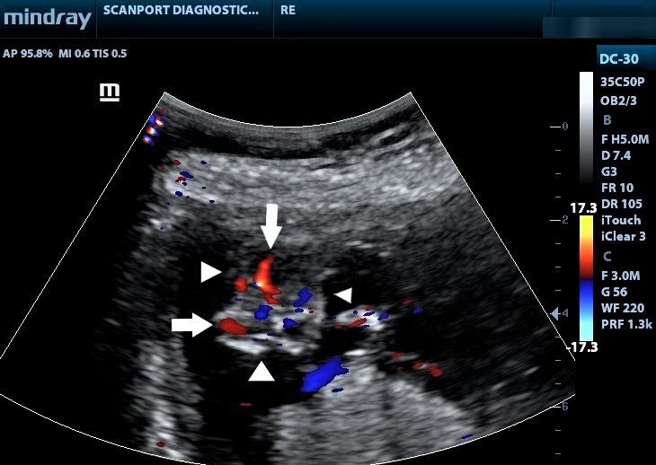
to 5.4 per 10,000 live births.1 When acrania occurs, the bulge freely into the amniotic fluid, with dysplastic in-
brain is exposed to the amniotic fluid with a risk of me- tracerebral vessels on colour Doppler (Figure 3).
chanical and chemical trauma through friction with the
uterine wall, placenta and foetal parts.2 Subsequently, the
brain may suffer progressive degenerations that could ul-
timately lead to partial or total disappearance of the brain
tissues, particularly from the 14th week of gestation.2,3
This presentation seeks to highlight a case of acrania di-
agnosed at 13 weeks to raise the index of suspicion
among obstetricians and radiologists.
CASE REPORT
A primigravida presented at a health facility for a first-
trimester ultrasound examination. She had been experi-
encing bleeding per vaginam with blood clots, and her
obstetricians were concerned she might have been abort- Figure 1 Coronal Ultrasound image showing a foetus at 13 weeks with
no calvaria. White arrowheads show the brain tissue without bony cov-
ing. There was no relevant past obstetric history. ering. The amniotic fluid index (AFI) was 17 cm.
165 www.ghanamedj.org Volume 55 Number 2 June 2021
Copyright © The Author(s). This is an Open Access article under the CC BY license.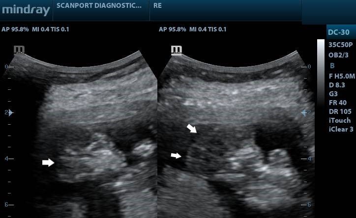
Case Report

                                                                              the placenta, suggesting a placental cyst (Figure 5). A di-
                                                                              agnosis of acrania with major low–lying placenta, pla-
                                                                              cental cyst and polyhydramnios was made. The patient
                                                                              was referred back to an obstetrician for counselling and
                                                                              continued management. Consent for the case to be pub-
                                                                              lished (including images, case history and data) was ob-
                                                                              tained from the patient.

Figure 2 Ultrasound images showing sagittal and parasagittal images
of the brain demonstrating the absence of the calvaria. See the margins
of the brain (white arrows) without bony margins.

                                                                              Figure 5 Ultrasound image of the lower uterine segment showing the
                                                                              placenta completely covering the internal cervical os. Note also the
                                                                              cystic lesion (white arrow) at the anterior part of the placenta, in keep-
                                                                              ing with a placental cyst.

                                                                              DISCUSSION
                                                                              The acrania – exencephaly – anencephaly spectrum se-
Figure 3 Axial Colour Doppler ultrasound images of the head showing
the absence of the calvaria. Note the dysplastic vessels (white arrows)
                                                                              quence is the most severe neural tube defect and results
within the brain. The arrowheads indicate the margins of the bare brain.      from a failure to close the rostral end of the neural tube
                                                                              and abnormal migration of mesenchymal tissue which
                                                                              normally covers the cerebral hemispheres.4 Embryologi-
                                                                              cally, this failure of migration occurs at the beginning of
                                                                              the 4th week, and the major insult is a lack of cranial de-
                                                                              velopment.

                                                                              The main mode of diagnosing acrania is by obstetric
                                                                              ultrasound.5 The calvaria was absent in the foetus in this
                                                                              case. The International Society of Ultrasound and Gynae-
                                                                              cology (ISUOG) has issued guidelines emphasizing the
                                                                              need to recognise the foetal head, cranial bones, choroid-
                                                                              plexus and cerebral ventricles for all first trimester
                                                                              scans.6 According to Santana et al. 6, from the 11th week
                                                                              onwards, cranial ossification ought to be evident. It was,
                                                                              however, reported in Denmark7 that detection rates for
                                                                              the acrania- exencephaly – anencephaly were low on
Figure 4 Lower uterine segment ultrasound image showing the urinary           basic scans done before the 11th week, requiring expert
bladder and internal cervical os (C) completely covered by the placenta       maternal – foetal sonologists for improved detection.7
from anterior to posterior. White arrowheads indicate placental mar-
gins. Note also the anterior placental cyst. UB refers to the urinary blad-
der.                                                                          The key sonographic features of acrania are absent cal-
                                                                              varia. The damaged brain tissue presents as a 'Mickey
The placenta completely covered the internal cervical os                      Mouse' sign3 in the early stages and then as echogenic
(Figure 4 and Figure 5). In addition, there was an ane-                       particles in the amniotic fluid in advanced stages of
choic cyst with an internal septum at the anterior part of                    the condition.

166                                                                                           www.ghanamedj.org Volume 55 Number 2 June 2021
                      Copyright © The Author(s). This is an Open Access article under the CC BY license.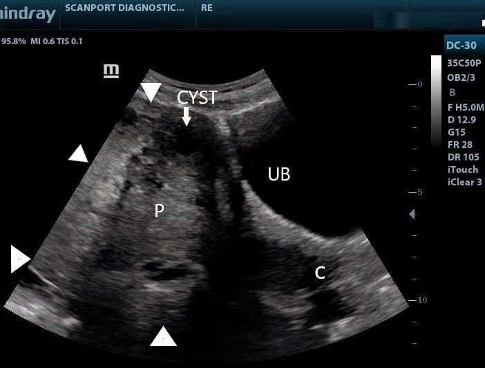
Case Report

These echogenic particles lead to an increased amniotic         Oppenheimer13 further asserted that because of normal
fluid texture, a feature reported by both Cafici5 and           trophotropism and first-trimester migration of the pla-
Santana6 as a likely very first indication of the spectrum.     centa, a diagnosis of placenta praevia should not be made
It has thus been suggested that this feature could be a         until after 15 weeks gestation. However, there may be
potential marker for first-trimester acrania. However,          spontaneous abortion or elective termination in cases of
such echogenic particles were not noted in this current         acrania shortly after diagnosis and before placental mi-
case, likely because much brain destruction had not yet         gration has occurred. Therefore, the early diagnosis of
occurred at the time of examination.                            placenta praevia becomes critical to help craft a delivery
                                                                plan that prevents maternal haemorrhage.14 In our opin-
It has also been suggested that the acrania – exencephaly       ion, therefore, whenever acrania and major low–lying
– anencephaly spectrum is actually the same disorder but        placenta occur in the same patient, the diagnosis of major
presents as acrania in the first trimester and later as anen-   placenta praevia must be made promptly, regardless of
cephaly after the brain destruction has occurred.5 For in-      gestational age and without waiting for placental migra-
stance, during one longitudinal study in the same cohort        tion or trophotropism (which may not be allowed to occur
of patients8, it was noted that exencephaly was the pre-        in such a patient). We further assert that such a clear
dominant finding in the first trimester. In contrast, in the    diagnosis of Major placenta praevia (even when made in
second trimester, the classic appearance of anencephaly         the early second trimester) helps quickly classify the
was seen more often. In fact, in one particular foetus, the     patient as high risk and appropriate interventions
transition from exencephaly to anencephaly was docu-            instituted. The differential diagnoses of acrania include
mented by serial ultrasound exams.8                             osteogenesis imperfecta and hypophosphatasia. In both
                                                                cases, however, bones are present around the brain but
Many types of the acrania–anencephaly spectrum have             are poorly mineralized. The presence of other bones with
been described. One retrospective study of 88 cases of          fractures may also help distinguish acrania from osteo-
the spectrum concluded that there are six subtypes;             genesis imperfecta.15
described as overhanging, elongated, bilobular, cystic,
foreshortened, and irregular, with the first three              Management of acrania is generally by elective termina-
accounting for 75% of all cases.9 In his pictorial essay,       tion. However, in all cases, early in - utero diagnosis
Santana et al1 showed images of acrania with cysts in the       helps manage maternal expectations and directs appropri-
brain, describing them as cystic acrania. Sepulveda et al10     ate counselling. In most cases, the prognosis is lethal,
also recently described a previously unknown first-             with 65% dying in –utero and 35% dying during delivery.
trimester presentation of the sequence in three foetuses.       Short term survival (minutes to days) has also been re-
There was a constriction ring noted around the external         ported as well as a 2 – 5% recurrence risk in future preg-
skull base with an enlarged globular head. It was,              nancies.16
therefore, suggested that this subtype of the sequence
may be due to segmental amniotic rupture with the               CONCLUSION
remnants entrapping the foetal head. All three cases were       Detailed ultrasound is required to detect acrania at 13
either aborted therapeutically or suffered intrauterine         weeks gestation. The diagnosis of acrania is required to
demise.                                                         help direct patient counselling and maternal expectation.
                                                                When acrania and major low–lying placenta occur in the
The current case was associated with polyhydramnios             same patient, both diagnoses must be made promptly and
(AFI of 17cm; reference range at 15 weeks – 8.7 -13.7           concurrently regardless of gestational age and without
cm, 5th -95th percentile)11, a feature also reported by         waiting for placental trophotropism and placental migra-
Santana.1 In Cameroun, Koaum12 described a case of              tion to occur first.
anencephaly complicated by acute polyhydramnios and
underscored the need to actively screen for anomalies in
all cases of polyhydramnios.
                                                                REFERENCES
                                                                 1.   Santana EFM, Araujo Júnior E, Tonni G, Costa
This current case showed acrania with major low–lying                 FDS, Meagher S. Acrania-exencephaly-anenceph-
placenta diagnosed at 13 weeks and a cystic area within               aly sequence phenotypic characterization using
the placenta. Oppenheimer13 also showed images of a                   two- and three-dimensional ultrasound between 11
patient with placenta percreta, placenta praevia and                  and 13 weeks and 6 days of gestation. J Ultrason.
anencephaly. However, whether a relationship exists                   2018;18(74):240-246.
between the acrania–exencephaly–anencephaly spectrum             2.   Gaillard F and Namdev R. Acrania anencephaly se-
and low–lying placenta is not clear.                                  quence. Retrieved from: https://radiopaedia.org/ar-

167                                                                          www.ghanamedj.org Volume 55 Number 2 June 2021
                  Copyright © The Author(s). This is an Open Access article under the CC BY license.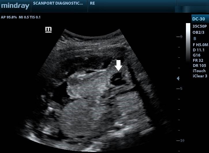
Case Report

      ticles/acrania-anencephaly-se-                         10. Sepulveda W, De La Maza F, Meagher S. An
      quence#:~:text=Acrania%20anencephaly%20se-                 Unusual First-Trimester Ultrasound Presentation of
      quence%20is%20the,brain%20tissue%20(anen-                  the Acrania-Anencephaly Sequence: The "Turkish
      cephaly)%201. Date accessed: 11/11/2020.                   Turban" Sign. J Ultrasound Med. 2020;39(4):829-
 3.   Chatzipapas IK, Whitlow BJ, Economides DL. The             832.
      'Mickey Mouse' sign and the diagnosis of anen-         11. Hallak M, Kirshon B, Smith EO, Smith EO & Cot-
      cephaly in early pregnancy. Ultrasound Obstet Gy-          ton DB. Amniotic fluid index. Gestational age-spe-
      necol. 1999;13 (3): 196-9.                                 cific values for normal human pregnancy. J Reprod
 4.   Greene ND, Copp AJ. Neural tube defects. Annu              Med. 1993;38 (11): 853-6.
      Rev Neurosci. 2014;37:221-242.                         12. Kouam L, Kamdom-Moyo J. L'anencéphalie asso-
 5.   Cafici D, Sepulveda W. First-trimester echogenic           ciée à l'hydramnios. A propos d'un cas diagnostiqué
      amniotic fluid in the acrania-anencephaly se-              tardivement par l'examen échographique au
      quence. J Ultrasound Med. 2003;22(10):1075-                troisième trimestre de la grossesse [Anencephaly
      1081.                                                      associated with hydramnios. A case diagnosed late
 6.   Salomon LJ, Alfirevic Z, Bilardo CM, Chalouhi              by ultrasonographic examination in the third tri-
      GE, Ghi T, Kagan KO. et al.: ISUOG practice                mester of pregnancy]. Rev Fr Gynecol Obstet.
      guidelines: Performance of first-trimester fetal ul-       1994;89(2):96-99.
      trasound      scan. Ultrasound    Obstet     Gyne-     13. Oppenheimer DC, Mazaheri P, Ballard DH, Yano
      col. 2013; 41: 102–113.                                    M, & Fowler KJ. Magnetic resonance imaging of
 7.   Fleurke-Rozema JH, van Leijden L, van de Kamp              the placenta and gravid uterus: a pictorial essay. Ab-
      K, Pajkrt E, Bilardo CM, Snijders RJ. Timing of            dominal radiology (New York), 2019;44(2):669–
      detection of anencephaly in The Netherlands.               684.
      Prenat Diagn. 2015 May; 35(5):483-5.                   14. Gibbins KJ, Einerson BD, Varner MW, Silver RM.
 8.   Beinder E, Grüner C, Erhardt I, Mauch E, Begon S.          Placenta previa and maternal hemorrhagic morbid-
      Die Exenzephalie-Anenzephalie-Sequenz. Ultras-             ity. J Matern Fetal Neonatal Med. 2018;31(4):494‐
      challdiagnostik in der Frühschwangerschaft [The            9.
      exencephaly-anencephaly sequence. Ultrasound di-       15. Kline-Fath B, Bulas D and Bahado–Singh R.
      agnosis in early pregnancy]. Ultraschall Med.              (2015). Fundamental and Advanced Fetal imaging.
      1995;16(4):192-195.                                        Wolters Kluwer Health.
 9.   Wertaschnigg D, Reddy M, Ramkrishna J, da Silva        16. Obeidi N, Russell N, Higgins JR, O'Donoghue K.
      Costa F, Sepulveda W, Rolnik DL, Meagher S. Ul-            The natural history of anencephaly. Prenat Diagn.
      trasound Appearances of the Acrania-Anencephaly            2010;30(4):357-360.
      Sequence at 10 to 14 Weeks' Gestation. Journal of
      Ultrasound in Medicine 2020;39(9):1695-1700.

168                                                                      www.ghanamedj.org Volume 55 Number 2 June 2021
                 Copyright © The Author(s). This is an Open Access article under the CC BY license.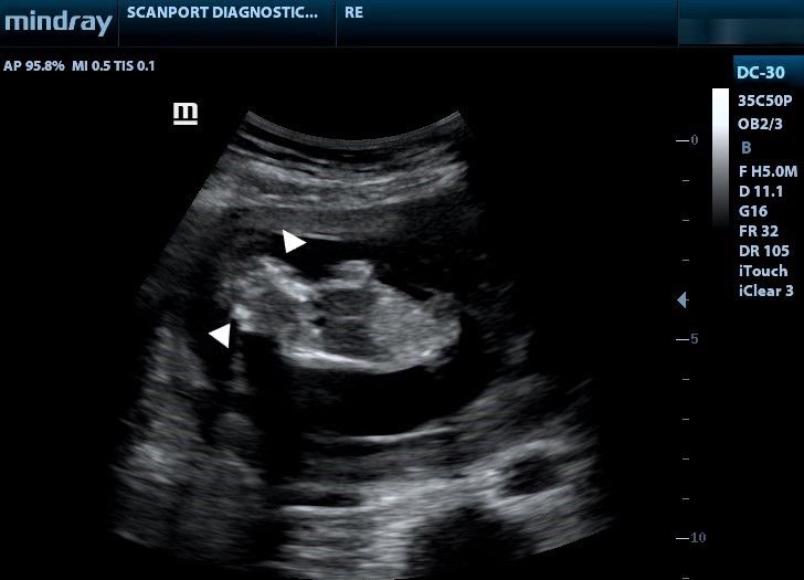
You can also read