Transcatheter aortic valve replacement for bicuspid aortic valve disease: does conventional surgery have a future?
←
→
Page content transcription
If your browser does not render page correctly, please read the page content below
Keynote Lecture Series

Transcatheter aortic valve replacement for bicuspid aortic valve
disease: does conventional surgery have a future?
Breandan B. Yeats1, Pradeep K. Yadav2, Lakshmi P. Dasi1, Vinod H. Thourani3
1
    Department of Biomedical Engineering, Georgia Institute of Technology and Emory University, Atlanta, GA, USA; 2Department of Cardiology,
Marcus Valve Center, Piedmont Heart Institute, Atlanta, GA, USA; 3Department of Cardiovascular Surgery, Marcus Valve Center, Piedmont Heart
Institute, Atlanta, GA, USA
Correspondence to: Vinod H. Thourani, MD. 95 Collier Road, Suite 5015, Atlanta, GA 30309, USA. Email: vinod.thourani@piedmont.org.

                  Bicuspid aortic valve (BAV) disease is the most common form of congenital heart valve defect. It is
                  associated with aortic stenosis (AS), aortic insufficiency, and aortopathy. Treatment of severe AS requires
                  valve replacement which historically has been performed with surgical aortic valve replacement (SAVR).
                  Recently, transcatheter aortic valve replacement (TAVR) has emerged as a promising alternative. However,
                  increased rates of adverse outcomes following TAVR have been shown in BAV patients with high amounts of
                  calcification. Comparison between TAVR and SAVR in low surgical risk BAV patients in a randomized trial
                  has not been performed and TAVR for BAV long-term performance is unknown due to lack of clinical data.
                  Due to the complexity of BAV anatomies and the significant knowledge gap from the lack of clinical data,
                  SAVR still has many benefits over TAVR in low surgical risk BAV patients. It also remains common for BAV
                  patients to have an aortopathy, which currently can be treated with surgical techniques. This review aims to
                  outline BAV associated diseases and their treatment strategies, the main TAVR adverse outcomes associated
                  with anatomically complex BAV patients, TAVR strategies for mitigating these risks and the current
                  state of cutting-edge 3D printing and computer modeling screening methods that can provide otherwise
                  unobtainable preoperative information during the BAV patient selection process for TAVR.

                  Keywords: Transcatheter aortic valve replacement (TAVR); surgical aortic valve replacement (SAVR); bicuspid
                  aortic valve disease

                                Submitted Feb 15, 2022. Accepted for publication Jun 07, 2022.
                                doi: 10.21037/acs-2022-bav-20
                                View this article at: https://dx.doi.org/10.21037/acs-2022-bav-20

Introduction                                                                    patients. This review aims to provide an overview of the
                                                                                management of calcific aortic stenosis (AS) associated with
Bicuspid aortic valve (BAV) disease affects people worldwide
                                                                                BAV. This will include the risks and benefits or SAVR and
as the most common congenital heart valve defect. Present
                                                                                TAVR and introduce strategies for reducing complications
in 2–4% of the population, BAV disease is associated with
high rates and early onset of calcific aortic valve disease                     by utilizing 3D printing and computer modeling techniques.
(CAVD), aortic insufficiency, and ascending aorta and/
or aortic root dilation compared to tricuspid aortic valve                      BAV anatomy and classification
patients (1-4). Historically, valve replacement has been with
surgical aortic valve replacement (SAVR) however, in the                        Due to the nature of congenital defects, the BAV anatomy
past decade transcatheter aortic valve replacement (TAVR)                       has many different types that differ in the way the cusps
as emerged as a safe and minimally invasive alternative.                        are fused. The most commonly used BAV classification
Currently, there is much debate on whether we can safely                        method was made by Sievers et al. and considers the specific
transition to TAVR for BAV patients due to the much more                        cusps that are fused and the number of fused raphe (5).
complex anatomy compared with tricuspid AV morphology                           Recently, Jilaihawi et al. created a classification method

© Annals of Cardiothoracic Surgery. All rights reserved.        Ann Cardiothorac Surg 2022;11(4):389-401 | https://dx.doi.org/10.21037/acs-2022-bav-20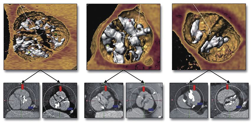
390                                                                                                            Yeats et al. SAVR or TAVR in BAV

                             Tricommissural                    Bicommissural raphe-type                   Bicommissural non raphe-type
                             21/91 (23.3%)                          50/91 (55.6%)                               19/91 (21.1%)
      morphology
        Leaflet
      orientation
        Leaflet

                      Coronary             Mixed             Coronary              Mixed                  Coronary              Mixed
                     cusp fusion         cusp fusion        cusp fusion          cusp fusion             cusp fusion         cusp fusion
                    13/21 (61.9%)       8/21 (38.1%)       44/50 (88.0%)        6/50 (12.0%)            4/19 (21.1%)        15/19 (78.9%)

Figure 1 BAV classification scheme factoring valve shape consisting of tricommissural, bicommissural raphe-type, and bicommissural non-
raphe-type groups with coronary cusp fusion and mixed cusp fusion subgroups (6). Reprinted by permission from Elsevier (6). BAV, bicuspid
aortic valve.

more relevant to TAVR that considers the shape of the                        stenosis or insufficiency and aortic aneurysms. The
valve opening including tricommissural, bicommissural                        following sections will outline these common diseases
raphe, and bicommissural non-raphe types with coronary                       associated with BAV disease and the available treatment
cusp fusion and mixed cusp fusion subtypes (6) (Figure 1).                   options.
The length of fusion can also vary as the valve can have
cusps that are partially fused. Common imaging modalities
                                                                             Aortic stenosis
include high temporal resolution echocardiography, which
can also provide hemodynamic information and high spatial                    AS is mainly caused by CAVD, which is a degenerative
resolution computed tomography (CT). Echocardiography                        disease involving the growth of calcific lesions within
alone has been shown to misdiagnose 88.5% of BAV                             the soft tissue. CAVD is commonly associated with
patients as being tricuspid who were later diagnosed with                    BAV disease—roughly 50% of the population of people
BAV disease from cardiac CT (7). This stresses the need                      effected by CAVD also have BAV disease (2). CAVD is also
for routine high resolution CT imaging for planning valve                    known to have an earlier onset in BAV patients compared
replacements. However, even with CT imaging it can still                     to tricuspid patients—many of them are younger and
be difficult to properly assess the fine structures of the                   have lower surgical risk (8). Often BAVs have elliptically
anatomy due to the calcium blooming effect when a large                      shaped openings and heavily calcified raphes making the
amount of calcium is present.                                                anatomy considerably more complicated compared to
                                                                             tricuspid patients. Treatment for CAVD is through full
                                                                             replacement of the valve and historically has been through
BAV associated diseases and treatment
                                                                             SAVR consisting of surgical excision of the native tissue
BAV disease is commonly associated with aortic valvular                      and implantation of a bioprosthetic or mechanical valve.

© Annals of Cardiothoracic Surgery. All rights reserved.     Ann Cardiothorac Surg 2022;11(4):389-401 | https://dx.doi.org/10.21037/acs-2022-bav-20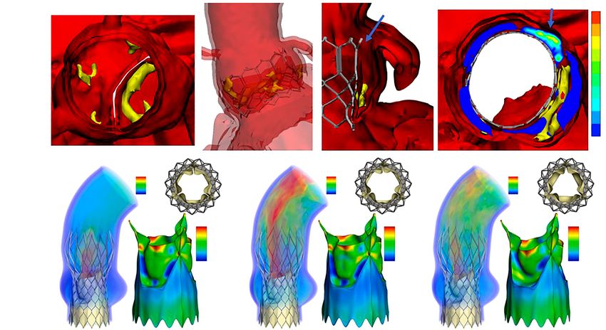
Annals of Cardiothoracic Surgery, Vol 11, No 4 July 2022 391
Tricuspid aortic valve Bicuspid aortic valve
No raphe Noncalcifed raphe Calcified raphe
(type 0) (type 1) (type 1)
A C E G
calcification
Mild leaflet
B D F H
Excess leaflet
calcification
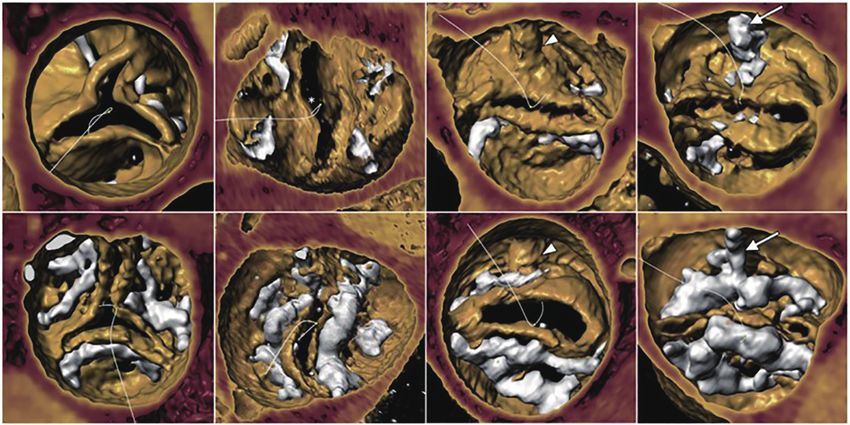
Figure 2 Study performed by Yoon et al. comparing TAVR outcomes with leaflet calcification volume and distribution across BAV types with
tricuspid control. Mild leaflet calcification on the top row and excess leaflet calcification on the bottom row with the tricuspid control, non-
raphe BAV, noncalcified raphe BAV, and calcified raphe BAV from left to right. Excess leaflet and raphe calcification BAV had highest rates
of PVR and aortic root rupture following TAVR. Reprinted by permission from Elsevier (13). TAVR, transcatheter aortic valve replacement;
BAV, bicuspid aortic valve; PVR, paravalvular regurgitation.
However, the field is continually transitioning to TAVR due respect to variance in BAV anatomy makes the TAVR for
to positive clinical comparisons to SAVR and fast patient BAV patient selection process very important.
recovery times. Recent clinical trials have shown similar
outcomes between SAVR and TAVR after five years in
Aortic insufficiency and aortopathy
tricuspid aortic valve patients leading to Food and Drug
Administration (FDA) approval of TAVR for low surgical Aortic insufficiency is defined by improper closing of the
risk patients (9,10). Some clinical data exists showing similar aortic valve cusps causing regurgitation during diastole.
outcomes between tricuspid and BAV patients which led to Aortic insufficiency has been found to be present in 47% of
FDA removal of the cautionary label of TAVR use in BAV BAV patients at the time of BAV diagnosis (3). Additionally,
patients (11,12). However, these studies were not long term aortopathy is known to be present in roughly 50% of BAV
and tended to be very patient selective by not encompassing patients (4). Vessel dilation is classified by the location of
the complex anatomy BAV cases. As of 2020, ~9% of TAVR dilation; type 1 is dilation of the ascending aorta, type 2 is
cases are for BAV patients (7). This number is expected to dilation of the aortic arch and type 3 is dilation of the aortic
rise yet there has not been a comprehensive clinical trial root (4). Aortic insufficiency in BAV patients can be caused
comparing TAVR for tricuspid and TAVR for BAV low by aortic dilation, cusp prolapse, or cusp restriction from
surgical risk patients. Yoon et al. showed that the presence of the raphe (14). Treatment strategy depends on the cause of
the calcified raphe and excess leaflet calcification increases insufficiency—aortic dilation requires replacement of the
the risk of aortic root rupture and paravalvular regurgitation ascending aorta and/or the aortic root and cusp prolapse/
(PVR) (13) (Figure 2). The difference in outcomes with restriction can commonly be repaired by resecting the
© Annals of Cardiothoracic Surgery. All rights reserved. Ann Cardiothorac Surg 2022;11(4):389-401 | https://dx.doi.org/10.21037/acs-2022-bav-20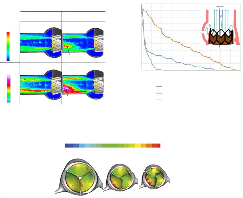
392                                                                                                          Yeats et al. SAVR or TAVR in BAV

raphe (14). Additionally, when the cause of insufficiency is               increases the risk of PVR. The decision between residual
aortic root dilation it is not necessary to replace the valve              PVR or root rupture remains the most important and
as valve sparing root replacement has been shown to be                     controversial aspect of TAVR in BAV patients.
safe and effective regardless of the severity (15). Although                  Currently, it is very difficult to determine the risk
TAVR has been performed in some high- and extreme-risk                     of aortic root rupture other than analyzing the calcium
patients with severe aortic insufficiency, the results have not            severity. This is due to high patient variability in calcium
been favorable and is not recommended.                                     distribution where calcium can be present on the cusp
                                                                           free edges, at the aortic annulus and in the left ventricular
                                                                           outflow tract making it difficult to determine which calcific
TAVR adverse outcomes: concern in complex
                                                                           nodules may puncture the tissue. Additionally, the patient
BAV anatomies
                                                                           tissue compliance has a significant amount of variability,
TAVR was originally applied in patients with tricuspid                     most closely associated with age, making it very difficult to
aortic valve morphologies with commonly symmetric root                     know how much stress is needed to rupture the tissue (24).
anatomy and calcific burden. However, BAVs are commonly                    Balloon volume is mostly determined procedurally where
elliptical and their diameter is not always constant with                  the operator may stop increasing volume if they feel too
respect to height (16). Additionally, the calcium distribution             much resistance from the native tissue which may leave the
can be more complex with the presence of a calcified                       patient with PVR. These counteracting strategies make it
raphe. The following sections will outline the common                      difficult to avoid moderate to severe PVR and aortic root
complications following TAVR and their relevance to BAV                    rupture in TAVR for complex BAV patients, and determine
patients including PVR, aortic root rupture, valve leaflet                 the risk of such complications prospectively due to the
thrombosis, increased permanent pacemaker implantation                     complex nature of the anatomy (Figure 3). Ultimately, BAV
(PPI), coronary access and durability.                                     patients with high amounts of calcium should be strongly
                                                                           considered for SAVR if they are low surgical risk, and if
                                                                           TAVR is necessary, a degree of caution should be used.
PVR and aortic root rupture

PVR is one of the most common complications associated
                                                                           Thrombosis and durability
with TAVR and is defined as backwards blood flow from the
aorta to the left ventricle during diastole due to improper                Thrombosis and valve durability are very important in the
device sealing and, when moderate to severe, has been                      low surgical risk population as they serve to benefit more
associated with increased patient mortality (17). Stent                    from a longer valve lifespan as to avoid re-intervention.
undersizing, high calcium volume, stent eccentricity and                   Leon et al. found higher thrombosis rates following TAVR
unfavorable positioning are all causes of PVR (18-20).                     (2.6%) compared to SAVR (0.7%) and similar valve
Specifically, this is one of the most common complications                 deterioration after two years in low surgical risk tricuspid
following TAVR in those with a bicuspid AV as the annulus                  patients (25). Valve thrombosis normally forms in the neo-
in these patients is eccentric and can have high volumes of                sinus, which is the pocket located between the bioprosthetic
calcium. Proper CT analysis and oversizing of the TAVR                     cusp and the native cusp, and is likely caused by reduced
prosthesis may mitigate PVR (21,22). The most dreaded                      blood flow in this region (26-29). Valve durability is thought
complication remains an aortic root rupture following                      to be related to the stress on the native leaflets which is
a balloon-expandable device or post-dilation in a self-                    dependent on the geometry of the deployed stent and the
expandable device deployment and has an increased risk                     hemodynamics of the device (30-32). Due to the complex
of occurring when high volumes of calcium are present.                     dynamic nature of the TAVR deployment it is common for
Aortic root rupture is a result of excess damage to the native             the stent to be asymmetrical post procedure. This includes
tissue from calcium protrusion (23). BAV patients with high                stent ellipticity, under expansion, and tilt with respect to
calcium volume and a calcified raphe have the highest risk of              the native sinus (29,33,34). Each of these asymmetrical
rupture (4.5%) as opposed to BAV patients with low calcium                 deployment types have been associated with adverse
volume (0.9%) (13). A common preventative strategy is                      hemodynamic environments demonstrated in three separate
for the operator to reduce the balloon filling volume as to                studies (Figure 4) (35-37). Stent eccentricity was found
reduce the stress on the native tissue (23). However, this                 to create localized regions of high Reynolds shear stress

© Annals of Cardiothoracic Surgery. All rights reserved.   Ann Cardiothorac Surg 2022;11(4):389-401 | https://dx.doi.org/10.21037/acs-2022-bav-20
Annals of Cardiothoracic Surgery, Vol 11, No 4 July 2022 393
PVR and aortic root rupture risk factors in BAV patients located below the membranous septum which is located
between the right and non-coronary sinuses (38,39). It is
PVR
Aortic root associated with device type, deployment depth, oversizing/
rupture
overexpansion, membranous septum length, preexisting
Undersizing/ Oversizing/
underexpansion Excess leaflet overexpansion
conduction abnormalities and severity of AS (38,40). It
calcification is also less common in younger healthy individuals (38).
No post balloon
Calcific raphe
Post balloon However, PPI occurrence can have increased consequences
dilation dilation
in young patients as it has various long-term adverse effects
Elliptical opening Low tissue on the heart (38). Thus, PPI risk should be considered when
compliance evaluating young low surgical risk BAV patients for TAVR
vs. SAVR as deploying above the membranous septum,
Figure 3 PVR and aortic root rupture risk factors in BAV patients. using a balloon-expandable TAVR and avoiding oversizing
Isolated PVR risks including valve undersizing/underexpansion, can help mitigate PPI risk.
absence of post balloon dilation, native elliptical opening, isolated
aortic root rupture risk factors including oversizing/overexpansion,
Coronary obstruction and access
post balloon dilation, and low tissue compliance, and factors
increasing both risks including excess leaflet calcification and Coronary access is always a concern during TAVR patient
presence of a calcific raphe. PVR, paravalvular regurgitation; BAV, selection. This is especially true in young patients when
bicuspid aortic valve. considering their lifetime management of AS. Coronary
obstruction is associated with low coronary heights and
small sinus of Valsalva diameters (41). BAV patients have
and increased turbulence intensity (35). Under-expanded been found to have larger sinus of Valsalva diameters and
deployments were found to increase bioprosthetic leaflet no difference in coronary heights compared to tricuspid
folding and increase neo-sinus blood residence time (36). patients (8). In some BAV patients the coronary ostium
Tilted stent deployment with respect to the native sinus location can be abnormal and located close to the native
was found to increase the fluid residence in the sinus commissure where in combination with heavy calcification
that the stent was angled away from (37). Each of these can decrease the gap between the native tissue and the
hemodynamic environments could increase thrombosis risk coronary ostium following TAVR. When considering
and lower device durability. secondary replacement, it is important to consider the type
Asymmetric deployment is caused by reduced balloon and position of the implant. TAVR deployment in SAVR
volume, native valve elliptical opening and increased calcium has a preventative strategy for coronary obstruction known
volume which is of particular concern for anatomically as BASILICA which involves intentional laceration of the
complex BAV patients who have elliptical openings, high bioprosthetic leaflet allowing for blood flow to the coronary
calcium volumes, and often have reduced balloon volume artery (42). However, if TAVR is performed as the first
deployments. Overall, BAV patients are more predisposed intervention it can increase the risk of coronary access issues
to asymmetrical deployments and adverse hemodynamic following the second TAVR procedure, especially if the first
environments with use of TAVR. This indicates thrombosis valve was a self-expanding prosthesis that had supra-annular
and valve deterioration will likely be a concern long-term. leaflets. The supra-annular bioprosthetic cusp suturing
However, because long-term data on TAVR for BAV is for the self-expandable device places the bioprosthetic
very limited it is not certain thrombosis and durability will cusps higher up on the frame and closer to the coronary
be a concern and requires additional long-term follow-up ostium. Similarly, increased deployment heights also place
clinical studies. the bioprosthetic cusps closer to the coronary ostium.
BASILICA has been shown to be much less effective for
TAVR in TAVR (43). When evaluating young low surgical
Pacemaker implantation
risk BAV patients for TAVR vs. SAVR the height and
PPI is one of the most common complications following location of the coronary artery should be measured and the
TAVR. PPI is caused by interaction between the stent risk of coronary obstruction following the first and second
frame and/or the catheter with the conduction system intervention should be considered.
© Annals of Cardiothoracic Surgery. All rights reserved. Ann Cardiothorac Surg 2022;11(4):389-401 | https://dx.doi.org/10.21037/acs-2022-bav-20394 Yeats et al. SAVR or TAVR in BAV
Hemodynamics in asymmetrical stent deployments
Eccentric Tilted
1.0
Ratio of particles remaining in the sinus
5.0 L/min
0.9
Circular Eccentric 0.8
Reynolds
shear 0.7
stress, N/m2
120 0.6
110
100
90
0.5
80
70 0.4
60
50
40 0.3
30
20 0.2
10 Localised area of high
0 Reynolds shear stress
Turbulent 0.1
kinetic
energy, N/m2 0
230 0 1 2 3 4 5 6 7 8 9 10
220
200 Cardiac cycles
180
160
140 With untilted Sapien
120
100 With Sapien tilted away from sinus
80
60
Region of elevated
40
turbulence intensity
With Sapien tilted towards sinus
20
Underexpanded
Blood residence time (i.e., stasis) on transcatheter aortic valve
leaflets after 100%, 90%, and 80% stent expansion
0 0.05 0.1 0.15 0.2 0.25 0.3 0.35 0.4 0.45 0.5 0.55 0.6 0.65 0.7 0.75 0.8 (s)
100%
90% 80%
Figure 4 Hemodynamics of asymmetrical stent deployments commonly seen post TAVR in BAV patients. Eccentric deployment showing
a localized area of high Reynolds shear stress and a region of elevated turbulence intensity compared to the circular case (left), tilted
deployment with respect to the native sinus showing increased fluid residence in the sinus that the TAV is tilted away from (right), and
underexpanded deployment showing increased leaflet folding and increased neo-sinus blood residence time (bottom) (35-37). The images
included in this figure are reprinted with the source acknowledged as following: Left: reprinted by permission from Springer Nature (35);
right: reprinted by permission from Springer Nature (37); bottom: reprinted by permission from Oxford University Press and European
Association for Cardio-Thoracic Surgery (EACTS) (36). TAVR, transcatheter aortic valve replacement; BAV, bicuspid aortic valve; TAV,
transcatheter aortic valve.
SAVR risks and benefits two years (25). The short-term recovery time and possibility
of psychological damage following open heart surgery is
SAVR for the management of AS has its inherent risks
and benefits. The main risks are associated with the also a factor when a patient is considering intervention.
physically demanding nature of the procedure where However, in anatomically complex BAV patients the main
rates of death, stroke, or rehospitalization of 17.4% have benefit of SAVR is significantly reducing the common risks
been seen in low surgical risk tricuspid patients after associated with TAVR that arise from the elliptical, heavily
© Annals of Cardiothoracic Surgery. All rights reserved. Ann Cardiothorac Surg 2022;11(4):389-401 | https://dx.doi.org/10.21037/acs-2022-bav-20Annals of Cardiothoracic Surgery, Vol 11, No 4 July 2022 395
calcified anatomy including aortic root rupture, PVR, and can be decreased with balloon expansion, but should be
asymmetrical deployments. The elliptical native leaflets performed with caution as to not damage the native tissue.
and heavy calcification are excised making it a non-factor in
the replacement. In young patients where future coronary
Importance of deployment depth
access is a concern for repeated intervention, SAVR also has
the benefit of the BASILICA technique which allows for The deployment depth for balloon-expandable valves is
blood flow to the coronary artery and is not as effective for often intra-annular—that is to deploy the stent just below
TAVR in TAVR procedures (42,43). A secondary benefit of the native annulus as to have the bioprosthetic leaflets
SAVR is it allows the parallel replacement of a dilated aortic be at the same depth as the native leaflets. However, this
root and/or aorta which is very relevant in BAV patients may not always be optimal especially for heavily calcified
due to the common association of valvular and aortic BAV patients who may have a circular annulus and an
disease. Lastly, in the case of aortic insufficiency, surgery elliptical leaflet opening which can lead to anchoring
can repair the malfunctioning native valve and restore it to issues (16). For self-expandable valves, the deployment
proper function without the need for replacement. Overall, depth can affect the risk of pacemaker implantation where
in complex BAV anatomies the risks of TAVR outweigh a decrease in deployment height can increase the risk of
the risks and physically demanding nature of SAVR in contacting the inferior border of the membranous septum
low surgical risk patients where the surgical device will which is the location of part of the conduction system (44).
likely have less adverse complications and have increased Measuring the membranous septum length preoperatively
durability, however, this has yet to be validated with a can assist in determining deployment depth with respect
clinical trial comparing TAVR vs. SAVR in low surgical risk to pacemaker implantation risk. Coronary access should
BAV patients. also be considered when deciding the deployment depth
in young patients where repeated intervention may be
necessary. Ensuring the top of the bioprosthetic cusps
Optimal TAVR for BAV strategy
are below the coronary ostium can help prevent future
In patients who are high or extreme surgical risk where coronary obstruction or coronary access issues. Overall, the
TAVR is often the only option, the effectiveness of various deployment depth is an important consideration for device
TAVR strategies and when they should be used can be a anchoring, PPI, and coronary access.
difficult decision. The operator has the choice between
balloon-expandable or self-expandable devices and can alter
Improving TAVR for BAV procedural strategies
the balloon filling volume and device placement and can
and planning
post dilate the stent in either device. Each of these choices
can impact the risk for PVR, aortic root rupture, pacemaker Given many BAV patients are low surgical risk, they often
implantation and the device hemodynamics. have the choice of TAVR or SAVR. TAVR outcomes are
very dependent on the patient anatomy making the patient
selection an extremely important decision especially
Use of balloon-expandable device and post balloon
when considering patient lifetime management in young
dilation
patients. It is known that increased native valve resistance
In severely calcified BAVs, self-expandable devices often from the raphe and calcium volume increases PVR, aortic
result in the stent being very deformed causing improper root rupture, and asymmetrical stent occurrence however,
opening and closing of the bioprosthetic leaflets and PVR. refined cutoff points and strict guidelines have not been
The use of a balloon-expandable device or post dilating established. This is mainly attributed to the lack of
with a balloon can help to mitigate PVR (19,20). Of structured clinical trials assessing TAVR vs. SAVR for BAV
course, these strategies should be used with caution in high safety. These factors make it difficult to evaluate the risk of
calcium volume patients where there is risk of aortic root TAVR during the patient planning process. The following
rupture. Currently there is no preventative mechanism to sections highlight new procedural TAVR strategies for
stop balloon expansion prior to damaging the tissue and is mitigating TAVR associated risks and the state of 3D
completely up to the operator’s experience to stop if they printing and computer modeling technologies to aid in
feel strong resistance from the tissue. Ultimately, PVR procedural planning.
© Annals of Cardiothoracic Surgery. All rights reserved. Ann Cardiothorac Surg 2022;11(4):389-401 | https://dx.doi.org/10.21037/acs-2022-bav-20396 Yeats et al. SAVR or TAVR in BAV
New procedural strategies for complication prevention material properties. 3D printed soft materials are often
very fragile and the material is not patient specific, making
Aortic root rupture is a very random event and is dependent
it difficult to accurately model structural deformation for
on many different factors that are not measurable with
assessing device deployment geometry and performing
medical imaging, making it very difficult to predict.
proper hemodynamic assessment in a flow loop. Additionally,
Preventative techniques are to reduce balloon filling volume
there are very high amounts of resources required. The
based on the operator’s sense of the tissue resistance and
process is very time-consuming consisting of manual
planned volume reduction based on observed high calcium
computer reconstruction of the anatomy, long printing
volume. Snir et al. recently evaluated the feasibility of
times of up to multiple days, many hours of tedious cleaning
calculating tissue stress from balloon pressure and using
of the support material, expensive 3D printing materials,
it as a guide to prevent root rupture (45). This is a step in
machines, engineering software and trained engineer labor
the right direction however, it does not capture high local
hours. Overall, 3D printing can accurately capture the
stresses from calcium piercing into the tissue as the pressure
geometry of the anatomy and can provide some geometric
measurement is measured globally throughout the balloon
and does not factor patient specific material properties and predictions of the procedure but currently has material
thickness. property and resource efficiency limitations preventing it
Manufacturers have recently added larger skirts and stent from widespread use.
covers to their devices in the SAPIEN 3 Ultra and Evolut
PRO+ which contribute to PVR reduction. Concerning Procedural planning: computer modeling
PPI, some studies have shown rates as low as 3% through
careful consideration of device placement with respect to Computer simulation for prospective planning has been
the conduction system and alternate fluoroscopic views to developing over the past decade. Many groups have
allow for optimal positioning (46,47). Device under-sizing developed accurate structural models for simulating the
is common in BAV patients who have cone shaped valve deployment of both balloon and self-expandable valves
openings. Preventative strategies have been developed (51,52). This has been shown to have better predictive
consisting of balloon pre-dilation and visualizing the power for coronary obstruction compared to traditional
diameter of the balloon along the height of the valve and methods (53). PPI has been shown to be accurately
measuring the valve diameter at various locations other predictable for self-expandable valves in BAV anatomies
than the annulus (16,33). As for preventing asymmetrical based on the contact area between the stent and the inferior
deployments in BAV patients, preventative strategies have border of the membranous septum (54). Computational
not yet been developed other than the use of balloon fluid dynamics has also been shown to accurately predict
dilation. PVR severity in self-expandable and balloon-expandable
TAVs (52,54-56). The prediction model process for balloon-
expandable valves is shown in Figure 5 including anatomical
Procedural planning: 3D printing reconstruction, stent deployment, and risk assessment for
Benchtop analysis in the past has mostly contributed coronary obstruction and PVR.
by providing more detailed information on valve and Originally these models were very resource expensive
device structural and hemodynamic function on a more including many manual hours for skilled engineers,
fundamental level. However, recently 3D printing has expensive software, and long simulation times. However,
made strides in its resolution and non-uniform material the models are becoming more efficient through the
property printing capabilities. This has allowed for accurate incorporation of artificial intelligence and machine learning.
reconstruction of patient’s anatomy from CT and can give This includes nearly automatic computer reconstruction
valuable insight for procedural planning (48). The main of the patient anatomy, reduced order simulation models
benefits are that it provides a 3D visual of the anatomy for the deployment of the stent, and machine learning risk
for the operator and has even shown correlation between assessment (57-59). This has enabled commercial availability
measured geometric gaps to observed PVR in balloon- of prospective modeling including companies such as
expandable and self-expandable valves (49,50). HeartFlow offering coronary artery stenting modeling,
One of the biggest challenges in 3D printing is the FEops offering TAVR and left atrial appendage occluder
© Annals of Cardiothoracic Surgery. All rights reserved. Ann Cardiothorac Surg 2022;11(4):389-401 | https://dx.doi.org/10.21037/acs-2022-bav-20Annals of Cardiothoracic Surgery, Vol 11, No 4 July 2022 397
Large area PVR velocity jet Velocity
for blood 3.6
flow 3.3
2.9
2.5
2.2
Current modeling of 1.8
stent deployment 1.5
assessing coronary 1.1
obstruction and PVR 0.7
0.4
0.0
[m/s]
Flow speed, Flow speed, Flow speed,
cm/s cm/s cm/s
125 125 125
62.5 62.5 62.5
0 0 0
MIPE MIPE MIPE
0.133 0.146 0.127
Future modeling 0.096 0.107 0.093
0.060 0.067 0.059
of hemodynamics 0.023 0.028 0.025
−0.014 −0.012 −0.010
assessing blood flow
and leaflet strain
Figure 5 Current and future state of computer modeling for TAVR outcome prediction. Anatomical reconstruction, stent deployment and
risk assessment for coronary obstruction and PVR (top) and future fluid-structure interaction approach modeling hemodynamics assessing
blood flow and bioprosthetic leaflet strain after deployment (bottom). Reprinted by permission from Elsevier (57). TAVR, transcatheter
aortic valve replacement; PVR, paravalvular regurgitation; MIPE, maximum in-plane strain.
modeling, and DASI Simulations with transcatheter stages where some existing models simulate the opening and
aortic and mitral modeling. These companies to date have closing of the device leaflets in the patient specific anatomy,
modeled thousands of patients to assist in patient planning. however these all have their limitations (57,61-63) (Figure 5).
Medical imaging currently cannot provide information Mainly, the geometric simplification of the anatomy, rigid
on the vessel thickness, intricate geometric calcium vessels, and very long simulation times up to multiple
integration, tissue fiber distribution, and residual tissue weeks. Overall, computational modeling is already being
stress from physiological loading. Geometric factors are used to aid in clinical planning and has shown accuracy in
particularly complex in BAV patients often due to increased predicting PVR, PPI, and coronary obstruction. However,
calcium volume, abnormal fiber distribution due to the modeling has increased limitations for simulating complex
nature of the congenital defect (60), and difficulty to anatomies, especially BAVs, due to the abnormal geometry
assess the precise length and degree of fusion at a raphe. and unknown patient specific properties making it difficult
These factors make it very difficult to accurately measure to assess aortic root rupture. Additionally, thrombosis and
the stress in the native tissues to assess for possible tissue long-term durability assessment is difficult due to the high
damage. Currently, most TAVR models do not incorporate complexity and low efficiency of fluid-structure interaction
both structural deformation and fluid responses together, simulations.
known as fluid-structure interaction, which is very difficult
to simulate. It would be very advantageous clinically to
Conclusions
simulate the opening and closing of the device for assessing
pressure gradient and fluid stagnation regions, which TAVR has changed the field of valve replacement and is
provide information on the device function and durability, currently projected to increase in use for BAV patients
especially in young patients being evaluated for TAVR. with AS over SAVR due to fast patient recovery times and
Fluid-structure interaction models are in their earlier recent FDA approval for low surgical risk patients and
© Annals of Cardiothoracic Surgery. All rights reserved. Ann Cardiothorac Surg 2022;11(4):389-401 | https://dx.doi.org/10.21037/acs-2022-bav-20398 Yeats et al. SAVR or TAVR in BAV
removal of the TAVR for BAV precautionary label. It is formal publication through the relevant DOI and the license).
known that calcification levels and complex BAV anatomical See: https://creativecommons.org/licenses/by-nc-nd/4.0/.
structures contribute to increased complication risks
following TAVR making SAVR the more optimal choice
References
in anatomically complex low surgical risk BAV patients.
SAVR also has advantages when considering dual treatment 1. Siu SC, Silversides CK. Bicuspid aortic valve disease. J Am
of aortopathy which is common in BAV patients. Cutoffs Coll Cardiol 2010;55:2789-800.
for acceptable TAVR in BAV patients based on calcium 2. Yutzey KE, Demer LL, Body SC, et al. Calcific aortic
volumes and distributions is an area of active investigation. valve disease: a consensus summary from the Alliance of
Additionally, the long-term side effects from asymmetrical Investigators on Calcific Aortic Valve Disease. Arterioscler
stent deployments associated with TAVR for BAV patients Thromb Vasc Biol 2014;34:2387-93.
are unknown. Future studies including a long-term clinical 3. Michelena HI, Desjardins VA, Avierinos JF, et al.
trial evaluating the differences between TAVR and SAVR Natural history of asymptomatic patients with normally
in low surgical risk BAV patients can assist in filling the functioning or minimally dysfunctional bicuspid aortic
knowledge gaps for a safer transition to TAVR. New patient valve in the community. Circulation 2008;117:2776-84.
screening techniques such as 3D printing and computer 4. Verma S, Siu SC. Aortic dilatation in patients with
modeling can provide more information on the suitability bicuspid aortic valve. N Engl J Med 2014;370:1920-9.
for highly calcific BAV patients for TAVR by predicting the 5. Sievers HH, Schmidtke C. A classification system for
risk of PVR, PPI, and coronary obstruction however, the the bicuspid aortic valve from 304 surgical specimens. J
determination of their effectiveness in their predictability Thorac Cardiovasc Surg 2007;133:1226-33.
of aortic root rupture, thrombosis, and durability requires 6. Jilaihawi H, Chen M, Webb J, et al. A Bicuspid Aortic
further studies. Valve Imaging Classification for the TAVR Era. JACC
Cardiovasc Imaging 2016;9:1145-58.
7. Kim WK, Liebetrau C, Fischer-Rasokat U, et al.
Acknowledgments
Challenges of recognizing bicuspid aortic valve in elderly
Funding: None. patients undergoing TAVR. Int J Cardiovasc Imaging
2020;36:251-6.
8. Philip F, Faza NN, Schoenhagen P, et al. Aortic annulus and
Footnote
root characteristics in severe aortic stenosis due to bicuspid
Conflicts of Interest: BBY has a patent pending as co-inventor aortic valve and tricuspid aortic valves: implications for
of patents related to computational predictive modeling transcatheter aortic valve therapies. Catheter Cardiovasc
of heart valves. PKY: consultant for Edwards Lifesciences, Interv 2015;86:E88-98.
Medtronic Inc., Abbott Vascular, Shockwave Medical. 9. Mack MJ, Leon MB, Thourani VH, et al. Transcatheter
VHT: consultant or research with Abbott Vascular, Boston Aortic-Valve Replacement with a Balloon-Expandable
Scientific, Cryolife, Edwards Lifesciences, Medtronic Valve in Low-Risk Patients. N Engl J Med
Corp., and Shockwave Medical and stakeholder in Dasi 2019;380:1695-705.
Simulations. LPD: stakeholder in Dasi Simulations, and 10. Popma JJ, Deeb GM, Yakubov SJ, et al. Transcatheter
has a patent pending as co-inventor of patents related to Aortic-Valve Replacement with a Self-Expanding Valve in
computational predictive modeling of heart valves. Low-Risk Patients. N Engl J Med 2019;380:1706-15.
11. Edwards Lifesciences Lsasuthv. Approval for modifying the
Open Access Statement: This is an Open Access article labeling to remove the precaution regarding patients with
distributed in accordance with the Creative Commons a congenital bicuspid aortic valve. In: Administration FaD,
Attribution-NonCommercial-NoDerivs 4.0 International editor. 2020. Available online: https://www.accessdata.fda.
License (CC BY-NC-ND 4.0), which permits the non- gov/scripts/cdrh/cfdocs/cfpma/pma.cfm?id=P140031S107
commercial replication and distribution of the article with 12. Medtronic Corevalve LLC. Medtronic CoreValve Evolut
the strict proviso that no changes or edits are made and the R System MCEPS, and Medtronic Evolut PRO+ System.
original work is properly cited (including links to both the Approval for modifying a precaution in the labeling
© Annals of Cardiothoracic Surgery. All rights reserved. Ann Cardiothorac Surg 2022;11(4):389-401 | https://dx.doi.org/10.21037/acs-2022-bav-20Annals of Cardiothoracic Surgery, Vol 11, No 4 July 2022 399
regarding patients with a congenital bicuspid aortic valve. prevention. JACC Cardiovasc Interv 2015;8:1-9.
In: Administration FaD, editor. 2020. Available online: 24. Azadani AN, Chitsaz S, Matthews PB, et al. Comparison
https://www.accessdata.fda.gov/scripts/cdrh/cfdocs/cfpma/ of mechanical properties of human ascending aorta and
pma.cfm?id=P130021S076 aortic sinuses. Ann Thorac Surg 2012;93:87-94.
13. Yoon SH, Kim WK, Dhoble A, et al. Bicuspid Aortic Valve 25. Leon MB, Mack MJ, Hahn RT, et al. Outcomes 2 Years
Morphology and Outcomes After Transcatheter Aortic After Transcatheter Aortic Valve Replacement in Patients
Valve Replacement. J Am Coll Cardiol 2020;76:1018-30. at Low Surgical Risk. J Am Coll Cardiol 2021;77:1149-61.
14. Boodhwani M, de Kerchove L, Glineur D, et al. Repair of 26. Mangione FM, Jatene T, Gonçalves A, et al. Leaflet
regurgitant bicuspid aortic valves: a systematic approach. J Thrombosis in Surgically Explanted or Post-Mortem
Thorac Cardiovasc Surg 2010;140:276-284.e1. TAVR Valves. JACC Cardiovasc Imaging 2017;10:82-5.
15. Beckerman Z, Kayatta MO, McPherson L, et al. 27. Yanagisawa R, Hayashida K, Yamada Y, et al. Incidence,
Bicuspid aortic valve repair in the setting of severe aortic Predictors, and Mid-Term Outcomes of Possible Leaflet
insufficiency. J Vis Surg 2018;4:101. Thrombosis After TAVR. JACC Cardiovasc Imaging 2016.
16. Liu X, He Y, Zhu Q, et al. Supra-annular structure [Epub ahead of print].
assessment for self-expanding transcatheter heart valve size 28. De Marchena E, Mesa J, Pomenti S, et al. Thrombus
selection in patients with bicuspid aortic valve. Catheter formation following transcatheter aortic valve replacement.
Cardiovasc Interv 2018;91:986-94. JACC Cardiovasc Interv 2015;8:728-39.
17. Kodali S, Pibarot P, Douglas PS, et al. Paravalvular 29. Makkar RR, Fontana G, Jilaihawi H, et al. Possible
regurgitation after transcatheter aortic valve replacement Subclinical Leaflet Thrombosis in Bioprosthetic Aortic
with the Edwards sapien valve in the PARTNER trial: Valves. N Engl J Med 2015;373:2015-24.
characterizing patients and impact on outcomes. Eur 30. Gunning PS, Vaughan TJ, McNamara LM. Simulation of
Heart J 2015;36:449-56. self expanding transcatheter aortic valve in a realistic aortic
18. Généreux P, Head SJ, Hahn R, et al. Paravalvular leak after root: implications of deployment geometry on leaflet
transcatheter aortic valve replacement: the new Achilles' deformation. Ann Biomed Eng 2014;42:1989-2001.
heel? A comprehensive review of the literature. J Am Coll 31. Abbasi M, Azadani AN. Leaflet stress and strain
Cardiol 2013;61:1125-36. distributions following incomplete transcatheter aortic
19. Mauri V, Deuschl F, Frohn T, et al. Predictors of valve expansion. J Biomech 2015;48:3663-71.
paravalvular regurgitation and permanent pacemaker 32. Xuan Y, Dvir D, Wang Z, et al. Stent and leaflet stresses
implantation after TAVR with a next-generation self- across generations of balloon-expandable transcatheter
expanding device. Clin Res Cardiol 2018;107:688-97. aortic valves. Interact Cardiovasc Thorac Surg
20. Khalique OK, Hahn RT, Gada H, et al. Quantity and 2020;30:879-86.
location of aortic valve complex calcification predicts 33. Tchetche D, de Biase C, van Gils L, et al. Bicuspid Aortic
severity and location of paravalvular regurgitation and Valve Anatomy and Relationship With Devices: The
frequency of post-dilation after balloon-expandable BAVARD Multicenter Registry. Circ Cardiovasc Interv
transcatheter aortic valve replacement. JACC Cardiovasc 2019;12:e007107.
Interv 2014;7:885-94. 34. Mangels DR, Siki M, Menon R, et al. Hemodynamic
21. Daneault B, Koss E, Hahn RT, et al. Efficacy and safety of Effects of Valve Asymmetry in Sapien 3 Transcatheter
postdilatation to reduce paravalvular regurgitation during Aortic Valves. J Invasive Cardiol 2018;30:138-43.
balloon-expandable transcatheter aortic valve replacement. 35. Gunning PS, Saikrishnan N, McNamara LM, et al. An
Circ Cardiovasc Interv 2013;6:85-91. in vitro evaluation of the impact of eccentric deployment
22. Fonseca P, Figueiredo B, Almeida C, et al. Aortic Valve on transcatheter aortic valve hemodynamics. Ann Biomed
Calcium Volume Predicts Paravalvular Regurgitation Eng 2014;42:1195-206.
and the Need for Balloon Post-Dilatation After 36. Khodaee F, Barakat M, Abbasi M, et al. Incomplete
Transcatheter Aortic Valve Implantation. J Interv Cardiol expansion of transcatheter aortic valves is associated with
2016;29:117-23. propensity for valve thrombosis. Interact Cardiovasc
23. Pasic M, Unbehaun A, Buz S, et al. Annular rupture during Thorac Surg 2020;30:39-46.
transcatheter aortic valve replacement: classification, 37. Hatoum H, Dollery J, Lilly SM, et al. Sinus
pathophysiology, diagnostics, treatment approaches, and Hemodynamics Variation with Tilted Transcatheter Aortic
© Annals of Cardiothoracic Surgery. All rights reserved. Ann Cardiothorac Surg 2022;11(4):389-401 | https://dx.doi.org/10.21037/acs-2022-bav-20400 Yeats et al. SAVR or TAVR in BAV
Valve Deployments. Ann Biomed Eng 2019;47:75-84. 49. Qian Z, Wang K, Liu S, et al. Quantitative Prediction
38. Chen S, Chau KH, Nazif TM. The incidence and impact of Paravalvular Leak in Transcatheter Aortic Valve
of cardiac conduction disturbances after transcatheter Replacement Based on Tissue-Mimicking 3D Printing.
aortic valve replacement. Ann Cardiothorac Surg JACC Cardiovasc Imaging 2017;10:719-31.
2020;9:452-67. 50. Reiff C, Zhingre Sanchez JD, Mattison LM, et al.
39. Young Lee M, Chilakamarri Yeshwant S, Chava S, et al. 3-Dimensional printing to predict paravalvular
Mechanisms of Heart Block after Transcatheter Aortic regurgitation after transcatheter aortic valve replacement.
Valve Replacement - Cardiac Anatomy, Clinical Predictors Catheter Cardiovasc Interv 2020;96:E703-10.
and Mechanical Factors that Contribute to Permanent 51. Tzamtzis S, Viquerat J, Yap J, et al. Numerical analysis of
Pacemaker Implantation. Arrhythm Electrophysiol Rev the radial force produced by the Medtronic-CoreValve
2015;4:81-5. and Edwards-SAPIEN after transcatheter aortic valve
40. Hamdan A, Guetta V, Klempfner R, et al. Inverse implantation (TAVI). Med Eng Phys 2013;35:125-30.
Relationship Between Membranous Septal Length and 52. Bianchi M, Marom G, Ghosh RP, et al. Patient-specific
the Risk of Atrioventricular Block in Patients Undergoing simulation of transcatheter aortic valve replacement:
Transcatheter Aortic Valve Implantation. JACC Cardiovasc impact of deployment options on paravalvular leakage.
Interv 2015;8:1218-28. Biomech Model Mechanobiol 2019;18:435-51.
41. Ribeiro HB, Webb JG, Makkar RR, et al. Predictive 53. Heitkemper M, Hatoum H, Azimian A, et al. Modeling
factors, management, and clinical outcomes of coronary risk of coronary obstruction during transcatheter
obstruction following transcatheter aortic valve aortic valve replacement. J Thorac Cardiovasc Surg
implantation: insights from a large multicenter registry. J 2020;159:829-838.e3.
Am Coll Cardiol 2013;62:1552-62. 54. Dowling C, Bavo AM, El Faquir N, et al. Patient-Specific
42. Khan JM, Greenbaum AB, Babaliaros VC, et al. The Computer Simulation of Transcatheter Aortic Valve
BASILICA Trial: Prospective Multicenter Investigation of Replacement in Bicuspid Aortic Valve Morphology. Circ
Intentional Leaflet Laceration to Prevent TAVR Coronary Cardiovasc Imaging 2019;12:e009178.
Obstruction. JACC Cardiovasc Interv 2019;12:1240-52. 55. Lavon K, Marom G, Bianchi M, et al. Biomechanical
43. Khan JM, Bruce CG, Babaliaros VC, et al. TAVR modeling of transcatheter aortic valve replacement
Roulette: Caution Regarding BASILICA Laceration for in a stenotic bicuspid aortic valve: deployments
TAVR-in-TAVR. JACC Cardiovasc Interv 2020;13:787-9. and paravalvular leakage. Med Biol Eng Comput
44. Chen YH, Chang HH, Liao TW, et al. Membranous 2019;57:2129-43.
septum length predicts conduction disturbances following 56. Mao W, Wang Q, Kodali S, et al. Numerical Parametric
transcatheter aortic valve replacement. J Thorac Cardiovasc Study of Paravalvular Leak Following a Transcatheter
Surg 2022;164:42-51.e2. Aortic Valve Deployment Into a Patient-Specific Aortic
45. Snir A, Wilson MK, Ju LA, et al. Novel Pressure- Root. J Biomech Eng 2018.
Regulated Deployment Strategy for Improving the Safety 57. Wu MCH, Muchowski HM, Johnson EL, et al.
and Efficacy of Balloon-Expandable Transcatheter Aortic Immersogeometric fluid-structure interaction modeling
Valves. JACC Cardiovasc Interv 2021;14:2503-15. and simulation of transcatheter aortic valve replacement.
46. Jilaihawi H, Zhao Z, Du R, et al. Minimizing Permanent Comput Methods Appl Mech Eng 2019;357:112556.
Pacemaker Following Repositionable Self-Expanding 58. Lalys F, Esneault S, Castro M, et al. Automatic aortic root
Transcatheter Aortic Valve Replacement. JACC Cardiovasc segmentation and anatomical landmarks detection for
Interv 2019;12:1796-807. TAVI procedure planning. Minim Invasive Ther Allied
47. Tang GHL, Zaid S, Michev I, et al. "Cusp-Overlap" Technol 2019;28:157-64.
View Simplifies Fluoroscopy-Guided Implantation of 59. Galli V, Loncaric F, Rocatello G, et al. Towards patient-
Self-Expanding Valve in Transcatheter Aortic Valve specific prediction of conduction abnormalities induced
Replacement. JACC Cardiovasc Interv 2018;11:1663-5. by transcatheter aortic valve implantation: a combined
48. Kohli K, Wei ZA, Yoganathan AP, et al. Transcatheter mechanistic modelling and machine learning approach.
Mitral Valve Planning and the Neo-LVOT: Utilization of European Heart Journal-Digital Health 2021;2:606-15.
Virtual Simulation Models and 3D Printing. Curr Treat 60. Aggarwal A, Ferrari G, Joyce E, et al. Architectural
Options Cardiovasc Med 2018;20:99. trends in the human normal and bicuspid aortic valve
© Annals of Cardiothoracic Surgery. All rights reserved. Ann Cardiothorac Surg 2022;11(4):389-401 | https://dx.doi.org/10.21037/acs-2022-bav-20Annals of Cardiothoracic Surgery, Vol 11, No 4 July 2022 401
leaflet and its relevance to valve disease. Ann Biomed Eng the Modeling of Patient-Specific Transcatheter Aortic
2014;42:986-98. Valve Replacement: A Fluid-Structure Interaction
61. Emendi M, Sturla F, Ghosh RP, et al. Patient-Specific Approach. Cardiovasc Eng Technol 2019;10:437-55.
Bicuspid Aortic Valve Biomechanics: A Magnetic 63. Lee JH, Rygg AD, Kolahdouz EM, et al. Fluid-Structure
Resonance Imaging Integrated Fluid-Structure Interaction Interaction Models of Bioprosthetic Heart Valve Dynamics
Approach. Ann Biomed Eng 2021;49:627-41. in an Experimental Pulse Duplicator. Ann Biomed Eng
62. Luraghi G, Migliavacca F, García-González A, et al. On 2020;48:1475-90.
Cite this article as: Yeats BB, Yadav PK, Dasi LP, Thourani
VH. Transcatheter aortic valve replacement for bicuspid aortic
valve disease: does conventional surgery have a future? Ann
Cardiothorac Surg 2022;11(4):389-401. doi: 10.21037/acs-2022-
bav-20
© Annals of Cardiothoracic Surgery. All rights reserved. Ann Cardiothorac Surg 2022;11(4):389-401 | https://dx.doi.org/10.21037/acs-2022-bav-20You can also read