Toll-like receptor 3 expression in myeloid cells is essential for efficient regeneration after acute pancreatitis in mice
←
→
Page content transcription
If your browser does not render page correctly, please read the page content below
Eur. J. Immunol. 2021. 0: 1–13                             DOI: 10.1002/eji.202048771                                              Ana Hidalgo-Sastre et al.     1

                                                                                                                                                               Basic
                                                                                                  Allergy and inflammation

Research Article
Toll-like receptor 3 expression in myeloid cells is
essential for efficient regeneration after acute
pancreatitis in mice
Ana Hidalgo-Sastre ∗1 , Ludwig A. Kuebelsbeck ∗1 , Leonie S. Jochheim ∗1 ,
Lina M. Staufer1 , Felicitas Altmayr2 , Widya Johannes2 , Katja Steiger3 ,
Monica Ronderos3 , Daniel Hartmann2 , Norbert Hüser2 ,
Roland M. Schmid1 , Bernhard Holzmann ∗∗2 and Guido von Figura ∗∗1

1
  School of Medicine, Medizinische Klinik und Poliklinik II, Klinikum rechts der Isar, Technical
  University of Munich, Munich, Germany
2
  Technical University of Munich, School of Medicine, Klinikum rechts der Isar, Department of
  Surgery, Munich, Germany
3
  Technical University of Munich, School of Medicine, Department of Pathology, Munich,
  Germany

Stringent regulation of the inflammatory response is crucial for normal tissue regenera-
tion. Here, we analyzed the role of Toll-like receptor 3 (TLR3) in pancreatic regeneration
after acute pancreatitis (AP). AP was induced by caerulein treatment in mice with global
TLR3 deficiency (TLR3OFF ) or in mice re-expressing TLR3 exclusively in the myeloid cell lin-
eage (TLR3Mye ). Compared to WT mice, TLR3OFF mice had a markedly increased formation
of acinar-to-ductal metaplasia (ADM) that persisted until day 7 after initiation of AP. Pan-
creatic tissue of WT mice was completely regenerated after 5 days with no detectable ADM
structures. The enhancing effect of TLR3-deficiency on ADM formation was closely linked
with an increased and prolonged accumulation of macrophages in pancreata of TLR3OFF
mice. Importantly, the phenotype of TLR3OFF mice was rescued in TLR3Mye mice, demon-
strating the causative role of myeloid cell selective TLR3 signaling. Moreover, in vitro stim-
ulation of macrophages through TLR3 initiated cell death by a caspase-8-associated mech-
anism. Therefore, these findings provide evidence that TLR3 signaling in myeloid cells is
sufficient to limit inflammation and ADM formation and to promote regeneration after AP.
Notably, resolution of inflammation after AP was associated with macrophage sensitivity
to TLR3-mediated cell death.

Keywords: acute pancreatitis       r   ADM   r   macrophages   r   myeloid cells    r   TLR3

      Additional supporting information may be found online in the Supporting Information section
       at the end of the article.

                                                                             ∗
Correspondence: Guido von Figura                                                 These authors contributed equally to this work.
                                                                             ∗∗
e-mail: gvfigura@tum.de                                                           Both authors share last authorship.

© 2021 The Authors. European Journal of Immunology published by Wiley-VCH GmbH                                                          www.eji-journal.eu
This is an open access article under the terms of the Creative Commons Attribution-NonCommercial License, which permits use, distribution and
reproduction in any medium, provided the original work is properly cited and is not used for commercial purposes.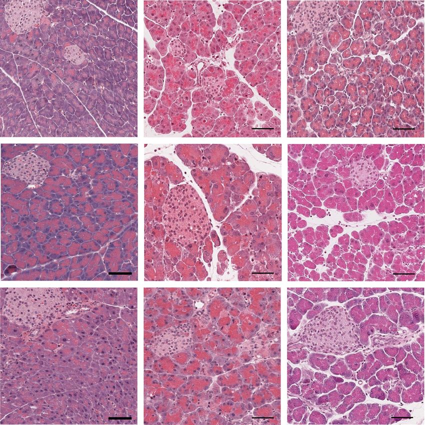
2   Ana Hidalgo-Sastre et al.                                                                                    Eur. J. Immunol. 2021. 0: 1–13

    Introduction                                                           of TLR3 [32, 39]. TRIF subsequently activates the transcription
                                                                           factors IRF3, AP-1, and NF-κB, which potentiates the secretion of
    Acute pancreatitis (AP) is an inflammatory disorder of the pan-        proinflammatory cytokines, like TNF and IL-6, and type 1 IFNs
    creas, caused by premature zymogene-activation resulting in pan-       [32, 40, 41]. Additionally, TLR3 induces the formation of cleaved
    creatic autodigestion [1, 2]. Within gastrointestinal diseases, AP     caspase-8 via TRIF and thereby initiates cell death [42–47].
    is the most common reason for hospitalization with an increasing           In this study, we aimed to analyze the cell specific function of
    global incidence rate of currently 34 cases per 100 000 individuals    TLR3 in acute pancreatitis, induced by caerulein treatment. In the
    [3–5]. The main causative factors include gallstones and alcohol       early phase of AP, LDH and amylase serum levels were increased
    abuse [6, 7].                                                          in TLR3OFF - or TLR3Mye -deficient mice, while morphologically no
        In AP, pancreatic autodigestion evokes severe tissue necrosis,     differences in necrosis, edema, or immune cell filtration were
    resulting in the release of damage-associated molecular patterns       detected. In late stages of AP, we observed enhanced and per-
    (DAMPs) like nucleic acids from dead cells [1]. The activation of      sistent ADM, accompanied by an increased and sustaining high
    DAMP receptors recruits immune cells to the pancreas, thereby          immune cell infiltrate of predominantly macrophages. We were
    initiating and orchestrating the inflammatory response [8].            able to trace back these findings to TLR3 exclusively expressed in
    As part of the subsequent regeneration process, acinar cells           myeloid cells. We showed that TLR3 induces cell death of murine
    reversibly transdifferentiate to a ductal-like progenitor cell state   macrophages in vitro, which could provide a new mechanism
    [9–11]. This transient so-called acinar-to-ductal metaplasia           for the termination of immune responses. Thus, TLR3 dysfunc-
    (ADM) enables the recovery of the exocrine pancreas [11]. In           tion may cause the continuous inflammatory response observed
    contrast, in the context of oncogenic signaling, ADM persists and      in TLR3-deficient mice after AP.
    represents the initial step of pancreatic intraepithelial neoplasia
    development and pancreatic ductal adenocarcinoma (PDAC)
    carcinogenesis [12–15]. Of note, chronic pancreatitis represents       Results
    a risk factor for pancreatic cancer, demonstrating that pancreatic
    inflammation has an important role in PDAC development [16,            Influence of TLR3 deficiency on the early phase of
    17].                                                                   acute pancreatitis
        Immune cells have a bipartite function in AP [6, 18–22].
    Neutrophils and macrophages support tissue repair by the tightly       The present study investigates the role of TLR3 in acute pancre-
    regulated secretion of pro- and anti-inflammatory cytokines [6].       atitis using mouse strains with global TLR3-deficiency (TLR3OFF ),
    Macrophage-secreted cytokines potentiate immune cell infiltra-         generated by the insertion of a floxed transcriptional termination
    tion in the pancreas and therefore enhance the inflammatory            element in the gene locus of Tlr3, or selective TLR3 expression
    response [23]. Accordingly, macrophage activation directly cor-        in myeloid cells (TLR3Mye ) by lysozyme M Cre-mediated deletion
    relates with the severity of AP, while depletion of macrophages        of the transcriptional termination element. TLR3 deficiency or its
    reduces AP severity [24, 25]. Furthermore, macrophages directly        exclusive expression in myeloid cells did not affect pancreatic tis-
    promote acinar transdifferentiation by the secretion of tumor          sue architecture as shown by H&E staining (Fig. 1A). Acinar and
    necrosis factor (TNF) and chemokine (C-C motif) ligand 5 (CCL5)        duct cells were morphologically normal as shown by CPA1 and
    [18]. However, dysbalanced or sustained cytokine secretion coun-       Krt19 staining (Supporting Information Fig. S1). In order to ana-
    teracts tissue regeneration and leads to severe inflammation and       lyze the role of TLR3 during the early phase of acute pancreati-
    chronic diseases [26, 27]. Therefore, the balanced activation and      tis, we examined the effect of TLR3 deficiency (TLR3OFF ) or re-
    termination of the immune response is essential for restoring the      expression in the myeloid lineage (TLR3Mye ) 8 h and 24 h after
    tissue function. Recently, the DAMP receptor Toll-like receptor        caerulein challenge. In untreated mice, serum concentrations of
    3 (TLR3) was described as a new modulator of various tissue            amylase and LDH were slightly higher in TLR3Mye mice as com-
    regenerative processes [28–31].                                        pared with WT controls (Supporting Information Fig. S2). In both,
        TLR3 is an endosomal receptor for double-stranded RNA              TLR3OFF and TLR3Mye mice, the serum concentrations of amy-
    (dsRNA) [32]. Originally discovered as receptor of the innate          lase and LDH were increased 8 h after AP induction compared to
    immune response for sensing viral infections, it is now evident        WT mice, while after 24 h the concentrations decreased to basal
    that TLR3 is also activated by endogenous dsRNA liberated by           values (Supporting Information Fig. S2). There was no signifi-
    necrotic cells [33, 34]. TLR3 is expressed by immune cells, like       cant difference in the pancreas-to-body weight ratios in untreated
    macrophages or DCs, but also in various other cell types includ-       controls and after 24 h AP between the various mouse strains
    ing pancreatic epithelial cells [30, 35, 36]. While TLR3 is pri-       (Fig. 1B). In TLR3Mye mice, the pancreas-to-body weight ratio
    marily localized in endosomes, it was also shown to be present         was elevated after 8 h AP. Neither the histological examination of
    on the surface of human epithelial cells and fibroblasts [37]. The     the tissue nor the Spormann index (combined score for necrosis,
    horseshoe-shaped ectodomain of TLR3 consists of 23 leucine rich        edema, leukocytes, and hemorrhage) revealed a major influence
    repeats, which are essential for ligand binding [38], leading to the   of global TLR3 deficiency or myeloid cell-specific re-expression of
    recruitment of Toll/IL-1 receptor-domain adapter-inducing IFN-β        TLR3 in the early phase of AP (Fig. 1A and C, Supporting Infor-
    (TRIF) to the cytosolic Toll/IL-1 receptor (TIR) homology domain       mation Table S1).

    © 2021 The Authors. European Journal of Immunology published by                                                       www.eji-journal.eu
    Wiley-VCH GmbH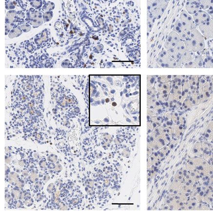
Eur. J. Immunol. 2021. 0: 1–13                                                                                                                                                               Allergy and inflammation           3

                    A
                                                                     ctr                                                 8h                                            24 h

                            WT
                           TLR3OFF
                           TLR3Mye

                    B                                                                                                          C
                                             20                                                                                                     8
                                                                                  *
                        PW/BW ratio (mg/g)

                                                                                                                                   Spormann Score

                                             15                                                                                                     6

                                             10                                                                                                     4

                                             5                                                                                                      2

                                             0                                                                                                      0
                                                               TLR3Mye

                                                                                          TLR3Mye

                                                                                                                   TLR3Mye
                                                       OFF

                                                                                TLR3OFF

                                                                                                         TLR3OFF

                                                                                                                                                                        TLR3Mye

                                                                                                                                                                                                 TLR3Mye
                                                                                                                                                             TLR3OFF

                                                                                                                                                                                       TLR3OFF
                                                  WT

                                                                           WT

                                                                                                    WT

                                                                                                                                                        WT

                                                                                                                                                                                  WT
                                                        TLR3

                                                  Untreated                8 h Caerulein            24 h Caerulein                                      8 h Caerulein             24 h Caerulein

Figure 1. Influence of TLR3 deficiency on the early phase of acute pancreatitis. (A) Representative H&E pictures of pancreatic sections from WT,
TLR3OFF , and TLR3Mye mice at 8 h and 24 h after repetitive caerulein injections compared to untreated controls. Scale bars indicate 100 μm. (B)
Pancreas to body weight ratio of TLR3OFF and TLR3Mye mice compared to WT controls at the specified time points. (C) Spormann scoring of WT,
TLR3OFF , and TLR3Mye mice at the specified time points. (B and C) For each time point and group, samples from N ≥ 3 mice were analyzed representing
pooled data from at least three independent experiments and were analyzed using the Mann–Whitney test. Data are presented as mean ± SEM.
*p < 0.05.

TLR3-deficient mice show a defective regenerative                                                                             with no histologically detectable ADM structures (Fig. 2A and C).
response                                                                                                                      In contrast, ADM formation was significantly increased in TLR3OFF
                                                                                                                              mice 2 days after AP (Fig. 2A and C). Strikingly, these ADM struc-
Next, we investigated the role of TLR3 at later time points after                                                             tures persisted and were still elevated even 7 days after AP induc-
AP induction. At 2 days after AP induction, H&E staining of the                                                               tion, indicating a crucial role of TLR3 for pancreatic regeneration
pancreas from WT mice showed apparent ADM, characterized by                                                                   in the late phase of AP.
acinar cells with ductal-like cell morphology (Fig. 2A). After 5 and                                                              Exclusive expression of TLR3 in myeloid cells partially rescued
7 days, pancreatic tissue of WT mice was completely regenerated                                                               the regenerative defects observed for global TLR3 deficiency on

© 2021 The Authors. European Journal of Immunology published by                                                                                                                                            www.eji-journal.eu
Wiley-VCH GmbH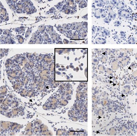
4   Ana Hidalgo-Sastre et al.                                                                                                          Eur. J. Immunol. 2021. 0: 1–13

      B                                                                  C
                                                  **
                                                                          % ADM area / exocrine tissue

                                  10
                                                                                                         100
                                                                                                                **         **
             PW/BW ratio (mg/g)

                                                                                                         80              *** **     ** **
                                                                                                         60
                                  5
                                                                                                         40

                                                                                                         20

                                  0                                                                                                          ND
                                                                                                          0
                                                              WT

                                                           TLR3Mye
                                                           TLR3OFF
                                          WT

                                                    WT
                                       TLR3Mye

                                                 TLR3Mye
                                       TLR3OFF

                                                 TLR3OFF

                                                                                                                  WT

                                                                                                                            WT

                                                                                                                                      WT

                                                                                                                                                 WT
                                                                                                               TLR3Mye

                                                                                                                         TLR3Mye

                                                                                                                                   TLR3Mye

                                                                                                                                              TLR3Mye
                                                                                                               TLR3OFF

                                                                                                                         TLR3OFF

                                                                                                                                   TLR3OFF

                                                                                                                                              TLR3OFF

                                         2d        5d        7d                                                 2d        5d         7d         21 d

    Figure 2. TLR3-deficient mice show a defective pancreatic regeneration after AP. (A) Representative H&E pictures of pancreatic sections from
    TLR3OFF and TLR3Mye mice compared to WT controls at 2, 5, 7, and 21 days after induction of acute pancreatitis. Scale bars indicate 200 μm.
    (B) Pancreas-to-body weight ratio of WT, TLR3OFF , and TLR3Mye mice at the specified time points. (C) Quantification of ADM area in the exocrine
    pancreas from TLR3OFF and TLR3Mye mice compared to WT controls at 2, 5, 7, or 21 days after caerulein injection. Because the pancreas from WT mice
    was completely regenerated after 7 days, ADM area after 21 days was not further analyzed. (B and C) For each time point and group, samples from
    N ≥ 5 mice were analyzed representing pooled data from at least five independent experiments and were analyzed using the Mann–Whitney test.
    Data are presented as mean ± SEM. *p < 0.05, **p < 0.01, ***p < 0.001. ND = not determined.

    © 2021 The Authors. European Journal of Immunology published by                                                                             www.eji-journal.eu
    Wiley-VCH GmbH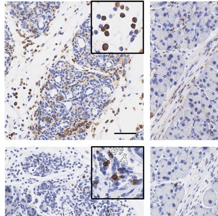
Eur. J. Immunol. 2021. 0: 1–13 Allergy and inflammation 5
days 2 and 5 after AP induction, while a full rescue was observed strains at 2 and 5 days after AP induction (Supporting Information
on day 7. There were significantly less ADM areas compared to Fig. S3A and B).
TLR3OFF mice after 5 days and the pancreatic tissue was com- Taken together, these results demonstrate the pro-regenerative
pletely regenerated after 7 days, comparable to the WT controls function of myeloid cell specific TLR3 in AP.
(Fig. 2A and C). These results demonstrate that pancreatic regen-
eration following AP is dependent on the expression of TLR3 in
myeloid cells.
TLR3 expression in myeloid cells is required for
At 21 days after AP induction, the pancreata of all mouse
immune cell clearance after AP
strains were fully recovered (Fig. 2A). Only one animal from the
TLR3OFF cohort developed an area of less than 5 % of the tis-
Immune cells play a pivotal role in pancreatic regeneration [6]. To
sue with signs of chronic pancreatitis. There was no difference in
further corroborate the importance of TLR3 expression in myeloid
pancreas-to-body weight ratio between the mouse strains, except
cells for pancreatic regeneration, the AP-induced immune cell
for a decreased ratio in TLR3OFF mice 5 days after AP, which is in
infiltration was characterized immunohistochemically.
line with the observed delay in regeneration (Fig. 2B).
In the untreated state and 8 h after AP induction, low num-
Therefore, global deletion of TLR3 resulted in a markedly
bers of pancreatic immune cells were detected and the numbers
increased formation of ADM and a delayed regeneration of the
did not significantly differ between the various mouse strains
pancreas after AP. This defect was rescued by myeloid cell selective
(Fig. 5A). Notably, TLR3OFF mice had significantly increased
TLR3 expression, demonstrating the importance of TLR3 expres-
amounts of F4/80, CD3, and MPO positive cells compared to
sion in myeloid cells for pancreatic regeneration after AP.
WT mice on day 2 (Fig. 5A and B). Accordingly, mRNA levels
of IL-6 and TNF were elevated in TLR3OFF as compared with
TLR3Mye and WT mice (Supporting Information Fig. S4). Signif-
icantly increased accumulation of F4/80 and CD3 positive cells
Increased ADM marker expression after AP caused by
in TLR3OFF mice persisted until day 5 after AP (Fig. 5A and C).
systemic TLR3 deficiency
In contrast, the numbers of infiltrating immune cells in TLR3Mye
mice were similar to those of WT mice both 2 and 5 days after AP
To further corroborate the role of myeloid cell TLR3 in aci-
induction. Accordingly, immune cell infiltration was significantly
nar regeneration, pancreatic differentiation markers were inves-
reduced in TLR3Mye as compared to TLR3OFF mice. Interestingly,
tigated at protein and RNA levels in pancreata of mice at 2 and
quantification of pancreatic immune cells revealed macrophages
5 days after AP induction.
to be the predominant cell type (Fig. 5).
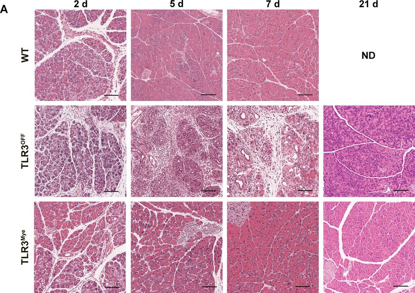
At 2 days after AP induction, expression of CPA1 in com-
These results show that TLR3 signaling exclusively in myeloid
bination with high numbers of Krt19 and SOX9 positive meta-
cells is sufficient to resolve the inflammatory immune cell infil-
plastic cells was detected, confirming ADM on a molecular basis
trate after AP and may therefore play a central role in the termi-
(Fig. 3A and B). In agreement with the increased formation of
nation of the immune response after acute pancreatitis, which, in
ADM observed in TLR3OFF mice, global TLR3 deficiency lead to
turn, may limit ADM formation and reduce ADM persistence.
increased mRNA expression of the ductal markers Krt19 and
SOX9, as well as to an increased number of nuclear SOX9 positive
metaplastic cells, compared to WT and TLR3Mye mice (Fig. 3A and
B). TLR3 signaling induces cell death of murine
After 5 days, acinar cells were fully restored in WT mice as macrophages
shown by the normal CPA1 expression and the restriction of Krt19
and nuclear SOX9 to ductal and centroacinar cells (Fig. 4A and Since macrophages represent the main cell population in the
B). These results confirm the complete tissue regeneration in WT pancreas after AP (Fig. 5) and due to their central importance for
mice 5 days after AP induction. In contrast, TLR3OFF mice pre- the regulation of the immune response, we further investigated
sented ADM areas with a significantly reduced number of CPA1 the effect of TLR3 on macrophages. To characterize the effect
positive acinar cells and a significantly higher number of Krt19 of TLR3 in macrophages, conditionally immortalized murine
positive cells with nuclear SOX9 compared to WT mice, demon- BM-derived progenitor cells (BMDCs) from WT mice were in vitro
strating ongoing tissue regeneration and the persistence of ADM differentiated to macrophages. The differentiation status was
(Fig. 4A and B). In TLR3Mye mice, mRNA expression of CPA1, verified via flow cytometry (Supporting Information Fig. S5A). To
Krt19, and SOX9 was similar to WT mice, while the number of determine the effect of TLR3 signaling on murine macrophages,
Krt19 positive cells was increased but lower than in TLR3OFF the cells were stimulated with different concentrations of the
mice, confirming the partial rescue of the regenerative defect by TLR3 agonist polyinosinic:polycytidylic acid (poly(I:C)). After 48
myeloid cell specific TLR3. h of stimulation, WT macrophages showed a dosage-dependent
Examination of the proliferation marker Ki67 and the apop- reduction of cell viability as detected by MTT assay (Fig. 6A).
tosis marker cleaved caspase-3 (CC3) in the pancreatic tissue Notably, even low concentrations of poly(I:C) were sufficient to
showed no significant differences between the different mouse cause a reduction of cell viability.
© 2021 The Authors. European Journal of Immunology published by www.eji-journal.eu
Wiley-VCH GmbH
6 Ana Hidalgo-Sastre et al. Eur. J. Immunol. 2021. 0: 1–13
A
WT TLR3OFF TLR3Mye
CPA1 day 2 IHC quantification
0.8
positive acinar cells (%)
0.6
CPA1
0.4
0.2
0.0
WT TLR3OFF TLR3Mye
Krt19 day 2 IHC quantification
0.3 n.s. *
positive cells (%)
Krt19
0.2
0.1
0.0
WT TLR3OFF TLR3Mye
SOX9 day 2 IHC quantification
0.8 * *
SOX9
positive cells (%)
0.6
0.4
0.2
0.0
WT TLR3OFF TLR3Mye
B
Krt19 day 2
CPA1 day 2 * * SOX9 day 2
0.15 0.25 0.00020
relative mRNA expression
relative mRNA expression
relative mRNA expression
n.s. * * *
0.20
0.00015
0.10
0.15
0.00010
0.10
0.05
0.00005
0.05
0.00 0.00 0.00000
WT TLR3OFF TLR3Mye WT TLR3OFF TLR3Mye WT TLR3
OFF
TLR3
Mye
Figure 3. Immunohistochemical analysis of cell identity markers 2 days after AP induction. (A) Representative pictures and quantification of the
immunohistochemical staining of CPA1, Krt19, and SOX9 from TLR3OFF and TLR3Mye mice compared to WT controls at 2 days after AP induction.
Scale bars indicate 200 μm. (B) Pancreatic mRNA expression levels of CPA1, Krt19, and SOX9 normalized to Rps13 at 2 days after AP induction
analyzed by qPCR. For each group, samples from N ≥ 4 mice were analyzed representing pooled data from at least four independent experiments
and were analyzed using the t-test. Data are presented as mean ± SEM. *p < 0.05. n.s. = not significant.
To exclude effects of other dsRNA receptors, conditionally it was absent in TLR3OFF macrophages (Supporting Informa-
immortalized BMDCs from TLR3OFF and TLR3Mye mice were gen- tion Fig. S5C). Consistent with the lack of TLR3, macrophages
erated and in vitro differentiated to macrophages. There were from TLR3OFF mice did not produce cytokines in response to
no differences in the differentiation status of WT as compared poly(I:C) (Supporting Information Fig. S5B). While stimulation
with TLR3OFF and TLR3Mye progenitor cell-derived macrophages of WT and TLR3Mye macrophages with 1 μg/mL poly(I:C) led
as shown by FACS analysis of macrophage-specific differentia- to a substantial reduction in cell viability, viability of TLR3OFF
tion markers (Supporting Information Fig. S5A). TLR3 expres- macrophages was not significantly reduced (Fig. 6B). Immunoblot
sion in TLR3Mye and WT macrophages was comparable, while analysis showed an induced formation of cleaved caspase-8 at
© 2021 The Authors. European Journal of Immunology published by www.eji-journal.eu
Wiley-VCH GmbH
Eur. J. Immunol. 2021. 0: 1–13 Allergy and inflammation 7
A
WT TLR3OFF TLR3Mye
CPA1 day 5 IHC quantification
0.4
** n.s.
positive acinar cells (%)
0.3
CPA1
0.2
0.1
0.0
WT TLR3OFF TLR3Mye
Krt19 day 5 IHC quantification
0.6 *
**
positive cells (%)
Krt19
0.4
0.2
0.0
WT TLR3OFF TLR3Mye
SOX9 day 5 IHC quantification
0.4 * *
SOX9
positive cells (%)
0.3
0.2
0.1
0.0
WT TLR3OFF TLR3Mye
B
CPA1 day 5
0.25 * ** 0.04 Krt19 day 5 0.00015 SOX9 day 5
relative mRNA expression
relative mRNA expression
relative mRNA expression
0.20
0.03
0.00010
0.15
0.02
0.10
0.00005
0.01
0.05
0.00 0.00 0.00000
OFF Mye
WT TLR3OFF TLR3Mye WT TLR3OFF TLR3Mye WT TLR3 TLR3
Figure 4. Immunohistochemical analysis of cell identity markers 5 days after AP induction. (A) Representative pictures and quantification of the
immunohistochemical staining of CPA1, Krt19, and SOX9 from TLR3OFF and TLR3Mye mice compared to WT controls at 5 days after AP induction.
Scale bars indicate 200 μm. (B) Pancreatic mRNA expression levels of CPA1, Krt19, and SOX9 normalized to Rps13 at 5 days after AP induction
analyzed by qPCR. For each group, samples from N ≥ 5 mice were analyzed representing pooled data from at least five independent experiments
and were analyzed using the t-test. Data are presented as mean ± SEM. *p < 0.05, **p < 0.01. n.s. = not significant
48 h after stimulation of WT macrophages with poly(I:C), indicat- regeneration. Interestingly, this effect seems to be associated with
ing a TLR3-mediated induction of caspase-8 dependent cell death the sensitivity of macrophages to TLR3-induced cell death.
by poly(I:C) (Fig. 6C; Supporting Information Fig. S6).
Collectively, stimulation of murine macrophages with
poly(I:C) led to a TLR3-dependent reduction of cell viability Discussion
by induction of cleaved caspase-8-mediated cell death. Because
macrophages are of central importance for the recruitment of Acute pancreatitis is associated with tissue necrosis resulting in
other immune cells and the resolution of the immune response the liberation of DAMPs, like nucleic acids [1]. Various studies
[23, 27, 48], TLR3-dependent resolution of the immune reac- have shown that TLR3 signaling is not only induced by viral
tion in AP appears to be crucial for terminating pancreatic dsRNA but also by RNA released from damaged tissue [28, 29, 33,
© 2021 The Authors. European Journal of Immunology published by www.eji-journal.eu
Wiley-VCH GmbH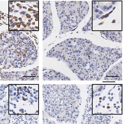
8 Ana Hidalgo-Sastre et al. Eur. J. Immunol. 2021. 0: 1–13
A
250 * * 50 ** * 100 * *
F4/80 positive cells / HPF
MPO positive cells / HPF
CD3 positive cells / HPF
** ** WT
WT 40 WT
200 80
* * TLR3OFF
TLR3OFF TLR3OFF
150 TLR3Mye 30 TLR3Mye 60 TLR3Mye
100 20 40
50 10 20
0 0 0
ctr 8h 2d 5d ctr 8h 2d 5d ctr 8h 2d 5d
B C
2 days after AP induction: 5 days after AP induction:
WT TLR3 OFF
TLR3 Mye
WT TLR3OFF TLR3Mye
F4/80
F4/80
CD3
CD3
MPO
MPO
Figure 5. Characterization of the immune cell infiltrate 2 days and 5 days after AP. (A) IHC-based quantification of F4/80-, CD3-, and MPO-positive
cells in the pancreas of WT, TLR3OFF , and TLR3Mye mice, at 8 h, 2 days and 5 days after AP induction, compared to untreated controls. (B) Represen-
tative pictures of the immunohistochemical staining of CD3, MPO, and F4/80 in WT, TLR3OFF , and TLR3Mye mice at 2 days after AP induction. Scale
bars indicate 100 μm. (C) Representative pictures of the immunohistochemical staining of CD3, MPO, and F4/80 in WT, TLR3OFF , and TLR3Mye mice
at 5 days after AP induction. Scale bars indicate 100 μm. (A–C) For each time point and group, samples from N ≥ 3 independent mice were analyzed.
Data were pooled from at least three independent experiments and were analyzed using the Mann–Whitney test. Data are presented as mean ±
SEM. *p < 0.05, **p < 0.01.
34]. Thereby, TLR3 fulfills versatile physiological functions includ- TLR3 in murine BM-derived macrophages. Thus, TLR3-mediated
ing the regulation of tissue regeneration after myocardial infarc- cell death provides a potential mechanism for the clearance
tion or skin neogenesis after injury [28, 29]. This pro-regenerative of macrophages from the pancreas and the termination of the
role of TLR3 is in agreement with the results in the present study, immune response after AP.
where the myeloid cell specific importance of TLR3 for pancre- Macrophages fulfill a central role for the recruitment of
atic regeneration in the late phase of acute pancreatitis has been immune cells, for example, CD4+ T cells, and thereby regulate
shown. Global TLR3 deficiency in mice led to increased forma- inflammatory processes [23]. The degree of macrophage activa-
tion of ADM, which persisted for up to 7 days after AP. This effect tion plays a determining role for the severity of AP [24, 25]. Folias
was partially rescued by myeloid cell selective expression of TLR3. et al. demonstrated that macrophage depletion reduced formation
These findings therefore highlight the functional importance of and persistence of acinar dedifferentiation [21]. It was further
TLR3 in myeloid cells for pancreatic regeneration. shown that macrophages promote ADM formation by the secre-
Induction of AP in TLR3OFF mice was accompanied by a signif- tion of TNF and CCL5 [18]. Both studies are in accordance with
icantly increased infiltration of macrophages and, at lower num- our data, showing that highly increased ADM formation was asso-
bers, also T cells and neutrophils in the pancreas. This effect ciated with increased macrophage infiltration in TLR3OFF mice.
was completely rescued by the expression of TLR3 in myeloid Furthermore, we demonstrated that TLR3 directly induces the
cells, showing that TLR3 signaling in myeloid cells limits the cell death of macrophages in vitro and that this effect is associated
number of pancreatic immune cells. Furthermore, we demon- with the activation of caspase-8. The induction of cleaved caspase-
strated the induction of cleaved caspase-8-associated cell death by 8 by TLR3 is well documented in literature [42–45, 47]. Activated
© 2021 The Authors. European Journal of Immunology published by www.eji-journal.eu
Wiley-VCH GmbH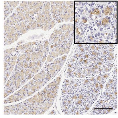
Eur. J. Immunol. 2021. 0: 1–13 Allergy and inflammation 9
A B
**** **** **** **** ****
1.00
Cell viability (% of control)
1.25
Cell viability (% of control)
** **
1.00
0.75
0.75
0.50
0.50
0.25
0.25
0.00 0.00
l
co ol
l
r d
d
tr e d
po co ol
µg l
( I: µ l
r
r.
/m
ro
l
l
l
l
l
)1 m
ly ) 1 /m
3 M nt e
te
ct
/m
/m
/m
/m
/m
s. ntr
. tr
ct
R u at
un eat
C g/
nt
ea
po I:C µg
3M s n
µg
µg
µg
µg
µg
TL 3 OFF ntre
s.
R po o
.c
po
y( ) 1
1
5
10
50
0
10
R u
3 OF s
R po I:C
ye
R po
TL WT
TL OFF ly(
F
ye
poly(I:C)
TL T
3M l
W
R po
ye
TL WT
TL
3
C
24 h 48 h
poly(I:C)
(100 µg/ml) - + - - + -
pos. control - - + - - +
43 kDa C. Caspase-8
18 kDa C. Caspase-8
GAPDH
Figure 6. TLR3 signaling induces cell death of murine macrophages in vitro. (A) Cell viability of murine WT macrophages stimulated with different
concentrations of poly(I:C) for 48 h. (B) Cell viability of macrophages from WT, TLR3OFF , and TLR3Mye mice stimulated with 1 μg/mL poly(I:C) for
48 h. (C) Representative immunoblot showing the formation of cleaved caspase-8 after 48 h stimulation of murine macrophages with 100 μg/mL
poly(I:C) or brefeldin A, as positive control. GAPDH was used as loading control. (A and B) As positive control, apoptosis was induced by brefeldin A.
Data were pooled from N ≥ 4 independent experiments and were analyzed using the t-test. Data are presented as mean ± SEM. *p < 0.05, **p < 0.01,
***p < 0.001, ****p < 0.0001.
TLR3 binds TRIF, which subsequently may recruit the ripoptosome that is based on the activity of TLR3 in myeloid cells. The results
complex, formed by caspase-8, RIP1, FADD, and cFLIP isoforms suggest that the termination of inflammation requires clearance
[47]. Depending on the molecular composition of the ripopto- of macrophages by TLR3-induced cell death, which allows for
some, it then induces apoptosis or necroptosis, the later medi- the resolution of ADM and successful pancreatic regeneration.
ated by RIP3 [49]. Thus, the increased cell death of macrophages, Hence, these findings are of potential relevance for the design of
observed in the present study occurs likely via the described for- pro-resolution therapies that could be applied to directly initiate
mation of the ripoptosome complex. the termination of the immune response, especially for chronic
While the initiation of ADM has been extensively studied, inflammatory diseases like chronic pancreatitis, with decisive
comparatively little is known about the termination of ADM and benefits compared to anti-inflammatory treatments [48].
acinar re-differentiation. Macrophages not only secrete proin- An incomplete resolution of inflammation not only may cause
flammatory cytokines that promote tissue infiltration by immune chronic disease [27], but, in the case of chronic pancreatitis, it can
cells to initiate inflammation, but they are also crucial for the also contribute to pancreatic carcinogenesis [17]. In the context
resolution of inflammation. Thus, macrophages are involved in of oncogenic signaling, ADM is assumed to be the initial step in
the phagocytic clearance of cell debris and apoptotic immune pancreatic intraepithelial neoplasia formation and accordingly, in
cells by efferocytosis and are subsequently cleared from the tissue PDAC carcinogenesis [12, 14, 15]. Therefore, the molecular path-
by lymphatic drainage or apoptosis [48, 50]. Both, a dysregulated ways involved in ADM also provide detailed insight into the car-
efferocytosis by macrophages, as well as impaired tissue clearance cinogenesis of PDAC and potentially offer new therapeutic targets.
of macrophages may lead to persistent inflammatory diseases Because systemic TLR3 deficiency leads to pronounced and persis-
and autoimmunity [23]. The in vivo depletion of macrophages tent ADM, TLR3 may also influence PDAC initiation. The role of
consequently was shown to reduce ADM [21]. In the present TLR3 on cancer development and progression is controversially
report, we provide evidence for a previously unrecognized mech- discussed in literature [44, 51–55]. It is conceivable, however,
anism for the termination of inflammation in the late phase of AP that diverse cell type specific functions of TLR3 underlay these
© 2021 The Authors. European Journal of Immunology published by www.eji-journal.eu
Wiley-VCH GmbH
10   Ana Hidalgo-Sastre et al.                                                                                    Eur. J. Immunol. 2021. 0: 1–13

     divergent results. Thus, cell-specific analysis of TLR3 may also        pancreatitis [59, 60]. Mice were sacrificed 8 h, 24 h, 2 days, 5
     provide decisive insights in its role in pancreatic carcinogenesis.     days, 7 days, and 21 days after the first caerulein injection for
         A recent study suggests that TLR3 deficiency may also enforce       analysis.
     AP severity in the early phase of AP by a NF-κB-regulated induc-
     tion of acinar cell necrosis, but did not address the role of TLR3 in
     pancreatic regeneration at later time points [30]. In contrast, the     Body and pancreatic weigh analysis
     present report focuses on the role of TLR3 in pancreatic regenera-
     tion and reveals a novel and myeloid cell specific function of TLR3     Mice were sacrificed and immediately weighted on a high preci-
     for downregulating ADM formation. Moreover, in our experimen-           sion scale to obtain the body weight. Directly afterward, the whole
     tal set up, we could not detect any dominant TLR3-dependent             pancreas was carefully excised without any adjacent tissue and
     effects, both 8 and 24 h after AP induction. The deviating results      straightaway weighed in a precision scale.
     may be explained by the different protocols used for AP induction,
     varying mouse models and the divergent time points of investi-
     gation after AP induction. It should be noted, however, that our        Serum analysis
     results are in agreement with those from Hoque et al., who also
     found no significant alteration of AP severity caused by TLR3 defi-     Mice were sacrificed and exsanguinated by vena cava. Blood was
     ciency [56].                                                            transferred to a tube (Microvette, Sarstedt) and centrifuged at
         In summary, our results demonstrate the key role of TLR3 in         10 000 rpm for 5 min. Serum was collected and stored at −80°C
     myeloid cells for the termination of ADM after AP and pancre-           until assayed. Serum levels of Amylase and LDH were measured
     atic regeneration. TLR3 reduces the infiltration of immune cells        using a cobas 8000 modular analyzer (Roche, Germany) at the
     and terminates the inflammatory response possibly via the induc-        Institute of Clinical Chemistry and Pathobiochemistry of the Tech-
     tion of cell death in macrophages. Thus, TLR3 represents a cen-         nical University of Munich, Germany.
     tral regulatory element for pancreatic regeneration and for the
     restoration of physiological tissue function.
                                                                             Immunohistochemical staining

                                                                             For immunohistochemical staining, sections of paraffin-embedded
      Materials and methods
                                                                             pancreata were rehydrated and antigens were retrieved using
                                                                             Antigen Unmasking Solution (Vector Laboratories). Overnight
     Mouse strains                                                           incubation with the following primary antibodies was performed:
                                                                             anti-CPA1 (1:300; RD Systems AF2765), anti-Krt19 (1:1000;
     TLR3OFF mice were generated by insertion of a floxed transcrip-         Abcam ab133496), anti-Clusterin (1:200; Santa Cruz sc-6419),
     tional termination element between exons 3 and 4 of the Tlr3            anti-SOX9 (1:1500; Millipore AB5535), anti-Ki67 (1:400; BD
     gene resulting in global TLR3 deficiency [57]. TLR3Mye mice             Pharmingen 550609), anti-cleaved caspase-3 (1:200; Cell Sig-
     were generated by crossing LysMcre mice [58] into the TLR3OFF           nalling 9661), anti-CD3 (1:200; BD Pharmingen 145-2C11), anti-
     strain. Due to the cell-specific expression of the Cre recombinase,     MPO (1:350; Santa Cruz Biotechnology sc-390109), anti-F4/80
     TLR3Mye mice show a selective expression of TLR3 in cells of the        (1:300; Abcam ab16911). Biotin-conjugated secondary antibod-
     myeloid lineage. WT C57BL/6N mice were used as controls. All            ies were incubated for 1 h at room temperature, following devel-
     mouse models were housed under specified pathogen-free condi-           opment with ABC and DAB kits (both Vector Laboratories). SOX9
     tions and maintained on a pure C57BL/6N background either at            and Krt19 were stained using the envision kit from Dako. Nuclear
     the Klinikum rechts der Isar, Technical University of Munich (Ger-      counterstaining was done using hematoxylin. CD3, MPO, and
     many) or at the Charles Rivers facility in Calco, Italy. All animal     F4/80 were stained using a Leica bond XRm staining robot.
     experiments were institutionally approved by the government of
     Upper Bavaria (license 55.2-1-54-2532-130-2016) and were per-
     formed in accordance with the guidelines and regulations.               Morphometric quantification

                                                                             For the evaluation of ADM area, pancreatic sections were stained
     Caerulein injection                                                     with H&E and then analyzed using Aperio ImageScope (Leica
                                                                             Biosystems). All slides were quantified blindly. The histological
     Induction of pancreatitis was carried out in 8-week-old female          evaluation of pancreatitis severity was based on the scoring sys-
     mice who were fasting overnight prior to the caerulein                  tem of Spormann by generating a combined score for necrosis,
     (Bachem) treatment with 8 hourly intraperitoneal injections of 2        edema, leukocyte infiltration, and hemorrhage as described [61].
     μg/injection, using a previously described protocol [11] modified       For quantification of Ki67 positive cells and nuclear SOX9 positive
     to one day. This protocol induces a mild form of acute pancreatitis     cells, five randomly distributed high-power fields (HPF) across the
     with less exocrine cell necrosis compared to protocols for severe       tissue (250 μm × 250 μm, 20× magnification) were counted.

     © 2021 The Authors. European Journal of Immunology published by                                                        www.eji-journal.eu
     Wiley-VCH GmbH
Eur. J. Immunol. 2021. 0: 1–13 Allergy and inflammation 11
For cleaved caspase-3, 10 HPF were counted. For CD3, MPO, and Statistical analysis
F4/80, 10 HPF (500 μm × 500 μm, 40× magnification) were
counted. For CPA1 and Krt19, the whole slide was quantified. Statistical analysis was performed using Graph Pad Prism6
(GraphPad Software Inc). Unless otherwise stated, the Mann–
Whitney test for nonnormal distributed unpaired data was used
for intergroup comparison. Differences with a p-value lower than
Reverse transcription quantitative PCR 0.05 were considered significant, and the following scale was
applied: *p < 0.05, **p < 0.01, ***p < 0.001. Data are presented
Pancreas sections were immediately stored at -80°C in RNAlater as mean value ± SEM.
(Qiagen) until use. RNA was isolated using the Maxwell RSC sim-
plyRNA Tissue Kit (Promega) and cDNA was synthesized using
the QuantiTect Reverse Transcription Kit (Qiagen). For the quan-
titative real-time PCR, the Universal Probe library (Roche Diag-
nostics) was used with the following primers (specified in 5
to 3 orientation): mCCL5-fwd: tgcagaggactctgagacagc, mCCL5- Acknowledgements: We thank Marion Mielke, Olga Seelbach,
rev: gagtggtgtccgagccat, mTNF-fwd: tgcctatgtctcagcctcttc, mTNF- and Nils Wirges from the Institute of Pathology at the Technical
rev: gaggccatttgggaacttct, mTLR3-fwd: gatacagggattgcacccata, University of Munich for performing the automated staining of
mTLR3-rev: tcccccaaaggagtacattaga, mIL-6-fwd: gctaccaaactg- the immune cell markers, scanning of whole pancreatic cross sec-
gatataatcagga, mIL-6-rev: ccaggtagctatggtactccagaa, mCPA1-fwd: tions, and for executing the pancreatitis scoring. We also thank
tcccatcaatgtgctgaagt, mCPA1-rev: ggatgccagtgtcaatcca. The SYBR Annett Dannemann for excellent technical help. This project has
green mastermix (Roche Diagnostics) was used for the following been funded by the Deutsche Forschungsgemeinschaft (DFG, Ger-
primers: mKrt19-fwd: accctcccgagattacaacc; mKrt19-rev: caag- man Research Foundation) Project number 329628492-SFB 1321
gcgtgttctgtctcaa, mSOX9-fwd: ccacgtgtggatgtcgaag, mSOX9-rev: to GVF, BH, and KS and with the Emmy Noether Program to GVF
ctcagctgctccgtcttgat. (FI 1719/2-1).
Conflict of interest: The authors declare no commercial or finan-
Cell lines cial conflict of interest.
Murine BM-derived progenitor cells were isolated from the femur Peer review: The peer review history for this article is available
of WT, TLR3OFF , and TLR3Mye mice and conditionally immor- at https://publons.com/publon/10.1002/eji.202048771
talized by retroviral transduction with a plasmid encoding for
estrogen receptor-coupled Hoxb8 according to Wang et al. [62]. Data availability statement: The data that support the findings
Differentiation to macrophages was induced by M-CSF stimu- of this study are available from the corresponding author upon
lation and simultaneous estradiol withdrawal for 4 days. Suc- reasonable request.
cessful differentiation was verified using flow cytometry analysis
using directly fluorescence-labeled antibodies for CD11b (Invit-
rogen RM2805), CD115 (eBioscience AFS98), and F4/80 (Invit- References
rogen MF48004). The protein amount of cleaved caspase-8 (Cell
Signaling D5B2) and GAPDH (Cell Signaling 14C10) was deter- 1 Kang, R., Lotze, M. T., Zeh, H. J., Billiar, T. R. and Tang, D., Cell death and
mined by immunoblot analysis using the antibodies at 1:1000 in DAMPs in acute pancreatitis. Mol. Med. 2014. 20: 466–477.
5% milk in PBS with 0.1% Tween-20. As positive control for apop- 2 Banks, P. A. and Freeman, M. L., Practice guidelines in acute pancreatitis.
tosis induction, cells were stimulated with 4.5 μg/mL brefeldin A Am. J. Gastroenterol. 2006. 101: 2379–2400.
for the indicated time points. 3 Peery, A. F., Dellon, E. S., Lund, J., Crockett, S. D., McGowan, C. E.,
Bulsiewicz, W. J., Gangarosa, L. M. et al., Burden of gastrointestinal dis-
ease in the United States: 2012 update. Gastroenterology 2012. 143: 1179–
1187.e3.
4 Xiao, A. Y., Tan, M. L. Y., Wu, L. M., Asrani, V. M., Windsor, J. A., Yadav, D.
Cell viability assay
and Petrov, M. S., Global incidence and mortality of pancreatic diseases:
a systematic review, meta-analysis, and meta-regression of population-
Cells were seeded in 96-well plates and stimulated with different based cohort studies. Lancet Gastroenterol. Hepatol. 2016. 1: 45–55.
concentrations of poly(I:C). After 48 h, MTT solution was added 5 Krishna, S. G., Kamboj, A. K., Hart, P. A., Hinton, A. and Conwell, D. L., The
for 4 h with subsequent cell lysis and solubilization of the for- changing epidemiology of acute pancreatitis hospitalizations: a decade of
mazan salt complex in 0.1 M HCl with 10% SDS over night at trends and the impact of chronic pancreatitis. Pancreas 2017. 46: 482–488.
37°C. The amount of complex formed was determined by measur- 6 Habtezion, A., Inflammation in acute and chronic pancreatitis. Curr. Opin.
ing the absorbance at 580 nm. Gastroenterol. 2015. 31: 395–399.
© 2021 The Authors. European Journal of Immunology published by www.eji-journal.eu
Wiley-VCH GmbH12 Ana Hidalgo-Sastre et al. Eur. J. Immunol. 2021. 0: 1–13
7 Lee, P. J. and Papachristou, G. I., New insights into acute pancreatitis. Nat. 26 Jiang, D., Liang, J., Fan, J., Yu, S., Chen, S., Luo, Y. Prestwich, G. D. et al.,
Rev. Gastroenterol. Hepatol. 2019. 16: 479–496. Regulation of lung injury and repair by Toll-like receptors and hyaluro-
nan. Nat. Med. 2005. 11: 1173–1179.
8 Hoque, R., Malik, A. F., Gorelick, F. and Mehal, W. Z., Sterile inflammatory
response in acute pancreatitis. Pancreas 2012. 41: 353–357. 27 Fullerton, J. N. and Gilroy, D. W., Resolution of inflammation: a new ther-
apeutic frontier. Nat. Rev. Drug Discov. 2016. 15: 551–567.
9 Elsässer, H. P., Adler, G. and Kern, H. F., Time course and cellular source of
pancreatic regeneration following acute pancreatitis in the rat. Pancreas. 28 Nelson, A. M., Reddy, S. K., Ratliff, T. S., Hossain, M. Z., Katseff, A. S., Zhu,
1986. 1: 421-429. A. S., Chang, E. et al., dsRNA released by tissue damage activates TLR3 to
drive skin regeneration. Cell Stem Cell. 2015. 17: 139–151.
10 Fendrich, V., Esni, F., Garay, M. V. R., Feldmann, G., Habbe, N., Jensen, J. N.,
Dor, Y. et al., Hedgehog signaling is required for effective regeneration of 29 Wang, X., Ha, T., Liu, L., Hu, Y., Kao, R., Kalbfleisch, J., Williams, D.
exocrine pancreas. Gastroenterology 2008. 135: 621–631. et al., TLR3 mediates repair and regeneration of damaged neonatal heart
through glycolysis dependent YAP1 regulated miR-152 expression. Cell
11 Jensen, J. N., Cameron, E., Garay, M. V. R., Starkey, T. W., Gianani, R. and
Death Differ. 2018. 25: 966–982.
Jensen, J., Recapitulation of elements of embryonic development in adult
mouse pancreatic regeneration. Gastroenterology 2005. 128: 728–741. 30 Regel, I., Raulefs, S., Benitz, S., Mihaljevic, C., Rieder, S., Leinenkugel, G.,
Steiger, K. et al., Loss of TLR3 and its downstream signaling accelerates
12 Schmid, R. M., Acinar-to-ductal metaplasia in pancreatic cancer develop-
acinar cell damage in the acute phase of pancreatitis. Pancreatology 2019.
ment. J. Clin. Invest. 2002. 109: 1403–1404.
19: 149–157.
13 Wagner, M., Greten, F. R., Weber, C. K., Koschnick, S., Mattfeldt, T., Dep-
31 Stöß, C., Laschinger, M., Wang, B., Lu, M., Altmayr, F., Hartmann, D.,
pert, W., Kern, H. et al., A murine tumor progression model for pancreatic
Hüser, N. et al., TLR3 promotes hepatocyte proliferation after partial hep-
cancer recapitulating the genetic alterations of the human disease. Genes
atectomy by stimulating uPA expression and the release of tissue-bound
Dev. 2000. 15: 286–293.
HGF. FASEB J Off. Publ. Federation Am. Soc. Exp. Biol. 2020. 34: 10387–10397.
14 Reichert, M., Blume, K., Kleger, A., Hartmann, D. and von Figura, G. Devel-
32 Alexopoulou, L., Holt, A. C., Medzhitov, R. and Flavell, R., Recognition
opmental pathways direct pancreatic cancer initiation from its cellular
of double-stranded RNA and activation of NF-kB by Toll-like receptor 3.
origin. Stem Cells Int. 2016. 2016. https://doi.org/10.1155/2016/9298535
Nature. 2001. 413:732–738.
15 Kopp, J. L., Figura, G., von Mayes, E., Liu, F.-F., Dubois, C. L., Morris, J. P.,
33 Karikó, K., Ni, H., Capodici, J., Lamphier, M. and Weissman, D., mRNA
Pan, F. C. et al., Identification of Sox9-dependent acinar-to-ductal repro-
is an endogenous ligand for Toll-like receptor 3. J. Biol. Chem. 2004. 279:
gramming as the principal mechanism for initiation of pancreatic ductal
12542–12550.
adenocarcinoma. Cancer Cell. 2012. 22: 737–750.
34 Cavassani, K. A., Ishii, M., Wen, H., Schaller, M. A., Lincoln, P. M., Lukacs,
16 Braganza, J. M., Lee, S. H., McCloy, R. F. and McMahon, M. J., Chronic pan-
N. W., Hogaboam, C. M. et al., TLR3 is an endogenous sensor of tissue
creatitis. Lancet North Am. Ed. 2011. 377: 1184–1197.
necrosis during acute inflammatory events. J. Exp. Med. 2008. 205: 2609–
17 Lowenfels, A. B., Maisonneuve, P., Cavallini, G., Ammann, R. W., Lankisch, 2621.
P. G., Andersen, J. R., Dimagno, E. P. et al., Pancreatitis and the risk of
35 Muzio, M., Bosisio, D., Polentarutti, N., D’amico, G., Stoppacciaro, A.,
pancreatic cancer. N. Engl. J. Med. 1993. 328: 1433–1437.
Mancinelli, R., van’t Veer, C. et al., Differential expression and regulation
18 Liou, G.-Y., Döppler, H., Necela, B., Krishna, M., Crawford, H. C., Raimondo, of toll-like receptors (TLR) in human leukocytes: selective expression of
M. and Storz, P., Macrophage-secreted cytokines drive pancreatic acinar- TLR3 in dendritic cells. J. Immunol. 2000. 164: 5998–6004.
to-ductal metaplasia through NF-κB and MMPs. J. Cell Biol. 2013. 202: 563–
36 Murakami, Y., Fukui, R., Motoi, Y., Kanno, A., Shibata, T., Tanimura, N.,
577.
Saitoh, S. et al., Roles of the cleaved N-terminal TLR3 fragment and cell
19 Merza, M., Hartman, H., Rahman, M., Hwaiz, R., Zhang, E., Renström, E., surface TLR3 in double-stranded RNA sensing. J. Immunol. 2014. 193: 5208–
Luo, L. et al., Neutrophil extracellular traps induce trypsin activation, 5217.
inflammation, and tissue damage in mice with severe acute pancreati-
37 Matsumoto, M., Kikkawa, S., Kohase, M., Miyake, K. and Seya, T., Estab-
tis. Gastroenterology. 2015. 149: 1920–1931.e8.
lishment of a monoclonal antibody against human Toll-like receptor 3
20 Bedrosian, A. S., Nguyen, A. H., Hackman, M., Connolly, M. K., Malhotra, that blocks double-stranded RNA-mediated signaling. Biochem. Biophys.
A., Ibrahim, J., Cieza-Rubio, N. E. et al., Dendritic cells promote pancreatic Res. Commun. 2002. 293: 1364–1369.
viability in mice with acute pancreatitis. Gastroenterology. 2011. 141: 1915–
38 Liu, L., Botos, I., Wang, Y., Leonard, J. N., Shiloach, J., Segal, D. M. and
1926.e1-e14.
Davies, D. R., Structural basis of Toll-like receptor 3 signaling with double-
21 Folias, A. E., Penaranda, C., Su, A. L., Bluestone, J. A. and Hebrok, M., Aber- stranded RNA. Science 2008. 320: 376–379.
rant innate immune activation following tissue injury impairs pancreatic
39 Bell, J. K., Botos, I., Hall, P. R., Askins, J., Shiloach, J., Segal, D. M. and Davies,
regeneration. PLoS One. 2014. 9: e102125.
D. R., The molecular structure of the Toll-like receptor 3 ligand-binding
22 Watanabe, T., Kudo, M. and Strober, W., Immunopathogenesis of pancre- domain. Proc. Natl. Acad. Sci. 2005. 102: 10976–10980.
atitis. Mucosal Immunol. 2017. 10: 283–298.
40 Doyle, S. E., Vaidya, S. A., O’Connell, R., Dodgostar, H., Dempsey, P. W.,
23 Das, A., Sinha, M., Datta, S., Abas, M., Chaffee, S., Sen, C. K. and Roy, S., Wu, T. T., Rao, G. et al., IRF3 mediates a TLR3/TLR4-specific antiviral gene
Monocyte and macrophage plasticity in tissue repair and regeneration. program. Immunity 2002. 17:251–263.
Am. J. Pathol. 2015. 185: 2596–2606.
41 Oshiumi, H., Matsumoto, M., Funami, K., Akazawa, T. and Seya, T.,
24 Xue, J., Sharma, V. and Habtezion, A., Immune cells and immune-based TICAM-1, an adaptor molecule that participates in Toll-like receptor 3-
therapy in pancreatitis. Immunol. Res. 2014. 58: 378–386. mediated interferon-beta induction. Nat. Immunol. 2003. 4: 161–167.
25 Saeki, K., Kanai, T., Nakano, M., Nakamura, Y., Miyata, N., Sujino, T., Yam- 42 Estornes, Y., Toscano, F., Virard, F., Jacquemin, G., Pierrot, A., Vanbervliet,
agishi, Y. et al., CCL2-induced migration and SOCS3-mediated activation B., Bonnin, M. et al., dsRNA induces apoptosis through an atypical death
of macrophages are involved in cerulein-induced pancreatitis in mice. complex associating TLR3 to caspase-8. Cell Death Differ. 2012. 19: 1482–
Gastroenterology 2012. 142: 1010–1020.e9. 1494.
© 2021 The Authors. European Journal of Immunology published by www.eji-journal.eu
Wiley-VCH GmbHEur. J. Immunol. 2021. 0: 1–13 Allergy and inflammation 13
43 Sun, R., Zhang, Y., Lv, Q., Liu, B., Jin, M., Zhang, W., He, Q. et al., Toll-like 55 Chew, V., Tow, C., Huang, C., Bard-Chapeau, E., Copeland, N. G., Jenkins,
receptor 3 (TLR3) induces apoptosis via death receptors and mitochon- N. A., Weber, A. et al., Toll-like receptor 3 expressing tumor parenchyma
dria by up-regulating the transactivating p63 isoform alpha (TAP63alpha). and infiltrating natural killer cells in hepatocellular carcinoma patients.
J. Biol. Chem. 2011. 286: 15918–15928. J. Natl. Cancer Inst. 2012. 104: 1796–1807.
44 Paone, A., Starace, D., Galli, R., Padula, F., Cesaris, P., de Filippini, A., 56 Hoque, R., Sohail, M., Malik, A., Sarwar, S., Luo, Y., Shah, A., Barrat, F. et al.,
Ziparo, E. et al., Toll-like receptor 3 triggers apoptosis of human prostate TLR9 and the NLRP3 inflammasome link acinar cell death with inflamma-
cancer cells through a PKC-alpha-dependent mechanism. Carcinogenesis. tion in acute pancreatitis. Gastroenterology 2011. 141: 358–369.
2008. 29: 1334–1342.
57 Garcias López, A., Bekiaris, V., Müller Luda, K., Hütter, J., Ulmert, I.,
45 Weber, A., Kirejczyk, Z., Besch, R., Potthoff, S., Leverkus, M. and Häcker, Getachew Muleta, K., Nakawesi, J. et al., Migration of murine intestinal
G., Proapoptotic signalling through Toll-like receptor-3 involves TRIF- dendritic cell subsets upon intrinsic and extrinsic TLR3 stimulation. Eur.
dependent activation of caspase-8 and is under the control of inhibitor of J. Immunol. 2020. 50: 1525–1536.
apoptosis proteins in melanoma cells. Cell Death Differ. 2010. 17: 942–951.
58 Clause, B. E., Burkhardt, C., Reith, W., Renkawitz, R. and Förster, I., Con-
46 Salaun, B., Coste, I., Rissoan, M.-C., Lebecque, S. J. and Renno, T., TLR3 ditional gene targeting in macrophages and granulocytes using LysMcre
can directly trigger apoptosis in human cancer cells. J. Immunol. 2006. 176: mice. Transgenic Res. 1999. 8: 265–277.
4894–4901.
59 Lerch, M. M. and Gorelick, F. S., Models of acute and chronic pancreatitis.
47 Feoktistova, M., Geserick, P., Kellert, B., Dimitrova, D. P., Langlais, C., Hupe, Gastroenterology. 2014. https://doi.org/10.1053/j.gastro.2012.12.043
M., Cain, K. et al., cIAPs block ripoptosome formation, a RIP1/caspase-
60 Silva-Vaz, P., Abrantes, A. M., Castelo-Branco, M., Gouveia, A., Botelho,
8 containing intracellular cell death complex differentially regulated by
M. F. and Tralhão, J. G., Murine models of acute pancreatitis: a critical
cFLIP isoforms. Mol. Cell. 2011. 43: 449–463.
appraisal of clinical relevance. Int. J. Mol. Sci. 2019. 20: 2794.
48 Sugimoto, M. A., Vago, J. P., Perretti, M. and Teixeira, M. M., Mediators
61 Spormann, H., Sokolowski, A. and Letko, G., Effect of temporary ischemia
of the resolution of the inflammatory response. Trends Immunol. 2019. 40:
upon development and histological patterns of acute pancreatitis in the
212–227.
rat. Pathol.-Res. Pract. 1989. 184: 507–513.
49 He, S., Liang, Y., Shao, F. and Wang, X., Toll-like receptors activate
62 Wang, G. G., Calvo, K. R., Pasillas, M. P., Sykes, D. B., Häcker, H. and Kamps,
programmed necrosis in macrophages through a receptor-interacting
M. P., Quantitative production of macrophages or neutrophils ex vivo
kinase-3-mediated pathway. Proc. Natl. Acad. Sci. USA 2011. 108: 20054–
using conditional Hoxb8. Nat. Methods 2006. 3: 287–293.
20059.
50 Gilroy, D. W., Colville-Nash, P. R., McMaster, S., Sawatzky, D. A., Abbreviations:ADM: acinar-to ductal metaplasia · AP: acute pan-
Willoughby, D. A. and Lawrence, T., Inducible cyclooxygenase-derived creatitis · BMDCs: BM-derived progenitor cells · DAMP: Damage-
15-deoxy12-14PGJ2 brings about acute inflammatory resolution in rat
associated molecular pattern · PanIN: pancreatic intraepithelial
pleurisy by inducing neutrophil and macrophage apoptosis. FASEB J. 2003:
2269–2271. neoplasia · PDAC: pancreatic ductal adenocarcinoma · poly(I:C):
polyinosinic:polycytidylic acid
51 Schwartz, A. L., Malgor, R., Dickerson, E., Weeraratna, A. T., Slominski,
A., Wortsman, J., Harii, N. et al., Phenylmethimazole decreases Toll-like
receptor 3 and noncanonical Wnt5a expression in pancreatic cancer and
melanoma together with tumor cell growth and migration. Clin. Cancer Full correspondence: Dr. Guido von Figura, School of Medicine,
Res. 2009. 15: 4114–4122. Medizinische Klinik und Poliklinik ΙΙ, Technical University of Munich,
Ismaninger Str. 22, 81675 Munich, Germany
52 Shojaei, H., Oberg, H.-H., Juricke, M., Marischen, L., Kunz, M., Mundhenke,
e-mail: gvfigura@tum.de
C., Gieseler, F. et al., Toll-like receptors 3 and 7 agonists enhance tumor
cell lysis by human gammadelta T cells. Cancer Res. 2009. 69: 8710–8717.
53 Jia, D., Yang, W., Li, L., Liu, H., Tan, Y., Ooi, S., Chi, L. et al., β-Catenin and
NF-κB co-activation triggered by TLR3 stimulation facilitates stem cell- Received: 28/5/2020
like phenotypes in breast cancer. Cell Death Differ. 2015. 22: 298–310. Revised: 12/11/2020
Accepted: 29/1/2021
54 Pradere, J.-P., Dapito, D. H. and Schwabe, R. F., The Yin and Yang of Toll-
Accepted article online: 1/2/2021
like receptors in cancer. Oncogene 2014. 33: 3485–3495.
© 2021 The Authors. European Journal of Immunology published by www.eji-journal.eu
Wiley-VCH GmbHYou can also read