Time course of histopathological changes after bleomycin sclerotherapy in rabbit gallbladders as a model for simple hepatic cysts
←
→
Page content transcription
If your browser does not render page correctly, please read the page content below
BIOMEDICAL REPORTS 15: 75, 2021

                      Time course of histopathological changes after
                      bleomycin sclerotherapy in rabbit gallbladders
                           as a model for simple hepatic cysts
                                      LONG LI1*, YONG‑HAO LAO1* and NAN ZHANG2

            1
             Division of Interventional Radiology, Department of Medical Imaging, Guangdong Provincial Corps Hospital
                of Chinese People's Armed Police Forces, Guangzhou Medical University; 2Department of Pathology,
                       Liwan Central Hospital of Guangzhou City, Guangzhou, Guangdong 510507, P.R. China

                                           Received April 17, 2021; Accepted July 1, 2021

                                                       DOI: 10.3892/br.2021.1451

Abstract. Bleomycin sclerotherapy is used in the treatment            with no visible inflammation or fibrosis. In the experimental
of cystic lesions; however, the histopathological changes             group, epithelial cells were swollen and necrotic on the first
are undefined. Present animal models of cystic diseases are           day, and were replaced gradually by single‑layer flat cells from
not adequate for the study of sclerotherapy of hepatic cysts,         day 56. Inflammatory infiltration was found in the submucosa,
primarily because the established cysts in these models are           and the IOD of T cells, B cells and macrophages were highest
too small in size. The aim of the present study was to estab‑         on day 1, and these parameters declined gradually, eventually
lish a new animal model of simple hepatic cysts, and assess           disappearing. The APSAP of collagen was highest on day 7,
the histopathological changes after bleomycin sclerotherapy.          and gradually declined thereafter. The results suggest that
Rabbit gallbladder, with ligaturing of the cholecystic duct           histopathological changes after bleomycin sclerotherapy of a
whilst preserving cholecystic vessels, was used as a model            simple hepatic cyst model were characterized by sequential
for simple hepatic cysts. Bleomycin (2 mg dissolved in 1 ml           epithelial destruction, inflammatory cell infiltration, collagen
saline) was injected into the aspirated gallbladder, gallbladder      proliferation and epithelial partial regeneration.
tissue was harvested (after 1, 7, 14, 28, 42, 56 and 84 days)
and histopathological changes were evaluated (n=4 per group).         Introduction
Additionally, control rabbit gallbladders were injected with
1 ml saline and sampled after 14 days (n=4). Histopathological        Percutaneous aspiration with sclerotherapy is a minimally
changes were evaluated using hematoxylin‑eosin and Masson's           invasive, simple, safe, inexpensive and reasonably effec‑
trichrome staining, and immunohistochemistry for CD20‑,               tive treatment for cystic diseases, such as simple hepatic,
CD43‑ and CD68‑positive cells was performed. The inte‑                renal, splenic, pancreatic and ovarian cysts, as well as for
grated optical density (IOD) of immunohistochemical staining          lymphoceles (1‑4). Bleomycin has been used successfully for
and average positive stained area percentage (APSAP) of               management of cysts (3,4); it is an antibiotic chemotherapeutic
collagen were quantitatively analyzed. The results revealed           agent derived from Streptomyces verticillus, which was first
gallbladders in the control group had regular epithelial cells        used as a sclerosant for malignant pleural effusions in 1976 (5).
                                                                      Intralesional bleomycin injection has been used successfully
                                                                      for the treatment of cystic diseases, including lymphoceles
                                                                      since 1977 (6), cystic craniopharyngiomas since 1985 (7),
                                                                      bronchogenic cysts since 1992 (8), simple renal cysts since
Correspondence to: Dr Long Li, Division of Interventional             2012 (9) and simple hepatic cysts since 2015 (10). Advantages
Radiology, Department of Medical Imaging, Guangdong Provincial        of percutaneous bleomycin sclerotherapy for treatment of
Corps Hospital of Chinese People's Armed Police Forces,               cystic diseases are minimal inflammatory reactions, modest
Guangzhou Medical University, 268 Yanling Road, Guangzhou,
                                                                      cost as only a single treatment session is required, good toler‑
Guangdong 510507, P.R. China
E‑mail: radiolilong@hotmail.com
                                                                      ance as it causes little pain and no pulmonary toxicity when
                                                                      performed with the proper precautions (3,9‑11).
*
    Contributed equally                                                   The sclerosing mechanisms of intracyst bleomycin injec‑
                                                                      tion are not understood. For example, they may be similar to
Abbreviations: APSAP, average positive stained area percentage;       those of pulmonary fibrosis induced by intratracheal bleo‑
HE, hematoxylin eosin; IOD, integral optical density                  mycin instillation (12); however, that inference has not been
                                                                      confirmed by histopathological examination, and there are no
Key words: bleomycin, sclerotherapy, simple hepatic cyst, pathology   animal models for evaluation of percutaneous sclerotherapy of
                                                                      hepatic cystic disease. Rodent models of drug‑induced poly‑
                                                                      cystic diseases (including liver, kidney and ovary) have been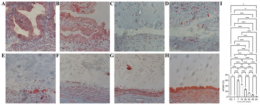
2 LI et al: BLEOMYCIN SCLEROTHERAPY FOR EXPERIMENTAL SIMPLE HEPATIC CYSTS
widely used, but they have limitations. For example, their patho‑
genesis is different from that of simple cysts, their modeling
time is long and they have multiple small cysts (13‑15). Thus,
such animal models are not suitable for studying percutaneous
sclerotherapy of cysts. The animal model of simple ovarian
cysts, established by unilateral total salpingectomy, has been
used successfully to evaluate the effectiveness and safety of
aspiration sclerotherapy, but only 70% of experimental animals
formed macroscopic ovarian cysts of ~10 mm (16). Therefore,
in the present study, a new animal model of a simple hepatic
cyst was established, and the chronological histopathological
changes after intracyst bleomycin injection was assessed.
Materials and methods
Ethical considerations. The present study was approved by
the Biomedical Ethics Committee of Animal Experiments of
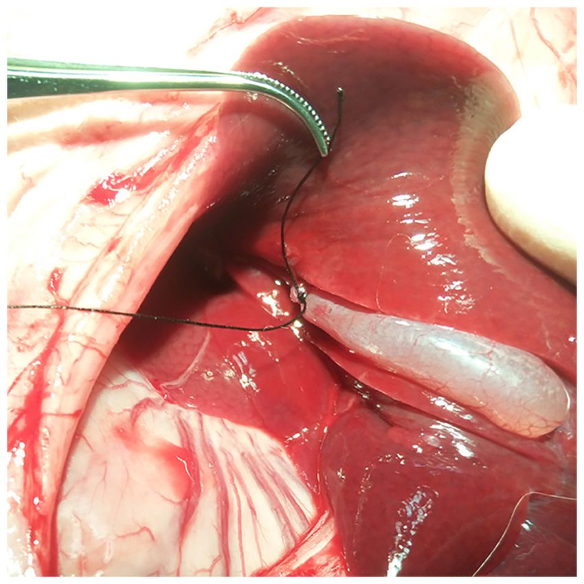
Guangdong Medical Laboratory Animal Center (Guangdong, Figure 1. Animal model of simple hepatic cysts. The cholecystic duct was
China; approval no. B201610‑5). An Accreditation Certificate ligated, with preservation of cholecystic vessels. The gallbladder was then
from the China National Accreditation Service for Conformity irrigated with normal saline until the cystic fluid became clear.
Assessment has been granted to this facility. All experimental
procedures were performed in accordance with the Guide for
the Care and Use of Laboratory Animals (National Institutes evacuated gallbladder. Rabbits received penicillin 10 million
of Health) (17). Every effort was made to minimize animal units/kg per day intramuscularly for three consecutive days
suffering and to use only the number of animals necessary for after completion of the procedure, and were sacrificed at the
the acquisition of reliable data. end of the experimental period. At 1, 7, 14, 28, 56 and 84 days
after the bleomycin injection, animals in experimental groups
Animal model. A total of 32 New Zealand rabbits of either sex were euthanized by intravenous injection of 150 mg/kg potas‑
(11 males, 21 females), weighing 2.6 kg (range, 2‑3 kg) at the sium chloride (Shanghai Chemical Reagent Company; China
beginning of the study, were obtained from Guangdong Medical National Pharmaceutical Group Corporation) under general
Laboratory Animal Center, China (order no. 44411600002998). anesthesia induced by intravenous injection of 30 mg/kg
Rabbits were housed in separate cages in the animal experi‑ pentobarbital sodium via the marginal ear vein; animals in the
mental center, with an ambient temperature of 18‑24˚C and a control group were euthanized at day 14. The gallbladders and
12 h light/dark cycle. Rabbits were provided ad libitum access the surrounding liver parenchyma were resected and evaluated
to food and water during the experimental period. Rabbits were histologically and immunohistochemically.
anesthetized by injection of 30 mg/kg sodium pentobarbital
in the ear vein (Shanghai Chemical Reagent Company, China Histopathological processing. The cholecystic tissue speci‑
National Pharmaceutical Group Corporation) and intramus‑ mens were fixed in 4% paraformaldehyde for 24 h at room
cular injection of 5 mg/kg xylazine hydrochloride (Shanghai temperature, trimmed appropriately, dehydrated in graded
Chemical Reagent Company, China National Pharmaceutical series of alcohol solutions and embedded in paraffin according
Group Corporation) after 12 h of fasting. By means of surgical to standard procedures (18). Paraffin sections of 4 µm thick‑
laparotomy, the cholecystic duct was ligated with a 3/0 surgical ness were cut and mounted on glass microscope slides.
suture, whereas cystic vessels were retained to ensure good Hematoxylin and eosin (H&E) staining was performed
blood supply and to avoid ischemic necrosis (Fig. 1). Direct using a H&E Staining kit (cat. no. G1005; Wuhan Servicebio
gallbladder puncture was performed as far as possible from Technology Co., Ltd.), as described previously (14). Masson's
gallbladder vessels using a 22‑gauge butterfly needle. Bile trichrome staining was performed using a Masson's Trichrome
was aspirated, and the gallbladder was irrigated 3‑4 times Stain kit (cat. no. G1006; Wuhan Servicebio Technology Co.,
with saline until clear. The surgical time ran between 45 and Ltd.) as described previously (19).
60 min. Anesthesia with a standard dose was well tolerated. Infiltration by T‑lymphocytes, B‑lymphocytes and macro‑
phages into the tissue was detected immunohistochemically
Experimental design. The rabbits were randomly divided into using a panel of monoclonal antibodies against CD20, CD43
a control group and seven experimental groups according to and CD68 (20). Immunohistochemical staining was performed
different time points after bleomycin sclerotherapy (1, 7, 14, using the Streptavidin‑Biotin‑Complex method, according
28, 42, 56 and 84 days; n=4 per group). In animals in the to the Sixth Edition of Dako's Educational Guidebook to
experimental group, after gallbladder content was aspirated as Immunohistochemical Staining Methods (21). Sections were
thoroughly as possible, 2 mg dissolved in 1 ml saline bleo‑ deparaffinized in xylene (2x10 min) and rehydrated using
mycin hydrochloride (Takasaki Plant; Nippon Kayaku Co., an alcohol gradient: 100% ethanol (2x10 min), 95% ethanol
Ltd.) was injected and allowed to remain in the evacuated gall‑ (1x8 min), 80% ethanol (1x5 min) and 70% ethanol (1x5 min),
bladder until the experiment ended. For animals in the control followed double‑distilled H2O (1x10 min). Nonspecific back‑
group, 1 ml normal saline was injected, which remained in the ground staining was blocked using Background Sniper (cat.
BIOMEDICAL REPORTS 15: 75, 2021 3
Figure 2. Time course of cyst epithelial changes after bleomycin sclerotherapy in the rabbit model of simple hepatic cysts (hematoxylin and eosin staining; mag‑
nification, x200). (A) In the control group, the integrity of the mucosal structure and the epithelial cells is visibly maintained. (B) A total of 1 day after bleomycin
sclerotherapy, the epithelial cells had ballooned and degenerated. (C) At 7 days after bleomycin sclerotherapy, the mucosa was visibly disrupted. (D‑H) From
days 14‑84, the mucosal structures were absent, and edema of the lamina propria edema gradually decreased and disappeared. (G) At 56 days after bleomycin
sclerotherapy, a single layer of scattered flat cells was present. (H) At 84 days, the gallbladder mucosa had been replaced by a single layer of flat epithelial cells.
no. BS966M; Biocare Medical, LLC), 3% BSA (Sigma‑Aldrich; immunohistochemical staining and Masson's trichrome staining
Merck KGaA) in PBS, and 5% normal goat‑serum solution were performed using a one‑way ANOVA followed by a post‑hoc
(cat. no. SP‑9000; OriGene Technologies, Inc.) for 30 min at Tukey's test. All statistical analyses were performed using SPSS
room temperature. Slides were incubated overnight at 4˚C version 20 (IBM, Corp.) Graphs were generated using GraphPad
for 1 h at room temperature with primary antibodies in 3% Prism version 6 (GraphPad Software, Inc.). P15 years of working experience, and who was blinded to morphological changes (Fig. 2A). Immunohistochemical
the experimental conditions. At a magnification of x200, two staining of CD20, CD43 and CD68 demonstrated no
sections in five random fields of view in each region of interest inflammatory‑cell infiltration in the cyst wall, whether by
were selected for further analysis. Images were taken using B‑lymphocytes 2 (Fig. 3A), T‑lymphocytes (Fig. 4A) or macro‑
a Nikon Eclipse Ti inverted fluorescence microscope (Nikon phages (Fig. 5A). Masson staining revealed no collagenous
Corporation) equipped with the Nikon NIS‑Elements imaging fibers in the mucous layer, and the basal side of epithelial
analysis software version 4.60.00. The average positive cells was fixed tightly by a small amount of fibrin; similarly,
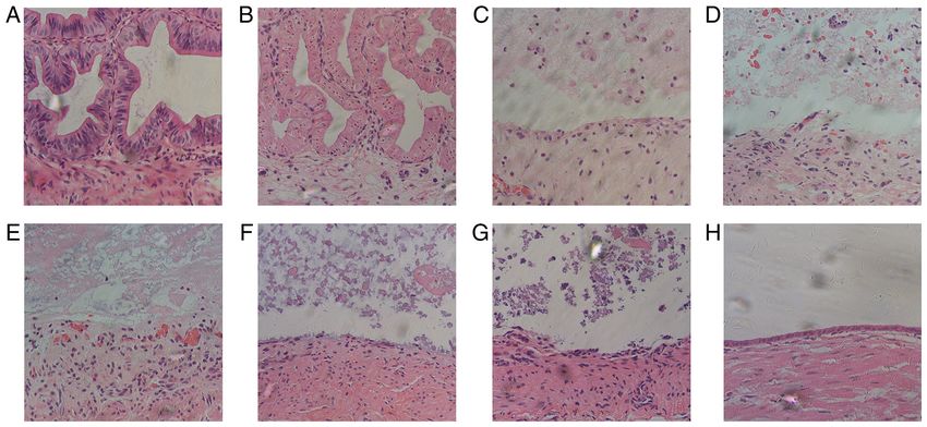
stained area percentage (APSAP) of collagen fiber revealed by collagen fibers in the lamina propria mucosae and submucous
Masson's trichrome staining was quantitatively analyzed using layer were lined up tightly (Fig. 6A).
Image‑Pro Plus version 7.0 (Media Cybernetics, Inc.) (22).
The immunohistochemical images with integrated optical Time course of cyst epithelial changes after bleomycin sclero-
density (IOD) of positive expression, which reflected the therapy in the rabbit model of simple hepatic cysts. All 28 rabbits
dynamic changes and distribution characteristics of T cells, in the experimental group survived the entire duration of the
B cells and macrophages in gallbladder tissues, were quantita‑ experimental procedure, with normal behavior, mental state and
tively analyzed using Image‑Pro Plus (23). body weight, and no complications were observed.
The first day after bleomycin sclerotherapy, the epithelial
Statistical analysis. Data are presented as the mean ± standard cells were swollen and had balloon‑like degeneration; the
error of the mean. Statistical comparisons of the intensities of nuclei were pyknotic and deformed, appearing as small circles.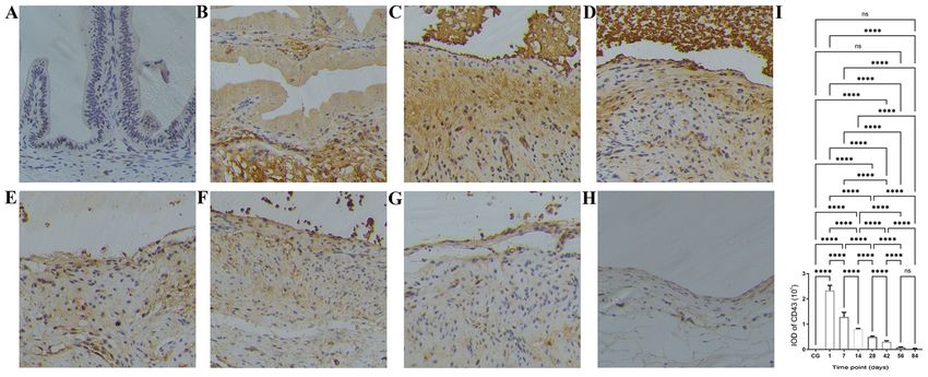
4                            LI et al: BLEOMYCIN SCLEROTHERAPY FOR EXPERIMENTAL SIMPLE HEPATIC CYSTS

Figure 3. Time course of B‑lymphocyte infiltration following bleomycin sclerotherapy in the rabbit model of simple hepatic cysts based on anti‑CD20 antibody
immunohistochemical staining. (A) In the control group, there were no B‑lymphocytes in the cyst wall. (B) Following intracyst bleomycin injection, after
1 day, B‑lymphocytes had extensively infiltrated the submucosal layer and lamina propria. (C and D) At 7 and 14 days after intracyst bleomycin injection,
B‑lymphocytes extensively infiltrated the submucosal layer, lamina propria, and the destroyed mucosal areas. (E‑H) At 28 and 84 days after intracyst bleomycin
injection, B‑lymphocyte infiltration decreased but persisted throughout the experiment. (I) Quantitative analysis of IODs of anti‑CD20 immunohistochemical
staining at the different time‑points. Detailed data and statistics to produce this graph are listed in Table SI. Magnification, x200. **P
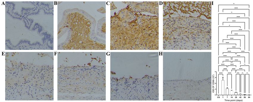
BIOMEDICAL REPORTS 15: 75, 2021                                                                       5

Figure 5. Time course of macrophage infiltration after bleomycin sclerotherapy in the rabbit model of simple hepatic cysts based on anti‑CD68 antibody
immunohistochemical staining. (A) Control group, no macrophages were present in the gallbladder wall. (B) At 1 day after intracyst bleomycin injection,
macrophages extensively infiltrated the submucosal layer and lamina propria. (C and D) At 7 and 14 days after intracyst bleomycin injection, macrophages
extensively infiltrated the submucosal layer, lamina propria and the destroyed mucosal areas. (E‑H) At 28 and 84 days after intracyst bleomycin injection, mac‑
rophages gradually decreased, but exhibited a persistent presence throughout the duration of the experiment. (I) Quantitative analysis of IODs of anti‑CD68
immunohistochemical staining at the different time‑points. Detailed data and statistics to produce this graph are listed in Table SIII. Magnification, x200.
*
 P
6                       LI et al: BLEOMYCIN SCLEROTHERAPY FOR EXPERIMENTAL SIMPLE HEPATIC CYSTS

    From 28 to 84 days after intracyst bleomycin injec‑              Bleomycin, in the presence of iron and oxygen, can generate
tion, inflammatory cell infiltration in the submucosal layer         reactive oxygen species and damage DNA by causing single‑
decreased gradually, but was present throughout the experi‑          and double‑strand breaks, a mechanism thought to be the
ment (Figs. 3E‑H, 4E‑H and 5E‑H).                                    basis for its antitumor activity and presumed mechanism of
                                                                     cytotoxicity and tissue injury (29). However, the doses used in
Time course of collagen proliferation after bleomycin sclero-        the rodent model of fibrosis do not appear to cause significant
therapy in the rabbit model of simple hepatic cysts. The time        DNA damage (12), thus making it unclear if this is the basis
course of the APSAP of collagen fibers stained with Masson's         for of lung fibrosis induced by bleomycin (12,28). A bleomycin
trichrome in the cyst wall at the various time points after          concentration of 2 mg/ml used in the present study is based on
intracyst bleomycin injection is given in Fig. 6 and Table SIV.      the experimental model of bleomycin‑induced lung and skin
Maximum staining was recorded on day 7, which gradually              fibrosis (29,30). The total amount of bleomycin saline solution
decreased and almost disappeared by day 84 (Fig. 6I, P
BIOMEDICAL REPORTS 15: 75, 2021 7
present study showed that the collagen fibers were abundant Availability of data and materials
at day 7, then gradually decreased and eventually disappeared
from 14 to 84 days. The different types of inflammatory cells The datasets used and/or analyzed during the present study are
contribute to the activation of fibroblasts and myofibroblasts available from the corresponding author on reasonable request.
and the deposition of collagen, and activated T‑lymphocytes,
B‑lymphocytes and macrophages likely collaborate to orches‑ Authors' contributions
trate post‑injury tissue remodeling and fibrosis (32). The
present study also revealed that collagen fibrous proliferation LL and YHL conceived and designed the study. LL, YHL and
is preceded by inflammatory cell infiltration, with the latter NZ acquired, analyzed and interpreted the data, and wrote and
lasting longer throughout the experiment. revised the manuscript. LL and YHL performed the statistical
The late epithelial regeneration by a single layer of flat cells analysis. All authors have read and approved the final manu‑
after chemical ablation of the gallbladder is like that reported script. LL and YHL confirm the authenticity of all the raw data.
in animal experiments with different sclerosants (25‑27). In
animal models of bleomycin‑induced pulmonary fibrosis, Ethics approval and consent to participate
alveolar epithelial regeneration is hypothesized to an impor‑
tant repair process following lung injury, which is regulated The present study was approved by the Biomedical Ethics
by various cellular and physiological mechanisms (34). In Committee of Animal Experiments of Guangdong Medical
the present study, the regenerating epithelium consisted of a Laboratory Animal Center (Guangdong, China; approval no.
single layer of flat cells that were different from the original B201610‑5). An Accreditation Certificate from the China
columnar epithelium of the gallbladder, and thus did not National Accreditation Service for Conformity Assessment
possess secretory function. Therefore, it is reasonable that has been granted to this facility. All experimental procedures
epithelial regeneration after bleomycin sclerotherapy is the were performed in accordance with the Guide for the Care and
repair response to bleomycin‑induced epithelial injury. Use of Laboratory Animals (National Institutes of Health).
The present study has some limitations. First, it had few Every effort was made to minimize animal suffering and to
early observation time points, so early or ultra‑early histopatho‑ use only the number of animals necessary for the acquisition
logical changes, such as the epithelial injury and inflammatory of reliable data.
processes, may have been undetected. Second, the molecular
and cellular mechanisms of bleomycin sclerotherapy for cystic Patient consent for publication
diseases were not investigated; thus, the mechanisms of bleo‑
mycin‑induced pulmonary and dermal fibrosis may be different Not applicable.
from those of bleomycin sclerotherapy for cystic diseases.
Finally, the gallbladder is a hypervascular structure, particularly Competing interests
when compared with simple hepatic cysts, with thick muscular
layers beneath the epithelium, which may result in substantial The authors declare that they have no competing interests.
differences in the complex inflammatory processes.
In conclusion, the present study described a rabbit model of References
simple hepatic cyst, established by ligating the cholecystic duct
1. Dietrich CF, Chiorean L, Potthoff A, Ignee A, Cui X and
and preserving cholecystic vessels. The time course of histo‑ Sparchez Z: Percutaneous sclerotherapy of liver and renal cysts,
pathological changes occurring after bleomycin sclerotherapy comments on the EFSUMB guidelines. Z Gastroenterol 54:
consisted of an epithelial destruction phase, inflammatory 155‑166, 2016.
2. Wijnands TF, Görtjes AP, Gevers TJ, Jenniskens SF, Kool LJ,
infiltration phase, collagenous proliferation phase and epithelial Potthoff A, Ronot M and Drenth JP: Efficacy and safety of aspi‑
regeneration phase. Information derived from this model may ration sclerotherapy of simple hepatic cysts: A systematic review.
help in understanding the cellular mechanisms involved in the AJR Am J Roentgenol 208: 201‑207, 2017.
3. Lam SC and Yuen HK: Medical and sclerosing agents in the
sclerosis induced by intracyst bleomycin injection of hepatic treatment of orbital lymphatic malformations: What's new? Curr
cysts and the time course of their resolution. Furthermore, these Opin Ophthalmol 30: 380‑385, 2019.
findings suggest that the pathogenesis of complications after 4. Eissa A, El Sherbiny A, Martorana E, Pirola GM, Puliatti S,
Scialpi M, Micali S, Rocco B, Liatsikos E, Breda A, et al;
percutaneous bleomycin sclerotherapy for treatment of cystic European Section of Uro‑Technology (ESUT): Non‑conservative
diseases is related to the bleomycin‑induced inflammatory management of simple renal cysts in adults: A comprehensive
reaction. The optimal follow‑up time of the therapeutic outcome review of literature. Minerva Urol Nefrol 70: 179‑192, 2018.
5. Paladine W, Cunningham TJ, Sponzo R, Donavan M, Olson K
assessment should be after inflammation has been resolved. and Horton J: Intracavitary bleomycin in the management of
malignant effusions. Cancer 38: 1903‑1908, 1976.
Acknowledgements 6. Yura J, Hashimoto T, Tsuruga N and Shibata K: Bleomycin
treatment for cystic hygroma in children. Nippon Geka Hokan 46:
607‑614, 1977.
Not applicable. 7. Takahashi H, Nakazawa S and Shimura T: Evaluation of
postoperative intratumoral injection of bleomycin for craniopha‑
ryngioma in children. J Neurosurg 62: 120‑127, 1985.
Funding 8. Johnston SR, Adam A, Allison DJ, Smith P and Ind PW: Recurrent
respiratory obstruction from a mediastinal bronchogenic cyst.
The present study was funded by the Medical Scientific Thorax 47: 660‑662, 1992.
9. Li L, Chen CC and Zeng XQ: One‑year results of single‑session
Research Foundation of Guangdong Province of China (grant sclerotherapy with bleomycin in simple renal cysts. J Vasc Interv
no. A2015530). Radiol 23: 1651‑1656, 2012.8 LI et al: BLEOMYCIN SCLEROTHERAPY FOR EXPERIMENTAL SIMPLE HEPATIC CYSTS
10. Souftas VD, Kosmidou M, Karanikas M, Souftas D, Menexes G 23. Varghese F, Bukhari AB, Malhotra R and De A: IHC Profiler: An
and Prassopoulos P: Symptomatic abdominal simple cysts: Is open source plugin for the quantitative evaluation and automated
percutaneous sclerotherapy with hypertonic saline and bleomycin scoring of immunohistochemistry images of human tissue
a treatment option? Gastroenterol Res Pract 2015: 489363, 2015. samples. PLoS One 9: e96801, 2014.
11. Li L, Zeng XQ and Li YH: CT‑guided percutaneous large‑needle 24. Terada T, Nakanuma Y, Ohta T, Nagakawa T, Motoo Y, Harada A,
aspiration and bleomycin sclerotherapy for bronchogenic cyst: Hamato N and Inaba T: Mucin‑histochemical and immunohisto‑
Report of four cases. J Vasc Interv Radiol 21: 1045‑1049, 2010. chemical profiles of epithelial cells of several types of hepatic cysts.
12. Della Latta V, Cecchettini A, Del Ry S and Morales MA: Virchows Arch A Pathol Anat Histopathol 419: 499‑504, 1991.
Bleomycin in the setting of lung fibrosis induction: From 25. Uchiyama N, Stridbeck H and Stenram U: Chemical sclerosis of
biological mechanisms to counteractions. Pharmacol Res 97: the gallbladder. An experimental study in pigs of the effect of
122‑130, 2015. absolute ethanol and polidocanol on gallbladder epithelium. Acta
13. Temmerman F, Chen F, Libbrecht L, Vander Elst I, Windmolders P, Radiol 30: 427‑431, 1989.
Feng Y, Ni Y, De Smedt H, Nevens F and van Pelt J: Everolimus halts 26. Aagaard BD, Wetter LA, Montgomery CK and Gordon RL: Heat
hepatic cystogenesis in a rodent model of polycystic‑liver‑disease. ablation of the normal gallbladder in pigs. J Vasc Interv Radiol 5:
World J Gastroenterol 23: 5499‑5507, 2017. 331‑339, 1994.
14. Yu Y, Shumway KL, Matheson JS, Edwards ME, Kline TL and 27. Lee JH, Won JH, Bae JI, Kim JH, Lee HS and Jung SM:
Lyons LA: Kidney and cystic volume imaging for disease presen‑ Chemical ablation of the gallbladder with acetic acid. J Vasc
tation and progression in the cat autosomal dominant polycystic Interv Radiol 20: 1471‑1476, 2009.
kidney disease large animal model. BMC Nephrol 20: 259, 2019. 28. Williamson JD, Sadofsky LR and Hart SP: The pathogenesis of
15. Divyashree S, Janhavi P, Ravindra PV and Muthukumar SP: bleomycin‑induced lung injury in animals and its applicability to
Experimental models of polycystic ovary syndrome: An update. human idiopathic pulmonary fibrosis. Exp Lung Res 41: 57‑73, 2015.
Life Sci 237: 116911, 2019. 29. Liu T, De Los Santos FG and Phan SH: The bleomycin model of
16. Atilgan R, Ozkan ZS, Kuloglu T, Kocaman N, Baspinar M, pulmonary fibrosis. Methods Mol Biol 1627: 27‑42, 2017.
Can B, Şimşek M and Sapmaz E: Impact of intracystic ethanol 30. Yamamoto T: Intradermal injections of bleomycin to model skin
instillation on ovarian cyst diameter and adjacent ovarian tissue. fibrosis. Methods Mol Biol 1627: 43‑47, 2017.
Eur J Obstet Gynecol Reprod Biol 174: 133‑136, 2014. 31. Hadjicharalambous MR and Lindsay MA: Idiopathic pulmonary
17. National Research Council (US): Committee for the Update of fibrosis: Pathogenesis and the emerging role of long non‑coding
the Guide for the Care and Use of Laboratory Animals: Guide for RNAs. Int J Mol Sci 21: 524, 2020.
the Care and Use of Laboratory Animals. 8th edition. National 32. Heukels P, Moor CC, von der Thüsen JH, Wijsenbeek MS and
Academies Press, Washington, DC, pp1‑154, 2011. Kool M: Inflammation and immunity in IPF pathogenesis and
18. Feldman AT and Wolfe D: Tissue processing and hematoxylin treatment. Respir Med 147: 79‑91, 2019
and eosin staining. Methods Mol Biol 1180: 31‑43, 2014. 33. MacIntosh PW, Yoon MK and Fay A: Complications of
19. Goldner J: A modification of the masson trichrome technique for intralesional bleomycin in the treatment of orbital lymphatic
routine laboratory purposes. Am J Pathol 14: 237‑243, 1938. malformations. Semin Ophthalmol 29: 450‑455, 2014.
20. Rehg JE, Bush D and Ward JM: The utility of immunohistochemistry 34. Basil MC, Katzen J, Engler AE, Guo M, Herriges MJ, Kathiriya JJ,
for the identification of hematopoietic and lymphoid cells in normal Windmueller R, Ysasi AB, Zacharias WJ, Chapman HA, et al: The
tissues and interpretation of proliferative and inflammatory lesions cellular and physiological basis for lung repair and regeneration:
of mice and rats. Toxicol Pathol 40: 345‑374, 2012. Past, present, and future. Cell Stem Cell 26: 482‑502, 2020.
21. Taylor CR and Rudbeck L (eds): Dako Education Guide:
Immunohistochemical Staining Methods. 6th edition. pp1-110, 2013.
22. Schmidt MJ, Tschoeke A, Noronha L, Moraes RS, Mesquita RA,
Grégio AM, Alanis LR, Ignácio SA, Santos JN, Lima AA, et al: This work is licensed under a Creative Commons
Histochemical analysis of collagen fibers in giant cell fibroma Attribution-NonCommercial-NoDerivatives 4.0
and inflammatory fibrous hyperplasia. Acta Histochem 118: International (CC BY-NC-ND 4.0) License.
451‑455, 2016.You can also read