Three-Day Continuous Oxytocin Infusion Attenuates Thermal and Mechanical Nociception by Rescuing Neuronal Chloride Homeostasis via Upregulation ...
←
→
Page content transcription
If your browser does not render page correctly, please read the page content below
ORIGINAL RESEARCH
                                                                                                                                            published: 24 March 2022
                                                                                                                                      doi: 10.3389/fphar.2022.845018

                                            Three-Day Continuous Oxytocin
                                            Infusion Attenuates Thermal and
                                            Mechanical Nociception by Rescuing
                                            Neuronal Chloride Homeostasis via
                                            Upregulation KCC2 Expression and
                                            Function
                           Edited by:
                             Xin Luo,       Xiyuan Ba 1†, Chenqiu Ran 2†, Wenjun Guo 3, Jing Guo 4, Qian Zeng 1, Tao Liu 5, Wuping Sun 1,
     Guangdong-Hong Kong-Macao              Lizu Xiao 1, Donglin Xiong 1, Yelan Huang 2, Changyu Jiang 1* and Yue Hao 2*
   Greater Bay Area Center for Brian
                                            1
         Science and Brain-Inspired           Department of Pain Medicine and Shenzhen Municipal Key Laboratory for Pain Medicine, Shenzhen Nanshan People’s Hospital,
                  Intelligence, China       Shenzhen, China, 2School of Pharmaceutical Sciences, Health Science Center, Shenzhen University, Shenzhen, China,
                                            3
                                              Department of Pain Medicine, Shenzhen, China, 4Department of Endocrinology and Metabolism, Shenzhen University General
                         Reviewed by:       Hospital and Shenzhen University Academy of Clinical Medical Sciences, Shenzhen University, Shenzhen, China, 5Department of
                             Ping Dong,     Pediatrics, The First Affiliated Hospital of Nanchang University, Nanchang, China
       Duke University, United States
                         Pascal Darbon,
    Université de Strasbourg, France        Oxytocin (OT) and its receptor are promising targets for the treatment and prevention of the
                  Jorge Baruch Pineda,
University of Pittsburgh, United States
                                            neuropathic pain. In the present study, we compared the effects of a single and continuous
                      Pierrick Poisbeau,    intrathecal infusion of OT on nerve injury-induced neuropathic pain behaviours in mice and
    Université de Strasbourg, France        further explore the mechanisms underlying their analgesic properties. We found that three
                    *Correspondence:        days of continuous intrathecal OT infusion alleviated subsequent pain behaviours for
                              Yue Hao
                    yuehao@szu.edu.cn       14 days, whereas a single OT injection induced a transient analgesia for 30 min, suggesting
                        Changyu Jiang       that only continuous intrathecal OT attenuated the establishment and development of
         changyujiang@email.szu.edu.cn
                                            neuropathic pain behaviours. Supporting this behavioural finding, continuous intrathecal
     †
         These authors have contributed
                    equally to this work
                                            infusion, but not short-term incubation of OT, reversed the nerve injury-induced
                                            depolarizing shift in Cl− reversal potential via restoring the function and expression of
                      Specialty section:    spinal K+-Cl- cotransporter 2 (KCC2), which may be caused by OT-induced enhancement
           This article was submitted to
                                            of GABA inhibitory transmission. This result suggests that only continuous use of OT may
                    Neuropharmacology,
                 a section of the journal   reverse the pathological changes caused by nerve injury, thereby mechanistically blocking
              Frontiers in Pharmacology     the establishment and development of pain. These findings provide novel evidence
         Received: 29 December 2021         relevant for advancing understanding of the effects of continuous OT administration on
            Accepted: 11 March 2022
           Published: 24 March 2022
                                            the pathophysiology of pain.
                              Citation:     Keywords: neuropathic pain, oxytocin, chloride homeostasis, K+-Cl-cotransporter 2, continuous intrathecal drug
Ba X, Ran C, Guo W, Guo J, Zeng Q,          delivery
Liu T, Sun W, Xiao L, Xiong D, Huang Y,
Jiang C and Hao Y (2022) Three-Day
          Continuous Oxytocin Infusion
  Attenuates Thermal and Mechanical
                                            INTRODUCTION
    Nociception by Rescuing Neuronal
Chloride Homeostasis via Upregulation
                                            Neuropathic pain is a debilitating condition that affects 7–10% of the general population (Colloca
       KCC2 Expression and Function.        et al., 2017). Unlike opioids and non-steroidal anti-inflammatory drugs for nociceptive pain, the
        Front. Pharmacol. 13:845018.        medications used to treat neuropathic pain tend to only be modestly effective and can potentially
      doi: 10.3389/fphar.2022.845018        cause multiple adverse reactions (Baron et al., 2010). Developing mechanism-based therapies for

Frontiers in Pharmacology | www.frontiersin.org                                    1                                         March 2022 | Volume 13 | Article 845018
Ba et al.                                                                                                       Oxytocin Attenuates Pain via KCC2

neuropathic pain remains a major challenge. A growing body of               its phosphorylation and insertion/stabilization at the neuronal
literature has demonstrated the analgesic effects of the                    surface in an early developmental time window (Leonzino et al.,
neuropeptide oxytocin (OT) in both humans and rodents (see                  2016). However, little is known on how OT affects chloride
reviews by Oxytocin and pain perception: from animal models to              homeostasis and the function of KCC2 in neuropathic pain.
human research) (Gimpl and Fahrenholz, 2001; Honda and                          In addition, the current understanding of mechanisms
Takano, 2009; Koshimizu and Tsujimoto, 2009; Stoop, 2014;                   underlying OT analgesia is mainly based on studies using
Boll et al., 2018; Herpertz et al., 2019). Electrical stimulation           single or multiple injections of OT in animals. Little is known
of the anterior part of the hypothalamic paraventricular nucleus            about the effects of continuous OT administration on pain
increased OT concentration in the cerebrospinal fluid (CSF) and              processing. In this study, we adopted intrathecal drug delivery
produced antinociception in rats (Martinez-Lorenzana et al.,                technique to administer OT centrally in nerve injured mice.
2008), and intraperitoneal or intrathecal (i.t.) injection of OT            Chronic intrathecal drug infusion through an implantable
was shown to block neuropathic pain in rats (Yang et al., 2007).            pump is a clinically available strategy to treat a number of
Clinical data suggested that administration of OT in the                    neurological diseases (Ganguly et al., 2001; Kästner, 2010).
cerebrospinal fluid (CSF) reduces surgical recovery time while               Findings based on continuous intrathecal OT delivery in mice
decreasing pain and hypersensitivity in patients after injury               may provide more information on how OT targets the
(Wang et al., 2013). Considering it also plays a key                        pathophysiology of pain and better implications for human
modulatory role in emotions, stress and anxiety, which are                  therapy.
well known to substantially influence pain perception                            Thus, in the present study we adopted intrathecal drug
(Apkarian et al., 2005; Apkarian, 2008; Baron et al., 2010;                 delivery technique to compare the effects of a single or
Peters, 2015; Tracy et al., 2015), OT has become a promising                continuous intrathecal infusion of OT on pain behaviours in
target for therapeutic interventions for pain.                              mice; we determined whether they block neuropathic pain by
    Excitation/inhibition imbalance along the entire nociceptive            preventing the disruption of the intracellular Cl− homeostasis in
pathway is considered a main driver in the development of                   the spinal superficial dorsal horn, and whether it is mediated by
neuropathic pain (Kahle et al., 2014). One of the mechanisms                restoring the KCC2 expression and function.
proposed for this imbalance involves compromised inhibition in
the superficial dorsal horn of the spinal cord, leading to
hyperactivity of spinal dorsal horn circuit, which is the main              MATERIALS AND METHODS
target for primary nociceptive afferents (Prescott, 2015). γ-
aminobutyric acid (GABA) is the most critical inhibitory                    Animals
neurotransmitter in the central nervous system. The inhibitory              All animal procedures were conducted in strict adherence to the
efficiency of GABAergic transmission is determined primarily by              guidelines of the International Association for the Study of Pain
the electrochemical gradient for Cl−, which is depended by the              and were approved by the Animal Care and Use Committee of
intra and extracellular concentration of Cl− (Ganguly et al., 2001).        Health Science Center at Shenzhen University. 80 male C57BL/6
It has been demonstrated that Cl− homeostasis is collapsed and              mice (5–8 weeks of age) were purchased from Guangdong
Cl− levels are elevated in spinal cord neurons under the                    Province Laboratory Animal Center (Guangzhou, China). 20
pathophysiology of pain disorders (Coull et al., 2003).                     vGAT-ires-cre mice and 20 td-Tomato (Ai9) mice were
Recently, a body of evidence showed that compromised spinal                 purchased from Jackson Laboratory. The animals were housed
inhibition resulted from downregulation of K+-Cl- cotransporter             in plastic cages (5 per cage) in a temperature-controlled
2 (KCC2) and the subsequent disruption of intracellular chloride            environment on a 12 h/12 h light/dark cycle. Food and water
homeostasis (Coull et al., 2003; Price et al., 2009; Li et al., 2016;       were available ad libitum.
Mapplebeck et al., 2019). In mature central neurons, KCC2 is
responsible for the low intracellular Cl− concentration ([Cl−]i)            Reagents
that forms the basis for hyperpolarizing GABAA receptor-                    Oxytocin (catalogue: H-2510) and [d(CH2)51,Tyr(Me)2,
mediated responses. It regulates the formation (Li et al., 2007),           Thr4,Orn8,des–Gly–NH29]–vasotocin (dVOT, catalogue: H-
functional maintenance and plasticity of glutamatergic synapses             2510) were purchased from Bachem AG (Bubendorf,
(Fiumelli et al., 2005; Gauvain et al., 2011; Chevy et al., 2015;           Switzerland). TC OT39 (catalogue: 1078) was obtained from
Llano et al., 2015). Indeed, Modol’s results indicate that nerve            Tocris (Minnesota, United States).
injury results in a reduction in the expression of KCC2 in the
spinal dorsal horn that accompanies chronic pain, but prevention            Neuropathic Pain Model
of the downregulation of KCC2 along the central sensory                     The partial sciatic nerve ligation (pSNL) pain model was
pathways relieves neuropathic pain after peripheral nerve                   established according to previously described procedures
injury (Modol et al., 2014). Loss of activity of this transporter           (Seltzer et al., 1990). Briefly, the animals were anaesthetized
is a key mechanism for chronic pain, and different groups                   with sodium pentobarbital (50 mg/kg, i.p.) and a tight ligation
demonstrated that renormalization of impaired KCC2                          of approximately one-third to one-half the diameter of the
alleviated nerve injury-induced neuropathic pain (Gagnon                    right sciatic nerve (ipsilateral) was performed with 6–0 silk
et al., 2013; Kitayama, 2017). Leonzino et al. found that OT                suture. In sham-operated mice, the nerve was exposed without
directly modulates the functional activity of KCC2 by promoting             ligation.

Frontiers in Pharmacology | www.frontiersin.org                         2                                  March 2022 | Volume 13 | Article 845018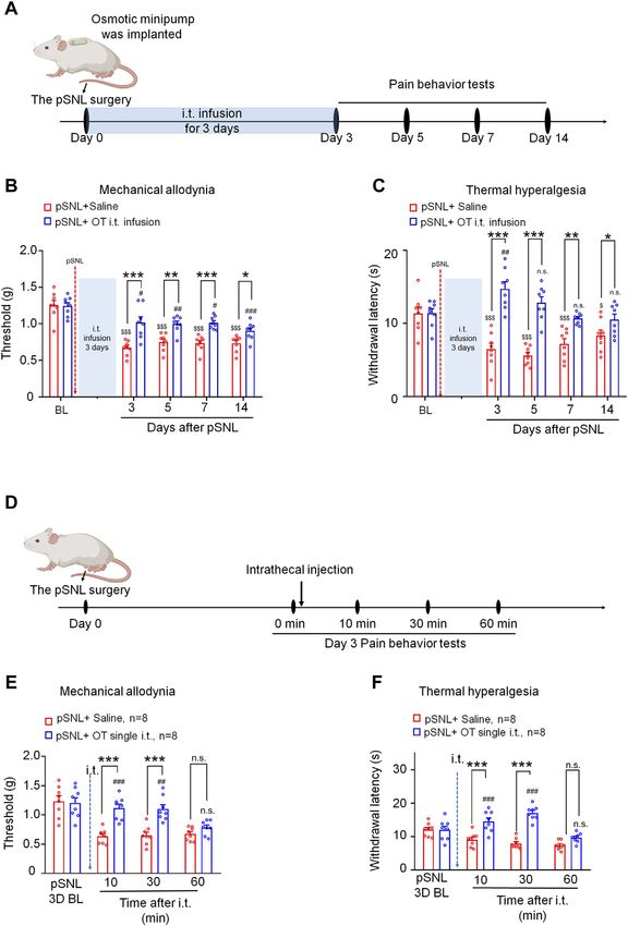
Ba et al.                                                                                                     Oxytocin Attenuates Pain via KCC2

Behavioural Testing                                                       Western Blotting
Von Frey testing was performed to assess mechanical                       The animals were sacrificed, and the L4-6 spinal cord segments
allodynia. The mice were habituated to the environment for                were removed and stored at −80°C until assayed. The samples
2 days before the testing began. All the behaviours were tested           were homogenized and centrifuged to extract the protein, and the
blindly. For testing mechanical allodynia, the mice were                  resulting preparations were saved. Equal amounts of protein were
confined separately in boxes (14 × 18 × 12 cm) placed on                   separated by 10% Tris-Tricine SDS-PAGE and transferred onto
an elevated metal mesh floor, and their hind paws were                     polyvinylidene difluoride membranes. The membranes were then
stimulated with a series of von Frey hairs with                           blocked in 5% non-fat milk for 1 h at room temperature, followed
logarithmically increasing stiffness (0.16–2.00 g, Stoelting)             by overnight incubation with rabbit anti-KCC2 antibody (1:1000;
situated perpendicularly to the central plantar surface. The              ab49917, Abcam, United States) and ß-actin (1:2000; Sigma,
50% paw withdrawal threshold was determined by Dixon’s up-                United States) primary antibody. Immunoblots were then
down method. The hot plate test (Hot/Cold Plate, Cat. 35150,              incubated for 1 h at room temperature with goat anti-rabbit
Ugo Basile, Italy) was used to examine thermal hyperalgesia.              polyclonal IgG (1:3000, ab205718, Abcam, MA, United States).
Each mouse was placed on the hot plate, and the latency of paw            Immunoblots were developed by chemiluminescent substrate and
withdrawal from the heat stimulus was measured twice                      quantified using ImageJ software.
separated by a 5-min interval. The average value was used
as the latency of response. All behavioural testing was done              Immunohistochemistry
with the experimenters blinded to the treatment conditions.               The mice were deeply anesthetized with isoflurane and
                                                                          transcardially perfused with PBS followed by 4% PFA. Lumbar
                                                                          L4-6 spinal cord segments sections were blocked and then
Intrathecal Injection and Continuous                                      incubated overnight at 4°C with rabbit antibodies against
Intrathecal Infusion of Drugs                                             KCC2 (Abcam, ab49917, United States). The sections were
OT (0.1 μg in 10 μL) or dVOT (0.1 μg/10 μL) was injected into the         then incubated for 30 min at 37°C with AF488-conjugated
subarachnoid space through the intervertebral foramen between             secondary antibodies (donkey, 1:500, Jackson Immuno-
L4 and L6 (Hylden and Wilcox, 1980). For the intrathecal                  Research, West Grove, PA, United States), and the nuclei were
infusion of drugs, an osmotic minipump (model 1003D,                      stained with DAPI. The sections were viewed under Zeiss 880
ALZET, Cupertino, CA, United States) connected with a                     inverted confocal microscopy, and images were collected using
polyethylene catheter was deposited in a subcutaneous pocket              identical acquisition parameters and quantified using Image-Pro
following partial sciatic nerve ligation. The other end of the            Plus 6.0 software (Media Cybernetics, Silver spring, MD,
catheter was inserted from the atlanto-occipital membrane                 United States) by experimenters blinded to treatment groups.
into the subarachnoid space until the tip of the catheter
reached the lumbar spinal enlargement. OT and other reagents              In Situ Hybridization
were then delivered continuously with a flow rate of 1 μL/h for            In situ hybridization was performed using the RNAscope system
3 days from days 0 to 2 after pSNL surgery. The final dose of OT           (Advanced Cell Diagnostics) following the manufacturer’s
intrathecal infusion is 0.3 μg in 100 μL. (The volume delivery rate       protocol. Pre-treatment consisted of dehydration, followed by
and the delivery duration of ALZET pumps are fixed at                      incubation with hydrogen peroxide and protease IV at room
manufacture).                                                             temperature. The Multiplex Fluorescent Kit v2 protocol was
                                                                          followed using commercial probes for the OT receptor (Oxtr,
Quantitative RT-PCR                                                       NM_001081147.1, #402658-C3). Images were captured by Zeiss
The animals were sacrificed and L4–6 spinal cord segments were             880 inverted confocal microscopy. Visualized cells with more
collected in tubes with RNAlater (Qiagen Inc., Valencia, CA,              than 5 puncta per cell were classified as positive neurons.
United States) and stored at −80°C until RNA isolation. Total
RNA was isolated from these tissues according to Chomczynski’s            Electrophysiological Recordings
method (Chomczynski and Sacchi, 1987) and reverse transcribed             Adult (5–7 weeks) male mice were anaesthetized with urethane
using Omniscript reverse transcriptase (Qiagen Inc., Valencia,            (1.5–2.0 g/kg, i.p.). The lumbosacral spinal cord was removed and
CA, United States) at 37°C for 60 min. The reaction was                   submerged into ice-cold dissection solution saturated with 95%
performed in the presence of the RNase inhibitor rRNAsin                  O2 and 5% CO2 at room temperature. Transverse slices
(Promega, Madison, WI, United States) and an oligo (dT16)                 (300–400 μm) were cut in a vibrating microslicer (VT1200s
primer (Qiagen) to selectively amplify the mRNA. For                      Leica). The slices were incubated at 32°C for at least 30 min in
quantitative PCR, 45 ng of cDNA was used as a template.                   regular artificial cerebrospinal fluid (aCSF) equilibrated with 95%
Reactions were performed using Assay-On-Demand TaqMan                     O2 and 5% CO2.
probes and TaqMan Universal PCR Master Mix (Applied                          The following solutions were used: dissection solution
Biosystems, Foster, CA, United States) according to the                   containing (in mM) 240 sucrose, 25 NaHCO3, 2.5 KCl, 1.25
manufacturer’s protocol. Reactions were run on a Real-Time                NaH2PO4, 0.5 CaCl2, and 3.5 MgCl2 at pH 7.4; regular artificial
PCR iCycler IQ (Bio-Rad, Hercules, CA, United States) with                CSF containing 135 NaCl, 2.5 KCl, 3 MgCl2, 1 CaCl2, 10 HEPES, 1
software version 3.0. The expression levels of Kcc2 were                  NaH2PO4, and 10 glucose at pH 7.4; and normal intrapipette
normalized to ß-actin.                                                    solution for perforated recording containing 115 K-

Frontiers in Pharmacology | www.frontiersin.org                       3                                  March 2022 | Volume 13 | Article 845018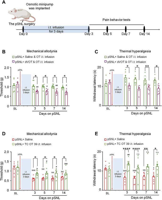
Ba et al.                                                                                                     Oxytocin Attenuates Pain via KCC2

methylsulfate, 25 KCl, 2 MgCl2, 10 HEPES, 0.4 GTP-Na and                 RESULTS
5 Mg-ATP at pH 7.2 and 310 mOsm.
    To measure the reversal potential of GABA-evoked currents, a         Three-Day Continuous Intrathecal Infusion,
slice was placed in the recording chamber and completely                 but Not Short-Term Application of OT,
submerged and superfused at a rate of 2–4 ml/min with aCSF.
                                                                         Attenuated the Establishment and
A perforated patch-clamp was applied to avoid changes in the
[Cl−]i. To measure the chloride equilibrium potential (ECl),             Development of Nerve Injury-Induced
gramicidin D (80 μg/ml with an 0.8% DMSO final                            Nociceptive Behaviours in pSNL Mice
concentration from an 8 mg/ml stock in DMSO) was added to                pSNL-induced nerve injury produced mechanical allodynia and
the intrapipette solution, and 6-cyano-7- nitroquinoxaline-2,3-          thermal hyperalgesia in mice. This mechanical and thermal
dione (CNQX, 10 μM), DL-2-amino-5-phosphonovaleric acid                  hypersensitivity started on day 1 and remained relatively stable
(APV, 50 μM) and tetrodotoxin (TTX, 0.5 μM) were added to                from days 3 to 14 after nerve ligation (Supplementary Figures
the aCSF solution. The tip of the patch pipette was filled with the       S1A,B).
normal intrapipette solution, while the rest of the pipette                 An osmotic minipump was implanted immediately following
contained the gramicidin-containing solution. After forming a            partial sciatic nerve ligation. OT was then delivered with a flow
seal on the membrane, we waited 30 min for the gramicidin to             rate of 1 μL/h for 3 days from days 0–2 after pSNL surgery.
effectively reduce the series resistance to below 100 MΩ.                Mechanical allodynia and thermal hyperalgesia were tested at
Membrane potential measurements were corrected for liquid                days 3, 5, 7 and 14 after pSNL surgery (Figure 1A). As shown in
junction potential, which was measured as in(Guo et al.,                 Figures 1B,C, infusion of OT (0.3 μg, 100 μL) for 3 days before
2014). GABA (1 mM) was puffed locally and instantaneously,               the behavioural tests decreased nerve injury-induced
and the puff pipette was aimed toward the recording pipette.             nociceptive behaviours in mice. Compared with the vehicle,
Voltage ramps were applied from +8 to −92 mV over 200 ms at a            3-days continuous infusion of OT increased the mechanical
holding potential of −42 mV. Since the voltage ramp might evoke          threshold in the von Frey test [F(1,14) = 61.57, p < 0.001;
a basal current, a control voltage ramp was first applied to record       Figure 1B, n = 8] and paw withdrawal latency in the hot-plate
the basal current; 1 min later, GABA was puffed, followed by             test [F(1,14) = 50.74, p < 0.001; Figure 1C, n = 8] for 14 days,
another voltage ramp, and then the GABA-evoked currents were             which was the longest period we tested, indicating that 3-days
recorded (Billups and Attwell, 2002). The reversal potential was         continuous intrathecal OT infusion may attenuate the
analysed as in (Billups and Attwell, 2002).                              establishment and development of nerve injury-induced
    Excitatory and inhibitory post-synaptic currents (EPSCs and          neuropathic pain.
IPSCs) recordings were made from lamina II inhibitory                       In comparison, the effect of a single injection of OT on pSNL-
neurons. The patch-pipette solution contained (in mM)                    induced mechanical and thermal hypersensitivity was also tested
K-gluconate 135, KCl 5, CaCl2 0.5, MgCl2 2, EGTA 5,                      on day 3 after nerve ligation, when the pain behaviours were well
HEPES 5, an Mg-ATP 5; or Cs2SO4 110, CaCl2 0.5, MgCl2 2,                 established (Figure 1D). Single intrathecal OT (0.1 μg/10 μL)
EGTA 5, HEPES 5, Mg-ATP5, tetraethylammonium (TEA)-Cl 5                  significantly alleviated pSNL-induced mechanical allodynia
(pH = 7.2) (Jiang et al., 2014). The former and latter solutions         [F(1,14) = 42.59, p < 0.001; Figure 1E] and thermal
were used to record EPSCs and IPSCs, respectively. EPSC                  hyperalgesia [F(1,14) = 29.66, p < 0.001; Figure 1F] at 10 [p <
recordings were made at a holding potential (VH) of −70 mV,              0.001] and 30 min [p < 0.001] after injection. This effect of OT
where no IPSCs were observed, since the reversal potential for           was not observed at 60 min after the injection [p > 0.05; Figures
IPSCs was near −70 mV. IPSCs were recorded at a VH of 0 mV,              1E,F], indicating that the analgesic effect of a single intrathecal
where EPSCs were invisible as reversal potential for EPSCs was           OT administration on nerve injury-induced pain behaviours is
close to 0 mV. Cs+ and TEA were used to block K+ channels                transient. OT at the doses used in the present study had no effect
expressed in the recorded neurons, and thus to easily shift VH           on the locomotor activity or motor coordination in mice (date not
from −70 to 0 mV. GABAergic IPSCs were obtained in the                   shown).
presence of the glycine-receptor antagonist strychnine (1 mM).              We found no significant differences between male and female
EPSC and IPSC events were detected and analysed using Mini               mice in the analgesic effects of oxytocin [p > 0.05; Supplementary
Analysis Program 6.0. Signals were acquired using an Axopatch            Figure S4].
700B amplifier and analysed with pCLAMP 10.3 software. Only
neurons with resting membrane potential < −50 mV and stable
access resistance were included.
                                                                         The Effects of 3-days OT Infusion on Nerve
                                                                         Injury-Induced Nociceptive Behaviours
Statistical Analysis                                                     Were Mediated by Oxtrs
The data are expressed as means ± SEM and analysed with a t-test         To determine whether the effects of 3-days OT infusion on
or variance (ANOVA) using one-way or mixed factorial designs             neuropathic pain were mediated by Oxtrs, its agonist or
as appropriate, followed by Bonferroni’s post hoc test or simple-        antagonist was administrated (Figure 2A). Co-intrathecal
effects ANOVA. All statistical analyses were performed using             infusion (100 μL) of a selective Oxtr antagonist, dVOT
GraphPad Prism 8.0. (GraphPad Inc., La Jolla, CA,                        (0.3 μg), with OT (0.3 μg) blocked the analgesic effect of OT
United States). Significance was defined as p < 0.05.                      on nerve injury-induced mechanical [F(1,13) = 25.04, p =

Frontiers in Pharmacology | www.frontiersin.org                      4                                   March 2022 | Volume 13 | Article 845018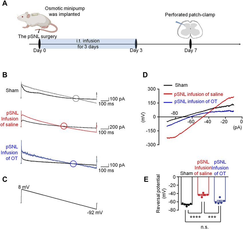
Ba et al.                                                                                                                              Oxytocin Attenuates Pain via KCC2

  FIGURE 1 | Three-day continuous intrathecal infusion, but not short-term application of OT, attenuated the establishment and development of nerve injury-induced
  nociceptive behaviours in pSNL mice. (A) A schematic of the experimental design. (B,C) Continuous intrathecal OT infusion (0.3 μg/100 μL) for 3 days before
  behavioural tests decreased pSNL-induced mechanical allodynia (A) and thermal hyperalgesia (B) for 14 days. (D) A schematic of the experimental design. (E,F) A single
  intrathecal OT injection (0.1 μg/10 μL) relieved pSNL-induced mechanical allodynia (E) and thermal hyperalgesia (F) in mice. Two-way repeated-measures ANOVA
  with group as the between-subjects factor and day/time as the within-subjects factor. Data are expressed as mean ± SEM. *p < 0.05, **p < 0.01, ***p < 0.001 OT vs.
  saline; $p < 0.05, $$$p < 0.001 vs. baseline; #p < 0.05, ##p < 0.01, ####p < 0.0001 vs. baseline.

0.0002; Figure 2B, n = 7–8] and thermal hypersensitivity                                 F(1,14) = 15.42, p = 0.0015; Hot-plat test F(1,14) = 29.80, p <
[F(1,12) = 28.92, p < 0.001; Figure 2C, n = 7]. The selective                            0.0001; Figures 2D,E; n = 8]. There results suggested that the 3-
Oxtr agonists TC OT (0.3 μg/100 μL) produced significant                                  days intrathecal infusion of OT induced analgesic effect is
analgesic effects which were equivalent to OT [von Frey test                             mediated by the Oxtrs in the spinal cord.

Frontiers in Pharmacology | www.frontiersin.org                                      5                                           March 2022 | Volume 13 | Article 845018
Ba et al.                                                                                                                            Oxytocin Attenuates Pain via KCC2

  FIGURE 2 | The effects of 3-days OT infusion on nerve injury-induced nociceptive behaviours were mediated by OXTRs. (A) A schematic of the experimental
  design. (B,C) OT’s effect on mechanical allodynia (B) and thermal hyperalgesia (C) was completely blocked by its selective antagonist, dVOT (0.3 μg/100 μL). (D,E)
  Selective OT receptor agonists, TC OT (0.3 μg/100 μL, intrathecal infusion) showed similar effects on mechanical allodynia (D) and thermal hyperalgesia (E) in pSNL
  mice. Two-way repeated-measures ANOVA with group as the between-subjects factor. Data are expressed as mean ± SEM. *p < 0.05, **p < 0.01, ***p < 0.001 TC
  OT vs. saline; OT vs. dVOT and OT. $$$p < 0.001, $$$$p < 0.0001 vs. baseline; #p < 0.05, ##p < 0.01, ###p < 0.001 vs. baseline.

Three-Day Continuous Intrathecal Infusion,                                             (Figure 3A). Since GABAA receptor (GABAAR) is the
but Not Short-Term Application of OT,                                                  dominant chloride ion channel on the membrane of neurons
                                                                                       in the superficial dorsal horn, GABA was puffed briefly to the
Renormalized Neuronal Chloride                                                         recorded neuron to trigger transient chloride influx or efflux.
Equilibrium Potential in Spinal Superficial                                                As voltage ramps were applied from +8 to −92 mV
Dorsal Horn                                                                            (Figure 3C), the GABA-evoked currents were recorded to
It was reported that neuronal intracellular chloride concentration                     evaluate chloride equilibrium potential (ECl-). These currents
was increased in the superficial dorsal horn after nerve injury                         were completely blocked by a selective GABAAR antagonist,
(Yeo et al., 2021), we performed perforated patch-clamp                                bicuculline (10 μM), confirming that they were mediated by
recording in spinal cord slices derived from each group to                             GABAAR (data not shown). The ECl- in sham mice was
investigate the effects of OT on chloride homeostasis                                  −66.68 ± 1.22 mV (Figures 3B–E, n = 5–6, 3 mice per group),

Frontiers in Pharmacology | www.frontiersin.org                                    6                                           March 2022 | Volume 13 | Article 845018
Ba et al. Oxytocin Attenuates Pain via KCC2
FIGURE 3 | Three-day continuous intrathecal OT infusion renormalized EGABA in spinal dorsal horn. (A) The schematics of the electrophysiological recording.
(B,C) As voltage ramps applied from +8 to −92 Mv (C), basal and GABA-evoked currents were recorded (B). (D,E) Representative (D) and statistical (E) reversal
potential of EGABA recorded from slices of sham and pSNL mice treated with continuous OT or saline. One-way ANOVA followed by Bonferroni’s post hoc test. Data
are expressed as mean ± SEM. ***p < 0.001 sham vs. pSNL; ***p < 0.001 OT vs. saline infusion.
whereas that value in pSNL mice shifted to a more positive value of expression levels of KCC2 in the spinal cord. Compared with the
−43.54 ± 1.67 mV [ p < 0.001 vs. sham group; F(2,12) = 36.26, p < sham group, quantitative PCR data revealed a significant decrease
0.001; Figures 3B–E, n = 5 from 3-4 mice]. Continuous intrathecal in spinal Kcc2 mRNA levels at both days 7 and 14 after pSNL
infusion of OT reversed the value of ECl- to −59.02 ± 2.69 mV, which surgery [p < 0.001 vs. sham; F(2,16) = 3.818, p = 0.0441;
was much closer to that of the sham mice [p > 0.05 vs. sham; Figures Figure 5A, n = 5 per group]. Intrathecal infusion of OT
3B–E, n = 5 from 3-4 mice], suggesting that 3-days infusion of OT was increased spinal Kcc2 mRNA levels in pSNL mice compared
able to restore [Cl−]i in pSNL mice. with saline group [p < 0.01; F(2,16) = 3.818, p = 0.0441;
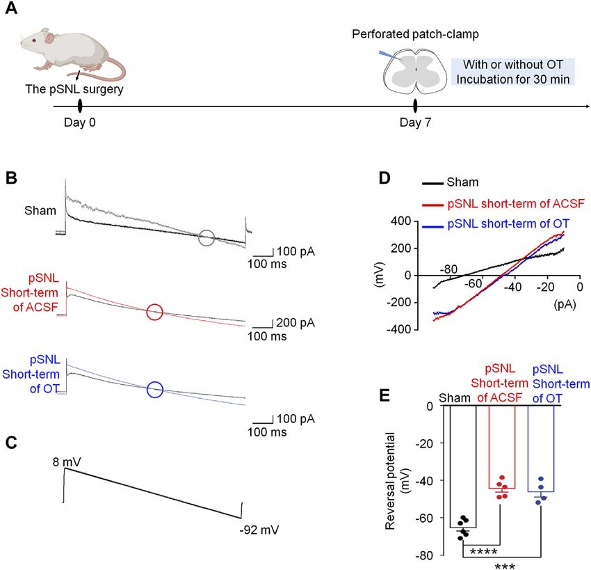
In comparison, we also recorded the ECl- using the spinal cord Figure 5A, n = 5 per group].
slices incubated with saline or OT for 30 min (short-term Western blotting data also showed that nerve injury-induced a
application, Figure 4A), and the reversal potentials were significant decrease in the protein levels of KCC2 in the spinal
−44.34 ± 2.91 mV and −46.10 ± 3.10 mV, respectively [p > dorsal horn at days 7 and 14 after pSNL surgery [p < 0.0001 vs.
0.05 vs. saline; F(2,12) = 31.71, p < 0.0001; Figures 4B–E]. sham; F(2,16) = 8.982, p = 0.0024; Figures 5B,C, n = 5 per group].
Incubation of the spinal cord slices with OT for a relatively short Intrathecal infusion of OT restored the protein levels of KCC2 but
time failed to restore the value of ECl- in pSNL mice, suggesting that did not completely reverse this decrease [p < 0.01 vs. saline;
the effect of OT on ECl- required relatively long-term application. F(2,16) = 8.982, p = 0.0024; Figures 5B,C, n = 5 per group].
Immunohistochemistry (IHC) of spinal slices from laminae II
further supported the western blotting data, which showed that
Three-Day Continuous Intrathecal OT the KCC2 signal was widely expressed throughout the spinal
Infusion Upregulated Spinal KCC2 dorsal horn in sham mice (Figure 5D). Nerve injury-induced a
Expression reduction in KCC2 expression at days 7 and 14 after pSNL
Given that the shift of ECl- in pSNL animals may be due to surgery [p < 0.0001 vs. sham; F(2,12) = 8.119, p = 0.0059;
depressed function of KCC2, we analysed the transcriptional and Figures 5D,E]. Infusion of OT reversed this reduction [p <
Frontiers in Pharmacology | www.frontiersin.org 7 March 2022 | Volume 13 | Article 845018Ba et al. Oxytocin Attenuates Pain via KCC2
FIGURE 4 | Short-term OT incubation failed to renormalize EGABA in spinal dorsal horn. (A) The schematics of the electrophysiological recording. (B,C) As voltage
ramps were applied from +8 to −92 Mv (C), basal and GABA-evoked currents (B) were recorded. (D,E) The reversal potential of EGABA recorded from slices of naïve and
pSNL mice incubated with OT or saline. One-way ANOVA followed by Bonferroni’s post hoc test. Data are expressed as mean ± SEM. ***p < 0.001, ****p < 0.0001 naïve
vs. pSNL incubated with saline or OT.
0.01 vs. sham; F(2,12) = 8.119, p = 0.0059; Figures 5D,E, n = 4 per fluorescence. As shown in Figures 6A,B, about 30% of vGAT +
group] to some extent. neurons (inhibitory neurons) expressed Oxtrs mRNA signalling
in the in the spinal dorsal horn. Oxtr mRNAs were also found
Oxtrs Are Functionally Expressed in expressed in vGAT negative interneurons in the superficial
Inhibitory Interneurons and OT Enhanced dorsal horn.
We then performed whole-cell voltage clamp on the vGAT
GABAergic Inhibitory Transmission positive interneurons in the superficial dorsal horn. About 72%
Through Activation of Oxtrs in the recorded vGAT+ neurons (n = 18) produced an inward current
Superficial Dorsal Horn when OT (0.5 μM) was perfused for 3 min at the VH of −70 mV
To further explore the underlying mechanism of OT on the with an average of −10.40 ± 1.27 pA (upper trace in Figures
regulation of ECl-, we performed a novel in situ hybridization 6C–E), but OT did not change the frequency and amplitude of
assay (RNAscope) to investigate the feature of Oxtr mRNA spontaneous EPSCs in all of the examined vGAT+ neurons [t-test,
expression. Firstly, we used a novel in situ hybridization assay p = 0.0663, t (34) = 1.963 for frequency; p = 0.6311, t (34) = 0.4890
(RNAscope) to detect the properties of Otxr mRNA distributions for amplitude; Figure 6F]. In the presence of the Oxtr antagonist
in the superficial dorsal horn. As shown in Supplementary dVOT (1 μM), OT failed to induce an inward current in all
Figure S2, Oxtrs mRNA (white) were not expressed on recorded vGAT positive interneurons in the superficial dorsal
microglia (green) and astrocytes (red), suggesting that majority horn (Figures 6G,H, n = 12). In comparison, OT perfusion
of Oxtrs are located in the neurons. To test whether that Oxtrs produced an inward current in 38% recorded vGAT negative
were expressed on the inhibitory neurons in the spinal dorsal neurons (Supplementary Figures 3B,C, n = 13).
horn. Spinal cord slices derived from the vGAT-tdTomato mice Due to OT produced inward currents in some vGAT positive
were used, in which the inhibitory neurons were visualized by red interneurons, we tested the effects of OT on GABAergic
Frontiers in Pharmacology | www.frontiersin.org 8 March 2022 | Volume 13 | Article 845018Ba et al. Oxytocin Attenuates Pain via KCC2
FIGURE 5 | Three-day continuous intrathecal OT infusion increased KCC2 expression in the spinal dorsal horn in pSNL mice. (A) Continuous intrathecal OT infusion
increased spinal KCC2 mRNA on days 7 and 14 after pSNL. (B,C) Continuous intrathecal OT infusion upregulated spinal KCC2 protein levels on days 7 and 14 after
pSNL. (B) Representative western blots of KCC2 and the loading control (β-actin) are presented for each group. (D) Representative image shows the staining of KCC2
(red) in naïve mice and in pSNL mice treated with saline or OT. DAPI was used to stain the cell nuclei (blue) (E) The intensity of KCC2 staining. One-way repeated
measures ANOVA was used to analyse differences across days within each group. Simple effects ANOVA was used to confirm differences between groups at each time
point. Data are expressed as mean ± SEM. #p < 0.05, ##p < 0.01, ###p < 0.001 vs. saline; ***p < 0.001, ****p < 0.0001 vs. sham.
transmission in the spinal cord in the presence of a glycine-receptor depolarizing shift in Cl− reversal potential, which was
antagonist, strychnine (1 μM). OT (0.5 μM) perfusion for 3 min mediated by improving the function and expression of spinal
increased the frequency and amplitude of spontaneous GABAergic K+-Cl- cotransporter 2 (KCC2). This result suggests that only
IPSCs at the VH of 0 mV from 5.02 ± 0.49 Hz to 13.61 ± 1.72 Hz continuous use of OT may reverse the pathological changes
and 9.40 ± 0.68 pA to 13.17 ± 1.30 pA, respectively (t-test, p = caused by nerve injury, thereby mechanistically blocking the
0.0009, t (12) = 6.026 for frequency; p = 0.0080, t (12) = 3.899 for establishment and development of pain.
amplitude; n = 7; Figures 7A,B). Expectedly, OT enhanced Pain is a multidimensional experience that includes not only
GABAergic spontaneous transmission was total blocked by pre- nociceptive and nocifensive components but also emotional-
treatment with a selective Oxtr antagonist, dVOT (1 μM, p = affective and cognitive components. As OT is involved in a
0.2498, t (12) = 1.274 for frequency; p = 0.2987, t (12) = 1.138 wide range of behaviours, it is a promising target for the
for amplitude; n = 7; Figures 7C,D). therapeutic pain intervention. The number of studies
supporting that OT has antinociceptive effects grows steadily.
Animal studies in particular have delivered robust evidence
DISCUSSION supporting this idea. Unfortunately, these findings have not
been translated into therapeutics. We believe at least two
In this study, we demonstrated that three days of continuous issues have hampered the clinical use of OT. One is the poorly
intrathecal OT infusion alleviated subsequent pain behaviours for defined mechanisms of action of OT, and the other is difficulty
14 days, whereas a single OT injection induced a transient with OT delivery to the central nervous system. Here, we adopted
analgesia for 30 min in mice. Supporting this behavioural intrathecal drug delivery technique to administer OT centrally in
finding, only continuous intrathecal infusion, but not short- nerve injured mice to understand how continuous use of OT acts
term incubation of OT, reversed the nerve injury-induced on the pathological changes caused by nerve injury.
Frontiers in Pharmacology | www.frontiersin.org 9 March 2022 | Volume 13 | Article 845018Ba et al. Oxytocin Attenuates Pain via KCC2 FIGURE 6 | OT produced an inward current in vGAT+ neurons through activation of Oxtrs in the superficial dorsal horn. (A) RNAscope showed that Oxtrs (pink) were expressed on the inhibitory neurons (red) in the spinal dorsal horn. Co-expression of a sample inhibitory neuron (red) and the puncta representing Oxtrs (pink) in the enlarged image. DAPI was used to stain the cell nuclei (blue). (B) percentage of Oxtrs expressed in the vGAT + neurons. (C) The vGAT+ interneurons in the superficial dorsal horn. (D,E) OT perfusion produced an inward current in 72% recorded vGAT + neurons (n = 18). (F) The frequency and amplitude of spontaneous EPSCs in all examined vGAT + neurons. Paired t-test. Data are expressed as mean ± SEM. (G,H) Selective Oxtr antagonist dVOT (1 μM) blocked OT induced inward currents in all recorded vGAT positive interneurons in the superficial dorsal horn (n = 12). As the results showed in this study, continuous intrathecal OT et al., 2003), and Yang reported that the effects of intraventricular infusion for three days alleviated subsequent pain behaviours or intrathecal injection of OT lasted about 30 min in intact rats induced by nerve injury. It is noteworthy that the pSNL mice that (Yang et al., 2007). received the OT perfusion in advance showed continuous relief in We also observed that intrathecal OT infusion not only pain behaviours for 14 days, which was as long as we tested, reverse thermal hyperalgesia but induces analgesia one day although the OT perfusion has stopped during behavioural tests. after OT continuous infusion. A single injection of OT also This result suggested that continuous intrathecal OT infusion showed an analgesia effect in the hotplate test 30 min after may attenuate the establishment and development of nerve injection. This analgesic effect of OT may be related to injury-induced neuropathic pain. In comparison, a single presynaptic TRPV1 inhibition in the spinal cord (Sun et al., intrathecal injection of OT in intact or neuropathic pain 2018). Since we found no significant differences between male model mice only induced a transient analgesia for 30 min. The and female mice in the analgesic effects of OT on day 3 after short-term analgesic effect of a single administration of OT pSNL surgery (Supplementary Figure S4).We conducted the revealed in this study was compatible with the results derived experiments using male mice in the present study. However, we from other pain models. For example, Yu found that the duration cannot rule out sex differences in the effect of intrathecal OT of analgesia of OT was within 1 hour in inflammatory pain (Yu infusion. Frontiers in Pharmacology | www.frontiersin.org 10 March 2022 | Volume 13 | Article 845018
Ba et al.                                                                                                                        Oxytocin Attenuates Pain via KCC2

  FIGURE 7 | OT enhanced GABAergic inhibitory transmission through activation of OXTRs in the superficial dorsal horn. (A,B) OT perfusion increased the frequency
  and amplitude of spontaneous GABAergic IPSCs. (C,D) The selective Oxtr antagonist dVOT blocked OT-enhanced GABAergic spontaneous transmission. Paired t-test.
  Data are expressed as mean ± SEM. **p < 0.01, ***p < 0.001 vs. control.

    All the behavioural tested were conducted within 14 days after                   (Kuner, 2010). The broken of neuronal intracellular Cl−
the pSNL surgery. Since inflammatory component existed post-                          homeostasis is a major cause for the loss of inhibition in
surgery, the current results cannot rule out that anti-                              spinal dorsal horn. In order to investigate the underlying
inflammatory mechanisms are involved in the analgesic effect                          mechanisms of continuous intrathecal OT infusion on pain
of OT.                                                                               processing, we tested whether they block neuropathic pain by
    OT plays its effects by activating OT receptors, which belongs                   preventing the disruption of the intracellular Cl− homeostasis in
to the G protein-coupled receptor superfamily, together with the                     the spinal superficial dorsal horn, a key region in nociceptive
three structurally related arginine-vasopressin (AVP) receptors                      information transmission; and whether it is mediated by restoring
(V1aR, V1bR and V2R), forms a small receptor sub-family. All of                      the KCC2 expression and function.
these receptors bind to OT albeit with different affinities and                          Firstly, we found that the chloride equilibrium potential (ECl-)
eliciting different responses. Selective activating OXTRs by its                     in pSNL mice was significantly shifted to a more positive value by
agonist, TC OT produced significant analgesic effects which were                      using whole-cell patch-clamp technique, indicating an elevated
equivalent to OT, whereas antagonizing OXTR by its antagonist,                       level of [Cl−]i in pSNL animals. The result was consistent with the
dVOT blocked the analgesic effect of OT in pSNL mice,                                previous finding that neuronal intracellular chloride
indicating that intrathecal OT infusion induced analgesic effect                     concentration was increased in the superficial dorsal horn after
is mediated by the OXTRs in the spinal cord.                                         nerve injury (Yeo et al., 2021). Only 3-days continuous
    The current understanding of mechanisms underlying OT                            intrathecal infusion, but not a short-term incubation of OT,
analgesia is mainly based on studies using single or multiple                        restored the value of ECl-, suggesting that only continuous
injections of OT. The acute analgesic mechanisms of OT involve                       intrathecal OT infusion was able to restore [Cl−]i. Considering
GABA, potassium channels, sodium channels and TRPV                                   neuronal chloride homeostasis plays important role in pain
channels (Breton et al., 2008; Jiang et al., 2014). Little is                        processing, this result indicated that continuous oxytocin
known about the actions of continuous, relatively long-term                          infusion renormalized neuronal chloride homeostasis to
OT administration on pain processing. It is proposed that                            attenuates neuropathic pain.
nerve injury causes an imbalance between excitatory and                                 KCC2 (Cl− extrusion) and NKCC1 (Cl− uptake) are the most
inhibitory control in the nervous system, which is partially                         important chloride transporters in cortical neurons and therefore
caused by a loss of inhibition in the dorsal horn of the spinal                      represent the main regulators of chloride homeostasis (Kaila,
cord and which is in turn responsible for neuropathic pain                           1994; Delpire, 2000). The elevated level of [Cl−]i in neurons

Frontiers in Pharmacology | www.frontiersin.org                                 11                                         March 2022 | Volume 13 | Article 845018
Ba et al. Oxytocin Attenuates Pain via KCC2
suggested a downregulation of KCC2 or an upregulation of NKCC1. evidence relevant for advancing understanding of the effects of
Only continuous intrathecal infusion, but not a short-term continuous OT administration on the pathophysiology of pain.
incubation of OT, restored chloride homeostasis, and suggested Many factors may mediate OT-induced KCC2 upregulation. It has
the altered function of KCC2 or NKCC1 in pSNL animals. been reported that BDNF may be the cause of the reduction in KCC2.
Since it is reported that lack of Oxtr in neurons affects specifically As a neurotrophic factor, BDNF is produced and secreted mainly by
KCC2 without impairing NKCC1 (Leonzino et al., 2016), we then microglia (Fujita et al., 2008). This study showed that Oxtrs were
used quantitative PCR, western blotting and immunohistochemistry mainly expressed in the neurons, but not glia cells. So we speculate that
to test whether the continuous intrathecal OT infusion upregulated OT did not upregulated of KCC2 through BDNF. In this study, we
spinal KCC2 expression and rescued the decrease in KCC2 also found that OT enhanced GABAergic inhibitory transmission
expression by nerve injury. As the results showed, nerve injury through activation of Oxtrs in the spinal dorsal horn, which may help
induced a significant decrease in the expression levels of KCC2 after us to understand the mechanisms underlying continuous OT’s action
pSNL. Intrathecal infusion of OT restored the expression levels of on KCC2. We first confirmed by RNAscope that Oxtr mRNA was
KCC2 in the spinal dorsal horn. expressed on some of the inhibitory neurons in the spinal dorsal horn,
Coull and his colleagues have shown that the inhibitory although it was also observed in vGAT negative neurons. We then
control in GABAergic neurons in the spinal dorsal horn can performed whole-cell voltage clamps to record the spontaneous EPSC
be lost when KCC2 activity is impaired, which can eventually lead in the inhibitory interneurons. OT perfusion produced an inward
to neuropathic pain (Coull et al., 2003). In mature central current without affecting the frequency and amplitude of spontaneous
neurons, KCC2 is responsible for the low [Cl−]i that forms the EPSCs in the inhibitory neurons. This result suggested that OT
basis for hyperpolarizing GABAA receptor-mediated responses. produced a depolarization in some inhibitory neurons without
Changes in KCC2 function and expression have been observed affecting glutamatergic transmission. As a result of the
under various physiological and pathophysiological conditions. depolarization of inhibitory neurons, GABA may be released,
Nerve ligation often tends to decrease spinal KCC2 expression, which was further confirmed by the finding that OT enhanced
which contributes to the development of neuropathic pain. Nerve GABAergic spontaneous transmission by increasing both the
injury-induced brain-derived neurotrophic factor (BDNF) release frequency and amplitude of spontaneous GABAergic IPSCs. These
may account for the reduction in KCC2 (Kitayama, 2017). effects of OT on GABAergic inhibitory transmission were completely
Therefore, it is indicated that spinal KCC2 expression is blocked by perfusion of a selective OTXR antagonist, dVOT. Ganguly
responsible for the development and maintenance of et al. reported that GABAergic activity drove the increase in the level of
neuropathic pain. Continuous infusion of OT may attenuate KCC2 mRNA in mature neurons (Ganguly et al., 2001). Heubl et al.
the development and maintenance of neuropathic pain by further demonstrated that enhancing GABAAR-mediated inhibition
restoring the alternations of KCC2. confines KCC2 to the plasma membrane, while antagonizing
As a small polypeptide, oxytocin is rapidly broken down in the inhibition reduces KCC2 surface expression by increasing the
gastrointestinal system. It has a very short half-life of 3–5 min in lateral diffusion and endocytosis of the transporter. This
the blood. Although the half-life of OT is much longer in CSF mechanism utilizes Cl− as an intracellular secondary messenger
(~28 min) than in the blood, it is known to penetrate the blood and is dependent on the phosphorylation of KCC2 at threonines
brain barrier only sparingly (Kang and Park, 2000), making oral or 906 and 1007 by the Cl−-sensing kinase WNK1. Taken together, we
parenteral administration untenable. Thus, human OT effects on hypothesis that OT up-regulated KCC2 in neuropathic pain through
pain sensitivity have most frequently been investigated using the the activation of GABAergic inhibitory transmission. However, this
intranasal administration route. However, there are many hypothesis is based on the transient actions of OT on the inhibitory
constraints to the intranasal application of this neuropeptide neurons. Long-term application (3-days infusion) of OT may have
that might contribute to the rather inconsistent findings in many consequences on receptor binding, trafficking and expression.
human studies. In one study, the elevation of OT levels in the Therefore, we cannot rule out that the effect of OT on inhibitory
CSF was observed only in one out of the six macaques that received neurons may be different when applied for a relatively long time, and
intranasal OT (Lee et al., 2018). In 1984, Penn and Kroin that there are other mechanisms involved in OT-induced upregulation
introduced intrathecal administration of baclofen in humans to of KCC2.
alleviate spasticity in severe cases (Penn and Kroin, 1984). Since
then, intrathecal drug delivery has become an important treatment
option for individuals with severe spasticity, dyskinetic cerebral CONCLUSION
palsy, stiff-man syndrome, and chronic pain (Penn and Mangieri,
1993; Saval and Chiodo, 2008; Eek et al., 2018). Drugs can be To conclude, this study used an intrathecal delivery technique to
administered via an intrathecal route that allows for the placement demonstrate that continuous intrathecal OT infusion attenuated
of the medication in close proximity to the target receptors so that a the subsequent establishment and development of nerve injury-
much lower dose is needed. By using continuous intrathecal induced neuropathic pain and renormalized neuronal chloride
delivery, a steady drug concentration can be maintained within homeostasis via upregulation of KCC2 expression and function,
the central nervous system (Mathur et al., 2014). In a long-term which may be caused by OT-induced activation of GABA
(>10 years) clinical study where Baclofen was administrated inhibitory transmission. These findings provide novel evidence
intrathecally, patients reported a high level of treatment and life relevant for advancing the understanding of the effects of
satisfaction (McCormick et al., 2016). These findings provide novel continuous OT administration on the pathophysiology of pain.
Frontiers in Pharmacology | www.frontiersin.org 12 March 2022 | Volume 13 | Article 845018Ba et al. Oxytocin Attenuates Pain via KCC2
DATA AVAILABILITY STATEMENT editing, Supervision. YuH: Conceptualization, Data curation,
Writing—original draft, Writing—review and editing,
The raw data supporting the conclusion of this article will be Supervision, Project administration, Funding acquisition.
made available by the authors, without undue reservation.
ETHICS STATEMENT FUNDING
This work was supported by the National Natural Science
The animal study was reviewed and approved by the Animal Care
Foundation of China (Grant 81971065, 82171221 and
and Use Committee of Health Science Center at Shenzhen
81900517), the Science Foundation of Shenzhen (Grant
University.
JCYJ20190808154603578 and JCYJ20190808151805516), the
Research Foundation of Medical Science and Technology of
Guangdong Province (Grant A2021102), and Natural Science
AUTHOR CONTRIBUTIONS Foundation of Shenzhen University General Hospital (Grant
SUGH2019QD015).
XB: Investigation, Methodology, Validation, Formal analysis. CR:
Investigation, Methodology, Validation, Formal analysis. WG:
Investigation, Methodology. JG: Investigation, Methodology. QZ:
Investigation, Methodology. TL: Investigation, Methodology. SUPPLEMENTARY MATERIAL
WS: Investigation, Methodology. LX: Investigation,
Methodology. DX: Investigation, Methodology. YeH: The Supplementary Material for this article can be found online at:
Investigation, Methodology. CJ: Conceptualization, Data https://www.frontiersin.org/articles/10.3389/fphar.2022.845018/
curation, Funding acquisition, Resources, Writing—review and full#supplementary-material
Eek, M. N., Olsson, K., Lindh, K., Askljung, B., Påhlman, M., Corneliusson, O.,
REFERENCES et al. (2018). Intrathecal Baclofen in Dyskinetic Cerebral Palsy: Effects on
Function and Activity. Dev. Med. Child. Neurol. 60 (1), 94–99. doi:10.1111/
Apkarian, A. V., Bushnell, M. C., Treede, R. D., and Zubieta, J. K. (2005). Human dmcn.13625
Brain Mechanisms of Pain Perception and Regulation in Health and Disease. Fiumelli, H., Cancedda, L., and Poo, M. M. (2005). Modulation of GABAergic
Eur. J. Pain 9 (4), 463–484. doi:10.1016/j.ejpain.2004.11.001 Transmission by Activity via Postsynaptic Ca2+-dependent Regulation of
Apkarian, A. V. (2008). Pain Perception in Relation to Emotional Learning. Curr. KCC2 Function. Neuron 48 (5), 773–786. doi:10.1016/j.neuron.2005.10.025
Opin. Neurobiol. 18 (4), 464–468. doi:10.1016/j.conb.2008.09.012 Fujita, R., Ma, Y., and Ueda, H. (2008). Lysophosphatidic Acid-Induced Membrane
Baron, R., Binder, A., and Wasner, G. (2010). Neuropathic Pain: Diagnosis, Ruffling and Brain-Derived Neurotrophic Factor Gene Expression Are
Pathophysiological Mechanisms, and Treatment. Lancet Neurol. 9 (8), Mediated by ATP Release in Primary Microglia. J. Neurochem. 107 (1),
807–819. doi:10.1016/S1474-4422(10)70143-5 152–160. doi:10.1111/j.1471-4159.2008.05599.x
Billups, D., and Attwell, D. (2002). Control of Intracellular Chloride Concentration Gagnon, M., Bergeron, M. J., Lavertu, G., Castonguay, A., Tripathy, S., Bonin, R. P.,
and GABA Response Polarity in Rat Retinal ON Bipolar Cells. J. Physiol. 545 et al. (2013). Chloride Extrusion Enhancers as Novel Therapeutics for
(1), 183–198. doi:10.1113/jphysiol.2002.024877 Neurological Diseases. Nat. Med. 19 (11), 1524–1528. doi:10.1038/nm.3356
Boll, S., Almeida de Minas, A. C., Raftogianni, A., Herpertz, S. C., and Grinevich, V. Ganguly, K., Schinder, A. F., Wong, S. T., and Poo, M. (2001). GABA Itself
(2018). Oxytocin and Pain Perception: From Animal Models to Human Promotes the Developmental Switch of Neuronal GABAergic Responses from
Research. Neuroscience 387, 149–161. doi:10.1016/j.neuroscience.2017.09.041 Excitation to Inhibition. Cell 105 (4), 521–532. doi:10.1016/s0092-8674(01)
Breton, J. D., Veinante, P., Uhl-Bronner, S., Vergnano, A. M., Freund-Mercier, M. 00341-5
J., Schlichter, R., et al. (2008). Oxytocin-induced Antinociception in the Spinal Gauvain, G., Chamma, I., Chevy, Q., Cabezas, C., Irinopoulou, T., Bodrug, N., et al.
Cord Is Mediated by a Subpopulation of Glutamatergic Neurons in Lamina I-II (2011). The Neuronal K-Cl Cotransporter KCC2 Influences Postsynaptic
Which Amplify GABAergic Inhibition. Mol. Pain 4, 19. doi:10.1186/1744- AMPA Receptor Content and Lateral Diffusion in Dendritic Spines. Proc.
8069-4-19 Natl. Acad. Sci. U S A. 108 (37), 15474–15479. doi:10.1073/pnas.1107893108
Chevy, Q., Heubl, M., Goutierre, M., Backer, S., Moutkine, I., Eugène, E., et al. Gimpl, G., and Fahrenholz, F. (2001). The Oxytocin Receptor System: Structure,
(2015). KCC2 Gates Activity-Driven AMPA Receptor Traffic through Cofilin Function, and Regulation. Physiol. Rev. 81 (2), 629–683. doi:10.1152/physrev.
Phosphorylation. J. Neurosci. 35 (48), 15772–15786. doi:10.1523/JNEUROSCI. 2001.81.2.629
1735-15.2015 Guo, J., Wang, Y., Sachs, F., and Meng, F. (2014). Actin Stress in Cell
Chomczynski, P., and Sacchi, N. (1987). Single-step Method of RNA Isolation by Reprogramming. Proc. Natl. Acad. Sci. U S A. 111 (49), E5252–E5261.
Acid Guanidinium Thiocyanate-Phenol-Chloroform Extraction. Anal. doi:10.1073/pnas.1411683111
Biochem. 162 (1), 156–159. doi:10.1006/abio.1987.9999 Herpertz, S. C., Schmitgen, M. M., Fuchs, C., Roth, C., Wolf, R. C., Bertsch, K., et al.
Colloca, L., Ludman, T., Bouhassira, D., Baron, R., Dickenson, A. H., Yarnitsky, D., (2019). Oxytocin Effects on Pain Perception and Pain Anticipation. J. Pain 20
et al. (2017). Neuropathic Pain. Nat. Rev. Dis. Primers 3, 17002. doi:10.1038/ (10), 1187–1198. doi:10.1016/j.jpain.2019.04.002
nrdp.2017.2 Honda, K., and Takano, Y. (2009). New Topics in Vasopressin Receptors and
Coull, J. A., Boudreau, D., Bachand, K., Prescott, S. A., Nault, F., Sík, A., et al. Approach to Novel Drugs: Involvement of Vasopressin V1a and V1b Receptors
(2003). Trans-synaptic Shift in Anion Gradient in Spinal Lamina I Neurons as a in Nociceptive Responses and Morphine-Induced Effects. J. Pharmacol. Sci. 109
Mechanism of Neuropathic Pain. Nature 424 (6951), 938–942. doi:10.1038/ (1), 38–43. doi:10.1254/jphs.08r30fm
nature01868 Hylden, J. L., and Wilcox, G. L. (1980). Intrathecal Morphine in Mice: a New
Delpire, E. (2000). Cation-Chloride Cotransporters in Neuronal Communication. Technique. Eur. J. Pharmacol. 67 (2-3), 313–316. doi:10.1016/0014-2999(80)
News Physiol. Sci. 15, 309–312. doi:10.1152/physiologyonline.2000.15.6.309 90515-4
Frontiers in Pharmacology | www.frontiersin.org 13 March 2022 | Volume 13 | Article 845018Ba et al. Oxytocin Attenuates Pain via KCC2
Jiang, C. Y., Fujita, T., and Kumamoto, E. (2014). Synaptic Modulation and Inward Penn, R. D., and Kroin, J. S. (1984). Intrathecal Baclofen Alleviates Spinal Cord
Current Produced by Oxytocin in Substantia Gelatinosa Neurons of Adult Rat Spasticity. Lancet 1 (8385), 1078. doi:10.1016/s0140-6736(84)91487-9
Spinal Cord Slices. J. Neurophysiol. 111 (5), 991–1007. doi:10.1152/jn.00609. Penn, R. D., and Mangieri, E. A. (1993). Stiff-man Syndrome Treated with
2013 Intrathecal Baclofen. Neurology 43 (11), 2412. doi:10.1212/wnl.43.11.2412
Kästner, S. (2010). Intrathekale Baclofen-Therapie bei gehfähigen Patienten mit Peters, M. L. (2015). Emotional and Cognitive Influences on Pain Experience. Mod.
spastischer Hemiparese nach Schlaganfall. Nervenarzt 81 (8), 1003–1006. Trends Pharmacopsychiatry 30, 138–152. doi:10.1159/000435938
doi:10.1007/s00115-010-3029-1 Prescott, S. A. (2015). Synaptic Inhibition and Disinhibition in the Spinal Dorsal
Kahle, K. T., Khanna, A., Clapham, D. E., and Woolf, C. J. (2014). Therapeutic Horn. Prog. Mol. Biol. Transl. Sci. 131, 359–383. doi:10.1016/bs.pmbts.2014.
Restoration of Spinal Inhibition via Druggable Enhancement of Potassium- 11.008
Chloride Cotransporter KCC2-Mediated Chloride Extrusion in Peripheral Price, T. J., Cervero, F., Gold, M. S., Hammond, D. L., and Prescott, S. A. (2009).
Neuropathic Pain. JAMA Neurol. 71 (5), 640–645. doi:10.1001/jamaneurol. Chloride Regulation in the Pain Pathway. Brain Res. Rev. 60 (1), 149–170.
2014.21 doi:10.1016/j.brainresrev.2008.12.015
Kaila, K. (1994). Ionic Basis of GABAA Receptor Channel Function in the Nervous Saval, A., and Chiodo, A. E. (2008). Effect of Intrathecal Baclofen Concentration on
System. Prog. Neurobiol. 42 (4), 489–537. doi:10.1016/0301-0082(94)90049-3 Spasticity Control: Case Series. J. Spinal Cord Med. 31 (4), 394–397. doi:10.
Kang, Y. S., and Park, J. H. (2000). Brain Uptake and the Analgesic Effect of 1080/10790268.2008.11760742
Oxytocin-Iits Usefulness as an Analgesic Agent. Arch. Pharm. Res. 23 (4), Seltzer, Z., Dubner, R., and Shir, Y. (1990). A Novel Behavioral Model of
391–395. doi:10.1007/BF02975453 Neuropathic Pain Disorders Produced in Rats by Partial Sciatic Nerve
Kitayama, T. (2017). The Role of K+-Cl−-Cotransporter-2 in Neuropathic Pain. Injury. Pain 43 (2), 205–218. doi:10.1016/0304-3959(90)91074-s
Neurochem. Res. 43, 110–115. doi:10.1007/s11064-017-2344-3 Stoop, R. (2014). Neuromodulation by Oxytocin and Vasopressin in the central
Koshimizu, T. A., and Tsujimoto, G. (2009). New Topics in Vasopressin Receptors Nervous System as a Basis for Their Rapid Behavioral Effects. Curr. Opin.
and Approach to Novel Drugs: Vasopressin and Pain Perception. J. Pharmacol. Neurobiol. 29, 187–193. doi:10.1016/j.conb.2014.09.012
Sci. 109 (1), 33–37. doi:10.1254/jphs.08r18fm Sun, W., Zhou, Q., Ba, X., Feng, X., Hu, X., Cheng, X., et al. (2018). Oxytocin
Kuner, R. (2010). Central Mechanisms of Pathological Pain. Nat. Med. 16 (11), Relieves Neuropathic Pain through GABA Release and Presynaptic TRPV1
1258–1266. doi:10.1038/nm.2231 Inhibition in Spinal Cord. Front. Mol. Neurosci. 11, 248. doi:10.3389/fnmol.
Lee, M. R., Scheidweiler, K. B., Diao, X. X., Akhlaghi, F., Cummins, A., Huestis, M. 2018.00248
A., et al. (2018). Oxytocin by Intranasal and Intravenous Routes Reaches the Tracy, L. M., Georgiou-Karistianis, N., Gibson, S. J., and Giummarra, M. J. (2015).
Cerebrospinal Fluid in Rhesus Macaques: Determination Using a Novel Oxytocin and the Modulation of Pain Experience: Implications for Chronic
Oxytocin Assay. Mol. Psychiatry 23 (1), 115–122. doi:10.1038/mp.2017.27 Pain Management. Neurosci. Biobehav. Rev. 55, 53–67. doi:10.1016/j.neubiorev.
Leonzino, M., Busnelli, M., Antonucci, F., Verderio, C., Mazzanti, M., and Chini, B. 2015.04.013
(2016). The Timing of the Excitatory-To-Inhibitory GABA Switch Is Regulated Wang, Y. L., Yuan, Y., Yang, J., Wang, C. H., Pan, Y. J., Lu, L., et al. (2013). The
by the Oxytocin Receptor via KCC2. Cell Rep. 15 (1), 96–103. doi:10.1016/j. Interaction between the Oxytocin and Pain Modulation in Headache Patients.
celrep.2016.03.013 Neuropeptides 47 (2), 93–97. doi:10.1016/j.npep.2012.12.003
Li, H., Khirug, S., Cai, C., Ludwig, A., Blaesse, P., Kolikova, J., et al. (2007). KCC2 Yang, J., Yang, Y., Chen, J. M., Liu, W. Y., Wang, C. H., and Lin, B. C. (2007).
Interacts with the Dendritic Cytoskeleton to Promote Spine Development. Central Oxytocin Enhances Antinociception in the Rat. Peptides 28 (5),
Neuron 56 (6), 1019–1033. doi:10.1016/j.neuron.2007.10.039 1113–1119. doi:10.1016/j.peptides.2007.03.003
Li, L., Chen, S. R., Chen, H., Wen, L., Hittelman, W. N., Xie, J. D., et al. (2016). Yeo, M., Chen, Y., Jiang, C., Chen, G., Wang, K., Chandra, S., et al. (2021).
Chloride Homeostasis Critically Regulates Synaptic NMDA Receptor Activity in Repurposing Cancer Drugs Identifies Kenpaullone Which Ameliorates
Neuropathic Pain. Cel Rep 15 (7), 1376–1383. doi:10.1016/j.celrep.2016.04.039 Pathologic Pain in Preclinical Models via Normalization of Inhibitory
Llano, O., Smirnov, S., Soni, S., Golubtsov, A., Guillemin, I., Hotulainen, P., et al. Neurotransmission. Nat. Commun. 12 (1), 6208. doi:10.1038/s41467-021-
(2015). KCC2 Regulates Actin Dynamics in Dendritic Spines via Interaction 26270-3
with β-PIX. J. Cel Biol. 209 (5), 671–686. doi:10.1083/jcb.201411008 Yu, S. Q., Lundeberg, T., and Yu, L. C. (2003). Involvement of Oxytocin in Spinal
Mapplebeck, J. C. S., Lorenzo, L. E., Lee, K. Y., Gauthier, C., Muley, M. M., De Antinociception in Rats with Inflammation. Brain Res. 983 (1-2), 13–22. doi:10.
Koninck, Y., et al. (2019). Chloride Dysregulation through Downregulation of 1016/s0006-8993(03)03019-1
KCC2 Mediates Neuropathic Pain in Both Sexes. Cel. Rep. 28 (3), 590–596.e4.
doi:10.1016/j.celrep.2019.06.059 Conflict of Interest: The authors declare that the research was conducted in the
Martínez-Lorenzana, G., Espinosa-López, L., Carranza, M., Aramburo, C., Paz- absence of any commercial or financial relationships that could be construed as a
Tres, C., Rojas-Piloni, G., et al. (2008). PVN Electrical Stimulation Prolongs potential conflict of interest.
Withdrawal Latencies and Releases Oxytocin in Cerebrospinal Fluid, Plasma,
and Spinal Cord Tissue in Intact and Neuropathic Rats. Pain 140 (2), 265–273. Publisher’s Note: All claims expressed in this article are solely those of the authors
doi:10.1016/j.pain.2008.08.015 and do not necessarily represent those of their affiliated organizations, or those of
Mathur, S. N., Chu, S. K., McCormick, Z., Chang Chien, G. C., and Marciniak, C. the publisher, the editors and the reviewers. Any product that may be evaluated in
M. (2014). Long-term Intrathecal Baclofen: Outcomes after More Than this article, or claim that may be made by its manufacturer, is not guaranteed or
10 Years of Treatment. PM R. 6 (6), 506–513.e1. doi:10.1016/j.pmrj.2013.12.005 endorsed by the publisher.
McCormick, Z. L., Chu, S. K., Binler, D., Neudorf, D., Mathur, S. N., Lee, J., et al.
(2016). Intrathecal versus Oral Baclofen: A Matched Cohort Study of Spasticity, Copyright © 2022 Ba, Ran, Guo, Guo, Zeng, Liu, Sun, Xiao, Xiong, Huang, Jiang and
Pain, Sleep, Fatigue, and Quality of Life. PM R. 8 (6), 553–562. doi:10.1016/j. Hao. This is an open-access article distributed under the terms of the Creative
pmrj.2015.10.005 Commons Attribution License (CC BY). The use, distribution or reproduction in
Mòdol, L., Cobianchi, S., and Navarro, X. (2014). Prevention of NKCC1 other forums is permitted, provided the original author(s) and the copyright owner(s)
Phosphorylation Avoids Downregulation of KCC2 in central Sensory are credited and that the original publication in this journal is cited, in accordance
Pathways and Reduces Neuropathic Pain after Peripheral Nerve Injury. Pain with accepted academic practice. No use, distribution or reproduction is permitted
155 (8), 1577–1590. doi:10.1016/j.pain.2014.05.004 which does not comply with these terms.
Frontiers in Pharmacology | www.frontiersin.org 14 March 2022 | Volume 13 | Article 845018You can also read