The Role of Microelectrode Recording and Stereotactic Computed Tomography in Verifying Lead Placement During Awake MRI-Guided Subthalamic Nucleus ...
←
→
Page content transcription
If your browser does not render page correctly, please read the page content below
Journal of Parkinson’s Disease 12 (2022) 1269–1278                                                                        1269
DOI 10.3233/JPD-223149
IOS Press

Research Report

The Role of Microelectrode Recording and
Stereotactic Computed Tomography in
Verifying Lead Placement During Awake
MRI-Guided Subthalamic Nucleus Deep
Brain Stimulation for Parkinson’s Disease
R. Saman Vinkea,∗ , Ashok K. Selvaraja , Martin Geerlingsa , Dejan Georgievb,c , Aleksander Sadikovc ,
Pieter L. Kubbend , Jonne Doorduine , Peter Praamstrae , Bastiaan R. Bloeme , Ronald H.M.A. Bartelsa
and Rianne A.J. Esselinke
a Donders   Institute for Brain, Cognition and Behaviour, Department of Neurosurgery, Radboud University
Medical Center, Nijmegen, The Netherlands
b Department of Neurology, University Medical Center Ljubljana, Ljubljana, Slovenia
c Faculty of Computer and Information Science, University of Ljubljana, Ljubljana, Slovenia
d Department of Neurosurgery, Maastricht University Medical Center, Maastricht, The Netherlands
e Donders Institute for Brain, Cognition and Behaviour, Department of Neurology, Radboud University Medical

Center, Nijmegen, The Netherlands
Accepted 9 March 2022
Pre-press 29 March 2022

Abstract.
Background: Bilateral deep brain stimulation of the subthalamic nucleus (STN-DBS) has become a cornerstone in the
advanced treatment of Parkinson’s disease (PD). Despite its well-established clinical benefit, there is a significant variation
in the way surgery is performed. Most centers operate with the patient awake to allow for microelectrode recording (MER)
and intraoperative clinical testing. However, technical advances in MR imaging and MRI-guided surgery raise the question
whether MER and intraoperative clinical testing still have added value in DBS-surgery.
Objective: To evaluate the added value of MER and intraoperative clinical testing to determine final lead position in awake
MRI-guided and stereotactic CT-verified STN-DBS surgery for PD.
Methods: 29 consecutive patients were analyzed retrospectively. Patients underwent awake bilateral STN-DBS with MER
and intraoperative clinical testing. The role of MER and clinical testing in determining final lead position was evaluated.
Furthermore, interobserver variability in determining the MRI-defined STN along the planned trajectory was investigated.
Clinical improvement was evaluated at 12 months follow-up and adverse events were recorded.
Results: 98% of final leads were placed in the central MER-track with an accuracy of 0.88 ± 0.45 mm. Interobserver variability
of the MRI-defined STN was 0.84 ± 0.09. Compared to baseline, mean improvement in MDS-UPDRS-III, PDQ-39 and LEDD
were 26.7 ± 16.0 points (54%) (p < 0.001), 9.0 ± 20.0 points (19%) (p = 0.025), and 794 ± 434 mg/day (59%) (p < 0.001)
respectively. There were 19 adverse events in 11 patients, one of which (lead malposition requiring immediate postoperative
revision) was a serious adverse event.

  ∗ Correspondence to: R. Saman Vinke, MD, Department of

Neurosurgery (633), Radboud University Medical Center, P.O.        361 64 04; Fax: +31 24 363 51 17; E-mail: Saman.Vinke@
Box 9101, 6500 HB Nijmegen, The Netherlands. Tel.: +31 24          radboudumc.nl.

ISSN 1877-7171 © 2022 – The authors. Published by IOS Press. This is an Open Access article distributed under the terms
of the Creative Commons Attribution-NonCommercial License (CC BY-NC 4.0).
1270               R.S. Vinke et al. / The Role of MER and Stereotactic CT in Awake MRI-Guided STN-DBS for PD

Conclusion: MER and intraoperative clinical testing had no additional value in determining final lead position. These results
changed our daily clinical practice to an asleep MRI-guided and stereotactic CT-verified approach.

Keywords: Deep brain stimulation, microelectrode recording, MRI-guided, subthalamic nucleus, Parkinson’s disease

INTRODUCTION                                                       on an immediate post-operative CT or MRI. These
                                                                   procedures were initially performed with the patient
   Bilateral deep brain stimulation (DBS) of the sub-              awake, and later under general anesthesia [12, 13].
thalamic nucleus (STN) has become a cornerstone in                 Long-term clinical outcome has been shown to be cor-
the advanced treatment of Parkinson’s disease (PD)                 related with the location of the stimulation site within
[1, 2]. Although the clinical benefit of STN-DBS is                the MRI-defined STN [14], suggesting that a surgical
well-established, there is a significant variation in              procedure under general anaesthesia based on ‘direct
the way the surgical procedure is performed across                 anatomical targeting’ that allows for accurate and pre-
centers [3]. Most centers adopt a more traditional                 cise lead placement could be a good alternative for
approach under local anesthesia with the patient                   the awake and MER-guided procedure. Furthermore,
awake and in the OFF-medication state, to allow for                direct targeting has the advantage of being able to
intraoperative microelectrode recording (MER) and                  optimally correct for inter- and intraindividual vari-
clinical testing. The intended target is usually deter-            ation in location, orientation and size of the STN
mined based on standard brain atlas coordinates of                 [15].
the STN in relation to anatomical landmarks, such as                  These technical advancements raise the question
the anterior and posterior commissure, which were                  whether MER and intraoperative clinical testing still
formerly visualized on ventriculography and later on               have an added value if the quality of MR imaging
computed tomography (CT) or magnetic resonance                     and the accuracy of the operative method are suffi-
imaging (MRI) [4]. Since this method of indirect tar-              cient. In this study, we retrospectively evaluated the
geting does not account for intra- and interindividual             added value of MER and intraoperative clinical test-
variability in position, size, and orientation of the              ing in determining final lead position in an awake
STN [5], MER and intraoperative clinical testing are               MRI-guided and stereotactic CT-verified approach.
used to further refine the final target.                           Despite MER can be performed in both awake and
   MER involves insertion of several microelectrodes               asleep surgery, this study will focus on the added
into the brain and is used as a tool to indirectly iden-           value of MER only.
tify the neurophysiological location and borders of
the STN based on typical neuronal firing patterns.                 METHODS
Moreover, it provides the opportunity to apply test
stimulation to immediately assess the clinical benefit             Patient selection
of stimulation and the threshold for adverse effects.
Although it is generally accepted that MER improves                   Data from twenty-nine consecutive patients with
the accuracy of lead placement, some reports ques-                 PD, who underwent awake bilateral STN-DBS
tion the anatomical accuracy of this approach based                between March 2018 and July 2019 at the Radboud
on post-mortem histopathological evaluation of lead                University Medical Center, were reviewed retrospec-
placement [6–8]. Furthermore, the use of MER is                    tively. Eligible patients had PD, with an unequivocal
associated with a longer operative time, an increase               reduction of at least 30% in OFF-phase symptoms
in costs and some studies report an increased risk of              on levodopa, and at least one of the following
hemorrhage [9–11].                                                 symptoms despite optimal pharmacologic treatment:
   With the advancements in MRI-techniques, func-                  cumbersome motor fluctuations, dyskinesia, and/or
tional neurosurgeons can now rely on direct                        drug-resistant tremor. Before inclusion, they all
visualization of the iron-containing STN on 1.5 Tesla              underwent an extensive multidisciplinary screen-
(T), 3.0T and 7.0T MR scanners. Therefore, some                    ing to decide on suitability of bilateral STN-DBS.
centers started to adopt an MRI-guided approach,                   The screening consisted of a levodopa challenge
without the use of MER and intraoperative clini-                   test to confirm levodopa-responsiveness based on
cal testing, and with verification of lead placement               the Movement Disorder Society Unified Parkinson’s
R.S. Vinke et al. / The Role of MER and Stereotactic CT in Awake MRI-Guided STN-DBS for PD 1271
Disease Rating Scale (MDS-UPDRS) part III, a
battery of questionnaires including all other parts
of the MDS-UPDRS and the Parkinson’s Disease
Questionnaire-39, both neuropsychological and psy-
chiatric evaluation to exclude patients with significant
cognitive impairment and/or psychiatric comorbidity,
and a structural MRI to rule out surgical contraindi-
cations. The study was approved by the medical
ethics committee and all patients gave their written
informed consent for their anonymized data to be
used for research.
Surgical procedure
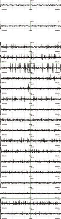
Fig. 1. Visualization of the STN, MRI-guided targeting and imme-
diate postoperative verification of final electrode position. Axial
All patients were operated under local anesthesia stereotactic 3D T2-weighted SPACE MRI at 3.0 T through the
and in the OFF-medication state to allow for MER and inferior portion of the STN. This sequence is used for both target-
intraoperative clinical testing for therapeutic benefit ing the STN and localization of the Leksell Vantage frame. Blue
and red bullets are indicating the patient-specific intended target
and side-effects. Anti-parkinsonian medication was at the left and right side respectively, with the corresponding lines
ceased 12 h prior to surgery. The first nine patients indicating the planned trajectories. The orange metal artefacts indi-
underwent a series of MRI sequences the day prior cate the position of the final electrodes of the same patient, verified
to surgery on a 3T Siemens Prisma MRI scanner by co-registering an immediate postoperative stereotactic CT to the
3D T2-weighted SPACE MRI.
(Siemens, Erlangen, Germany). Sequences included
a 3D T2 SPACE to visualize the STN and a 3D
T1 after administration of intravenous gadolinium defined at the level of the maximal rubral diameter
to plan the trajectory. For scanning parameters, see (5–6 mm below the AC-PC plane) and just postero-
the Supplementary Material. On the day of surgery, lateral to the target point described by Bejjani et al.
a Leksell Vantage stereotactic frame (Elekta Instru- [16] (Fig. 1). The entry point was defined at the medial
ment AB, Stockholm, Sweden) was attached to the aspect of the middle frontal gyrus, at or just behind
head under local anesthesia and a total scalp block. the coronal suture. Care was taken that the trajectory
The patient was transferred to the intraoperative MRI would avoid sulci, subcortical vessels, the caudate
suite and a stereotactic 3D T1 MPRAGE was acquired nucleus, and the ventricular system. General anesthe-
on a 3T Siemens Skyra (Siemens, Erlangen, Ger- sia was reversed after the MRI scan and the patient
many). The frame was subsequently localized by was positioned on the operating table, with the frame
co-registering the preoperative MRI dataset to the mounted and fixed to the table.
intraoperative stereotactic 3D T1 MPRAGE. For the Surgery always commenced on the left side. After
other 20 patients, all imaging was performed intra- administering local anesthetic, a linear incision was
operatively. They were briefly put under general made to accommodate a 14 mm frontal burrhole in
anesthesia with short-acting agents that would not line with the planned trajectory. A SureTek burrhole
interfere with MER. A Leksell Vantage stereotac- device (Boston Scientific, Valencia, CA, USA) was
tic frame was subsequently attached to the head and placed to fix the definitive lead later. The dura was
the patient was transferred to the intraoperative MRI opened only locally to facilitate a free pass of the
suite. MRI sequences were acquired on 3T Siemens guide tubes used. During insertion of the guide tubes
Skyra MRI scanner and included a tailored stereotac- copious irrigation with saline minimized CSF-loss
tic 3D T2 SPACE to visualize the STN and localize and the associated brain shift. After insertion the
the frame, and a 3D T1 MPRAGE after administra- burrhole was filled up with fibrin glue. MER was per-
tion of intravenous gadolinium to plan the trajectory. formed in 1 mm steps starting 10 mm above the target
See the Supplementary Material for MRI parame- and 0.5 mm steps starting 5 mm above the target,
ters. Target selection, planning of the trajectory, and until typical nigral activity was seen. The number of
localization of the frame was done by using Brain- microelectrodes used varied between 1 and 4 per side.
lab Elements software (Brainlab, Munich, Germany). The recordings were analyzed by a dedicated neuro-
The STN was defined as the T2-hypointense structure physiologist. Then, test stimulation was applied with
lateral to the red nucleus. The final target point was clinical testing by a DBS neurologist. The optimal1272 R.S. Vinke et al. / The Role of MER and Stereotactic CT in Awake MRI-Guided STN-DBS for PD
target was determined by results of MER and intraop- condition. The optimal stimulation contact was
erative clinical testing. After determining the optimal defined as the contact with the largest therapeutic
target for stimulation, a Vercise Cartesia 8 contact window and permanent stimulation was initiated sub-
directional DBS-lead (Boston Scientific, Valencia, sequently. Stimulation and medication were further
CA, USA, Model DB-2202) was inserted. The depth titrated based on clinical response during the follow-
of the deepest contact was determined based on the up visits.
microelectrode recordings. After the insertion of both
definitive leads, the patient was placed under general Follow-up
anesthesia and internalization of the extension cables
and a right infraclavicular Vercise Gevia rechargeable All patients were assessed 12 months after surgery.
internal pulse generator (Boston Scientific, Valen- They were asked to complete the same battery of
cia, CA, USA, Model DB-1200-S) was performed. questionnaires as preoperatively (including PDQ-
Immediately following surgery, all patients had a 39) and an MDS-UPDRS III score was obtained in
stereotactic CT to confirm lead positions. Surgery four conditions (OFF-MED ON-STIM, OFF-MED
was only considered to be finished if an acceptable OFF-STIM, ON-MED OFF-STIM, and ON-MED
placement of both leads was confirmed. Prophylactic ON-STIM). The OFF-medication condition was
antibiotics (cefazoline 2000 mg) were administered defined as the condition of the patient after withhold-
intraoperatively and for three more times in the sub- ing antiparkinsonian drugs for 12 h overnight. The
sequent 24 h. ON-medication condition was defined as the condi-
tion 1 h after a suprathreshold levodopa dose was
Calculation of stereotactic targeting error and administered. The suprathreshold dose was calcu-
postoperative volume of intracranial air lated by multiplying the total levodopa equivalent
morning dose by 1.2. Clinical assessments in the
Since depth of the definitive lead was predom- ON-stimulation condition were done 15 min after
inantly determined by MER-findings, the error in switching on the stimulator. Improvement of motor
depth was considered less relevant than the scalar function was defined as the difference between pre-
error between the intended target and the trajectory of operative MDS-UPDRS-III (OFF medication) and
the definitive lead. The stereotactic targeting error (h) 12 months postoperative MDS-UPDRS-III (OFF
was therefore defined as the shortest (perpendicular) medication and ON stimulation). Pre and postopera-
distance between the intended target and the trajec- tive Levodopa Equivalent Daily Dose (LEDD) were
tory of the lead. For a more detailed and mathematical calculated according to the formula described by
description, see Holl et al. [17]. The postoperative Tomlinson et al. [18].
air volume was determined by volumetric segmen-
tation, using the Brainlab Elements software, on the Adverse events
immediate postoperative CT-scan.
Adverse events were recorded for 12 months after
Determination of the MRI-defined STN surgery. Serious adverse events were defined as any
events that lead to permanent disability, death, pro-
Two observers (RSV and AKS), who were blinded longed hospital stay or new hospital admissions.
to the MER-results, assessed the T2-weighted MR
imaging along the planned lead trajectory from Statistical analysis
10 mm above the target to 5 mm past the target, in
step sizes equal to those used for MER. Based on All data are presented as mean ± standard devia-
imaging alone, they determined whether the planned tion (SD). Ranges [min-max] were provided as well.
trajectory at that particular depth was indeed located To quantify interobserver variability, a mean Jac-
within the STN or not. card’s index of similarity (JI) [19, 20] was used to
quantify the overlap between MRI observer 1 and
Programming of stimulation 2 in determining the MRI-defined STN. Further-
more, JI was used to quantify the overlap between
Two weeks after surgery, all patients underwent the MER-defined STN and the MRI-defined STN for
structured monopolar review of all lead contacts in observer 1 and 2 separately. All data were tested for
ring mode and with the patient in the OFF-medication normality using a Shapiro-Wilk test. Differences inR.S. Vinke et al. / The Role of MER and Stereotactic CT in Awake MRI-Guided STN-DBS for PD 1273
mean changes between MDS-UPDRS-III and PDQ- of MER tracks used per patient was 2.9 ± 0.6 for the
39 scores and LEDD at baseline and at 12 months left side and 2.9 ± 0.7 for the right. In 97% of the
follow-up were compared using a two-tailed paired left-sided trajectories and 100% of the right-sided
sample t-test, since all data was normally distributed. trajectories, MER identified signals typical for the
Pearson’s r-coefficient was used to evaluate the cor- STN with a mean length of the longest STN trajec-
relation between air volume and scalar error. Test tory of 5.1 ± 1.0 mm and 4.8 ± 1.0 mm, respectively.
results with p-values less than 0.05 were considered In 57 out of 58 (98%) of the sides, the permanent
statistically significant. DBS lead was placed in the central track. Only in 1
out of 58 sides (2%), the permanent DBS lead was
RESULTS placed in the lateral track. In one patient there were no
MER signals on the left side, due to the interfering
Twenty-nine consecutive patients underwent effect of dexmedetomidine. Dexmedetomidine was
awake bilateral STN-DBS with MER and intraoper- stopped immediately and by the time attention was
ative clinical testing in the period from March 2018 turned to right side, good quality MER-signals could
to July 2019. Baseline demographic data and clinical be acquired.
characteristics are shown in Table 1.
Motor function, Quality of Life (QoL), and
Microelectrode recordings LEDD at 12 months follow-up
MER was performed in all 29 patients (58 sides) A four-condition test at 12 months follow-up
with a total of 170 MER tracks. The mean number was available for all patients. Mean improvement
between preoperative MDS-UPDRS-III (OFF medi-
Table 1 cation) and at 12 months follow-up (OFF medication
Baseline demographic data and clinical characteristics and ON stimulation) was 26.7 ± 16.0 points (54%)
Number of patients 29 (p < 0.001). Completed PDQ-39 questionnaires at
Sex – no. (%) baseline and at 12 months follow-up were available
Male 24 (82.8%)
Female 5 (17.2%) for 28 patients. QoL improved with a mean change
Age at implantation – y [Range] 62 ± 7.8 [42–77] from baseline to 12 months of 9.0 ± 20.0 points
Disease Duration – y [Range] 9 ± 4.3 [3–21] (19%) (p = 0.025). The mean reduction of the LEDD
Levodopa Equivalent Daily Dose – 1354 ± 480 [200–2545.5] from baseline to 12 months was 794 ± 434 mg/day
mg [Range]
MDS-UPDRS-III OFF Medication 49.5 ± 13.4 [29–90] (59%) (p < 0.001). Results are summarized in Table 2.
[Range]
MDS-UPDRS-III ON Medication 18.5 ± 10.0 [7–42] Stereotactic accuracy and air volume
[Range]
PDQ-39 [Range] 46.9 ± 21.5 [12–92]
The mean scalar error of all leads was 0.88 ±
MDS-UPDRS, Movement Disorders Society Unified Parkinson’s
0.45 mm. Mean scalar errors of the left and right leads
Disease Rating Scale; PDQ-39, Parkinson’s Disease Question-
naire. The values of age, disease duration, Levodopa Equivalent were 0.92 ± 0.39 mm and 0.82 ± 0.50 mm respec-
Daily Dose, MDS-UPDRS, and PDQ-39 are presented as tively. This difference was not statistically significant
mean ± standard deviation of the mean. (p = 0.291). The mean volume of air on the immediate
Table 2
Clinical Improvement in UPDRS-III, PDQ-39, and LEDD
Baseline 12 Months Change from baseline p
Mean ± SD Mean ± SD Mean ± SD
[Range] [Range] (%)
MDS-UPDRS III 49.5 ± 13.4 22.9 ± 9.2 26.7 ± 16.0 < 0.001
OFF medication state [29–90] [5–42] (54%)
(OFF meds) (OFF meds / ON stim)
PDQ-39 46.9 ± 21.5 39.2 ± 20.8 9.0 ± 20.0 0.025
[12–92] [10–97] (19%)
LEDD 1354 ± 480 560 ± 314 794 ± 434 < 0.001
mg/day [200–2545.5] [0–1275] (59%)
MDS-UPDRS, Movement Disorders Society Unified Parkinson’s Disease Rating Scale; PDQ-39, Parkinson’s
Disease Questionnaire; LEDD, Levodopa Equivalent Daily Dose.1274 R.S. Vinke et al. / The Role of MER and Stereotactic CT in Awake MRI-Guided STN-DBS for PD
Fig. 2. Evaluation of MER signals and inter-observer variability of the MRI-defined STN along the planned trajectory. Visualization of a
one-sided standard evaluation in a particular patient. Depth of the recordings ranges from 7 mm above target (T) to 3 mm below in steps
of 0.5 mm starting from 5 mm above target. MER signals of the central track were visualized at each depth and were interpreted by a
dedicated neurophysiologist. Characterization of the signals as being STN-specific was indicated in the ‘MER’ column. Two independent
observers assessed whether the planned trajectory was located within the MRI-defined STN at every depth. Subsequently, a Jaccard’s Index
of Similarity was calculated for MRI Observer 1 and Observer 2. The Jaccard’s Index of Similarity was defined as the number in both sets
divided by the number in either set (intersection over union; see formula). The ‘Lead’ column indicates the depth of the contacts of the final
lead with the active contact at 12 months follow-up marked in green.
postoperative CT was 5.26 ± 9.43 cm3 . There was no lap with the MRI-defined STN did not overlap with
correlation between air volume and the scalar errors the MER-defined STN either.
of the left (r = –0.140, p = 0.468) and right (r = 0.154,
p = 0.424).
Adverse events
Inter-observer variability in determination of Adverse events were recorded at 12 months follow-
MRI-defined STN, overlap between MER-defined up for all patients. One patient was re-operated after
STN and MRI-defined STN and active contacts a displacement in depth of the right lead was noted on
the immediate post-operative CT. The displacement
The mean JI between observers 1 and 2 in deter- appeared to be caused by an erroneous fixation of the
mining the MRI-defined STN along the planned lead in the burrhole device. The retaining clip was not
trajectory on both the left and right side for all patients properly locked, allowing for the lead to migrate. In
was 0.84 ± 0.09. The mean JI between the MER- the following patients, the retaining clip was locked
defined STN and the MRI-defined STN by observers meticulously and double checked to prevent more
1 and 2 were 0.59 ± 0.25 and 0.66 ± 0.25 respectively cases of lead migration from happening. There were
(Fig. 2). 1 out of 58 (2%) active contacts at 12 months no infections, intracerebral hemorrhages, epilepsy or
follow-up did not overlap with the MRI-defined STN, hardware failures. All other adverse events are listed
whereas 7 out of 58 (12%) did not overlap with the in Table 3. In total, there were 19 adverse events in
MER-defined STN. The one contact that did not over- 11 patients.R.S. Vinke et al. / The Role of MER and Stereotactic CT in Awake MRI-Guided STN-DBS for PD 1275
Table 3
Adverse Events
Mild Moderate Severe Total
No. of events No. of events No. of events No. of events (%)
Lead malposition requiring revision – – 1 1 (3%)
Traction on extension cable 1 0 0 1 (3%)
Dysarthria 4 0 0 4 (14%)
Gait disorder 1 1 0 2 (7%)
Balance disorder 2 0 0 2 (7%)
Dyskinesia 1 0 0 1 (3%)
Cognitive decline 2 1 0 3 (10%)
Depression 1 0 0 1 (3%)
Impulse control disorder 1 0 0 1 (3%)
Apathy 1 0 0 1 (3%)
Dysphagia 1 0 0 1 (3%)
Hypersalivation 1 0 0 1 (3%)
DISCUSSION found out that there was a short circuit between the
central and lateral channel. So, instead of stimulat-
This study evaluates whether MER with intraop- ing the central electrode, we were in fact stimulating
erative clinical testing has an added value in awake the lateral electrode. Therefore, the definitive lead
bilateral STN-DBS surgery when an MRI-guided was placed in the lateral track based on incorrect
and stereotactic CT-verified approach has been used. assumptions. This means that, even in experienced
Baseline patient demographics in the present study hands, the technical complexity of MER can induce
were similar to those in other comparable studies in errors. Several studies have reported the percentage
terms of disease duration and severity [1, 21, 22]. of definitive leads that were placed in the central track
[23–28]. These percentages vary from 32% to 73.5%
Clinical improvement and are evidently lower than the percentage reported
in this study. This difference may have several dif-
The mean overall improvement from baseline to 12 ferent explanations. The MRI-sequences we used are
months for the MDS-UPDRS-III motor score, PDQ- tailored to accommodate a realistic visualization of
39 summary index score and the mean reduction in the STN and are corrected for MRI distortion to
LEDD was consistent with other studies [1, 21, 22]. localize the stereotactic frame as accurately as possi-
ble. Performing the scans under general anesthesia,
MER tracks and stereotactic accuracy for the last 19 patients, evidently reduced movement
artefacts and therefore improved the quality of the
The definite position of the permanent DBS leads images. For these 19 patients targeting and localiza-
in this study was solely determined by MER and tion of the frame was performed on the 3D T2 SPACE
the results of intraoperative clinical testing. Based only, making CT-MRI or MRI-MRI co-registration
on these results, 98% of all leads were placed in unnecessary. Furthermore, care was taken to mini-
the central track. Only one lead (2%) was placed mize the amount of CSF loss during the procedure,
in the lateral track. During this particular procedure, with a mean air volume of 5.26 ± 9.43 cm3 on the
we noted that the stimulation over the central micro- immediate postoperative CT. All the items mentioned
macroelectrode did not work properly, despite good above contribute to a reduction of targeting error [29,
MER signals prior to the stimulation. Impedances 30]. Another explanation could be the difference in
were checked and revealed an impedance of 0.0 k mindset, regarding targeting, between MER-guided
of the central micro-macroelectrode. The electrode and MRI-guided surgeons. An MER-guided surgeon
was replaced with a new micro-macroelectrode, but would rely on the MER and results of clinical test-
the impedance remained 0.0 k. Finally, the central ing to determine and confirm the final position of
MER-cable was switched with one of the unused the definitive lead, whereas and MRI-guided sur-
channels. Subsequently, impedance was checked and geon would rely on immediate postoperative imaging
revealed a normal impedance. Stimulation and clini- to verify the correct position of the definitive lead.
cal testing were then performed in the usual fashion. Therefore, meticulous determination of the intended
Unfortunately, only right after the procedure we target on the preoperative MRI is likely to be more1276 R.S. Vinke et al. / The Role of MER and Stereotactic CT in Awake MRI-Guided STN-DBS for PD
essential for MRI-guided surgeons since correction can be determined reliably on tailored MRI sequences
of the final position based on MER and results of and that the operative technique is accurate enough
clinical testing is not possible. to get the definitive lead at the intended target. The
Long term clinical outcomes of DBS have shown key factor remains how the optimal target should be
to be highly dependent on accuracy of lead place- determined. MER has shown to increase the opera-
ment. Rolston et al. reported revision or removal of tive time by 3 h and more than doubles the cost of
leads in 15.2–34.0% of the patients. Up to 48.5% of STN-DBS-surgery, without improving clinical effi-
these revisions were likely due to improper targeting cacy [11, 33]. These higher costs are due to personnel,
despite the use of MER and intraoperative clinical operation time, equipment and anesthesia costs to
testing [31]. Furthermore, Okun et al. reported that perform the procedure awake with MER and intra-
up to 46% of all treatment failures appeared to be operative clinical testing. Furthermore, some studies
related to misplaced leads [32]. The accuracy of lead suggest that MER is associated with an increased risk
placement in the present study was similar to those of hemorrhagic complications [10]. Although DBS
published in other studies using an MRI-guided tech- surgeons who are used to MER might not feel com-
nique [13, 17]. This accuracy was well within the fortable without having the intraoperative feedback
2 mm interspacing of the different MER tracks. of MER and intraoperative clinical testing, omitting
MER may have several advantages. It will likely
MER vs. MRI-defined STN in the planned result in a reduction of hemorrhagic complications
trajectory and correlation with active contact and a significant reduction in operation time and
localization costs, especially since omitting MER and intraopera-
tive clinical testing allows for an asleep MRI-guided
In defining the STN along the planned trajec- and CT or MRI-verified approach. Moreover, patients
tory, there appeared to be relatively high overlap do not have to experience the burden of coming off
between the MRI-defined STN of observer 1 and their anti-parkinsonian medication 12–24 h prior to
observer 2. This means that the STN could be defined surgery and undergoing awake surgery. The latter
quite reliably based on MRI alone. The similarity consideration should, however, not be generalized,
of both observers on the one hand and MER-results since some patients may prefer to undergo an awake
on the other are lower, indicating a discrepancy procedure [34]. Based on the results of this study and
between the electrophysiologically-defined STN and the experience of our team, we changed our practice
the MRI-defined STN. Most likely, this is because to an asleep MRI-guided and stereotactic CT-verified
both the STN and the substantia nigra have a similar approach, which created the opportunity to operate
hypointense aspect on the targeting MRI. Therefore, two instead of one patients a day.
distinction between these two structures can only be
done by indirect anatomical landmarks in an MRI- Limitations of this study
guided approach. However, this difference did not
seem to be clinically relevant, since more active con- Although this study evaluates the added value of
tacts did not overlap with the MER-defined STN than MER in MRI-guided STN-DBS in a thorough man-
with the MRI-defined STN. ner, several limitations should be considered. This
study is limited by its relatively small study popula-
Added value of MER in an MRI-guided approach tion and the retrospective study design. Furthermore,
the results of our study are dependent on the quality
Ninety-eight percent of the final leads in this of the MR imaging, experience of the team with an
study were placed in the central track, despite MER MRI-guided approach and the accuracy of the oper-
and intraoperative clinical testing. Accuracy of lead ative technique. Therefore, generalizing the outcome
placement in this study was well within the 2 mm of this study should be done with caution.
interspacing of the different MER tracks. Consider-
ing our clinical outcome, medication reduction and CONCLUSION
incidence of adverse effects at 12 months follow-up
are consistent with other studies, this suggests that Since our team has experience in both awake
MER would not be of additional value if a solely MER-guided and asleep MRI-guided STN-DBS, we
MRI-guided and stereotactic CT-verified approach is got the unique opportunity to evaluate the value of
used. However, this approach requires that the target MER in MRI-guided STN-DBS. In this study weR.S. Vinke et al. / The Role of MER and Stereotactic CT in Awake MRI-Guided STN-DBS for PD 1277
demonstrated that MER and intraoperative clinical [5] Ashkan K, Blomstedt P, Zrinzo L, Tisch S, Yousry T,
testing are not of additional value in determination Limousin-Dowsey P, Hariz MI (2007) Variability of the sub-
thalamic nucleus: The case for direct MRI guided targeting.
of the final lead position, if an adequate MRI-guided Br J Neurosurg 21, 197-200.
and stereotactic CT-verified technique is used. These [6] Counelis GJ, Simuni T, Forman MS, Jaggi JL, Trojanowski
results changed our daily clinical practice to an asleep JQ, Baltuch GH (2003) Bilateral subthalamic nucleus deep
MRI-guided and stereotactic CT-verified approach. brain stimulation for advanced PD: Correlation of intraoper-
ative MER and postoperative MRI with neuropathological
However, further research should be directed towards findings. Mov Disord 18, 1062-1065.
a properly designed comparative trial that evaluates [7] Hariz M, Blomstedt P, Limousin P (2004) The myth of
the added value of MER and intraoperative clinical microelectrode recording in ensuring a precise location of
the DBS electrode within the sensorimotor part of the sub-
testing and towards giving more insight into optimal
thalamic nucleus. Mov Disord 19, 863-864.
and patient-specific target localization. [8] McClelland S, 3rd, Vonsattel JP, Garcia RE, Amaya MD,
Winfield LM, Pullman SL, Yu Q, Fahn S, Ford B, Goodman
RR (2007) Relationship of clinical efficacy to postmortem-
CONFLICT OF INTEREST determined anatomic subthalamic stimulation in Parkinson
syndrome. Clin Neuropathol 26, 267-275.
RSV acts as an independent consultant for Boston [9] Gorgulho A, De Salles AA, Frighetto L, Behnke E (2005)
Incidence of hemorrhage associated with electrophysi-
Scientific. The other authors have nothing to disclose. ological studies performed using macroelectrodes and
microelectrodes in functional neurosurgery. J Neurosurg
SUPPLEMENTARY MATERIAL 102, 888-896.
[10] Zrinzo L, Foltynie T, Limousin P, Hariz MI (2012) Reduc-
ing hemorrhagic complications in functional neurosurgery:
The supplementary material is available in the A large case series and systematic literature review. J Neu-
electronic version of this article: https://dx.doi.org/ rosurg 116, 84-94.
[11] McClelland S, 3rd (2011) A cost analysis of intraoperative
10.3233/JPD-223149. microelectrode recording during subthalamic stimulation
for Parkinson’s disease. Mov Disord 26, 1422-1427.
[12] Nakajima T, Zrinzo L, Foltynie T, Olmos IA, Taylor C, Hariz
REFERENCES
MI, Limousin P (2011) MRI-guided subthalamic nucleus
deep brain stimulation without microelectrode recording:
[1] Deuschl G, Schade-Brittinger C, Krack P, Volkmann J, Can we dispense with surgery under local anaesthesia?
Schafer H, Botzel K, Daniels C, Deutschlander A, Dill- Stereotact Funct Neurosurg 89, 318-325.
mann U, Eisner W, Gruber D, Hamel W, Herzog J, Hilker R, [13] Chen T, Mirzadeh Z, Chapple KM, Lambert M, Shill
Klebe S, Kloss M, Koy J, Krause M, Kupsch A, Lorenz D, HA, Moguel-Cobos G, Troster AI, Dhall R, Ponce FA
Lorenzl S, Mehdorn HM, Moringlane JR, Oertel W, Pinsker (2018) Clinical outcomes following awake and asleep deep
MO, Reichmann H, Reuss A, Schneider GH, Schnitzler A, brain stimulation for Parkinson disease. J Neurosurg 130,
Steude U, Sturm V, Timmermann L, Tronnier V, Trottenberg 109-120.
T, Wojtecki L, Wolf E, Poewe W, Voges J, German Parkinson [14] Wodarg F, Herzog J, Reese R, Falk D, Pinsker MO, Steiger-
Study Group, Neurostimulation Section (2006) A random- wald F, Jansen O, Deuschl G, Mehdorn HM, Volkmann
ized trial of deep-brain stimulation for Parkinson’s disease. J (2012) Stimulation site within the MRI-defined STN
N Engl J Med 355, 896-908. predicts postoperative motor outcome. Mov Disord 27,
[2] Schuepbach WM, Rau J, Knudsen K, Volkmann J, Krack P, 874-879.
Timmermann L, Halbig TD, Hesekamp H, Navarro SM, [15] Patel NK, Khan S, Gill SS (2008) Comparison of atlas-
Meier N, Falk D, Mehdorn M, Paschen S, Maarouf M, and magnetic-resonance-imaging-based stereotactic target-
Barbe MT, Fink GR, Kupsch A, Gruber D, Schneider ing of the subthalamic nucleus in the surgical treatment
GH, Seigneuret E, Kistner A, Chaynes P, Ory-Magne F, of Parkinson’s disease. Stereotact Funct Neurosurg 86,
Brefel Courbon C, Vesper J, Schnitzler A, Wojtecki L, 153-161.
Houeto JL, Bataille B, Maltete D, Damier P, Raoul S, Sixel- [16] Bejjani BP, Dormont D, Pidoux B, Yelnik J, Damier P,
Doering F, Hellwig D, Gharabaghi A, Kruger R, Pinsker Arnulf I, Bonnet AM, Marsault C, Agid Y, Philippon
MO, Amtage F, Regis JM, Witjas T, Thobois S, Mertens J, Cornu P (2000) Bilateral subthalamic stimulation for
P, Kloss M, Hartmann A, Oertel WH, Post B, Speelman Parkinson’s disease by using three-dimensional stereotac-
H, Agid Y, Schade-Brittinger C, Deuschl G, EARLYSTIM tic magnetic resonance imaging and electrophysiological
Study Group (2013) Neurostimulation for Parkinson’s dis- guidance. J Neurosurg 92, 615-625.
ease with early motor complications. N Engl J Med 368, [17] Holl EM, Petersen EA, Foltynie T, Martinez-Torres I,
610-622. Limousin P, Hariz MI, Zrinzo L (2010) Improving target-
[3] Abosch A, Timmermann L, Bartley S, Rietkerk HG, Whit- ing in image-guided frame-based deep brain stimulation.
ing D, Connolly PJ, Lanctin D, Hariz MI (2013) An Neurosurgery 67, 437-447.
international survey of deep brain stimulation procedural [18] Tomlinson CL, Stowe R, Patel S, Rick C, Gray R, Clarke
steps. Stereotact Funct Neurosurg 91, 1-11. CE (2010) Systematic review of levodopa dose equivalency
[4] Benabid AL, Krack PP, Benazzouz A, Limousin P, Koudsie reporting in Parkinson’s disease. Mov Disord 25, 2649-
A, Pollak P (2000) Deep brain stimulation of the subthala- 2653.
mic nucleus for Parkinson’s disease: Methodologic aspects [19] Real R, Vargas JM (1996) The probabilistic basis of Jac-
and clinical criteria. Neurology 55, S40-44. card’s Index of Similarity. Syst Biol 45, 380-385.1278 R.S. Vinke et al. / The Role of MER and Stereotactic CT in Awake MRI-Guided STN-DBS for PD
[20] Jaccard P (1908) Nouvelles recherches sur la distribution [28] Frequin HL, Bot M, Dilai J, Scholten MN, Postma M, Bour
florale. Bull Soc Vaud Sci Nat 44, 223-270. LJ, Contarino MF, de Bie RMA, Schuurman PR, van den
[21] Weaver FM, Follett K, Stern M, Hur K, Harris C, Marks Munckhof P (2020) Relative contribution of magnetic res-
WJ, Jr., Rothlind J, Sagher O, Reda D, Moy CS, Pahwa R, onance imaging, microelectrode recordings, and awake test
Burchiel K, Hogarth P, Lai EC, Duda JE, Holloway K, Samii stimulation in final lead placement during deep brain stim-
A, Horn S, Bronstein J, Stoner G, Heemskerk J, Huang GD, ulation surgery of the subthalamic nucleus in Parkinson’s
CSP 468 Study Group (2009) Bilateral deep brain stimu- disease. Stereotact Funct Neurosurg 98, 118-128.
lation vs best medical therapy for patients with advanced [29] Azmi H, Machado A, Deogaonkar M, Rezai A (2011)
Parkinson disease: A randomized controlled trial. JAMA Intracranial air correlates with preoperative cerebral atro-
301, 63-73. phy and stereotactic error during bilateral STN DBS surgery
[22] Odekerken VJ, van Laar T, Staal MJ, Mosch A, Hoffmann for Parkinson’s disease. Stereotact Funct Neurosurg 89,
CF, Nijssen PC, Beute GN, van Vugt JP, Lenders MW, 246-252.
Contarino MF, Mink MS, Bour LJ, van den Munckhof P, [30] Geevarghese R, O’Gorman Tuura R, Lumsden DE, Samuel
Schmand BA, de Haan RJ, Schuurman PR, de Bie RM M, Ashkan K (2016) Registration accuracy of CT/MRI
(2013) Subthalamic nucleus versus globus pallidus bilat- fusion for localisation of deep brain stimulation electrode
eral deep brain stimulation for advanced Parkinson’s disease position: An imaging study and systematic review. Stereo-
(NSTAPS study): A randomised controlled trial. Lancet tact Funct Neurosurg 94, 159-163.
Neurol 12, 37-44. [31] Rolston JD, Englot DJ, Starr PA, Larson PS (2016) An
[23] Bour LJ, Contarino MF, Foncke EM, de Bie RM, van den unexpectedly high rate of revisions and removals in deep
Munckhof P, Speelman JD, Schuurman PR (2010) Long- brain stimulation surgery: Analysis of multiple databases.
term experience with intraoperative microrecording during Parkinsonism Relat Disord 33, 72-77.
DBS neurosurgery in STN and GPi. Acta Neurochir (Wien) [32] Okun MS, Tagliati M, Pourfar M, Fernandez HH, Rodriguez
152, 2069-2077. RL, Alterman RL, Foote KD (2005) Management of
[24] Temel Y, Wilbrink P, Duits A, Boon P, Tromp S, referred deep brain stimulation failures: A retrospective
Ackermans L, van Kranen-Mastenbroek V, Weber W, analysis from 2 movement disorders centers. Arch Neurol
Visser-Vandewalle V (2007) Single electrode and multiple 62, 1250-1255.
electrode guided electrical stimulation of the subthalamic [33] Maiti TK, Konar S, Bir S, Kalakoti P, Nanda A (2016)
nucleus in advanced Parkinson’s disease. Neurosurgery 61, Intra-operative micro-electrode recording in functional neu-
346-355; discussion 355-347. rosurgery: Past, present, future. J Clin Neurosci 32, 166-172.
[25] Hamel W, Fietzek U, Morsnowski A, Schrader B, Herzog [34] LaHue SC, Ostrem JL, Galifianakis NB, San Luciano M,
J, Weinert D, Pfister G, Muller D, Volkmann J, Deuschl G, Ziman N, Wang S, Racine CA, Starr PA, Larson PS,
Mehdorn HM (2003) Deep brain stimulation of the subtha- Katz M (2017) Parkinson’s disease patient preference and
lamic nucleus in Parkinson’s disease: Evaluation of active experience with various methods of DBS lead placement.
electrode contacts. J Neurol Neurosurg Psychiatry 74, 1036- Parkinsonism Relat Disord 41, 25-30.
1046.
[26] Amirnovin R, Williams ZM, Cosgrove GR, Eskandar EN
(2006) Experience with microelectrode guided subthalamic
nucleus deep brain stimulation. Neurosurgery 58, ONS96-
102; discussion ONS196-102.
[27] Rola R, Tutaj M, Koziara H, Koziorowski D, Brodacki
B, Karliński M, Tykocki T, Krolicki B, Mandat T (2016)
Optimizing final electrode placement Based on intraopera-
tive neurophysiological evaluation during subthalamic deep
brain stimulation for Parkinson’s disease. J Neurol Neuro-
physiol 7, 4.You can also read