The Role of LIM Kinases during Development: A Lens to Get a Glimpse of Their Implication in Pathologies - MDPI
←
→
Page content transcription
If your browser does not render page correctly, please read the page content below
cells
Review
The Role of LIM Kinases during Development: A Lens to Get a
Glimpse of Their Implication in Pathologies
Anne-Sophie Ribba, Sandrine Fraboulet, Karin Sadoul and Laurence Lafanechère *

                                          Institute for Advanced Biosciences, Team Cytoskeletal Dynamics and Nuclear Functions,
                                          INSERM U1209, CNRS UMR5309, Université Grenoble Alpes, 38000 Grenoble, France;
                                          anne-sophie.ribba@univ-grenoble-alpes.fr (A.-S.R.); sandrine.fraboulet@univ-grenoble-alpes.fr (S.F.);
                                          karin.sadoul@univ-grenoble-alpes.fr (K.S.)
                                          * Correspondence: laurence.lafanechere@univ-grenoble-alpes.fr; Tel.: +33-(0)476-54-95-71

                                          Abstract: The organization of cell populations within animal tissues is essential for the morphogenesis
                                          of organs during development. Cells recognize three-dimensional positions with respect to the whole
                                          organism and regulate their cell shape, motility, migration, polarization, growth, differentiation, gene
                                          expression and cell death according to extracellular signals. Remodeling of the actin filaments is
                                          essential to achieve these cell morphological changes. Cofilin is an important binding protein for
                                          these filaments; it increases their elasticity in terms of flexion and torsion and also severs them. The
                                          activity of cofilin is spatiotemporally inhibited via phosphorylation by the LIM domain kinases 1
                                          and 2 (LIMK1 and LIMK2). Phylogenetic analysis indicates that the phospho-regulation of cofilin
                                          has evolved as a mechanism controlling the reorganization of the actin cytoskeleton during complex
                                          multicellular processes, such as those that occur during embryogenesis. In this context, the main
                                          objective of this review is to provide an update of the respective role of each of the LIM kinases

                                          during embryonic development.

Citation: Ribba, A.-S.; Fraboulet, S.;    Keywords: LIM kinase; development; cofilin
Sadoul, K.; Lafanechère, L. The Role
of LIM Kinases during Development:
A Lens to Get a Glimpse of Their
Implication in Pathologies. Cells 2022,   1. Introduction
11, 403. https://doi.org/10.3390/
                                                LIM kinase 1 (LIMK1) first appeared on the scientific scene in 1994, thanks to the work
cells11030403
                                          of a Japanese [1] and an Australian team [2]. The following year, the LIM kinase family
Academic Editors: Hélène Bénédetti        was extended with the description of LIM kinase 2 (LIMK2), which has an overall sequence
and Béatrice Vallée-Méheust               and a domain structure similar to that of LIMK1, but the overall identity is 50–51% at the
Received: 21 December 2021
                                          amino acid level [3]. As indicated by their denomination, they contain two LIM domains.
Accepted: 22 January 2022
                                          The name LIM is an acronym of the three genes in which such a domain was first identified
Published: 25 January 2022
                                          (LIN-11, Isl-1 and MEC-3). LIM domains are tandem zinc-finger structures that function
                                          as modular protein-binding interfaces [4]. The LIM domains of LIMKs are positioned in
Publisher’s Note: MDPI stays neutral
                                          the amino-terminal part of the protein. These domains are followed by a central PDZ
with regard to jurisdictional claims in
                                          domain, a proline/serine (P/S)-rich region, and a carboxyterminal kinase domain. Thus,
published maps and institutional affil-
                                          LIMKs belong to the PDZ-LIM protein family with which they share the common trait of
iations.
                                          influencing the actin cytoskeleton [5]. In addition, the PDZ-LIM family of proteins has
                                          been shown to mediate signals between the nucleus and the cytoskeleton, with a significant
                                          impact on organ development [6].
Copyright: © 2022 by the authors.
                                                In 1998, the physiological substrate of LIMK1 was identified; in two “back-to-back”
Licensee MDPI, Basel, Switzerland.        Nature publications, Arber and collaborators [7] and Yang and collaborators [8] provided
This article is an open access article    evidence that LIMK1 phosphorylates cofilin. The following year, it was shown that cofilin
distributed under the terms and           was also the substrate of LIMK2 [9]. While cofilin remains the best-characterized substrate
conditions of the Creative Commons        of LIM kinases (LIMKs), other substrates have been identified, extending the field of action
Attribution (CC BY) license (https://     of this family of kinases [10–12].
creativecommons.org/licenses/by/                Cofilin is a protein that plays an essential role in the regulation of actin dynamics.
4.0/).                                    It binds to actin filaments, which increases their elasticity in terms of both flexion and

Cells 2022, 11, 403. https://doi.org/10.3390/cells11030403                                                         https://www.mdpi.com/journal/cells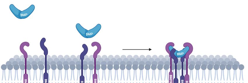
Cells 2022, 11, 403                                                                                           2 of 17

                      torsion, and severs them, leading to their depolymerization [13,14]. Cofilin is regulated by
                      phosphorylation of the serine residue at position 3, which inhibits its actin-binding and
                      depolymerization activities. Besides LIMKs, cofilin is phosphorylated and inactivated by
                      testicular protein kinases (TESKs) [15,16], Nck-interacting kinase-related kinases (NRK) [17]
                      and by Aurora kinase [18]. Cofilin is reverted to its basal unphosphorylated active state by
                      the phosphatases slingshot 1 (SSH) [19] and chronophin [20].
                            From a phylogenetic point of view, gene orthologs of LIMKs are present in vertebrates,
                      as well as in Drosophila and Anopheles, but are not found in yeast, Caenorhabditis elegans,
                      Dictyostelium, or in plants [6,21]. Therefore, it has been proposed by Kazumasa Ohashi in
                      his brilliant review on the role of cofilin in development, that “cofilin phosphoregulation
                      may have evolved as a mechanism for reorganizing the actin cytoskeleton during complex
                      multicellular processes in some higher organisms” [22].
                            The morphogenesis of organs and tissues during development involves the controlled
                      arrangement of multicellular processes. Depending on the extracellular signals they receive,
                      cells adapt their shape, motility and migration, polarization, growth, differentiation, and
                      cell death. Because of their ability to control actin remodeling, LIMKs have a central role in
                      many of these processes. The prominent role of LIMK1 during development is evidenced
                      by some of the features observed in Williams–Beuren syndrome. This rare genetic disorder,
                      first described by New Zealand’s Dr. John Cyprian Phipps Williams in 1961 and in the
                      following year by Germany’s Dr. Alois J. Beuren, is caused by the hemizygous deletion
                      of approximately 1.5–1.8 mega base pairs on the long arm of chromosome 7 (7q11.23),
                      encompassing 27 genes including elastin, CLIP115 and LIMK1. Some of the clinical charac-
                      teristics of Williams–Beuren syndrome [23], such as distinctive craniofacial abnormalities
                      (broad forehead, wide mouth, full cheeks and lips, and oval ears), impaired visuospatial
                      constructive cognition, and mild mental retardation have been specifically associated with
                      the deletion of LIMK1 [24–26].
                            In this review, we will focus on the identified roles of LIMKs in specific developmental
                      processes and on the LIMK-regulated signaling pathways, whose activation is central
                      during development. In addition to descriptions of the tissue expression of LIMKs, we will
                      base our review on studies of animal models with loss- or gain-of-function mutations in
                      LIMKs, as well as on studies where LIMK functions are probed with specific inhibitors of
                      LIMK activity.
                            Understanding the role of LIMKs during development may indeed shed light on
                      the LIMK-dependent pathological perturbations observed not only in Williams–Beuren
                      syndrome but also in other pathologies (autism, fragile X syndrome (see the recent review
                      of Ben Zablah and collaborators [25]) or carcinogenesis (see reviews of Lee and collabora-
                      tors [27] or of Fabrizio Manetti [28,29])).

                      2. Expression of LIMKs during Development
                            The expression patterns of LIMK1 and LIMK2 during mammalian early development
                      have been explored only recently, in a study investigating their role during porcine em-
                      bryonic development. Using first immunofluorescence staining and then validation of
                      LIMK1/2 expression by quantitative real-time polymerase chain reaction (qRT-PCR), it
                      was found that both LIMK1 and LIMK2 are expressed at an early point in development
                      and play a role during the cleavage and morula stages of embryos. Inactivation of LIMKs
                      through silencing with dsRNA or the use of the LIMK inhibitor LIMKi3, which shows a
                      good selectivity within the kinome (see the recent review by Chatterjjee and collabora-
                      tors [30]), induced the disruption of adherent junctions, resulting in the early termination
                      of development before the blastocyst stage [31]. A similar observation was made in an
                      analysis of early mouse embryogenesis: the use of the LIMKi3 inhibitor caused the failure
                      of early blastomere cleavage, compaction and blastocyst formation [32]. These results point
                      to a crucial role of LIMKs during the early stages of development. They also indicate that
                      LIMK activity is compensated for by the action of other kinases in LIMK knock-out mice,
                      as they give birth to viable pups.
Cells 2022, 11, 403 3 of 17
During the later stages of development (Table 1), LIMK1 is highly expressed in neu-
ronal tissues and is present in many epithelial tissues, where its expression pattern is
spatially and temporally regulated. LIMK1 is detected in embryonic skin epidermis, the
heart, lung, and kidney in varying amounts, depending on the stage of development. It
has been observed that LIMK1 is also found in specific cell types that undergo transitions
between epithelial and mesenchymal states [33].
Similar to LIMK1, the LIMK2 expression pattern is also dynamically regulated in space
and time during all developmental stages, and the expression of both LIMKs may overlap
in some embryonic tissues [3,34–38]. Although also present in neuronal tissues, LIMK2
seems to be more abundantly detected in epithelia such as the digestive tract [36] (Table 1).
A clear comprehension of the expression pattern of LIMKs is, however, complicated due to
the existence of several splice variants [39–41].
Table 1. Expression of LIMKs during development.
Embryonic/Adult
Publications LIMK Isoform Species Experimental Procedures Main Observations
Tissues—Cell Lines
Mizuno et al. adult High level in the rat brain.
Oncogene, 1994 LIMK human, rat rat brain, epithelial and Northern blot Expressed in human epithelial and
[1] hematopoietic cell lines hematopoietic cell lines
E13, E14, E15, E16, E18, P0 Northern blot,
Bernard et al. mouse brain, Identification of LIMK. Expressed in
Cell Growth and LIMK RNase protection assay
adult human brain, mouse brain, human and mouse brain and
Differentiation, 1994 human, mouse in situ hybridization,
heart, liver, muscle olfactory epithelial cell lines
[2] mouse olfactory epithelial cell lines immunohistochemistry
Ohashi et al. adult
Journal of Biochemistry, Expressed in lung, brain, kidney,
LIMK chicken lung, brain, kidney, liver, gizzard, Northern blot
1994 liver, spleen, gizzard and intestine
[42] intestine, spleen
Cheng and Robertson E8.5, E11.5, E15.5 brain, olfactory
Mechanisms of Northern blot, Variable expression rates depending on
LIMK mouse system, gut, trophoblast giant cells
Development, 1995 in situ hybridization the stage of development and the tissue
[43] adult brain, ovary, testis, skin, lung
LIMK1 expressed in all tissues, with
Okano et al. adult highest amounts in the brain. Two
Journal of Biological
LIMK1 LIMK2 human brain, skeletal muscle, Northern blot LIMK2 isoforms: longer in all tissues,
Chemistry, 1995
heart, placenta smaller only in skeletal
[34]
muscle and heart
Pröschel et al. adult Nervous system expression
Oncogene, 1995 LIMK1 mouse spinal cord, brain, cranial nerve, Northern blot,
in situ hybridization of LIMK1
[44] dorsal root ganglia
Nunoue et al.
LIMK1 LIMK2 rat adult LIMK1 in the brain, LIMK2 in
Oncogene, 1995 Northern blot
brain, various tissues various tissues
[3]
Ikebe et al. adult
LIMK2 mouse brain, thymus, lung, heart, stomach, RT-PCR LIMK2a and LIMK2b isoforms
Genomics, 1997
expressed in various tissues
[45] spleen, kidney, intestine, liver, testis
Koshimizu et al. E10 to E18
Biochemistry and embryos
LIMK2 mouse adult LIMK2a and LIMK2b isoforms
Biophysical Research Northern blot expressed in various tissues
Communications, 1997 brain, heart, lung, spleen, thymus,
[39] kidney, stomach
Mori et al. LIMK1 and LIMK2 expressed in brain.
Molecular Brain E12, E14, E16, E18 Differential expression of LIMK1 and
LIMK1 LIMK2 rat In situ hybridization
Research, 1997 embryo LIMK2 in epithelia. High expression
[36] in extra-embryonic tissues
Takahashi et al. Variable expression rates during
Developmental Stage 2 to 40 Northern blot,
LIMK1 xenopus development. Important role of
Dynamics, 1997 cleavage, gastrula, blastula, neurula in situ hybridization
[46] XLIMK1 in neural development
Ikebe et al.
Biochemistry and adult Identification of LIMK2c, a
Biophysical Research LIMK2 mouse liver, brain, thymus, lung, heart, Northern blot, RT-PCR brain-specific isoform, and LIMK2t,
Communications, 1998 stomach, testis a testis-specific isoform
[35]
Takahashi et al.
Biochemistry and adult LIMK2 expressed in all tissues,
LIMK1 LIMK2 mouse brain, thymus, lung, spleen, testis, Northern blot, in situ
Biophysical Research identification of a testis-specific
Communications, 1998 hybridization
kidney, stomach, heart isoform LIMK2t
[47]Cells 2022, 11, 403 4 of 17
Table 1. Cont.
Embryonic/Adult
Publications LIMK Isoform Species Experimental Procedures Main Observations
Tissues—Cell Lines
Identification of LIMK2a and
LIMK2b with tissue-specific
Nomoto et al. fetal and adult expression profile. LIMK2a
brain, stomach, colon, pancreas, liver, RT-PCR, RNase
Genes, 1999 LIMK2 human protection assay predominantly expressed in fetal
[37] lung, kidney, placenta
and adult tissues compared
to LIMK2b
LIMK1 KO mice,
Meng et al. immunohistochemistry, Dendritic spine morphology and
LIMK1 mouse adult
Neuron, 2002 primary neurons,
brain synaptic function alterations
[48]
brain sections
Takahashi et al. Abnormal spermatogenesis found in
LIMK2 KO mice, MEF cells,
Developmental Biology, LIMK2 mouse adult LIMK2-KO testis associated with an
immunofluorescence,
2002 testis increased number of apoptotic
histology
[41] germ cells
Meng et al. LIMK1, LIMK2 and Normal synaptic plasticity in
Neuropharmacology, adult LIMK1/2 KO mice,
LIMK1 LIMK2 mouse immunohistochemistry, LIMK2-KO mice, altered synaptic
2004 brain
[49] electrophysiology functions in double-LIMK1/2-KO mice
Expression in late larval and pupal
Chen et al. stages, suggesting a role in this
Current Biology, 2004 dLIMK drosophila from larvae to adult mRNA level transition. Defects in leg
leg morphogenesis
[50] morphogenesis. Role of the
Rho-dLIMK signaling pathway.
rat and chick embryos
Foletta et al. brain and spinal cord Expression of LIMK1 in liver,
Experimental Cell LIMK1 rat,
mouse, chicken mouse adult Western blot thymus, kidney, heart, lung, small
Research, 2004
[51] brain, heart, liver, lung, small intestine, stomach and brain
intestine, stomach, kidney
Ang et al. larvae dLIMK active/inactive, Role of LIMK in synapse
Developmental Biology, neuromuscular junctions (abdominal drosophila strains, development and in glomeruli of
dLIMK drosophila
2006 muscle fibers), antennal lobe immunohistochemistry, antennal lobe. LIMK is a
[52] glomeruli electrophysiology downstream effector of PAK
E14
olfactory epithelium, heart, liver,
Acevedo et al. intestine, urogenital sinus, thymus, Variable LIMK2 expression levels in
Journal of Embryo sections,
spinal cord embryonic and adult tissues, similar
Histochemistry and LIMK2 mouse immunohistochemistry,
Cytochemistry, 2006 adult expression pattern than LIMK1
brain, heart, spleen, stomach, western blot
[38] except in testis
intestine, lung, skin, kidney, ovary,
eyes, testes, uterus
PAK-LIMK-cofilin pathway are
Menzel et al. Genetic screen, mutant,
drosophila strains, involved in photoreceptor cell
Mechanism of adult
dLIMK drosophila immunohistochemistry of morphogenesis by regulating
Development, 2007 eyes
[53] photoreceptor cell adherent junctions and actin
dynamics
Ott et al. Temporal and spatial expression of
Gene Expression
LIMK1 LIMK2 zebrafish all embryonic stages In situ hybridization LIMK1 and LIMK2 during
Patterns, 2007
[54] embryogenesis
Lindström et al. E10.5 to E18.5 LIMK1 highly expressed in many
Gene Expression EMT- and MET-tissues, limb, eye, Embryo sections,
LIMK1 mouse neuronal and epithelial tissues
Patterns, 2011 heart, lung, skin, kidney, intestine, immunohistochemistry
[33] testes undergoing EMT and MET
E14.5 E15.5 E18.5 and newborns P1.5 LIMK2-KO mice, RT-PCR, Phenotype of EOB “eyes open at
Rice et al. in situ hybridization,
LIMK2 mouse ocular tissue birth” of LIMK2-KO mice, abnormal
PLoS ONE, 2012
adult western blot, migration of keratinocytes during
[40]
brain, testis, eyes, rate, lung immunohistochemistry eyelid development
Role of LIMK2 in growth cone
Andrews et al. In situ hybridization,
E13.5, E15.5 collapse in response to Sema3A by
Biology Open, 2013 LIMK2 mouse siRNA transfections, in
brain regulating
[55] utero electroporation
PlexinA1 expression level
LIMK1-KO mice, bone Bone mass reduction in LIMK1-KO
Kawano et al. histomorphometry, mice, abnormal osteoblast
LIMK1 mouse newborns PD3-PD5 microCT, primary
Bone, 2013 differentiation and defective
tibiae, femur
[56] osteoblasts, osteoclasts and osteoblastic and
bone marrow cells osteoclastic functions
Abe et al. Involvement of Rac-Sickie-SSH and
newborns P2-P3 Drosophila strains,
Development, 2014 dLIMK drosophila Rac-PAK-LIMK pathways in
brain immunohistochemistry
[57] axonal growth
Piccioli et al. Drosophila strains, live Role of BMPRII-LIMK-cofilin-actin
Journal of Neuroscience, dLIMK drosophila larvae imaging of synaptic growth signaling in potentialization of
2014 neuromuscular junctions
[58] and bouton budding neuromuscular junctions
Yang et al. siRNA transfections,
LIMK2 mouse newborns PD2-PD3 Contribution of LIMK2 in the
Bone, 2015 immunofluorescence, fluid
primary osteoblasts mechanosensitivity of osteoblasts
[59] shear stressCells 2022, 11, 403 5 of 17
Table 1. Cont.
Embryonic/Adult
Publications LIMK Isoform Species Experimental Procedures Main Observations
Tissues—Cell Lines
Xie et al. In utero electroporation, Altered neuronal migration and
Histochemistry and Cell LIMK1 mouse E15.5, E18.5, newborns P1
Biology, 2017 brain brain sections, number of neurites due to aberrant
[60] immunofluorescence expression of LIMK1
In vitro fertilization,
Duan et al. LIMK1 and LIMK2 are involved in
2, 4, 8 -cells embryo culture,
Cell Cycle, 2018 LIMK1 LIMK2 mouse early stages of embryo development
morula, blastocyst immunofluorescence,
[32] and regulate actin assembly
inhibition of LIMKs activity
Immunohistochemistry, Regulation of dendritic branching by
E11.5, E13.5, E15.5
Saxena et al. cortex and cortical neurons cell proliferation, in LIMK-mediated non-canonical BMP
Development, 2018 LIMK mouse signaling and involvement of both
postnatal P0, P6, P21 utero-electroporation,
[61] neurons canonical and non-canonical BMP
P-SMAD labeling signaling in neuronal migration
LIMK1-KO, LIMK2-KO Contribution of LIMK1 and LIMK2
Mao et al. E14.5 and double LIMK1/2-KO in progenitor cell proliferation and
Molecular Brain, 2019 LIMK1 LIMK2 mouse
brain mice, migration. Role of LIMK2 in
[62] immunohistochemistry embryonic cell apoptosis
Fang et al. LIMK1/2-KO mice, No alteration of cochlear
LIMK1 LIMK2 mouse E3 and from P3 to P30 immunohistochemistry, development and auditory function
Scientific Reports, 2019 cochlea
[63] auditory measurement in LIMK1/2-KO mice
Kwon et al. RT-QPCR, LIMK1/2
activity inhibition, Role of LIMK1 and LIMK2 in
Asia-Australasian embryo cleavage and compaction
LIMK1 LIMK2 porcine 1, 2, 4-cells LIMK1/2 dsRNA injection,
Journal of Animal
morula, blastocyst through actin regulation and the
Sciences, 2020 embryo culture,
[31] maintenance of cell–cell junctions
immunofluorescence
He et al. Control of actin assembly,
endodermal differentiation,
In Vitro Cellular and EMT-related genes expression and
siRNA transfection,
Developmental Biology, LIMK2 human Embryonic Stem Cells
2021 RT-QPCR, cell migration by LIMK2 in
immuno-fluorescence endodermal lineage
[64]
3. Signaling Pathways Involving LIMKs during Development
While LIMKs are actors of several signaling pathways, the best-described pathways
during development are the semaphorin, nerve growth factor and the non-canonical bone
morphogenetic protein (BMP) pathways. Within these pathways, LIMKs are downstream
effectors of RhoA/ROCK, cdc42/PAK, and Rac/PAK (for reviews, see [12,21,22]).
3.1. The Non-Canonical BMP Pathway
BMPs belong to the TGFβ superfamily and play a crucial role during the development of the
nervous system, where LIMK1 is abundantly expressed. Thus, the BMPs/LIMK1/cofilin axis has
been thoroughly investigated and has been shown to be essential for neuronal morphogenesis.
BMPs bind to a heterodimeric complex of type-I and type-II Ser/Thr kinase receptors
and, as a result, type-II receptors phosphorylate and thereby activate type-I receptors.
While BMP can signal through Smad-dependent (canonical) and Smad-independent (non-
canonical) pathways, LIMK1 is only involved in the non-canonical Smad-independent
pathway. LIMK1 interacts with a large cytoplasmic domain of 600 amino acids of BMP
type-II receptors (BMPRII) [65,66]. According to Foletta and collaborators, this interaction
prevents the activation of LIMK1 by PAK [65]. This downregulation of LIMK1 can be
relieved by the binding of BMP4 to its receptor, leading to the dissociation of LIMK1 from
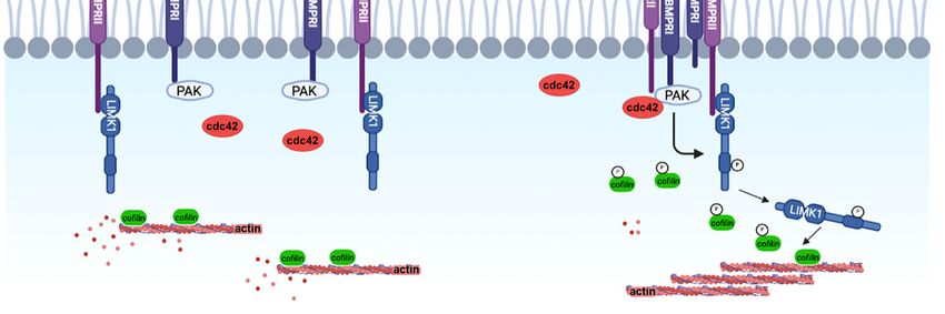
BMPRII and subsequently to cofilin phosphorylation (Figure 1).
Lee-Hoeflich and collaborators demonstrated that BMPs induce the activation of the
small GTPase cdc42, which cooperates with the binding of LIMK1 to BMPRII to prompt
high levels of LIMK1 activity and of phospho-cofilin [66]. This may be a useful mechanism
to control cofilin phosphorylation close to the plasma membrane, to locally regulate cortical
actin dynamics.
The activation of LIMK1 by BMPs has been further characterized during dendritogen-
esis, by experiments showing that PAK1 binds to BMPRI (BMPR1b and ALK2), resulting in
the close proximity of the different players in this signaling pathway [61,67,68] (Figure 1).Cells 2022, 10, x 7 of 19
Cells 2022, 11, 403 6 of 17
Figure1.1.The
Figure Thebinding
bindingofofBMPs
BMPsleads
leadstotothe
the formation
formation of of a BMP
a BMP receptor
receptor complex.
complex. PAK1,
PAK1, recruited
recruited by
by BMPRI, is thus in close proximity to its target LIMK1, bound to BMPRII. After being
BMPRI, is thus in close proximity to its target LIMK1, bound to BMPRII. After being activated by activated
by cdc42,
cdc42, PAK1PAK1 phosphorylates
phosphorylates LIMK1,
LIMK1, whichwhich induces
induces cofilin
cofilin phosphorylation,
phosphorylation, thereby
thereby inhibiting
inhibiting its
its activity and thus contributing to the regulation of actin dynamics required for dendrite extension.
activity and thus contributing to the regulation of actin dynamics required for dendrite extension.
Lee-Hoeflich
In and collaborators
order to establish demonstrated
a correct neuronal thatgrowth
circuit, the BMPs induce
rate andthe activation
length of the
of neurites
small GTPase cdc42, which cooperates with the binding of LIMK1
must be controlled in space and time during the different stages of neuronal development. to BMPRII to prompt
highimplies
This levels ofaLIMK1 activity and
fine regulation of of phospho-cofilin
actin dynamics by[66]. This may be a useful
BMP/LIMK1/cofilin mechanism
signaling, as
to control cofilin
illustrated by the phosphorylation
work of Wen andclose to the plasma
collaborators, whomembrane,
established tothat
locally
BMP regulate
gradientscor-
ticalrequired
are actin dynamics.
to trigger the rapid responses of neuronal growth cones via LIMK1- and
The activation
SSH-regulated cofilinofphosphorylation
LIMK1 by BMPs[69]. has been further
The BMP characterized
gradients during dendrito-
act as guidance cues to
genesis,
orient theby experiments
commissural showing
axons in thethat PAK1 binds
developing spinaltocord
BMPRI [70].(BMPR1b
Commissural and ALK2), result-
axons extend
ing infrom
away the close proximity
the roof plate inofresponse
the different players
to BMP in this signaling
chemorepellent pathway
activity, which[61,67,68]
controls(Fig-
the
ure 1). rate of commissural axons and their orientation by finely regulating LIMK1 activity.
growth
WhenInthe order to establish a correct
BMP/LIMK1/cofilin neuronal
signaling circuit,isthe
pathway growth rate
activated, and length
the growth rateof is neurites
slowed
must be
down, controlled
whereas when inLIMK1
space and timeisduring
activity reduced, theaxons
different
growstages
fasterof[70].
neuronal
Thus, development.
temporal and
This implies
spatial guidance a fine regulation
decisions resultoffrom
actinthe
dynamics
activation bystatus
BMP/LIMK1/cofilin
of cofilin and actinsignaling,
dynamics as illus-
in a
LIMK1-dependent
trated by the work manner
of Wen during development [68,70].
and collaborators, Frendo and
who established collaborators,
that BMP gradients usingarea
mouse
required LIMK1-KO
to trigger model, showed
the rapid that suchofa neuronal
responses regulationgrowth
of cofilincones
activity
viaand actin dynamics
LIMK1- and SSH-
by LIMK1 is
regulated relevant
cofilin in axon regeneration
phosphorylation [69]. Theafter
BMPsciatic nerve injury
gradients act as[71].
guidance cues to orient
Taken together,
the commissural thesein
axons studies highlight the
the developing fine modulation
spinal of LIMK1 activity
cord [70]. Commissural axons by extend
BMPs
to control actin dynamics during neuronal development.
away from the roof plate in response to BMP chemorepellent activity, which controls the
growthAlthough LIMK1 appears
rate of commissural axonsto be
andatheir
preferred partner
orientation by of BMPRII
finely in theLIMK1
regulating developing
activ-
neurons,
ity. When LIMK2 is also activated by BMPs.
the BMP/LIMK1/cofilin Its direct
signaling interaction
pathway with BMPRII
is activated, was, however,
the growth rate is
debated
slowed between Foletta and
down, whereas when collaborators [65] and
LIMK1 activity Lee-Hoeflich
is reduced, axonsandgrow
collaborators
faster [70].[66], but
Thus,
was demonstrated
temporal and spatialin the context decisions
guidance of cancer development
result from the [72]. The BMP-induced
activation activation
status of cofilin and
of LIMK1 and LIMK2 occurs via two distinct signaling pathways
actin dynamics in a LIMK1-dependent manner during development [68,70]. Frendo and that are both independentCells 2022, 11, 403 7 of 17
of SMAD. BMP-induced LIMK1 activation involves cdc42 and PAK signaling, whereas
BMP-induced LIMK2 activation occurs via RhoA and ROCK signaling. Other factors, such
as members of the TGF family (TGFbeta1 and Activin B), also activate LIMK2, via RhoA
and ROCK, to control cofilin phosphorylation and actin dynamics [73,74].
3.2. Nerve Growth Factor
A critical role of both LIMK1 and LIMK2 in controlling the cofilin activity during
nerve growth factor-induced neurite extension and growth cone motility was mentioned
by Endo and collaborators [75]. Interestingly, this study also revealed that the respective
roles of LIMK1 and LIMK2 are distinct and that the signaling mechanisms leading to their
activation, as well as their activation time-course, differ.
3.3. Semaphorins
The semaphorins 3A and 3F (Sema3A/3F) are chemorepulsive axonal guidance molecules,
acting through neuropilin-plexin receptors expressed at the surface of neurons.
By using a dominant-negative LIMK1, which cannot be activated by PAK or ROCK,
Aizawa and collaborators showed that LIMK1 activation is required for Sema3A-induced
growth cone collapse in dorsal root ganglia neurons. In addition, a synthetic cell-permeable
peptide containing a cofilin phosphorylation site acts as a competitive inhibitor of LIMK1,
which leads to a reduction of cofilin phosphorylation and the suppression of Sema3A-induced
growth cone collapse [76].
Further investigations by Duncan and collaborators provide evidence that Sema3F
signaling in cortical neurons sets up a pathway downstream of Rac1, involving the phos-
phorylation of PAK1-3, LIMK1/2 and cofilin, which leads to spine retraction [77]. The
role of LIMK2 was analyzed more precisely, in terms of the semaphorin response of cor-
tical interneurons during embryonic development, by Andrews and collaborators. By
combining in vitro and in vivo experiments of silencing LIMK2 with siRNA, the authors
showed that interneurons deficient in LIMK2 are not responsive to Sema3A signaling.
Furthermore, LIMK2 knockdown is concomitant with a reduction in the expression level
of the receptor PlexinA1, a co-receptor involved with neuropilin in Sema3A binding. The
molecular mechanism is not clearly elucidated but it appears that LIMK2 mediates the
response to Sema3A via the control of the expression level of PlexinA1 [55]. This results
in the abnormal migration of these interneurons and a higher number of neurites with
reduced length [55]. These studies suggest that both LIMK1 and LIMK2 are involved in the
semaphorin pathway, not only by acting on cofilin activity and actin dynamics but also via
different partners of each kinase. LIMK2 seems to be specifically involved in the SEMA3A
pathway, while LIMK1 is involved in SEMA3A and SEMA3F signaling.
4. The Role of LIMKs during Embryonic Cell Migration
Directed cell migration is an integrated process that is essential for embryonic devel-
opment and takes place throughout the developmental processes, from the earliest stages,
during gastrulation, and during important determination steps, leading to appropriate
differentiation in space and time. Although numerous studies have suggested that LIMKs
regulate cell motility through cofilin phosphorylation, evidence supportive of this function
during migration processes occurring in vivo are still limited to neural progenitors and the
keratinocytes of nascent eyelids.
4.1. Role of LIM Kinases in Neural Progenitor Migration
Although LIMKs are expressed in the central nervous system during development [36,51],
the precise role of LIMK signaling in cortical development has been investigated only re-
cently, using LIMK1 KO, LIMK2 KO or LIMK1/2 double-KO mice [62]. Whereas the overall
laminar organization of the cortex was not altered in newborn mice, a reduced number
of late-born pyramidal neurons was observed in all three KO mouse lines. Tracing the
outcomes at the birth of progenitors labeled with BrdU on the embryonic day 14, theCells 2022, 11, 403 8 of 17
authors showed that mice lacking LIMK exhibited significant deficits in neural progenitor
proliferation and migration. This study highlights the critical role of LIMKs in the prolifera-
tion and migration of neural progenitors, although the exact function of each LIMK in these
processes and the stage of their action remain to be discovered. It can be proposed from
the work of Das and Storey [78] and Kawaguchi [79] that LIMKs play a critical role during
the first delamination prior to migration, as this mechanism is dependent on actin-myosin
contraction and involves dynamic changes in actin and adherent junction organization.
Indeed, during cortical development, neurons and differentiating neuronal cells, called
intermediate progenitors, are generated by the division of neural progenitor cells from the
neuro-epithelial layer. The newborn neuronal daughter cell has to escape the epithelial tis-
sue, via a necessary delamination step that will lead to a retraction of the cellular processes
that link the neuroepithelial cell to the apical surface.
4.2. Role of LIM Kinases in Keratinocyte Migration
One specific role of LIMK2 in the control of keratinocyte migration has been demon-
strated in LIMK2-deficient mice. Indeed, in the absence of LIMK2, keratinocytes in nascent
eyelids differentiate and acquire a pre-migratory phenotype, but the leading keratinocytes,
emerging from the tip of the eyelid, fail to nucleate filamentous actin and subsequently do
not migrate [40].
This results in an eye-open at birth (EOB) phenotype that closely resembles the one
observed in ROCK KO mice [80,81]. This indicates that LIMK2 is the biochemical target
of ROCK that modulates actin dynamics during keratinocyte migration and is impor-
tant for eyelid closure. Interestingly, the EOB phenotype has never been described in
LIMK1-deficient mice (personal observations and [56,82]), which suggests that each LIMK
is regulated differently, to perform different functions.
Finally, this central role of LIMK2 in the migration of leading keratinocytes can be
related to a previous work showing that, during cell invasion in vitro, LIMKs are required
for path generation by leading the tumor cells and nontumor stromal cells during collective
tumor cell invasion [83]. This similarity reinforces the concept that mechanisms at work
during embryonic development are reactivated during the cancer process.
5. Role of LIMKs in Epithelial-Mesenchymal Transitions
The epithelial–mesenchymal transition (EMT), a physiological process by which ep-
ithelial cells lose their polarity and cell–cell adhesion and gain migratory and invasive
properties to become mesenchymal cells, is at work during specific developmental steps.
EMT is also observed in cancer, as a process that may also favor cell-invasive properties
and promote carcinoma progression [84,85].
The first EMT events taking place in the embryo, referred to as primary EMT, are
associated with major induction events, giving rise, for example, to mesoderm formation
during gastrulation or to neural crest cell delamination from the neural tube [84]. It is
difficult to establish indications regarding EMT events during the gastrulation step in vivo,
as major perturbation during this early event is usually lethal or is compensated for by the
upregulation of other signaling pathways.
Interestingly, an EMT accompanies the derivation of endoderm cells from human
pluripotent stem cells (hPSCs) in vitro and, therefore, allows researchers to mimic and
finely study the gastrulation events taking place during early vertebrate development [64].
In fact, knocking down LIMK2 by siRNA is sufficient to inhibit the EMT process during
endodermal lineage specification. This inhibition was accompanied by the absence of
upregulation of EMT-associated genes, such as SNAIL 1 and 2 transcription factors. Cell
migration was also impaired in the absence of LIMK2 in these induced hPSCs. This work
emphasizes one particular role of LIMK2 in endodermal lineage specification, via the
regulation of key EMT transcription factors through actin cytoskeletal assembly [64]. In
the same way, experiments based on a kinome-wide RNAi screen, to identify kinases
that regulate somatic cell reprogramming to iPSCs, have shown that the knockdown ofCells 2022, 11, 403 9 of 17
LIMK2 or the cofilin kinase TESK1 in mouse embryonic fibroblasts (MEFs) is an inducer
of mesenchymal to epithelial (MET) transition, the reverse process of EMT. Indeed, in
the absence of LIMK2 or TESK1 during iPSCs reprogramming, cofilin phosphorylation is
decreased and the actin cytoskeleton is disrupted [86]. Although still difficult to reconcile,
these results point to a central role of LIMK2 activity in the control of the transition between
the epithelial and the mesenchymal phenotypes.
Neural crest cells (NCC) are multipotent cells at the border of the neural plate, which
acquire a migration phenotype at the time of neural tube closure and will later give rise
to diverse cell lineages, contributing to most of the peripheral nervous system, the cranio-
facial cartilage and bones, as well as pigment cells [87,88]. Neural crest cells undergoing
EMT have been intensively studied to understand the mechanism of delamination. Chick
embryos provide a useful in vivo model to follow the first step of NCC de-epithelialization.
By manipulating LIMK1 expression in chicken embryos, through in ovo electroporation
in the neural tube prior to NCC delamination, Park and Gumbiner have shown that
LIMK1 overexpression induces de-epithelialization in the neural tube, whereas a dominant-
negative LIMK1 inhibits de-epithelialization [89]. More indirectly, in the same model of
chick embryo neural crest cells, matrix metalloproteinase 14 (MMP14) has been shown
to be required for NCC delamination [90,91]. Interestingly, MMP14 is a recently iden-
tified substrate of LIMK1/2, and its phosphorylation has been shown to play a role in
the endosome-mediated recycling of MMP14 to invadopodia and matrix degradation in
MDA-MB-231 breast carcinoma cells, thereby contributing to the functional machinery
required for invasion [92].
During the organogenesis processes that occur later, when tertiary EMT takes place [84],
LIMK1 is highly expressed in the organs where EMT or MET takes place [33].
Additionally, LIMK1 has been shown to co-localize via immunochemistry with Wilms’
tumor protein 1 [93], which regulates EMT in the epicardium via the activation of SNAIL
and the inhibition of E-cadherin expression.
Together, these results highlight an important role for LIMKs in the control of transi-
tions between epithelial and mesenchymal phenotypes during embryo development, with
possibly a preponderant involvement of LIMK2 in the early stages, while LIMK1 may act
later during development.
6. Impact of LIMKs in Cell Differentiation
Besides the differentiation of cells in epithelial and mesenchymal phenotypes, LIMKs
participate in other differentiation processes that occur during development or in the adult,
such as differentiation of neurons, bone cells, gametes, blood cells or gland morphogenesis.
6.1. Neuron Differentiation
The only LIM kinase present in Drosophila melanogaster was found to be primarily
involved in the development of olfactory and neuromuscular synapses [52], highlighting
the importance of this enzyme in neuronal differentiation.
One specific role for LIMK1 has been described in the control of dendritic spine
morphogenesis. Dendritic spines in the mammalian central nervous system are small,
specialized post-synaptic protrusions on the dendrites where excitatory chemical synapses
are formed. Actin dynamics are involved in the formation, morphological properties,
and motility of the dendritic spines, as well as in presynaptic neurotransmitter release,
post-synaptic receptor function and synaptic plasticity [94].
The first evidence of the involvement of LIMKs in synaptic function was provided by
Meng and collaborators, who have shown that LIMK1 knockout mice exhibited normal
anatomy of the brain but significant abnormalities in spine morphology and in synaptic
function, including enhanced hippocampal long-term potentiation (LTP), a persistent in-
crease in synaptic strength following high-frequency stimulation [48]. The LIMK1 knockout
mice showed altered fear responses and spatial learning, which are reminiscent of some ofCells 2022, 11, 403 10 of 17
the symptoms of Williams–Beuren syndrome. LIMK2 knockout mice, on the other hand,
exhibited only minimal LTP abnormalities [49].
In 2015, George and collaborators have further demonstrated that a conserved palmitoyl-
motif is necessary and sufficient to target and anchor LIMK1 in the spine. Using shRNA
knockdown, followed by rescue experiments, they revealed that LIMK1 palmitoylation is
essential for normal actin polymerization in spines, for spine-specific structural plasticity,
and for long-term spine stability [95]. Recently, a study performed by Chen and collabo-
rators has also shown that neuregulin, a transmembrane ligand of the plasma membrane
kinase receptor ErbB4, interacts with the LIM domain of LIMK1 to regulate its activity,
which affects spine density [96]. In addition, mutations or altered expression levels of
neuregulin are linked to increased susceptibility to schizophrenia and depression. Con-
versely, pharmacological LIMK inhibition restores normal behavior in a mouse model of
schizophrenia [97].
6.2. Bone Cell Differentiation
In addition to its role in neurogenesis, LIMK1 is required for proper bone formation.
LIMK1 is highly expressed in bone at equivalent levels in osteoclasts and osteoblasts, the
two cell types on which bone homeostasis depends. Osteoclasts resorb the bone matrix
and osteoblasts secrete the new bone matrix, allowing continuous renewal of the skeleton
throughout life. By examination of the skeletal phenotype of LIMK1-KO mice, Kawano and
collaborators observed a reduced bone mass at different skeletal sites [56]. Histomorpho-
metric analysis of LIMK1-KO bones revealed a significant decrease in osteoblast numbers,
whereas osteoclast numbers were normal. This reduced number of osteoblasts is due to a
low number of osteoblast progenitors, in LIMK1-KO bone marrow. This is explained by an
impaired osteoblastic differentiation of stromal stem cells and a decrease in cell viability in
the absence of LIMK1 [98]. LIMK1-KO osteoblasts are functionally abnormal, exhibiting
defective mineralizing capacity. In addition, the absence of LIMK1 alters the osteoclast
function in bone, with an observed increase of their bone resorptive capacity. LIMK1-KO
osteoclasts also showed a greater spreading response to colony-stimulating factor 1. In-
creased spreading is explained by an enhanced actin treadmilling in lamellipodia, due to
more active cofilin in the absence of LIMK1 [56]. Thus, the loss of LIMK1 activity results in
dramatically altered osteoblast and osteoclast functions, and osteoblast differentiation.
In contrast, the role of LIMK2 in bone homeostasis is less widely investigated. To date,
no in vivo studies have been performed on bone development in LIMK2-KO mice. Only
one in vitro study has been published, using normal primary mouse osteoblasts in which
the LIMK2 gene was silenced by siRNA [59]. In these experiments, Yang and collaborators
proposed that osteoblast mechanosensitivity and cell proliferation are increased when
LIMK2-deficient cells are under continuous mechanical stress [59]. Thus, by controlling
stress fiber formation in osteoblasts, LIMK2 regulates the osteoblast’s mechanosensitivity
and contributes to bone mass formation and osteogenesis.
Taken together, these data revealed that both LIMKs have central but distinct roles in
bone formation.
6.3. Gonadal Cell Differentiation
Spermatogenesis and oocyte maturation offer other examples of differentiation processes
in adults, wherein LIMKs play a regulatory role during important actin reorganizations.
Sperm differentiation encompasses a complex sequence of morphological changes that
takes place in the seminiferous epithelium. A testis-specific isoform of LIMK2 (tLIMK2) that
lacks LIM domains and part of the PDZ domain is specifically expressed in differentiated,
meiotic stages of spermatogenic cells [47]. Moreover, impaired spermatogenesis and germ
cell loss, in association with enhanced apoptosis in spermatocytes, have been observed
in LIMK2-deficient mice, with disruption of the three LIMK2 isoforms [41]. Interestingly,
LIMK2 ranks high in the comprehensive list of 654 human infertility-related genes that have
been sequenced in the study of nonobstructive azoospermia, the most common form ofCells 2022, 11, 403 11 of 17
azoospermia, performed by Li and collaborators [99]. This central role in spermatogenesis
is specific for LIMK2, as it has not been observed in LIMK1-deficient mice and is not
reported in the study by Li and collaborators. In contrast, LIMK1 is essential for the ability
of spermatozoa to fertilize. In fact, in mice, it has been shown that LIMK1 activity is central
to the correct execution of acrosomal exocytosis during the capacitation process [100].
Mammalian oocyte maturation is distinguished by asymmetric division and polar
body extrusion, which is regulated primarily by the cytoskeleton, including microtubules
and microfilaments. The first evidence of a functional role of LIMKs in oocyte maturation
was obtained in Xenopus. It was observed that the mRNAs encoding the Xenopus ho-
mologs of mammalian LIMKs, XLIMK1 and XLIMK2, are abundantly expressed in oocytes.
Microinjection experiments placing XLIMK1/2 mRNA into progesterone-treated oocytes
prevented Xenopus cofilin dephosphorylation (activation) and entry into meiosis. This is
countered by the co-injection of XLIMK1/2 with constitutively active cofilin [101]. Based
on these results, it was proposed that XLIMK is a regulator of cytoskeletal rearrangements
during oocyte maturation.
LIMK inhibition through the application of the LIMK inhibitor LIMKi3, or microinjec-
tion of a LIMK1 antibody into mouse oocytes, induces a defect in the positioning of the
microtubule-organizing center [102] and of the meiotic spindle positioning [103]. Moreover,
it has recently been shown that disruption of LIMK1/2 activity, either through LIMK1-
and LIMK2-specific siRNA microinjection or LIMKi 3 inhibitor treatment, significantly
decreases oocyte polar body extrusion and induces abnormal spindles. The small GT-
Pase RhoA is the upstream regulator of LIMK1/2 action on actin assembly and spindle
organization [104].
The role of cofilin and its regulators in the control of actin network dynamics, during
the different stages of mouse oocyte maturation, was explored in depth in a recent study.
It was found that cofilin activity varies depending on the stages of meiosis, with an
inactivation through its phosphorylation during prophase arrest. Successful maturation,
with the resumption of meiosis, involves a switch-like activation of cofilin [105].
6.4. Gland Morphogenesis
Branching morphogenesis is a developmental process used by the developing organs,
such as mammary and salivary glands. Using small interfering RNA (siRNA) knockdown
or a small-molecule LIMK inhibitor (LIMKi3), the function of LIMK 1 and 2 and their
downstream effectors in this morphogenetic process was investigated using organotypic ex
vivo cultures of embryonic mouse submandibular salivary glands. This study identified
a central role for LIMK 1/2, coordinating the actin and microtubule cytoskeleton in the
process of cleft formation during salivary gland branching morphogenesis [106].
Similarly, it has been demonstrated that a Cdk5-Pak1-LIMK-Cofilin kinase cascade
regulates intrahepatic biliary network branching in zebrafish [107].
6.5. Blood Cells
Many of the processes observed during embryonic development also take place
throughout adulthood, as illustrated by the differentiation of sexual cells described above,
or, for instance, the renewal of polarized cells in the epithelium of the gut, the endothelial
cell differentiation during angiogenesis in the context of wound healing, or the daily
renewal of hematopoietic cells.
The aberrant differentiation of megakaryocytes in the absence of the upstream LIMK
regulators RhoA and Cdc42 is an illustration of the importance of LIMKs to differentiation
processes in the adult organism. Megakaryocytes are the progenitors of blood platelets
and undergo a remarkable differentiation process that involves endomitoses and the mat-
uration of the cytoplasm, to allow the formation of long cytoplasmic processes called
proplatelets [108]. In fact, conditional double knock-out mice for the two small GTPases,
RhoA and Cdc42, have megakaryocytes with increased LIMK and cofilin expression levels.
These megakaryocytes are still able to undergo endomitoses but the normal maturationCells 2022, 11, 403 12 of 17
process of their cytoplasm does not take place, which results in a macrothrombocytope-
nia phenotype in these mice [109]. This observation reinforces previous studies in which
phosphorylated, inactive cofilin or cofilin deficiency correlates with altered proplatelet
formation and macrothrombocytopenia [110,111]. Interestingly, in two studies focusing
on megakaryocyte maturation, reduced LIMK expression and cofilin phosphorylation
are also accompanied by an abnormal actin cytoskeleton and less proplatelet formation,
suggesting that the right balance of activities might be essential for efficient megakaryocyte
differentiation [112,113].
Mast cell differentiation is also under the control of a signaling pathway that includes
LIMKs since their deficiency reduces the proliferation and maturation of bone marrow-
derived mast cells [114]. Similarly, LIMKs are part of the signaling cascade responsible for
monocyte to macrophage differentiation [115].
Besides differentiation processes as such, LIMKs also play a key role in the activation
processes of certain cell types, necessitating the reorganization of the cytoskeleton. For
instance, the activation of blood platelets is initiated by the coiling of marginal band
microtubules, as well as by actomyosin contraction; this is followed by spreading and the
formation of actin stress fibers [116]. It has been shown that LIMK1 and the LIMK2a isoforms
are expressed in platelets, and their presence/activity is essential for the platelet activation
and spreading process [117,118]. Furthermore, there appears to be an additional function of
LIMK1 in platelets that is independent of its role in actin polymerization, because LIMK1
deficiency leads to reduced thromboxane production following von Willebrand factor-induced
platelet activation, which is independent of the actin polymerization status [119].
Another example is the activation of microglia in the nervous system that undergo mor-
phological changes induced by a signaling cascade, including LIMK/cofilin actions [120].
LIMK expression is also essential for natural killer cell cytotoxicity by ensuring lytic granule
trafficking to the immune synapse [121].
7. Future Directions
Given their central role in the regulation of actin dynamics, one would expect that
the KO of LIMKs would be lethal. This is not the case, suggesting that compensatory
mechanisms may act to counteract LIMK deficiency. Furthermore, only the most obvious
KO phenotypes were identified, such as in problems with neuronal morphogenesis or
bone cell differentiation in LIMK1-deficient mice. More subtle disturbances in KO mice
could possibly be revealed by challenging them with particular environmental situations.
This could allow the identification of other implications of LIMKs and probably of specific
functions for each isoform.
Although it has long been known that LIMK1 and LIMK2 have different tissue dis-
tributions, it is still not clear what could be the purpose for higher eukaryotes to have
two paralogous forms of LIMKs. Selective inhibitors of LIMK1 have recently been de-
scribed [122]. Their use, as well as the development of selective LIMK2 inhibitors, may
provide some answers to this question [30]. Furthermore, although cofilin is the best-
described LIMK substrate, other substrates are beginning to be discovered, extending our
knowledge about the field of action of LIMKs. The possibility is not excluded that specific
substrates for each of the LIMKs will one day be discovered, unraveling their specific role.
Moreover, the study of the role of the LIMK’s PDZ and LIM modules in regulating
LIMK activity, subcellular localization, the formation of complexes and substrate phospho-
rylation remains a poorly explored, yet exciting, field.
The increasing number of publications describing a role for LIMKs at almost all stages
of development and in adults suggests an important role of LIMKs also in pathological sit-
uations. Indeed there are numerous reviews that report deregulation of the cofilin pathway,
including LIMK activity/expression in neurological and mental disorders [25,29,123], in
cancer and metastasis [28,124], and in several urogenital diseases such as erectile dysfunc-
tion and infertility [125].Cells 2022, 11, 403 13 of 17
The detailed study of the physiological processes in which both LIMKs are involved
may give us some clues on diagnostic and therapeutic approaches, in order to restore the
correct level of their activities when they are disturbed in pathological situations.
Author Contributions: A.-S.R., S.F., K.S. and L.L. wrote and approved the paper. All authors have
read and agreed to the published version of the manuscript.
Funding: This research was funded by la Ligue Contre le Cancer, Comité de l’Isère.
Institutional Review Board Statement: Not applicable.
Informed Consent Statement: Not applicable.
Data Availability Statement: Not applicable.
Conflicts of Interest: The authors declare no conflict of interest.
References
1. Mizuno, K.; Okano, I.; Ohashi, K.; Nunoue, K.; Kuma, K.; Miyata, T.; Nakamura, T. Identification of a human cDNA encoding a
novel protein kinase with two repeats of the LIM/double zinc finger motif. Oncogene 1994, 9, 1605–1612.
2. Bernard, O.; Ganiatsas, S.; Kannourakis, G.; Dringen, R. Kiz-1, a protein with LIM zinc finger and kinase domains, is expressed
mainly in neurons. Cell Growth Differ. 1994, 5, 1159–1171.
3. Nunoue, K.; Ohashi, K.; Okano, I.; Mizuno, K. LIMK-1 and LIMK-2, two members of a LIM motif-containing protein kinase
family. Oncogene 1995, 11, 701–710.
4. Kadrmas, J.L.; Beckerle, M.C. The LIM domain: From the cytoskeleton to the nucleus. Nat. Rev. Mol. Cell Biol. 2004, 5, 920–931.
[CrossRef]
5. Te Velthuis, A.J.W.; Bagowski, C.P. PDZ and LIM domain-encoding genes: Molecular interactions and their role in development.
Sci. World J. 2007, 7, 1470–1492. [CrossRef]
6. Krcmery, J.; Camarata, T.; Kulisz, A.; Simon, H.-G. Nucleocytoplasmic functions of the PDZ-LIM protein family: New insights
into organ development. BioEssays 2010, 32, 100–108. [CrossRef]
7. Arber, S.; Barbayannis, F.A.; Hanser, H.; Schneider, C.; Stanyon, C.A.; Bernard, O.; Caroni, P. Regulation of actin dynamics through
phosphorylation of cofilin by LIM-kinase. Nature 1998, 393, 805–809. [CrossRef]
8. Yang, N.; Higuchi, O.; Ohashi, K.; Nagata, K.; Wada, A.; Kangawa, K.; Nishida, E.; Mizuno, K. Cofilin phosphorylation by
LIM-kinase 1 and its role in Rac-mediated actin reorganization. Nature 1998, 393, 809–812. [CrossRef]
9. Sumi, T.; Matsumoto, K.; Takai, Y.; Nakamura, T. Cofilin phosphorylation and actin cytoskeletal dynamics regulated by rho- and
Cdc42-activated LIM-kinase 2. J. Cell Biol. 1999, 147, 1519–1532. [CrossRef]
10. Nikhil, K.; Chang, L.; Viccaro, K.; Jacobsen, M.; McGuire, C.; Satapathy, S.R.; Tandiary, M.; Broman, M.M.; Cresswell, G.; He, Y.J.; et al.
Identification of LIMK2 as a therapeutic target in castration resistant prostate cancer. Cancer Lett. 2019, 448, 182–196. [CrossRef]
11. Ou, S.; Tan, M.-H.; Weng, T.; Li, H.; Koh, C.-G. LIM kinase1 regulates mitotic centrosome integrity via its activity on dynein light
intermediate chains. Open Biol. 2018, 8, 170202. [CrossRef] [PubMed]
12. Prunier, C.; Prudent, R.; Kapur, R.; Sadoul, K.; Lafanechère, L. LIM kinases: Cofilin and beyond. Oncotarget 2017, 8, 41749–41763.
[CrossRef] [PubMed]
13. McCullough, B.R.; Grintsevich, E.E.; Chen, C.K.; Kang, H.; Hutchison, A.L.; Henn, A.; Cao, W.; Suarez, C.; Martiel, J.L.; Blanchoin, L.; et al.
Cofilin-linked changes in actin filament flexibility promote severing. Biophys. J. 2011, 101, 151–159. [CrossRef]
14. McCullough, B.R.; Blanchoin, L.; Martiel, J.L.; De la Cruz, E.M. Cofilin increases the bending flexibility of actin filaments:
Implications for severing and cell mechanics. J. Mol. Biol. 2008, 381, 550–558. [CrossRef]
15. Toshima, J.; Toshima, J.Y.; Takeuchi, K.; Mori, R.; Mizuno, K. Cofilin Phosphorylation and Actin Reorganization Activities of
Testicular Protein Kinase 2 and Its Predominant Expression in Testicular Sertoli Cells. J. Biol. Chem. 2001, 276, 31449–31458.
[CrossRef]
16. Toshima, J.; Toshima, J.Y.; Amano, T.; Yang, N.; Narumiya, S.; Mizuno, K. Cofilin phosphorylation by protein kinase testicular
protein kinase 1 and its role in integrin-mediated actin reorganization and focal adhesion formation. Mol. Biol. Cell 2001, 12,
1131–1145. [CrossRef]
17. Nakano, K.; Kanai-Azuma, M.; Kanai, Y.; Moriyama, K.; Yazaki, K.; Hayashi, Y.; Kitamura, N. Cofilin phosphorylation and actin
polymerization by NRK/NESK, a member of the germinal center kinase family. Exp. Cell Res. 2003, 287, 219–227. [CrossRef]
18. Ritchey, L.; Chakrabarti, R. Aurora A kinase modulates actin cytoskeleton through phosphorylation of Cofilin: Implication in the
mitotic process. Biochim. Biophys. Acta Mol. Cell Res. 2014, 1843, 2719–2729. [CrossRef]
19. Niwa, R.; Nagata-Ohashi, K.; Takeichi, M.; Mizuno, K.; Uemura, T. Control of actin reorganization by Slingshot, a family of
phosphatases that dephosphorylate ADF/cofilin. Cell 2002, 108, 233–246. [CrossRef]
20. Gohla, A.; Birkenfeld, J.; Bokoch, G.M. Chronophin, a novel HAD-type serine protein phosphatase, regulates cofilin-dependent
actin dynamics. Nat. Cell Biol. 2005, 7, 21–29. [CrossRef]You can also read