The popliteomeniscal fascicles: from diagnosis to surgical repair: a systematic review of current literature
←
→
Page content transcription
If your browser does not render page correctly, please read the page content below
D’Addona et al. Journal of Orthopaedic Surgery and Research                 (2021) 16:148
https://doi.org/10.1186/s13018-021-02290-z

 SYSTEMATIC REVIEW                                                                                                                                  Open Access

The popliteomeniscal fascicles: from
diagnosis to surgical repair: a systematic
review of current literature
Alessio D’Addona1,2* , Andrea Izzo1, Giovanni Di Vico3, Donato Rosa1 and Nicola Maffulli4,5

  Abstract
  Background: Popliteomeniscal fascicles (PMF) are considered the posterolateral meniscocapsular extensions which
  connect the lateral meniscus to the edge of the tibia. PMFs disruption leads to hypermobility of the lateral
  meniscus with pain and locking sensation. Recognition and treatment of PMFs tear remain very challenging. The
  aim of this systematic review is to collect and analyse the articles concerning popliteomeniscal fascicle disruption
  from diagnosis to surgical approach.
  Methods: PubMed, Scopus, Web of Science and EMBASE were searched. Various combinations of the keywords
  “Popliteomeniscal Fascicles”, “Lateral Meniscus”, “Popliteal Hiatus”, “Posterolateral Corner”, “Tear” and “Surgical Repair”
  were used. The original literature search identified a total of 85 articles comprising of duplicates. The PRISMA
  guidilines were followed. Studies in English language and published in peer-reviewed journals were included.
  Articles with level of evidence I to IV were included
  Results: A total of three articles were included in the qualitative analysis. All the articles included are retrospective
  case series, with a level of evidence IV. Studies concerning patients with pre-operative imaging MRI and clinical
  assessment, reporting surgical technique and clinical outcomes assessed by physical examination and/or subjective
  evaluation scales were analysed.
  Conclusions: MRI and the Figure-4 test allow to assess PMF tears pre-operatively. Arthroscopic evaluation
  constitutes the gold standard to confirm the diagnosis. Although surgery is considered resolutive for symptoms,
  there is still controversy about the most appropriate technique. Further higher quality studies are required.
  Keywords: Popliteomeniscal, Fascicles, Postero-lateral corner, Lateral meniscus, Arthroscopy, Repair

Background                                                                             tendon and the arcuate popliteal ligament. All of
The anatomy of posterolateral corner (PLC) of the                                      them are reinforced by the deep lateral collateral liga-
knee is complex and, given the variable injury pat-                                    ment [3]. The popliteomeniscal fascicles (PMFs) are
terns, controversy and confusion abound [1, 2]. The                                    one of the several structures of the PLC [1, 4, 5].
PLC is composed of several structures, including the                                   They are considered the posterolateral meniscocapsu-
lateral meniscal wall, the popliteus muscle with its                                   lar extensions directed inferiorly that allow the poplit-
                                                                                       eal tendon to pass from an intra-articular to an extra-
* Correspondence: alessio.daddona@gmail.com                                            articular compartment [4, 6]. PMFs are composed of
1
 A.O.U. Federico II, Department of Public Health, Section of Orthopaedics and          two distinct fascicles, the antero-inferior (aPMF) and
Trauma Surgery, Via S. Pansini 5, 80131 Naples, Italy
2
 Humanitas Clinical and Research Center-IRCCS, Via Alessandro Manzoni 56,
                                                                                       the postero-superior fascicle (sPMF). The superior
20089 Rozzano, MI, Italy                                                               fascicle arises from the medial fibers of the
Full list of author information is available at the end of the article

                                        © The Author(s). 2021 Open Access This article is licensed under a Creative Commons Attribution 4.0 International License,
                                        which permits use, sharing, adaptation, distribution and reproduction in any medium or format, as long as you give
                                        appropriate credit to the original author(s) and the source, provide a link to the Creative Commons licence, and indicate if
                                        changes were made. The images or other third party material in this article are included in the article's Creative Commons
                                        licence, unless indicated otherwise in a credit line to the material. If material is not included in the article's Creative Commons
                                        licence and your intended use is not permitted by statutory regulation or exceeds the permitted use, you will need to obtain
                                        permission directly from the copyright holder. To view a copy of this licence, visit http://creativecommons.org/licenses/by/4.0/.
                                        The Creative Commons Public Domain Dedication waiver (http://creativecommons.org/publicdomain/zero/1.0/) applies to the
                                        data made available in this article, unless otherwise stated in a credit line to the data.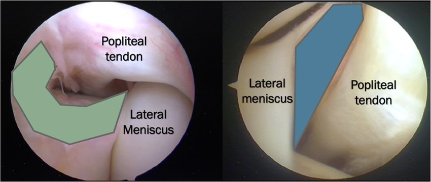
D’Addona et al. Journal of Orthopaedic Surgery and Research          (2021) 16:148                                                           Page 2 of 9

aponeurosis of the popliteus tendon, and the inferior                         concurrent damage to the popliteomeniscal fascicles
fascicle is a coronary ligament which connects the                            [30]. Tears of PMFs are the most frequent lesion occur-
meniscus to the edge of the tibia [7]. A third incostant                      ring in 80% of patients with grade III posterolateral in-
postero-inferior fascicle is sometime present [8–12].                         juries associated with ACL insufficiency [30–32]. The
PMFs, connecting the lateral meniscus at the popliteal                        risk is that an ACL injury could wrongly be identified as
hiatus [13], are thought to provide stability to the lat-                     the isolated cause of instability and knee pain [6].
eral meniscus, stabilizing the joint during internal rota-                      Unrecognised tears to the structure comprising the
tion of the tibia and sudden changes of direction (Fig.                       PLC have been cited as an important factor in post-
1) [4, 14–17]. In particular, working in conjunction                          surgical failure after cruciate ligament reconstruction
with the popliteus musculotendinous unit, the PMFs                            and in chronic instability and degenerative changes
prevent excessive lateral meniscal movement and pos-                          after knee trauma [6]. PMFs tears lead to lateral knee
sible entrapment [8]. For this reason, athletes, such as                      pain, painful squatting and locking sensation [9, 11,
martial artists, dancers, wrestlers and football players,                     16, 18, 20, 24, 30]. Several surgical options are avail-
whose activity is characterised by sudden changes of                          able: from open surgery to arthroscopic ones [2, 3,
direction, rotational stresses, repetitive twisting and                       32]. This systematic review identifies and analyses the
high jumps, are at higher risk of injuring the PMFs [3,                       articles on PMFs tears from diagnosis to surgical
8, 18]. The injuries which affect the lateral meniscus                        approach.
lead to an increase of contact pressure and rotational
instability, predisposing the joint to osteoarthritis as
observed on radiographs [1, 19]. A hypermobile lateral                        Methods
meniscus (HLM) may cause knee pain and a locking                              The PRISMA (Preferred Reporting Items for Systematic
sensation during deep knee flexion. One of the most                           Reviews and Meta-Analyses) guidelines were followed to
frequent cause of a HLM is thought to be a post-                              perform this systematic review [33] (Fig. 2).
traumatic injury of the PMFs [9, 20–26].
   Clinical and imaging diagnosis of these lesions is al-
ways challenging, even though MRI using proton density                        Search strategy
sequences may be useful [2, 7, 27–29]. In particular, it                      Four electronic databases (PubMed, Scopus, Web of Sci-
has been reported that the detection rate of PMFs on a                        ence and EMBASE) were used to search the scientific lit-
routine knee MRI on sagittal and coronal plane is                             erature using various combination of the keywords
approximatively 60% [27]. The clinical diagnosis is even                      “Popliteomeniscal Fascicles”, “Lateral Meniscus”, “Poplit-
more challenging, as most PMFs, tears occur in multi-                         eal Hiatus”, “Posterolateral Corner”, “Tear” and “Surgical
ligamentosus injuries [14]. When a tear of the PMFs                           Repair” for the years 1950–2020. The final search was
contributes to make the lateral meniscus unstable,                            performed on 1 December 2020 by two indipendent in-
arthroscopic observation and probing into the popliteo-                       vestigators. All the resulting titles were organised and
meniscal fascicle area is helpful to identify the damaged                     screened indipendently. In case of disagreement, a third
fascicle [16]. In particular, many knees with acute and                       senior investigator was asked to check and screened the
chronic ACL and/or posterolateral injuries have                               resulting titles.

 Fig. 1 a, b Arthoscopic anatomy of the PMFs. On the left, in green, it is underlined the antero-inferior fascicle (aPMF). On the right, in blue, the
 postero-superior fascicle (sPMF) is underlined. It is clear the connection between the posterior horn of lateral meniscus and the popliteal hiatus
 mediated by the PMFs
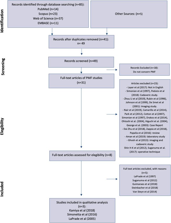
D’Addona et al. Journal of Orthopaedic Surgery and Research        (2021) 16:148                                           Page 3 of 9

 Fig. 2 PRISMA flow-chart. Methodology of selection used to screen and include articles for qualitative analysis

Selection criteria                                                          the surgical procedure performed, diagnosis, follow-up,
Only studies published in peer-reviewed journals were                       pre-operative imaging, arthroscopic or surgical assess-
included. Using the Oxford Center of Evidence-Based                         ment of PMFs and associated tears, pre-operative clinical
Medicine guidelines, level I to IV articles were identified.                examination and clinical postoperative outcomes were
Studies with patients assessed by pre-operative MRI and                     recorded.
then arthroscopically for post-traumatic PMFs tear were
included. Furthermore, studies reporting surgical repair                    Evaluation of the study quality
of PMFs outcomes assessed by clinical examination and/                      The methodological quality and bias of each study were
or subjective evaluation scales were included. Reviews,                     evaluated with the Coleman Methodology Score (CMS)
metanalyses, cadaveric and animal studies, biomechan-                       [34], which assesses methodology with 10 criteria, giving
ical studies, case report, commentaries, expert opinions                    a total score between 0 and 100. A score of 100 indicates
and operative techniques were excluded. We also de-                         that the study largely avoids chance, various biases, and
cided to exclude studies in which no information about                      confounding factors. The subsections that consistute the
D’Addona et al. Journal of Orthopaedic Surgery and Research            (2021) 16:148                                                  Page 4 of 9

CMS are based on the subsections of the Consolidated                            laboratory study; 1 article was a cadaveric and imaging
Standards of Reporting Trials (CONSORT) statement                               study; 2 articles were excluded because they described op-
(for randomized controlled trials). Each study was scored                       erative techniques without patient outcomes. A total of 8
by two indipendent reviewers and matched each other                             articles were therefore assessed. Finally, three articles were
(Table 1). In case of mismatch, a third investigator was                        included for the qualitative analysis: LaPrade et al. (2005)
asked to perform the CMS assessment indipendently.                              [30], Simonetta et al. (2016) [18] and Kamiya et al. (2018)
Possible disagreements were resolved by discussion.                             [20]. Five articles were excluded with reasons: LaPrade
                                                                                et al. (1997) [2] was excluded because, although an arthro-
Data extraction                                                                 scopic visualization of PMF tear with other associated le-
To avoid any bias of selection, the included articles with                      sions of PLC was reported, it is not specified whether and
all the relative list of references, and the articles ex-                       how PMFs were repaired; Steinbacher et al. (2019) [23]
cluded from the study were reviewed, assessed, and dis-                         and Van Steyn et al. (2014) [22] were excluded because,
cussed by all the authors. In case of disagreement                              although they report the surgical management of HLM, in
among the reviewers regarding the selection of articles                         their articles PMFs tears were not visualized and not iden-
based on inclusion and exclusion criteria, the senior in-                       tified as the cause of HLM; Guimaraes et al. (2018) [8]
vestigator made the final decision. The following data                          was excluded because the surgical technique is not dis-
were independently extracted by all the investigators:                          cussed; Suganuma et al. (2012) [9] was excluded because,
demographics, inlcuding mean age, sex, level of activity                        although an arthroscopic confirmation of PMF tears was
of population; mean follow-up; timing from symptoms                             performed, the surgical technique is not described.
to surgery; pre-operative MRI assessment; associated le-
sion reported; certainty of diagnosis by pre-operative clin-                    Study characteristics and quality assessment
ical examination and arthroscopic confirmation; surgical                        All the articles included for the qualitative analysis were
management and technique performed; clinical outcome                            published in the period 2005 to 2018, and their charac-
measurements by post-operative clinical examination and/                        teristics are summarized in the Table 1. All the articles
or subjective evaluation scales; recurrence of the lateral                      included are retrospective case series, and their level of
meniscus instability and/or pain and/or locking sensation,                      evidence according to the Oxford Center of Evidence-
and intra- and/or post-operative complications.                                 Based Medicine guidelines is IV. The mean follow-up
                                                                                was 37.1 months, ranging from 36 months [30] to 38.3
Results                                                                         months [18]. According to the Coleman Methodology
Study selection                                                                 Score (CMS), 2 articles [18, 30] were of poor quality (<
A total of three articles were included in the qualitative                      50), and 1 [20] of fair quality (59). The median CMS was
analysis. Figure 1 describes the methodology used for se-                       49.3 (43–59) of a possible 100 total score.
lection and inclusion of articles. The original literature
search identified a total of 85 articles comprising of dupli-                   Patient characteristics
cates. Another five articles were identified by other                           Patient characteristics and surgery indications are reported
sources. After removal of 41 duplicates, 49 articles were                       in Table 2. A total of 32 patients (17 males and 15 females;
assessed for eligibility. Eighteen articles were removed be-                    mean age 26.9 (range 15–67) years) from the included ar-
cause they did not concern PMF. Of the remaining 31                             ticles was analysed. The level activity was reported for 9 of
full-text articles, 22 articles were removed because do not                     32 patients: it ranged from recreational activity to semi-
meet inclusion criteria: 1 was excluded because was a re-                       professional and professional sport. In LaPrade et al. case
view in German language; 2 articles were excluded be-                           series [30], 2 patients were professional wrestlers, while in
cause they were cadaveric studies; 4 articles were excluded                     Simonetta et al. series [18], 5 were semi-pro soccer players.
because they were imaging studies; 9 articles were ex-                          The diagnosis and the indications for surgery were based
cluded because they were case reports; 3 were excluded                          on patient symptoms, pre-operative clinical examination
because they were reviews of literature; 1 article was a                        and imaging. In all the articles included, patients

Table 1 Study characteristics
Study                   Year of publication      Level of evidence     Study design                Mean follow-up (months)   Coleman Methodology
                                                                                                                             Score (CMS)
Kamiya et al. [20]      2018                     IV                    Retrospective case series   37                        59
Simonetta et al. [18]   2016                     IV                    Retrospective case series   38.3                      46
LaPrade et al. [30]     2005                     IV                    Retrospective case series   36                        43
The most significative data concerning the quality and the study methodology used are reported
D’Addona et al. Journal of Orthopaedic Surgery and Research (2021) 16:148 Page 5 of 9
Table 2 Patients characteristics and surgical indications
a a
A. Patient characteristics B. Surgical indications
N of Mean Sex (male/ Level of Symptoms Mean timing Pre-operative Pre-operative imaging
patients age female) activity symptoms to clinical
surgery (months) examination
Kamiya et al. [20] 20 37.7 9M/11F Locking sensation 25.3 3D-virtual load MRI
Simonetta et al. [18] 6 16.6 4M/2F 5/6 semi-pro athlete; Pain, locking 14.6 Figure-4 test MRI in the sagittal plane
1/6 recreational sensation +: 3/6 patients and T2 sequences
LaPrade et al. [30] 6 26.6 4M/2F 2/6 professional wrestler; Pain, locking 5.6 Figure-4 test MRI
1/6 football player sensation +: 6/6 patients
a
Blank cells indicate not available
On the left side of the table, number of patients, mean age, sex and activity level are summarized; on the right side: symptoms, mean timing from onset of
symptoms to surgery, pre-operative clinical examination and imaging are shown
experienced symptoms such as pain and locking sensation. Surgical approach and suturing technique
The pre-operative clinical examination was based on the In Table 3, the surgical technique is reported. To con-
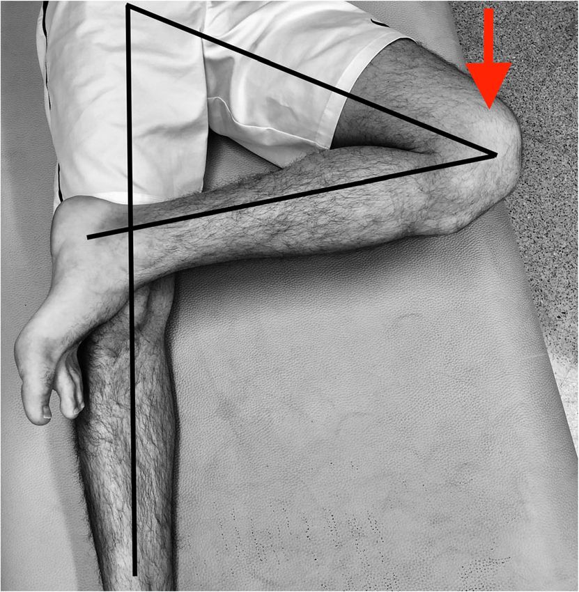
Figure-4 test [18, 30] (Fig. 3), which was positive in all pa- firm the diagnosis and assess the disruption of the PMF,
tients in LaPrade et al. study [30], and in 3 of 6 patients in arthroscopic evaluation is necessary. In all the articles
Simonetta et al. study [18]. Kamiya et al. [20] do not re- included, direct arthroscopic visualization confirmed the
port the results of any clinical examination. PMF tear between the posterior horn of the lateral me-
The imaging study to diagnose PMF tears was MRI in niscus and the popliteus hiatus.
for all included articles. Kamiya et al. [20] performed a Further evidence of PMF tear was given by the in-
3D-virtual load MRI, which allows, under dynamic load, creased mobility and forward translation of the intact
to verify the forward and medial translation of the lateral lateral meniscus on arthroscopic probing.
meniscus during knee flexion. Simonetta et al. [18] im- Different surgical procedures are reported: both
aging study was based on MRI, and the diagnosis of PMF Kamiya et al. [20] and Simonetta et al. [18] performed
tear was confirmed in the sagittal plane and T2 sequences an arthroscopic technique, while LaPrade et al. [30] per-
that demonstrate the disruption of PMFs. LaPrade et al. formed an open repair. Each article reports a different
[30] used plain MRI scan to demonstrate PMF tear. The suturing technique.
mean timing of symptoms to surgery, reported in all the Kamiya et al. [20] performed an inside-out repair of in-
articles, was 15.1 months (range 0.3–60). jured PMFs using a polyester non-absorbable suture on a
10-inch straight and/or curved cutting needle (Stryker,
Kalamazoo, CA, USA). An average of 5.0 (range 2–8)
double-stacked vertical stitches connecting the lateral me-
niscus and the meniscocapsular junction was reported. No
other associated lesions were noted at the arthroscopic
evaluation.
Simonetta et al. [18] used the FastFix meniscal repair
system (Smith & Nephew, Hull, UK) to fix disrupted
PMFs with an all-inside technique. Two or three stitches
in a vertical fashion on either side of the popliteal hiatus
were used. In their case series, 4 of 6 patients, in whom
timing from symptoms to surgery was greater than 12
months, presented a chondral lesion of the lateral femoral
condyle, but these associated lesions were not addressed.
La Prade et al. [30] performed an open repair. For this
purpose, after an antero-lateral capsular arthrotomy, the
midthird lateral capsular ligament was retracted and the
antero-inferior fascicle tear was exposed. After this, an
open repair using horizontal mattress sutures with non-
absorbable 0 sutures was performed.
Fig. 3 Figure-4 test. Illustration of figure-4 test drawn with black Outcome analysis
lines indicating the correct position, with the examined knee flexed Clinical outcome with evaluation subjective scales, peri-
upon the other leg. The red vector indicates the strength direction
operative complications and post-operative imaging stud-
to apply to the knee to provoke pain in the lateral compartment
ies are reported in Table 4. For all three articles included,D’Addona et al. Journal of Orthopaedic Surgery and Research (2021) 16:148 Page 6 of 9
Table 3 Surgical characteristics
Arthroscopic confirmation Surgical procedure Suturing technique Associated lesion
Kamiya et al. [20] Direct visualization of disruption Inside-out arthroscopic Polyester non-absorbable sutures on a Isolated PMFs
of PMF. repair 10-inch straight and/or curved cutting
Forward translation by probing needle (Stryker).
intact lateral meniscus An average of 5.0
(range, 2–8) double-stacked vertical
suture were used.
Simonetta et al. [18] Direct visualization of disruption All-inside arthroscopic Fastfix meniscal repair system (Smith & 4/6 chondral lesions LFC
of PMF. repair Nephew)
Forward translation by probing Two to three sutures are placed on
intact lateral meniscus either
side of the popliteal hiatus, in a
vertical fashion
LaPrade et al. [30] Direct visualization of disruption Open repair horizontal mattress non-absorbable Isolated PMFs
of PMF. 0 sutures
Forward translation by probing
intact lateral meniscus
Details of arthroscopic confirmation of the diagnosis, surgical and suturing technique used, and the associated lesion reported during the procedures
the clinical evidence at the final follow-up demonstrated a with an ACL tear or traumatic injury to the PLC [8, 30, 32,
complete resolution of pre-operative symptoms (pain and 35]. In symptomatic patients, surgery is the gold standard
locking feeling), but only LaPrade et al. [30] recorded at to manage these injuries [21, 36], although the most ap-
the final follow-up the result of clinical post-operative propriate surgical technique is still debated [3, 22, 30, 37].
examination, demonstrating a negative figure-4 test for all This systematic review shows surgery for symptomatic pa-
their patients. No subjective scales were used for outcome tients with PMF tears is effective and safe, with pain relief
assessment except for Kamiya et al. [20], who recorded a and resolution of locking sensation with no complications
mean Tegner activity level before and after surgery of 4.6 reported. Furthermore, although the diagnosis is not easy
(range, 2–8) and 4.7 (range, 2–8), respectively (p = 0.480). and an accurate clinical examination and imaging, by MRI
The Lysholm knee score significantly increased from 72.0 scan, is fundamental to plan the most appropriate treat-
(range, 48–85) to 97.8 (range, 78–100) (p < 0.01). None of ment, arthroscopic evaluation by probing the lateral me-
the analysed studies reported clinically relevant complica- niscus and the intra-articular visualization of disrupted
tions. Post-operative imaging assessment was performed PMFs remain the gold standard to accurately identify these
only by Kamiya et al. [20] using a 3D-virtual load MRI at injuries. The lack of high level-of-evidence studies about
4-month post-operatively, demonstrating the absence of a this topic and the paucity of articles regarding the manage-
mobile lateral meniscus during knee flexion with a suc- ment of PMFs tears does not allow to fully clarify which
cessfull repair of PMFs. could be the most appropriate surgical technique to
undertake.
Discussion According to our results, younger patients with high-
The diagnosis, by imaging and clinical examination or demanding activities are most suitable candidates for
arthroscopically, of disrupted PMFs remains very challen- surgery. Although Kamiya et al. [20] operated on pa-
ging [18, 32]. Tears of PMFs are present in 80% of patients tients older than 50, the mean age among the 32 patients
Table 4 Outcomea. Symptoms and post-operative clinical evaluation, evaluation scales, complications and post-operative imaging
study
Clinical outcome Evaluation scales Complications Post-operative imaging
Kamiya et al. [20] Absence of locking and pain at The mean Tegner activity level scales before None 3D-Virtual load MRI after
final F-U and after 4 months
surgery was 4.6 (range, 2–8) and 4.7 (range, 2–8),
respectively (p = 0.480).
The Lysholm knee scores were significantly
increased from 72.0 (range, 48–85) to 97.8
(range, 78–100) (p < 0.01)
Simonetta et al. [18] Absence of locking and pain at None
final F-U
LaPrade et al. [30] Figure-4 test -:6/6 None
Absence of locking and pain at
final F-U
a
Blank cells indicate not availableD’Addona et al. Journal of Orthopaedic Surgery and Research (2021) 16:148 Page 7 of 9
analysed was 26.9. Only for 9 patients the level of activ- characterized. While Kamiya et al. [20] and Simonetta
ity was specified [18, 30]: 7 of 9 patients were profes- et al. [18] performed two different arthroscopic tech-
sional or semi-professional athletes. High-demanding niques, an inside-out and an all-inside repair respect-
athletic patients are susceptible to PMFs tears, with the ively, LaPrade et al. [30] choose an open repair, with
common injury mechanism being twisting on their knee. three different suturing technique. All three articles re-
Full-contact sports, such as wrestling or soccer, are the ported good clinical outcome with resolution of symp-
most commonly involved. toms and no complications: only LaPrade et al. [30]
The indications for surgery included symptoms, clin- performed a new clinical examination by subjecting pa-
ical examination, except for Kamiya et al. [20], and im- tients to a post-operative figure-4 test, which always re-
aging: pain over the lateral aspect of the knee, pain sulted negative. Kamiya et al. [20] reported subjective
during active joint flexion and a locking sensation were evaluation scales and post-operative MRI imaging study:
reported in all the patients described in the studies in- the Tegner and Lysholm evaluations scores showed an
cluded in the present review. A specific clinical examin- improvement between pre- and post-operative values.
ation was performed by LaPrade et al. [30] and
Simonetta et al. [18]: the figure-4 test, first described by Study limitations
LaPrade [2], was chosen as diagnostic test to check the The limitations of the present systematic review are re-
integrity of PMF. Of 12 patients undergoing the figure-4 lated to the scanty quality of the studies available in the
test, 9 (75%) resulted positive, indicating that a positive literature. All the articles included were retrospective
figure-4 test is strongly suggestive of PMF tear. In all the case series, with limited number of patients. Further-
patients reported, the diagnosis of PMF disruption was more, the absence of subjective evaluation scales and no
confirmed arthroscopically. Arthroscopy is crucial to post-operative assessment by MRI imaging, except for
check the lateral meniscus stability [11, 18, 20, 22–24, Kamiya et al. [20], do not allow complete outcome ana-
26, 30, 36]. If the PMFs are disrupted, probing the lateral lysis. The level of evidence for all the articles included
meniscus produces an abnormally large forward transla- was low (IV). According to the Coleman Score, the study
tion of the meniscus with a direct visualization of PMF quality ranges from fair [20] to poor [18, 30].
tear on the lateral side of the joint space. In LaPrade et al.
[30] study, although they performed an open repair, an Conclusion
arthroscopic check to confirmation the diagnosis was per- Although the number of studies available in the literature
formed. The time from onset of symptoms to surgery sug- was limited and the methodological quality of the articles
gests that timing does not influence the healing potential included was questionable, the most relevant evidence
of PMF, and that the continuous forward movement of arising from this systematic review is that it is important
the lateral meniscus prevents the formation of a scar tissue to recognise PMF disruption in patients with pain and
that would stabilise the meniscus. PMF tears are often as- locking sensation, and no clear evidence of other causes of
sociated with other intra-articular injuries such as ACL internal derangement of the knee. In particular, it is neces-
tears, meniscal and chondral tears, and PLC injuries [2, 3, sary to evaluate PMF tears by pre-oeprative MRI imaging,
8, 32]. Furthermore, associated chondral lesions of the lat- and clinical examination using the figure-4 test. Arthro-
eral femoral condyle were reported in 4 of 32 (12.5%) pa- scopic confirmation of the diagnosis by direct visualization
tients. These four patients had reported symptoms for of the PMF tear and probing of the lateral meniscus is ne-
longer than 12 months. To prevent further damage to the cessary. When recognised, the evidence suggests to oper-
knee joint and protect other structures which have been ate on the lesion to stabilise the knee joint, especially in
injured or reconstructed (ACL, meniscal and chondral le- young athletic patients. There is no definite evidence
sions) or stabilize the lateral compartment, prompt sur- about the most appropriate surgical technique to perform,
gery in active patients with symptoms and a positive MRI and surgeon experience and confidence should guide the
and clinic examination is mandatory. choice of the appropriate technique. Further studies with
A pre-operative MRI is always performed and appears a higher number of patients, and higher methodological
the best imaging modality to demonstrate disruption of quality should be undertaken to better understand the
PMF on the sagittal plane and in T2 sequences [7, 8, pathogenesis and the evolution of PMF tears and to clarify
29]. Kamiya et al. [20] performed a virtual 3D-loading the best surgical technique.
MRI which demonstrated the forward translation of the
Abbreviations
lateral meniscus that during active knee flexion, a find- PMFs: Popliteomeniscal fascicles; PLC: Postero-lateral corner;
ing subsequently confirmed arthroscopically. HLM: Hypermobile lateral meniscus; MRI: Magnetic resonance imaging;
The appropriate surgical technique is still debated [3, ACL: Anterior cruciate ligament; CMS: Coleman Methodology Score
18, 30, 32]. In all the articles included in the present re- Acknowledgements
view, the surgical procedure is well described and Not applicableD’Addona et al. Journal of Orthopaedic Surgery and Research (2021) 16:148 Page 8 of 9
Authors’ contributions 11. Camarillo M, Johnson DL. Popliteomeniscal Fascicle Tears. Orthopedics.
A.D. and A.I. searched studies among literature, screened eligible articles and 2014;37(3):187–90.
wrote the review. G.D.V. checked the search strategy, intervened in case of 12. Nair R, Dubey N. MR imaging of the hypermobile lateral meniscus of the
mismatch and participated to the writing of the article. D.R. participated and knee: a case report. Acta Med Acad. 2019;48(2):225–9.
supervisioned the writing of the article. N.M. drafted the search strategy, 13. Cohn AK, Mains DB. Popliteal hiatus of the lateral meniscus. Anatomy and
corrected and approved the final review version. All authors read and measurement at dissection of 10 specimens. Am J Sports Med. 1979;7(4):221–6.
approved the final manuscript. 14. Shin RH, Zhao C, Zobitz ME, Amadio PC, An KN. Mechanical properties of
intrasynovial and extrasynovial tendon fascicles. Clin Biomech. 2008;23(2):236–41.
Funding 15. Speziali A, Placella G, Tei MM, Georgoulis A, Cerulli G. Diagnostic value of
No fundings were used for the writing of the article. the clinical investigation in acute meniscal tears combined with anterior
cruciate ligament injury using arthroscopic findings as golden standard.
Musculoskelet Surg. 2015;100(1):31–5.
Availability of data and materials
The datasets used and/or analysed during the current study are available 16. Shin H-K, Lee H-S, Lee Y-K, Bae K-C, Cho C, Lee K-J. Popliteomeniscal fascicle
from the corresponding author on reasonable request. tear: diagnosis and operative technique. Arthrosc Tech. 2012;1(1):e101–6.
17. Suganuma J, Inoue Y, Tani H, Sugiki T, Sassa T, Shibata R. Reconstruction of
the popliteomeniscal fascicles for treatment of recurrent subluxation of the
Ethics approval and consent to participate lateral meniscus. Arthrosc Tech. 2017;6(2):e283–90.
Not applicable 18. Simonetta R, Di Vico G, Papalia R, et al. Arthroscopic all-inside
treatment of popliteomeniscal fascicles tears: surgical technique and
Consent for publication results from the first 6 consecutive patients. J Biol Regul Homeost
All authors give their consent to publish this article. Agents. 2016;30(4):91–7.
19. Higuchi H, Kimura M, Kobayashi A, Hatayama K, Takagishi K. A novel
Competing interests treatment of hypermobile lateral meniscus with monopolar radiofrequency
All the authors declare that they have no competing interests. energy. Arthroscopy. 2004;20(6 SUPPL):1–5.
20. Kamiya T, Suzuki T, Otsubo H, et al. Midterm outcomes after arthroscopic
Author details surgery for hypermobile lateral meniscus in adults: Restriction of paradoxical
1 motion. J Orthop Sci. 2018;23(6):1000–4.
A.O.U. Federico II, Department of Public Health, Section of Orthopaedics and
Trauma Surgery, Via S. Pansini 5, 80131 Naples, Italy. 2Humanitas Clinical and 21. George M, Wall EJ. Locked knee caused by meniscal subluxation: magnetic
Research Center-IRCCS, Via Alessandro Manzoni 56, 20089 Rozzano, MI, Italy. resonance imaging and arthroscopic verification. Arthroscopy. 2003;19(8):
3 885–8.
Department of Orthopaedics and Trauma Surgery, Clinica San Michele,
Maddaloni, CE, Italy. 4Department of Musculoskeletal Disorders, University of 22. Van Steyn MO, Mariscalco MW, Pedroza AD, Smerek J, Kaeding CC, Flanigan
Salerno, Salerno, Italy. 5Centre for Sports and Exercise Medicine, Mile End DC. The hypermobile lateral meniscus: a retrospective review of
Hospital, Barts and The London School of Medicine and Dentistry, 275 presentation, imaging, treatment, and results. Knee Surgery, Sport Traumatol
Bancroft Road, London E1 4DG, UK. Arthrosc. 2016;24(5):1555–9.
23. Steinbacher G, Alentorn-Geli E, Alvarado-Calderón M, Barastegui D, Álvarez-
Received: 14 December 2020 Accepted: 10 February 2021 Díaz P, Cugat R. Meniscal fixation is a successful treatment for hypermobile
lateral meniscus in soccer players. Knee Surgery, Sport Traumatol Arthrosc.
2019;27(2):354–60.
References 24. Park J-H, Ro K-H, Lee D-H. Snapping knee caused by a popliteomeniscal
1. Akita K, Nimura A, Fujishiro H, Tsukada S, Mochizuki T, Nakamura T. Attachment fascicle tear of lateral meniscus in a professional taekwondo athelete.
area of fibres from the horns of lateral meniscus: anatomic study with special Orthopedics. 2012;35(7):e1104–7.
reference to the positional relationship of anterior cruciate ligament. Knee 25. Simonian PT, Sussmann PS, Van Trommel M, Wickiewicz TL, Warren RF.
Surgery, Sport Traumatol Arthrosc. 2015;25(2):368–73. Popliteomeniscal fasciculi and lateral meniscal stability. Am J Sports Med.
2. Laprade RF. Arthroscopic evaluation of the lateral compartment of knees 1997;25(6):849–53.
with grade 3 posterolateral knee complex injuries. Am J Sports Med. 1997; 26. Ohtoshi K, Kimura M, Kobayashi Y, Higuchi H, Kikuchi S. Arthroscopic
25(5):596–602. thermal shrinkage for hypermobile lateral meniscus. Am J Sports Med. 2004;
3. Papalia R, Simonetta R, Di Vico G, et al. Tears of popliteomeniscal fascicles, 32(5):1297–301.
diagnostic and clinical implications. A Review of the Evidence. J Biol Regul 27. Sakai H, Sasho T, Wada Y, et al. MRI of the Popliteomeniscal Fasciculi. Am J
Homeost Agents. 2016;30(4):99–106. Roentgenol. 2006;186(2):460–6.
4. Kimura M, Shirakura K, Hasegawa A, Kobayashi Y, Udagawa E. Anatomy and 28. Laundre BJ, Collins MS, Bond JR, Dahm DL, Stuart MJ, Mandrekar JN. MRI
pathophysiology of the popliteal tendon area in the lateral meniscus: 2. accuracy for tears of the posterior horn of the lateral meniscus in patients
Clinical investigation. Arthroscopy. 1992;8(4):424–7. with acute anterior cruciate ligament injury and the clinical relevance of
5. Fineberg MS, Duquin TR, Axelrod JR. Arthroscopic visualization of the missed tears. Am J Roentgenol. 2009;193(2):515–23.
popliteus tendon. Arthroscopy. 2008;24(2):174–7. 29. De Smet AA, Asinger DA, Johnson RL. Abnormal superior popliteomeniscal
6. Peduto AJ, Nguyen A, Trudell DJ, Resnick DL. Popliteomeniscal fascicles: fascicle and posterior pericapsular edema: indirect MR imaging signs of a
anatomic considerations using MR arthrography in cadavers. AJR Am J lateral meniscal tear. Am J Roentgenol. 2001;176(1):63–6.
Roentgenol. 2008;190(2):442–8. 30. LaPrade RF, Konowalchuk BK. Popliteomeniscal fascicle tears causing
7. Johnson RL, De Smet AA. MR visualization of the popliteomeniscal fascicles. symptomatic lateral compartment knee pain diagnosis by the figure-4 test
Skelet Radiol. 1999;28(10):561–6. and treatment by open repair. Am J Sports Med. 2005;33:1231–6.
8. Guimaraes JB, Facchetti L, Schwaiger BJ, Gersing AS, Li X, Link TM. Natural 31. Temponi EF, de Carvalho Júnior LH, Saithna A, Thaunat M, Sonnery-Cottet B.
evolution of popliteomeniscal fascicle tears over 2 years and its association Incidence and MRI characterization of the spectrum of posterolateral corner
with lateral articular knee cartilage degeneration in patients with traumatic injuries occurring in association with ACL rupture. Skelet Radiol. 2017;46(8):
anterior cruciate ligament tear. Eur Radiol. 2018;28(8):3542–9. 1063–70.
9. Suganuma J, Mochizuki R, Inoue Y, Yamabe E, Ueda Y, Kanauchi T. Magnetic 32. Zappia M, Reginelli A, Chianca V, et al. MRI of popliteo-meniscal fasciculi of
resoncance imaging and arthroscopic findings of the popliteomeniscal the knee: A pictorial review. Acta Biomed. 2018;89:7–17.
fascicles with and without recurrent subluxation of lateral meniscus. 33. Liberati A, Altman DG, Tetzlaff J, et al. The PRISMA statement for reporting
Arthroscopy. 2012;28(4):507–16. systematic reviews and meta-analyses of studies that evaluate health care
10. Aman ZS, DePhillipo NN, Storaci HW, et al. Quantitative and qualitative interventions: explanation and elaboration. J Clin Epidemiol. 2009;62(10):e1–34.
assessment of posterolateral meniscal anatomy: defining the popliteal 34. Robertson G, Ang KK, Maffulli N, Simpson CK, Rust PA. Return to sport
hiatus, popliteomeniscal fascicles, and the lateral meniscotibial ligament. following surgical management of triangular fibrocartilage tears: a
Am J Sports Med. 2019;47(8):1797–803. systematic review. Br Med Bull. 2019;130(1):89–103.D’Addona et al. Journal of Orthopaedic Surgery and Research (2021) 16:148 Page 9 of 9
35. Di Vico G, Di Donato SL, Balato G, Correra G, D'Addona A, Maffulli N, Rosa D.
Correlation between time from injury to surgery and the prevalence of
ramp and hidden lesions during anterior cruciate ligament reconstruction. A
new diagnostic algorithm. Muscles Ligaments Tendons J. 2018;7(3):491–7.
36. de Albornoz PM, Forriol F. The meniscal healing process. Muscles, ligaments
and Tendons J. 2012;2(1):10–8.
37. Frizziero A, Ferrari R, Giannotti E, Ferroni C, Poli P, Masiero S. The meniscus
tear. State of the art of rehabilitation protocols related to surgical
procedures. Muscles, Ligaments and Tendons J. 2012;2(4):295–301.
Publisher’s Note
Springer Nature remains neutral with regard to jurisdictional claims in
published maps and institutional affiliations.You can also read


