The influence of nasal microbiome diversity and inflammatory patterns on the prognosis of nasal polyps
←
→
Page content transcription
If your browser does not render page correctly, please read the page content below
www.nature.com/scientificreports

                OPEN             The influence of nasal microbiome
                                 diversity and inflammatory
                                 patterns on the prognosis of nasal
                                 polyps
                                 Weigang Gan, Hongting Zhang, Fengjuan Yang, Shixi Liu, Feng Liu & Juan Meng*
                                 To understand the inflammatory microenvironment and microbiome factors for prognosis of chronic
                                 rhinosinusitis with polyps (CRSwNP), we explored the difference in characteristics of the microbiome
                                 of the nasal sinuses and inflammatory cytokines between recurrent and non-recurrent groups. We
                                 collected nasal secretions and polyp tissue from 77 CRSwNP patients. Then, we extracted microbial
                                 DNA from cotton swabs, performed high-throughput sequencing based on 16S rRNA to detect
                                 bacterial community composition, and analyzed cytokines such as IL-5, IL-8, IL-17a, IL-17e, IL-18,
                                 IL-27 and INF-gamma from polyp tissue using Luminex. The eosinophil and neutrophil cells in the
                                 peripheral blood and polyp tissue were counted. Postoperative follow-up of patients with CRSwNP for
                                 1 year was conducted to record the recurrence of nasal polyps and analyze the correlation between the
                                 recurrence of nasal polyps and the characteristics of inflammatory cytokines, inflammatory cell count
                                 and nasal microbial diversity. After 1 year of follow-up, there were 12 recurrent patients, including 5
                                 males and 7 females. Postoperative recurrence of nasal polyps was not significantly correlated with
                                 age, sex, asthma, allergic rhinitis or other allergic diseases in CRSwNP patients. In terms of the total
                                 nasal symptom score, the recurrent group was significantly higher than the non-recurrent group. In
                                 nasal polyp tissues, eosinophils (40.83/HP) and neutrophils (30.83/HP) in patients with CRSwNP in
                                 the recurrent group were significantly higher than those in the non-recurrent group (13.72/HP), and
                                 neutrophils (18.5/HP) were also significantly higher in the recurrent group than the non-recurrent
                                 group. The expression levels of IFN-, IL-17A, IL-17E and IL-18 were significantly higher in the recurrent
                                 group than in the non-recurrent group, and the positive rates were not different. In Southwest China,
                                 Enterobacteria and anaerobic bacteria may be correlated with the inflammatory pattern expression of
                                 nasal polyps. The neutrophil-mediated inflammatory response plays an important role in patients with
                                 CRSwNP in Southwest China and is correlated with nasal polyp recurrence. Recurrence of nasal polyps
                                 after endoscopic sinus surgery may be potentially associated with a reduced abundance of protective
                                 microorganisms and an increased number of pathogenic microorganisms.

                                 Abbreviations
                                 CRSwNP	Chronic rhinosinusitis with polyps
                                 TNSS	Total nasal symptom score
                                 ESS	Endoscopic sinus surgery
                                 rRNA	Ribosomal RNA
                                 BMI	Body mass index
                                 SP method	Streptomyces antibiotinin protein-peroxidase conjugating method
                                 ECP	Eosinophilic cationic protein
                                 MPO	Myeloperoxidase
                                 NGS	Next-generation sequencing
                                 NCBI	National Center for Biotechnology Information
                                 SRA	Sequence Read Archive
                                 FLASH	Fast low-angle shot magnetic resonance imaging
                                 OTUs	Operational taxonomic units

                                 Department of Otorhinolaryngology–Head and Neck Surgery, West China Hospital, West China School of Medicine,
                                 Sichuan University, Chengdu, Sichuan, People’s Republic of China. *email: mjmelinda@163.com

Scientific Reports |   (2021) 11:6364               | https://doi.org/10.1038/s41598-021-85292-5                                           1

                                                                                                                                       Vol.:(0123456789)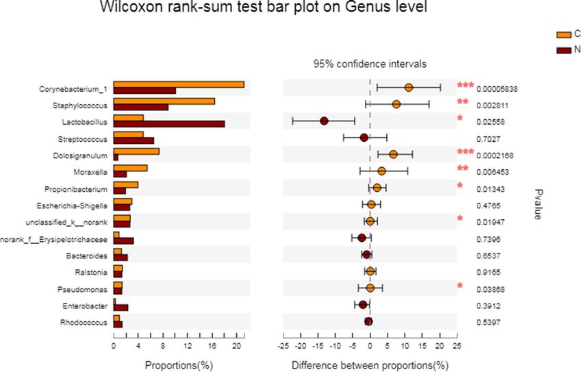
www.nature.com/scientificreports/
VAS Visual analogue scale
PCoA Principal coordinate analysis
FDR False discovery rate
KW Kruskal–Wallis
LDA Linear discriminant analysis
The incidence of chronic rhinosinusitis with nasal polyps (CRSwNP) is increasing yearly, and this condition is
a primary cause of morbidity and medical expenditure in European and American countries, which makes it of
research value from the perspective of both epidemiology and health economics1. Nasal polyps treatment has
been developed as a comprehensive approach including surgery, drugs, biological treatment and other means,
but the recurrence rate of nasal polyps or sinusitis after nasal endoscopy is still high. Some scholars have reported
recurrence rates reaching 80% and secondary surgery rates reaching 42%, and some have reported 57% and 46%,
respectively2. Therefore, increasing attention has been given to the factors affecting the recovery of nasal polyps
after surgery, among which the effects of the nasal microenvironment and immunophenotype on nasal polyps
are particularly prominent.
With the deepening of the research, people have gradually realized that the nasal microbes for nasal inflam-
matory diseases (including nasal polyps) have a certain impact, regardless of whether the disease is in the early
stage. In vitro research helps explore nasal sinus inflammatory disease pathogens, and modern molecular biology
techniques provide 16S ribosomal RNA(rRNA) sequencing data and reliable means of studying the nasal cavity
flora; however the latter approach is more accurate in reflection of the nasal cavity flora changes in nasal sinus
inflammatory disease. High-throughput sequencing technology showed that changes in the core microbiome
occurred in patients with chronic sinusitis compared to that in control s ubjects3–5. The inflammatory microen-
vironment was another key point in CRSwNP patients that we addressed. Based on previous studies related to
the inflammatory microenvironment of nasal polyp t issues6,7, seven factors, IL-5, IL-8, IL-17A, IL-17E, IL-18,
IL-27 and IFN-γ, were selected for analysis. We selected inflammatory cytokines as markers referring to the four
types of inflammation in nasal polyp tissues. Patients with CRSwNP were divided into different types according
to inflammatory patterns and the characteristics of their respective nasal microbiome. These results provide a
basis for recognizing the microbiome diversity of patients with different inflammatory patterns of CRSwNP
and exploring the effects of nasal microbial diversity and inflammatory types on the prognosis of nasal polyps.
Results
Comparison of microbiome composition between CRSwNP patients and the control sub-
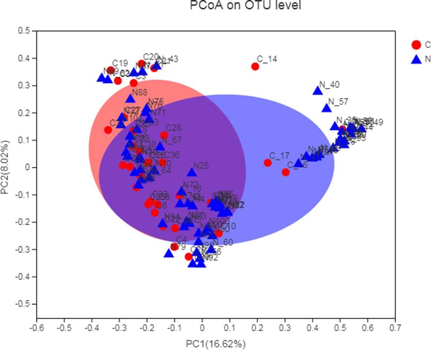
jects. Principal coordinates analysis (PCoA) plots reflect the beta diversity of the microbial community
among groups. In our study, we observed that between two groups most microbial distributions are overlapping
and only a minority of samples are specific to each group (Fig. 1). Wilcoxon rank sum test was used to compare
the nasal microbial diversity of CRSwNP patients and the control subjects. At the phylum level, the relative
abundance of Actinobacteria was significantly lower in the CRSwNP group (16.07%) than that in the control
group (31.42%) (FDR P = 0.041). At the genus level, the relative abundance of Corynebacterium in the CRSwNP
group was 10.07%, which was significantly lower than that in the non-CRS group (21.21%) (FDR P = 0.047). The
relative abundance of Dolosigranulum in the CRSwNP group was 0.69%, which was significantly lower than that
in the non-CRS group (7.38%) (FDR P = 0.012) (Fig. 2).
Clinical data of CRSwNP patients in the recurrent and non‑recurrent groups. For the 77 patients
with CRSwNP who underwent endoscopic sinus surgery (ESS), we conducted a 1-year follow-up and found
12 recurrent patients, including 5 males and 7 females. In the recurrent group, there were 3 CRSwNP patients
with asthma, 4 with allergic rhinitis, and 0 with eczema; however, in the non-recurrent group, there were 8 with
asthma, 10 with allergic rhinitis, and 6 with eczema. The epidemiological data of the two groups of patients are
listed in Table 1. According to the data, there was no difference between the two groups in age, gender, asthma,
allergic rhinitis or other allergic diseases. The Lund-Mackay scores and total nasal symptom scores (TNSS) of the
recurrent group was significantly higher than that of the non-recurrent group (P = 0.001, P = 0.000).
Comparison of inflammatory mediators between the recurrent group and the non‑recurrent
group. In terms of peripheral blood inflammatory cell count, the neutrophil count (median 3.82 × 10⁹/L) in
the recurrent group was higher than that in the non-recurrent group (median 3.59 × 10⁹/L). There was no sig-
nificant difference in peripheral blood eosinophil count between the two groups. In the nasal polyp tissues, the
number of eosinophils (40.83/HP) and neutrophils (30.83/HP) in the recurrent group was higher than that in
the non-recurrent group (13.72/HP), and the differences were statistically significant. Regarding the expression
level of inflammatory mediators, 2 patients with positive IL-5 and 10 patients with negative IL-5 in the recur-
rent group were compared, and the difference was not statistically significant (X2 = 0.242, P = 0.623). The median
expression of IL-8 in the recurrent group was 3,152.25 pg/ml and that in the non-recurrent group was 544.95 pg/
ml. The difference was statistically significant, that is, the recurrent group was significantly higher than the non-
recurrent group. The expression levels of IFN-, IL-17A, IL-17E and IL-18 were significantly higher in the recur-
rent group than in the non-recurrent group (IFN- (61.75 pg/ml in the recurrent group, 26.97 pg/ml in the non-
recurrent group), IL-17A (15.27 pg/ml in the recurrent group, 6.77 pg/ml in the non-recurrent group), IL-17E
(223.38 pg/ml in the recurrent group, 56.62 pg/ml in the non-recurrent group) and IL-18 (30.83 pg/ml in the
recurrent group, 19.07 pg/ml in the non-recurrent group)). However, from the perspective of expression level,
there was no significant difference in the positive rates of the four inflammatory factors between the recurrent
Scientific Reports | (2021) 11:6364 | https://doi.org/10.1038/s41598-021-85292-5 2
Vol:.(1234567890)
www.nature.com/scientificreports/

                                  Figure 1.  A principal coordinates analysis (PCoA) of bacterial communities was calculated with non-
                                  constrained data dimensionality reduction analysis method. The plots’ central tendency indicates the
                                  interpersonal variability of the two groups.

                                  group and the non-recurrent group. There was no significant difference in the positive rate or median expression
                                  of IL-27 between the recurrent and non-recurrent groups (Table 2).

                                  Comparison of microbiome composition between the recurrent group and the non‑recurrent
                                  group. The mean microbial richness was compared between the recurrent group and the non-recurrent
                                  group. The results showed that there was no statistically significant difference in the Sobs index between the
                                  recurrent group (462.75) and the non-recurrent group (433.66) (P = 0.852), indicating that there was no signifi-
                                  cant difference in nasal microbiome richness between the two groups (Fig. 3).
                                      For patients with CRSwNP in the recurrent group and the non-recurrent group, the most abundant bacte-
                                  rial categories were relatively concentrated in the following groups, including Firmicutes (non-recurrent group
                                  49.19% vs recurrent group 52.99%), Proteobacteria (non-recurrent group 17.33% vs recurrent group 24.58%),
                                  Actinobacteria (non-recurrent group 18.0% vs recurrent group 2.626%), Bacteroidetes (non-recurrent group
                                  8.31% vs recurrent group 14.65%), Fusobacteria (1.79% in non-recurrent group, 0.279% in recurrent group) and
                                  Spirochaetae (0.178% in non-recurrent group, 1.599% in recurrent group) (Table 3).
                                      At the genus level, we listed the dominant bacteria in the two groups as follows: The dominant bacteria in
                                  the non-recurrent group were categorized according to relative abundance: Lactobacillus (17.21%), Corynebac-
                                  terium (11.9%), Staphylococcus (8.167%), Dolosigranulum (7.38%), Moraxella (5.44%), Streptococcus (5.915%),
                                  Escherichia-Shigella (3.715%), Enterobacter (2.675%), Hemophilus (2.509%), Propionibacterium (2.235%), Bac-
                                  teroides (1.906%), Pseudomonades (1.552%), Escherichia-Shigella (2.98%, Ralstonia (1.522%), Ralstonia (1.42%)
                                  and Pseudomonades (1.38%). In contrast, the dominant bacteria in the relapsing group included Lactobacillus
                                  (22.47%), Moraxella (12.96%), Streptococcus (9.836%), Staphylococcus (8.987%), Neisseria (7.416%), Bacteroides
                                  (4.016%), Prevotella (4.008%), Dolosigranulum (1.166%) and Candidatus Arthromitus (1.13%) (Table 3).
                                      The relative abundance of bacterial groups recorded in our study is shown in Table 3. Welch’s T test was used
                                  to compare the differences in nasal microbiome diversity between the two groups at the phylum level and the
                                  genus level. At the phylum level, the relative abundance of Actinobacteria in the non-recurrent group was 18.0%,
                                  while that in the recurrent group was 2.626%. Initially, the difference between the two groups was statistically sig-
                                  nificant (P = 0.011), but after testing and correction, the difference was not statistically significant (RDF P > 0.05)
                                  (Fig. 4). At the genus level, the relative abundance of Corynebacterium was 11.9% in the non-recurrent group,
                                  which was significantly higher than that of the recurrent group (0.154%), but the difference was not statistically
                                  significant (RDF P = 0.638). The relative abundance of Staphylococcus in the non-recurrent group was 8.167%,
                                  which was significantly lower than that in the recurrent group (8.987%), but the difference was not statistically
                                  significant (RDF P = 0.638) (Table 3, Fig. 5).

Scientific Reports |   (2021) 11:6364 |                https://doi.org/10.1038/s41598-021-85292-5                                                      3

                                                                                                                                                  Vol.:(0123456789)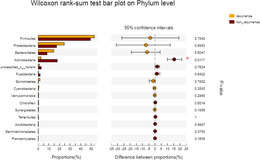
www.nature.com/scientificreports/
Figure 2. The Wilcoxon rank sum test was used to compare the difference in nasal microbial colony
composition between recurrent and non-recurrent nasal polyp patients at the general level. Note: The right side
of the figure shows the P value before FDR correction, *0.01 < P ≤ 0.05, **0.001 < P ≤ 0.01, ***P ≤ 0.001. After
correction by FDR, only Corynebacterium had a PDR P = 0.047, and Dolosigranulum had PDR P = 0.012; the
difference was statistically significant.
Recurrent Non-recurrent Statistics P value
Amount (proportion) 12(15.6%) 65(84.4%)
Male/female 5/7 29/36 x2 = 0.036 0.85a
Age 48.62 ± 6.33 49.73 ± 9.16 F = 0.605 0.437b
2
Asthma (amount, %) 3(14) 8(6) x = 1.768 0.184a
2
AR (amount, %) 4(18) 10(17) x = 2.194 0.139a
2
Eczema (amount, %) 0(8) 6(8) x = 1.201 0.273a
Bilateral CT score (Lund-Mackay) 22.50(14.00–24.00) 10.50(5.25–19.75) Z = 3.435 0.001c
VAS (TNSS) 42.3(30.2–67.1) 37.76(29.38–50.3) Z = − 4.93 0.000c
Table 1. Comparison of clinical characteristics of CRSwNP patients in the recurrent and non-recurrent
groups. a Chi-square test, bsingle factor analysis of variance, cMann-Whitney U test, P < 0.05.
Discussion
There are some differences in ESS postoperative nasal polyp recurrence rates among many countries around
the world. For example, the DeConde team from the United States found that the recurrence rates of CRSwNP
patients undergoing ESS surgery were 35%, 38%, and 40% at 6, 12, and 18 months, r espectively8. Wynn’s team,
also from the United States, found that up to 40% of patients with nasal polyps had recurrence 40 months after
ESS9. The Gevaert team from Belgium followed up a European nasal polyp population after ESS for 12 years and
found that 80% of the patients had recurrent nasal polyps, and 37% of the patients had to undergo ESS surgery
again10. In Asian countries, the team of Professor Zhang from Beijing Tongren Hospital found that nasal polyps
recurred in up to 55.3% of CRSwNP patients 34 months after surgery, and the recurrence rate of CRSwNP in
Southwest China was slightly lower than that in northeast China, presenting an obvious regional difference11.
The Nakayama team from Japan conducted a 17.5-month follow-up of CRSwNP patients and found that 22.9%
of patients with nasal polyps had r ecurrence12. The study in Southwest China in the West China Hospital of
Scientific Reports | (2021) 11:6364 | https://doi.org/10.1038/s41598-021-85292-5 4
Vol:.(1234567890)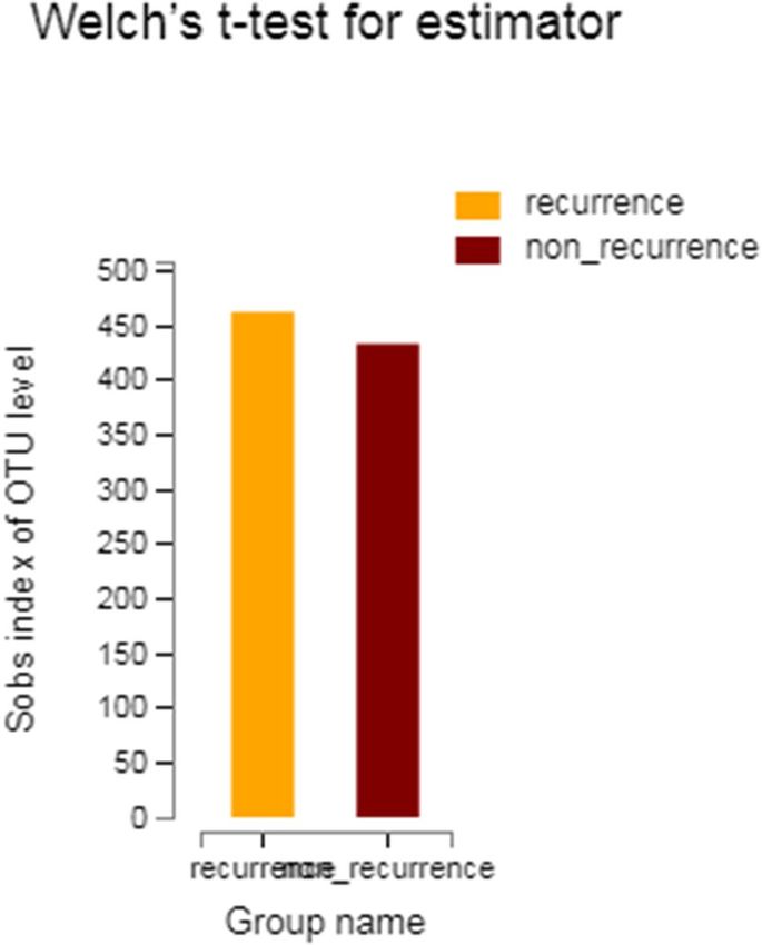
www.nature.com/scientificreports/
Non-recurrent Recurrent Statistic P value
Peripheral blood eosinophil count (× 109/L) 0.31(0.05–0.25) 0.40(0.22–0.60) Z = − 1.837 0.066
Peripheral blood neutrophil count (× 109/L) 3.82(2.99–5.37) 3.59(2.79–4.05) Z = − 2.226 0.026a
Eosinophil count in polyp tissue (/HP) 40.83(22.33–102.0) 13.72(13.5–48.33) Z = − 6.997 0.000a
Neutrophils count in polyp tissue (/HP) 30.83(20.33–56.44) 18.5(12.0–26.08) Z = − 8.243 0.000a
IL-5
Negative 10 50
x2 = 0.242 0.623
Positive 2 15
IL-8 (pg/ml)
Negative 3152.25 544.95
Z = − 17.885 0.000a
Positive (1627.0–5144.94) (520.72–1889.53)
IFN-γ (pg/ml)
Negative
61.75(30.11–90.47) 26.97(23.83–41.14) Z = − 3.424 0.001a
Positive
IFN-γ
Negative 2 10
x2 = 0.013 0.91
Positive 10 55
IL-17A (pg/ml) 15.27(6.77–20.2) 6.77(1.26–11.93) Z = − 5.27 0.000a
IL-17A
Negative 3 16
x2 = 0.001 0.977
Positive 9 49
IL-17E (pg/ml)
Negative 223.38 56.62
Z = − 21.04 0.000a
Positive (167.87–872.74) (56.62–219.29)
IL-17E
Negative 4 15
x2 = 0.573 0.449
Positive 8 50
IL-18 (pg/ml) 30.83(20.33–56.44) 19.07(12.0–26.08) Z = − 8.243 0.000a
IL-18
Negative 2 2
x2 = 3.799 0.051
Positive 10 63
IL-27 (pg/ml)
Negative 138.46 98.99
Z = − 0.941 0.347
Positive (86.11–248.12) (60.86–178.94)
IL-27
Negative 4 20
x2 = 0.031 0.86
Positive 8 45
Table 2. Comparison of inflammatory mediators between recurrent and non-recurrent CRSwNP patients.
a
P < 0.05.
Sichuan University of ESS surgery patients with CRSwNP with postoperative follow-up of 1 year found that the
nasal polyp recurrence rate was 15.6%; therefore, the postoperative nasal polyp recurrence rate of this region
is significantly lower than that of European and American countries and slightly closer to the Japanese team’s
findings, but as time goes on, the recurrence rate is likely to rise gradually, so we need to continue to track and
conduct a longer long-term follow-up.
In this study, beta diversity of microbial community distribution between CRSwNP patients and the control
subjects was calculated by PCoA. We found that most microbial distributions are overlapping and only a minority
of samples are specific to each group. At phylum and genus level, some microbial flora in non-CRS group were
higher than that in CRSwNP group, mainly concentrated in Actinobacteria, Corynebacterium and Dolosigranu-
lum. After 1-year follow-up, nasal polyp recurrence occurred in 12 patients, including 5 males and 7 females;
3 were asthma patients, 4 patients had allergic rhinitis, and 0 had eczema; there were 8 patients with asthma,
10 patients with allergic rhinitis, and 6 cases with eczema in the CRSwNP non-recurrent group. There was no
significant differences between the groups in proportion for age, gender, or allergic diseases such as asthma or
allergic rhinitis. In a 40-month follow-up, Wynn’s team found that CRSwNP patients with asthma were more
prone to nasal polyp recurrence after ESS9. Therefore, a longer postoperative follow-up is needed to observe
whether the accompanying allergic disease will increase the tendency of nasal polyp recurrence.
In terms of the Lund-Mackay scores and TNSS, the recurrent group was significantly higher than the non-
recurrent group, suggesting that patients with severe nasal symptoms or higher Lund-Mackay scores before
surgery may have a higher degree of disease severity. After the same ESS treatment, these patients may have a
higher risk of nasal polyp recurrence than patients with higher Lund-Mackay scores or lower TNSSs, identical
with some previous s tudies13–15. Based on the symptom scores of nasal inflammatory diseases and the distribu-
tion characteristics of nasal microorganisms, Copeland found that E. coli was positively correlated with the nasal
symptom scores of CRS patients16. Enterobacter pylori is involved in IL-8-mediated neutrophilic inflammation
and has a certain correlation in the pathogenesis of nasal polyps. In our study, Faecalibaculum was the only
Scientific Reports | (2021) 11:6364 | https://doi.org/10.1038/s41598-021-85292-5 5
Vol.:(0123456789)www.nature.com/scientificreports/
Figure 3. Differences in the Sobs index at the OTU level between the postoperative recurrent group and the
non-recurrent group.
bacterium that was negatively correlated with the overall nasal symptoms of CRSwNP patients. It is an obligate
anaerobic bacterium that has strong lactic acid fermentation ability and might become a new probiotic to replace
Lactobacillus and Bifidobacteria. The two genera have opposite functions and may have antagonistic effects in the
nasal cavity. Due to the influence of the sample quantity and the proportion of grouping, the current study haven’t
found that E. coli and Faecalibaculum differed between the recurrent group and the non-recurrent group, and
because of this phenomenon, we can consider expanding the sample size of patients with CRSwNP and having
a longer follow-up period, as far as possible further observation, and summarize surgical prognosis if possible
correlation with the microflora.
From the relationship between inflammatory cells and nasal polyp recurrence, the degree of eosinophil
infiltration in the recurrent group was higher than that in the non-recurrent group, and there was no difference
in eosinophil count in peripheral blood between the two groups. Prior study of nasal polyps in the organization
degree of eosinophil infiltration17 has no clear correlation with postoperative recurrence, and in recent years,
research has shown that CRSwNP patients with a higher degree of eosinophil infiltration were characterized by
more severe clinical symptoms and a higher recurrence rate, which is consistent with our research conclusion18,19.
At the same time, we compared the neutrophil count of patients with CRSwNP in peripheral blood and polyp
tissue and found that the neutrophil count of the recurrent group was significantly higher than that of the non-
recurrent group, which means that the neutrophil-mediated inflammation pattern plays an important role in
patients with CRSwNP in Southwest China and is correlated to postoperative recurrence of nasal polyps to
some extent.
For inflammatory mediators, the median expression levels of IFN-γ, IL-8, IL-17A, IL-17E and IL-18 were
significantly higher in the recurrent group than in the non-recurrent group, while the positive rates of all inflam-
matory factors, including IL-5 and IL-27, were not significantly different between the recurrent group and the
non-recurrent group. IL-8 is known to be a major cytokine produced by activated neutrophils and is positively
correlated with the severity of respiratory disease. Derycke et al.20 proved that IL-17A can effectively prolong
the survival rate of neutrophils in nasal polyp tissue and reduce its apoptosis rate. IFN-γ and IL-18 are activated
products and stimulating factors of macrophages, which are closely correlated with anti-inflammatory activi-
ties. Therefore, the expression levels of the above cytokines were higher in the recurrent group than in the non-
recurrent group, indicating that in this study, the neutrophils and phagocytes in the recurrent group showed a
higher degree of activation, mediated more severe respiratory inflammation, and presented an increased risk of
postoperative nasal polyp recurrence.
A small percentage of the ESS microbiota remained stable for a long time among the CRSwNP patients. Their
stability is mainly reflected in the limited species of dominant bacteria and the large number of low-abundance
bacteria. In general, they vary greatly in relative abundance, which constitutes the concept of an individual-
specific core sinus microbiome. Although its relative abundance fluctuates among different populations and
Scientific Reports | (2021) 11:6364 | https://doi.org/10.1038/s41598-021-85292-5 6
Vol:.(1234567890)www.nature.com/scientificreports/
Taxonomic level Non-recurrent (%) Recurrent (%) P value FDR P value
Phylum level
Firmicutes 49.19 ± 33.24 52.99 ± 39.04 0.784 0.961
Actinobacteria 18.0 ± 22.15 2.626 ± 3.099 0.011 0.336
Proteobacteria 17.33 ± 22.45 24.58 ± 36.44 0.888 0.987
Bacteroidetes 8.311 ± 13.59 14.65 ± 20.97 0.504 0.901
Fusobacteria 1.79 ± 7.677 0.279 ± 0.587 0.642 0.961
Cyanobacteria 0.263 ± 0.669 0.647 ± 1.123 0.258 0.901
Synergistetes 0.183 ± 0.585 0.346 ± 0.657 0.185 0.863
Tenericutes 0.392 ± 2.522 0.065 ± 0.089 0.873 0.994
Spirochaetae 0.178 ± 0.813 1.599 ± 5.493 0.788 0.916
Verrucomicrobia 0.281 ± 0.974 0.453 ± 0.907 0.297 0.901
Genera level
Corynebacterium 11.9 ± 20.31 0.154 ± 0.203 0.012 0.638
Lactobacillus 17.21 ± 30.05 22.47 ± 39.41 0.989 0.999
Staphylococcus 8.167 ± 27.7 8.987 ± 15.89 0.017 0.638
Streptococcus 5.915 ± 17.67 9.836 ± 20.99 0.478 0.882
Dolosigranulum 0.598 ± 2.506 1.166 ± 3.411 0.509 0.881
Escherichia-Shigella 3.715 ± 12.6 0.548 ± 1.743 0.707 0.892
Propionibacterium 2.235 ± 4. 646 0.213 ± 0.306 0.275 0.881
Erysipelotrichaceae 0.833 ± 2.628 0.038 ± 0.077 0.114 0.882
Moraxella 0.08 ± 0.459 12.96 ± 30.88 0.555 0.882
Ralstonia 1.522 ± 5.119 0.366 ± 0.48 0.983 0.999
Pseudomonas 1.552 ± 10.86 0.067 ± 0.134 0.125 0.881
Hemophilus 2.509 ± 12.56 0.155 ± 0.355 0.78 0.918
Faecalibacterium 0.589 ± 1.775 0.87 ± 1.708 0.55 0.881
Enterobacter 2.675 ± 10.43 0.327 ± 0.842 0.604 0.882
Peptostreptococcus 0.34 ± 1.679 0.409 ± 1.319 0.954 0.992
Candidatus Arthromitus 0.956 ± 7.197 1.13 ± 2.467 0.299 0.881
fusobacterium 1.197 ± 5.897 0.274 ± 0.589 0.751 0.914
Porphyromonas 1.592 ± 6.97 0.47 ± 1.604 0.869 0.961
Rhodococcus 1.501 ± 3.486 0.658 ± 1.205 0.921 0.981
Prevotella 0.474 ± 2.409 4.008 ± 13.69 0.496 0.881
Bacteroides 1.906 ± 4.836 4.016 ± 7.451 0.538 0.882
Neisseria 0.209 ± 1.191 7.416 ± 25.63 0.721 0.907
Table 3. Dominant groups and their average relative abundance in the recurrent and non-recurrent groups.
individuals, with the development of precision medicine, this concept has greatly enriched the hypothesis of the
role of bacteria in the pathological mechanism of CRS.
Some researchers found that the nasal bacterial community showed unstable changes after ESS, and the
nasal bacterial abundance increased after ESS. The average relative abundance and distribution of many taxa
varied, among which Staphylococcus was the only dominant taxon whose relative abundance increased signifi-
cantly. Some experts found that the relative abundance of a rare strain (Finegoldia magna) decreased after ESS,
the number of patients with higher nasal symptom scores increased, and the number of patients with reduced
overall bacterial load decreased. They argues that even with changes in nasal microbial abundance and colony
composition after surgery, these data do not support the notion that the prognosis of patients after ESS may be
directly attributable to changes in microbial distribution c omposition21. Based on this view, we believe that the
diversity of microbial colonies is different in the different follow-up stages, and relatively stable colony structure
may also be found in a particular period of time. We monitoring the next follow-up to verify the above idea.
From the perspective of nasal microbiome composition, we found that for both the recurrent and non-
recurrent CRSwNP patients in our study, the phyla with higher abundance were all concentrated in the follow-
ing groups: Firmicutes, Proteobacteria, Actinomycetes and Bacteroidetes. The dominant bacteria gerena mainly
included Lactobacillus, Staphylococcus, Streptococcus and Bacteroides, which was consistent with the previous
research results of our t eam22, indicating that the nasal microbial colony composition of CRSwNP patients in
a certain region was relatively stable. However, in terms of the nasal microbial richness of the two groups, we
found that there was no statistically significant difference in the Sobs index between the recurrent group and
the non-recurrent group, indicating that there was no significant difference in the nasal microbiome richness
between the two groups before surgery.
Scientific Reports | (2021) 11:6364 | https://doi.org/10.1038/s41598-021-85292-5 7
Vol.:(0123456789)www.nature.com/scientificreports/
Figure 4. The Wilcoxon rank sum test was used to compare the difference in nasal microbial colony
composition between the recurrent and non-recurrent nasal polyp patients at the phylum level.
Figure 5. The Wilcoxon rank sum test was used to compare the difference in nasal microbial colony
composition between recurrent and non-recurrent nasal polyp patients at the general level.
In this study, we compared the nasal microbial community structure differences between the CRSwNP
Scientific Reports | (2021) 11:6364 | https://doi.org/10.1038/s41598-021-85292-5 8
Vol:.(1234567890)www.nature.com/scientificreports/
population in the recurrent group and the non-recurrent group at both the phylum level and the genera level,
and the results showed that there were no statistically significant differences among all the microbial communi-
ties. Although the relative abundance of Actinomycetes and Corynebacterium in the non-recurrent group was
significantly higher than that in the recurrent group, the difference was not statistically significant; the relative
abundance of Staphylococci in the non-recurrent group was significantly lower than that in the recurrent group,
and the difference was also not statistically significant, which was consistent with the research results of Jain’s
team18. Corynebacterium in Actinomycetes phylum is a kind of bacteria that has a protective function in the upper
respiratory tract. Hoggard et al.23 observed that the bacterial distribution in patients with CRS was different from
that in healthy people, mainly manifested as the reduced content of Corynebacterium, the dominant strain, in
CRS patients compared with healthy adults. A breu3 found that Corynebacterium was relatively abundant in the
nasal cavity of CRS patients, and the severity of nasal symptoms of these patients (scored using the SNOT 20
scale) was positively correlated with the abundance of Corynebacterium. Staphylococcus aureus is considered the
pathogenic bacterium of sinusitis from the perspective of in vitro culture and high-throughput sequencing, and
it mainly induces chronic inflammation of the nasal and sinus mucosa through bacterial surface a ntigens24,25.
Uehara26 found a relationship between survival competition and inhibition between intranasal S. aureus and
Corynebacterium. The incidence of S. aureus colonization in Corynebacter nasal carriers (8.5%) was significantly
lower than that in noncarriers (44.5%). In addition, when the Corynebacterium strain was artificially implanted
into the nasal cavity of a S. aureus carrier, approximately 71% of the subjects had S. aureus removed from the nose.
In our study, we observed a decrease in Corynebacterium and an increase in Staphylococci in the recurrent
group of CRSwNP patients, which means that in the recurrent group, the abundance of bacteria with a protec-
tive function is relatively lower, while the abundance of S. aureus with pathogenic effects is relatively higher,
which is an important factor in the nasal microbiome composition in the recurrence of nasal polyps. We will
further analyze the differences in nasal microbial composition between the two groups by increasing the sample
size and extending the follow-up time, hoping to obtain more information about the correlation between nasal
microbiome diversity and CRSwNP recurrence.
We found that postoperative recurrence of nasal polyps was not associated with age, gender, asthma, allergic
rhinitis or other allergic diseases in CRSwNP patients. From the TNSSs, patients with more severe nasal symp-
toms before surgery may have a higher risk of nasal polyp recurrence. The neutrophil-mediated inflammatory
response plays an important role in patients with CRSwNP in Southwest China and may be associated with
nasal polyp recurrence. In the recurrent group, the abundance of protective bacteria was relatively lower, while
the abundance of pathogenic S. aureus was relatively higher, which is an important factor in nasal microbiome
composition for nasal polyp recurrence.
Materials and methods
Subjects. A total of 77 CRSwNP patients who underwent endoscopic sinus surgery (ESS) by the author (Liu
F.) between December 2017 and December 2018 were recruited into the study at the Department of Otorhino-
laryngology of the West China Hospital, Sichuan University, Chengdu, Sichuan. The surgery mainly involves
removal of polyps from the nasal cavity, removal of inflamed or dysfunctional mucosa, conservation of normal
mucosa and sinus open. All patients were advised to regularly use intranasal hormones and saline nasal irriga-
tion after surgery. Follow-up was performed at least 1 month, 3 months, 6 months and 1 year after surgery.
The diagnosis of CRSwNP or CRSsNP was based on patient history, clinical examination, nasal endoscopy
and computed tomography results according to the current EPOS g uidelines27. During the follow-up after ESS
and medication, if objective CT indicated abnormal density shadow in nasal meatus or sinuses, and endoscopy
showed mucosal thickening, mucinous purulent discharge, mucosal edema, or polypoid changes, recurrence was
considered. Patients who had received antibiotics and systemic or topical intranasal steroid treatments within
4 weeks before the operation were excluded. This time frame was based on a previous study showing that the gut
microbiome returned to a steady level within 4 weeks after the cessation of antibiotic u se28. Moreover, patients
with cystic fibrosis, an immunocompromised status or autoimmune disease, immunodeficiency, pregnancy or
a primary mucociliary impairment and those less than 18 years old were excluded from the study.
Patients who underwent ESS surgery and had no sinus inflammation diseases, including nasal septum devia-
tion, inverted papilloma, pituitary adenomas, chronic dacryocystitis, or optical canal fractures, were recruited
as the control group.
Clinical data of CRSwNP patients were collected, including age, gender, smoking history, alcohol consump-
tion history, body mass index (BMI), allergic disease history, total clinical symptom score (TNSS), Lund-Mackay
score, and Davos score.
The study was approved by the ethics committee of West China Hospital, Sichuan University. All patients
gave their written informed consent before participation.
Sample collection. Following the induction of general anaesthesia, sampling was immediately performed
prior to the removal of nasal secretions and the application of topical mucosal vasoconstrictors. Sterile swabs
(Gongdong Medical Technology, Taizhou, Zhejiang, CN) were endoscopically guided to the middle meatus
region, rotated for 5 full turns and kept in place for at least one minute until fully saturated. Care was taken to
avoid contamination from the anterior nasal cavity during swabbing. Any swabs that were contaminated through
contact with a nontarget region were discarded. The collected samples were placed in 2-ml sterile Corning freez-
ing tubes without enzyme on ice temporarily, transported to the laboratory within 2 h and stored at − 80 °C in
liquid nitrogen until DNA extraction was performed22,29.
Nasal polyp tissues were collected, stored in two parts: one was fixed with paraformaldehyde for HE and
immunohistochemical staining, and the other was frozen at − 80 °C and homogenized to detect the expression
Scientific Reports | (2021) 11:6364 | https://doi.org/10.1038/s41598-021-85292-5 9
Vol.:(0123456789)www.nature.com/scientificreports/
of IL-5, IL-8, IL-17A, IL-17E, IL-18, IL-27 and IFN-γ. Blood samples were obtained to count inflammatory cells
by flow cytometer.
During the 1-year postoperative follow-up, symptoms were recorded and nasal endoscopy and sinus CT
examination were performed to determine whether nasal polyps had recurred, and middle nasal meatus secre-
tions were collected at the same time.
Measurement of inflammatory factors. Samples for inflammatory factor assessment using immuno-
assays were weighed. A total of 1.0 ml of 0.9% NaCl was added per 0.1 g of each sample, and all samples were
homogenized at 1000 rpm for 5 min and centrifuged at 1500 g for 10 min at 4 °C. Supernatants were collected
and stored at − 20 °C until analysis. IL-5, IL-8, IL-17A, IL-17E, IL-18, IL-27 and IFN-γ concentrations were
assessed using a Luminex 100 system (Luminex, Austin, TX, USA).
Immunohistochemical staining. Streptomyces antibiotinin protein-peroxidase conjugating method (SP
method) was performed by eosinophilic cationic protein (ECP) and myeloperoxidase (MPO). The brownish
cells in the cytoplasm of various inflammatory cells are positive cells. 10 fields of 400-fold light microscopy were
randomly selected in each sample. A manual counter was used to count the number of positive cells, and then
the average value of the 10 counts was used to count the positive inflammatory cells in the samples. The two
researchers made the decision double-blind.
Microbial genomic DNA extraction. Microbial DNA was extracted from the swab samples using the
DNA Kit (Omega Bio-tek, Norcross, GA, USA) according to the manufacturer’s protocols. The V3-V4 region of
the bacteria’s 16S ribosomal RNA gene were amplified by PCR (95 °C for 3 min, followed by 27 cycles at 95 °C for
30 s, 55 °C for 30 s, 72 °C for 45 s and a final extension at 72 °C for 10 min) using the primers 338F 5′-barcode-
ACTCCTAGGGAGGCAGCAG)-3′ and 806R 5′-GGACTACHVGGGTWTCTAAT-3′, where the barcode is an
eight-base sequence unique to each sample. PCR reactions were performed in a triplicate 20 μL mixture contain-
ing 4 μL of 5 × FastPfu Buffer, 2 μL of 2.5 mM dNTPs, 0.8 μL of each primer (5 μM), 0.4 μL of FastPfu Polymerase,
and 10 ng of template DNA22,29.
Illumina MiSeq sequencing. Illumina MiSeq is a next-generation sequencing (NGS) method in which
high-quality 16S rRNA sequences are generated for the subject. Amplicons were extracted from 2% agarose
gels, purified using the AxyPrep DNA Gel Extraction Kit (Axygen Biosciences, Union City, CA, USA) according
to the manufacturer’s instructions and quantified using QuantiFluor-ST (Promega, USA). The purified ampli-
cons were pooled in equimolar amounts and paired-end sequenced (2 × 250) on the Illumina MiSeq platform
according to standard protocols. V3–V4 variable regions of the bacterial 16S rRNA gene were generated for the
amplicons22,29.
Statistical analysis. Demographic and clinical characteristics were analysed using the chi-square test for
binominal variables and the Mann–Whitney U test for continuous variables, such as Lund-Mackay score and
visual analogue scale (VAS) score. We used Welch’s t-test to analyse the microbiota richness index between
groups. We used the UPARSE calculation in the clustering process, which excluded single-read OTUs automati-
cally. Alpha diversity indices were calculated for all samples at 97% OTU similarity using mothur index analysis.
Alpha diversity indices include the Sobs, Shannon, and Simpson indices. The Sobs index was calculated to reflect
community richness, and the Shannon and Simpson indices were calculated to determine community diversity.
Principal coordinate analysis (PCoA) was used to explore and visualize data similarities in three-dimensional
space. Distance matrices were calculated using the Morisita-Horn index (vegdist function of the R package
vegan) and applied to the cmdscale function in R. The differences in the relative abundance between the two
groups were evaluated using the Wilcoxon rank sum test with the false discovery rate (FDR) multiple testing
correction. When P < 0.05 (2-sided), we considered the difference significant.
All methods were carried out in accordance with relevant guidelines and regulations.
Ethics approval and consent to participate. The study was approved by the ethics committee of West
China Hospital, Sichuan University (NO. 2017-448). All patients gave their written informed consent before
participation.
Consent to publish. All authors consent to publish this manuscript to Scientific Reports.
Data availability
The datasets generated and analysed during the current study are available in the National Center for Biotechnol-
ogy Information (NCBI) Sequence Read Archive (SRA) database(Accession Number: SRP162948, ftp://ftp.ncbi.
nlm.nih.gov/sra/sra-instant/reads/ByStudy/sra/SRP/SRP162/SRP162948/).
Received: 30 November 2020; Accepted: 26 February 2021
References
1. Bhattacharyya, N. Incremental health care utilization and expenditures for chronic rhinosinusitis in the United States. Ann. Otol.
Rhinol. Laryngol. 120, 423–427 (2011).
Scientific Reports | (2021) 11:6364 | https://doi.org/10.1038/s41598-021-85292-5 10
Vol:.(1234567890)www.nature.com/scientificreports/
2. Dalziel, K. et al. Endoscopic sinus surgery for the excision of nasal polyps: a systematic review of safety and effectiveness. Am. J.
Rhinol. 20, 506–519 (2006).
3. Abreu, N. A. et al. Sinus microbiome diversity depletion and Corynebacterium tuberculostearicum enrichment mediates rhinosi-
nusitis. Sci. Transl. Med. 4, 151ra124 (2012).
4. Biswas, K. et al. The nasal microbiota in health and disease: variation within and between subjects. Front. Microbiol. 9, 134 (2015).
5. Kim, R. J. et al. Paired analysis of the microbiota of surface mucus and whole-tissue specimens in patients with chronic rhinosi-
nusitis. Int. Forum Allergy Rhinol. 5, 877–883 (2015).
6. Ba, L. et al. The association between bacterial colonization and inflammatory pattern in Chinese chronic rhinosinusitis patients
with nasal polyps. Allergy 66, 1296–1303 (2011).
7. Wei, B. et al. Multivariate analysis of inflammatory endotypes in recurrent nasal polyposis in a Chinese population. Rhinology 56,
216–226 (2018).
8. DeConde, A. S. et al. Prevalence of polyp recurrence after endoscopic sinus surgery for chronic rhinosinusitis with nasal polyposis.
Laryngoscope 127, 550–555 (2017).
9. Wynn, R. & Har-El, G. Recurrence rates after endoscopic sinus surgery for massive sinus polyposis. Laryngoscope 114, 811–813
(2004).
10. Gevaert, P. et al. Allergic sensitization, high local IL-5 and IgE predict surgical outcome 12 years after endoscopic sinus surgery
for chronic rhinosinusitis with nasal polyposis. J. Allergy Clin. Immun. 135, Ab238–Ab238 (2015).
11. Lou, H. F. et al. Predictive significance of tissue eosinophilia for nasal polyp recurrence in the Chinese population. Am. J. Rhinol.
Allergy 29, 350–356 (2015).
12. Nakayama, T. et al. Prognostic factors for recurrence after endoscopic sinus surgery for chronic rhinosinusitis with nasal polyps.
Auris Nasus Larynx 43, 641–647 (2016).
13. Tao, X. et al. Prediction models for postoperative uncontrolled chronic rhinosinusitis in daily practice. Laryngoscope 128(12),
2673–2680 (2018).
14. Grgić, M. V., Ćupić, H., Kalogjera, L. & Baudoin, T. Surgical treatment for nasal polyposis: predictors of outcome. Eur. Arch. Oto-
Rhino-Laryngol. 272(12), 3735–3743 (2015).
15. Smith, T. L. et al. Predictive factors and outcomes in endoscopic sinus surgery for chronic rhinosinusitis. Laryngoscope 115(12),
2199–2205 (2005).
16. Copeland, E. et al. Chronic rhinosinusitis: potential role of microbial dysbiosis and recommendations for sampling sites. Front.
Cell. Infect. Microbiol. 8, 57 (2018).
17. Eweiss, A. et al. VCAM-1 and eosinophilia in diffuse sino-nasal polyps. Eur. Arch. Oto-Rhino-Laryngol. 266, 377–383 (2009).
18. Kountakis, S. E. Relationship between clinical measures and histopathologic findings in chronic rhinosinusitis. Otolaryngol. Head
Neck 142, 920–921 (2010).
19. Tokunaga, T. et al. Novel scoring system and algorithm for classifying chronic rhinosinusitis: the JESREC study. Allergy 70,
995–1003 (2015).
20. Derycke, L. et al. IL-17A as a regulator of neutrophil survival in nasal polyp disease of patients with and without cystic fibrosis. J.
Cyst. Fibros 11, 193–200 (2012).
21. Jain, R. et al. Changes in the bacterial microbiome of patients with chronic rhinosinusitis after endoscopic sinus surgery. Int. Forum
Allergy Rhinol. 7, 7–15 (2017).
22. Gan, W. et al. The difference in nasal bacterial microbiome diversity between chronic rhinosinusitis patients with polyps and a
control population. Int. Forum Allergy Rhinol. 9, 582–592 (2019).
23. Hoggard, M. et al. Evidence of microbiota dysbiosis in chronic rhinosinusitis. Int. Forum Allergy Rhinol. 7, 230–239 (2017).
24. Corriveau, M. N. et al. Detection of Staphylococcus aureus in nasal tissue with peptide nucleic acid-fluorescence in situ hybridiza-
tion. Am. J. Rhinol. Allergy 23, 461–465 (2009).
25. Sachse, F. et al. Staphylococcus aureus invades the epithelium in nasal polyposis and induces IL-6 in nasal epithelial cells in vitro.
Allergy 65, 1430–1437 (2010).
26. Uehara, Y. et al. Bacterial interference among nasal inhabitants: eradication of Staphylococcus aureus from nasal cavities by artificial
implantation of Corynebacterium sp. J. Hosp. Infect. 44, 127–133 (2000).
27. Fokkens, W. J. et al. European position paper on rhinosinusitis and nasal polyps 2012. Rhinol. Suppl. 23(3), 1–298 (2012).
28. Antonopoulos, D. A. et al. Reproducible community dynamics of the gastrointestinal microbiota following antibiotic perturbation.
Infect. Immun. 77, 2367–2375 (2009).
29. Gan, W. et al. The influence of nasal bacterial microbiome diversity on the pathogenesis and prognosis of chronic rhinosinusitis
patients with polyps. Eur. Arch. Oto-Rhino-Laryngol.: Off. J. Eur. Fed. Oto-Rhino-Laryngol. Soc. 9, 1–14 (2020).
Acknowledgements
The data presented herein were analyzed in part using free online cloud platform Majorbio I-Sanger (www.i-
sanger.com). National Nature Science Foundation of China (81800892).
Author contributions
W.G. and F.Y. were responsible for the data and sample collection and paper writing; H.Z. was responsible for
sample detection; WG was responsible for data analysis; S.L., F.L. and J.M. were responsible for the whole study
design and paper revision. All authors have read and approved the manuscript.
Funding
This work was supported by Grants to Juan Meng from the National Science Fund of China (81300815), the 12th
5-Year Science and Technology Support Program (2014BAI07B04) and the international cooperation Project of
Sichuan Provincial Science and Technology Department (2018HH0097), by Grants to Shixi Liu from the National
Science Fund (81570310000), and by Grants to Weigang Gan from the General Program of Sichuan Provincial
Department of Education (18ZB0213), the Youth Innovation Project of Sichuan Provincial Medical Association
(Q15025), the Open Fund of Key Laboratory of Deep Earth Science and Engineering (Sichuan University), Min-
istry of Education, Sichuan University, (DESEYU201901) and Post-Doctor Research Project (2020HXBH129),
West China Hospital, Sichuan University.
Competing interests
The authors declare no competing interests.
Scientific Reports | (2021) 11:6364 | https://doi.org/10.1038/s41598-021-85292-5 11
Vol.:(0123456789)www.nature.com/scientificreports/
Additional information
Correspondence and requests for materials should be addressed to J.M.
Reprints and permissions information is available at www.nature.com/reprints.
Publisher’s note Springer Nature remains neutral with regard to jurisdictional claims in published maps and
institutional affiliations.
Open Access This article is licensed under a Creative Commons Attribution 4.0 International
License, which permits use, sharing, adaptation, distribution and reproduction in any medium or
format, as long as you give appropriate credit to the original author(s) and the source, provide a link to the
Creative Commons licence, and indicate if changes were made. The images or other third party material in this
article are included in the article’s Creative Commons licence, unless indicated otherwise in a credit line to the
material. If material is not included in the article’s Creative Commons licence and your intended use is not
permitted by statutory regulation or exceeds the permitted use, you will need to obtain permission directly from
the copyright holder. To view a copy of this licence, visit http://creativecommons.org/licenses/by/4.0/.
© The Author(s) 2021
Scientific Reports | (2021) 11:6364 | https://doi.org/10.1038/s41598-021-85292-5 12
Vol:.(1234567890)You can also read