THE EFFECTS OF COMPOUND CENTELLA FORMULA ON OXINFLAMMATION DIET/STREPTOZOTOCIN INDUCED DIABETIC KIDNEY DISEASE RAT MODEL AND SILENT INFORMATION ...
←
→
Page content transcription
If your browser does not render page correctly, please read the page content below
EXPERIMENTAL AND THERAPEUTIC MEDICINE 22: 962, 2021

    The effects of compound centella formula on OxInflammation
            and silent information regulator 1 in a high‑fat
    diet/streptozotocin‑induced diabetic kidney disease rat model
                           QIN ZHU1, XIAO‑HONG LI1, HONG‑YU CHEN1 and QIN‑YANG JIN2

              1
               Department of Nephrology, Key Laboratory of Zhejiang Province, Management of Kidney Disease,
                      Hangzhou Hospital of Traditional Chinese Medicine, Hangzhou, Zhejiang 310007;
                   2
                     Department of Cardiology, Zhejiang Provincial People's Hospital, People's Hospital of
                             Hangzhou Medical College, Hangzhou, Zhejiang 310014, P.R. China

                                        Received March 7, 2020; Accepted April 14, 2021

                                                 DOI: 10.3892/etm.2021.10394

Abstract. The Chinese decoction compound centella                 35 mg/kg STZ on day 29. All rats were sacrificed on day 112.
formula (CCF) is clinically effective against diabetic kidney     High‑performance liquid chromatography was performed to
disease (DKD), but the exact mechanism remains unclear.           analyse asiaticoside, astragaloside and triptolide levels in CCF
The present study aimed to investigate the effects of CCF on      (0.3400, 0.0640 and 0.0001 mg/ml, respectively). Fasting blood
OxInflammation and silent information regulator 1 (SIRT1)         glucose, urine protein‑to‑creatinine ratio, serum creatinine
levels in rats with streptozotocin (STZ)‑induced diabetes.        and blood urea nitrogen were quantified. Periodic acid Schiff
Sprague‑Dawley rats were divided into CCF, losartan, diabetic     staining, H&E staining and transmission electron micros‑
control (DC) and normal control (NC) groups (n=7). Except         copy were used to examine kidney pathological changes.
for the NC, all subgroups of rats were fed a high‑fat diet for    The mRNA and protein expression levels of SIRT1 in renal
112 days and received a single intraperitoneal injection of       tissues were analysed by reverse transcription‑quantitative
                                                                  PCR, western blotting and immunohistochemistry. Oxidative
                                                                  stress was evaluated by measuring the levels of superoxide
                                                                  dismutase (SOD), malondialdehyde (MDA) and nicotinamide
                                                                  adenine dinucleotide phosphate oxidase 4 (NOX4) in renal
Correspondence to: Dr Qin‑Yang Jin, Department of Cardiology,     tissues. TNF‑α and NF‑κ B p65 subunit in renal tissues were
Zhejiang Provincial People's Hospital, People's Hospital of
                                                                  assessed for inflammation. Compared with the rats in the NC
Hangzhou Medical College, 158 Shangtang Road, Hangzhou,
                                                                  group, the rats in the DC group exhibited renal injury with
Zhejiang 310014, P.R. China
Email: jqy119@163.com                                             proteinuria, decreased expression levels of SIRT1 and SOD
                                                                  (P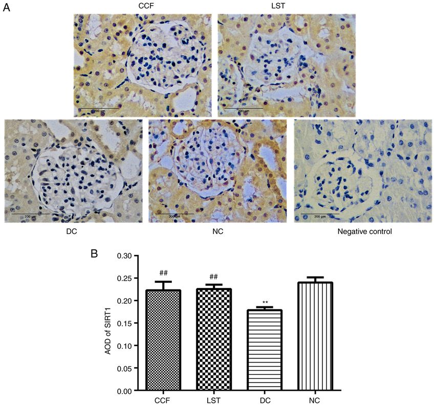
2                     ZHU et al: THE EFFECTS OF COMPOUND CENTELLA FORMULA IN A DIABETIC RAT MODEL

oxidative stress and cell apoptosis (3,4). An increasing body         by the Zhejiang Chinese Medical University Animal Ethics
of research has confirmed that oxidative stress and inflamma‑         Committee. The rats were reared in a standard experimental
tion play crucial roles in DKD (5,6). Inflammation can lead           animal laboratory with free access to food and water in a specific
to excessive reactive oxygen species (ROS), which can cause           pathogen‑free laboratory environment under the following
lipid peroxidation reactions and decreased cellular antioxidant       conditions: Temperature, 20‑25˚C; humidity, 50‑65% and a
capacity (7). In contrast, ROS and ROS‑induced DNA damage             12 h light/dark cycle. A total of 7 rats were fed ordinary feed to
can promote inflammatory responses and fibrotic processes (8).        represent the normal control (NC). Considering the mortality
The term ‘OxInflammation’ refers to a prepathological condi‑          rate of the pathological models, 23 rats were fed high‑fat feed
tion; it has been well‑documented that mild‑subclinical chronic       for 112 days (72.5% ordinary feed formula plus 10% lard,
inflammation is related to chronic and systemic oxidative             10% sucrose, 2.0% cholesterol, 0.5% cholic acid and 5% yolk
stress with in a vicious circle (9). Silent information regulator 1   powder). The feed was purchased from Beijing Boaigang
(SIRT1) is a nicotinamide adenosine dinucleotide‑dependent            Biotechnology Co., Ltd. On the 29th day of the experiment,
protein deacetylase with remarkable abilities to prevent              the same 23 rats all received a single intraperitoneal injection
diseases and even reverse aspects of ageing. It can regulate a        of STZ (MilliporeSigma Canada Co.) at a dose of 35 mg/kg;
variety of biological processes, including inflammatory and           two of the 23 rats gradually lost weight after injection of STZ
metabolic disorders (10,11). Renoprotective effects of SIRT1          and appeared to show slower activity, as well as eating less
have been found in experimental models of renal disorders,            food and grooming less. For ethical reasons, these two rats
including DKD (12,13).                                                were eventually sacrificed. It was speculated that the abnormal
    Although many interventions have been found to be                 response of the two rats was due to a faulty intraperitoneal
effective against DKD, including lifestyle adjustment,                injection procedure. The remaining 21 rats were randomly
glycaemic control, blood pressure control, renin‑angiotensin          divided into the diabetic control (DC) group (n=7), CCF group
system blockers, sodium‑dependent glucose transporter‑2               (n=7) and losartan (LST) group (n=7). As a classic drug for
inhibitors and glucagon‑like peptide‑1 agonists, few have been        the treatment of diabetic nephropathy, LST was used as the
established as optimal options (2). The discovery of new inter‑       positive control drug for centella formula in this experiment.
ventions, which overcome these limitations (such as restricted        Plasma fasting blood glucose (FBG) was measured from the
use in chronic renal failure and non‑specific effect of reducing      tail vein, and FBG levels >16.7 mmol/l (72 h after injection)
albuminuria) to delay the evelopment of DKD is warranted.             were considered diabetic rats (14). Subsequently, the drug
At present, the use of medicinal plants and their natural             intervention was carried out from the 32nd to the 112th day.
components as future drugs for the treatment of diabetes              The rats in the CCF group were treated with a concentrated
and its complications has received considerable interest from         solution of CCF at 2 ml/day per rat. The dose of losartan potas‑
researchers worldwide. The Chinese decoction compound                 sium was 4.5 mg/kg/day per rat (diluted to 2 ml with normal
centella formula (CCF) was designed by a famous contem‑               saline) in the LST group (Merck Sharp & Dohme‑Hoddesdon).
porary doctor of traditional Chinese medicine, Professor              The rats in the NC and DC groups received the same volume
Yongjun Wang (14). The composition and daily dose for adults          of normal saline. The drug intervention was administered by
of this decoction are as follows: Centella asiatica (L.) Urb.         gavage. All rats were sacrificed by decapitation on the 112th
(JiXueCao), 30 g; Astragalus Membranaceus Fish. (HuangQi),            day of the experiment after anaesthesia. Firstly, the midline
30 g; Tripterygium wilfordii Hook. f. (LeiGongTeng), 15 g. In a       of the abdomens of rats were cut open following local skin
previous study, it was observed that CCF reduced the quantity         disinfection. The kidneys were then carefully dissociated,
of proteins found in the 24 h urinary protein test, improved          and the kidney blood vessels were clamped with hemostatic
renal function, inhibited mesangial cell proliferation and            forceps and ligated. The kidneys were then removed, and the
mesangial matrix accumulation, and prevented sclerosis of             abdomens were sutured.
the glomerulus in stage 3‑4 patients with DKD treated for 3
months. A small number of patients (2/43) had a drug‑related          Preparation of CCF. One daily dose of CCF for each rat
increase in alanine transaminase, which was within 3 times            included 0.8 g of Centella asiatica (L.) Urb. (JiXueCao), 0.8 g
the normal value and returned to normal levels after symp‑            of Astragalus membranaceus Fish. (HuangQi), and 0.4 g of
tomatic treatment (15). Although CCF has been indicated to be         Tripterygium wilfordii Hook. f. (LeiGongTeng). The rat CCF
an effective treatment for DKD in a clinical setting, the exact       dose was calculated based on the human dose according to
mechanism is still unclear.                                           the body surface area formula (16): A=k (W2/3)/10,000, where
    In the present study, the protective effects of CCF against       k=9.1 (A, Body surface area, m2; K, A constant, which varies
renal injuries in streptozotocin (STZ)‑induced diabetic rats          with animal species; W, weight, g). The herbs were mixed in
were examined, along with the possible mechanisms associ‑             water, decocted for 45 min and then concentrated. The final
ated with the inhibition of crosstalk between inflammatory            volume of the concentrated decoction was 2 ml for one rat per
responses and oxidant stress and the regulation of SIRT1.             day. The decoction was stored at 4˚C.

Materials and methods                                                 Chemical analysis of CCF extracts by high‑performance
                                                                      liquid chromatography. To prepare the asiaticoside reference
Animals and experimental design. A total of 30 male Sprague           solution, 6.75 mg asiaticoside reference (National Institute
Dawley rats (specific‑pathogen‑free grade; 4‑5 weeks old,             for the Control of Pharmaceutical and Biological Products)
weighing 100±10 g) were purchased from Shanghai SIPPR‑Bk              was precisely weighed and added to methanol to obtain a
Laboratory Animal Co., Ltd. The experiment was approved               0.27 mg/ml solution. To prepare the sample solution, 5 ml of
EXPERIMENTAL AND THERAPEUTIC MEDICINE 22: 962, 2021 3
this solution was added to a test tube and heated (100˚C) in a flask. After shaking well, the extraction solution was filtered
water bath for evaporation until dry. Subsequently, the solution with a 0.45 µm filter membrane. For content analysis, the
was dissolved in methanol and placed in an ultrasonic bath precise absorption of the triptolide reference solution (10 µl)
for 30 min. The final volume was 2 ml in a volumetric flask. and sample solution (10 µl) were analysed by liquid chromatog‑
After shaking well, the extraction solution was filtered with raphy (Varian ProStar 230 Solvent delivery module; Varian)
a 0.45 µm filter membrane. For content analysis, the precise with the following conditions: Column, YMC ODS C18 (YMC
absorption of the reference asiaticoside solution (10 µl) and Co., Ltd.) (inner diameter: 4.6 mm, length: 250 mm; particle
sample solution (10 µl) was analysed by injecting the samples diameter: 5 µm); mobile phase, acetonitrile:water (30:70);
into a liquid chromatograph (Varian ProStar 230 Solvent detection wavelength, 220 nm; the other details were the same
delivery module; Varian) with the following conditions: as those aforementioned.
Column, YMC ODS C18 (YMC Co., Ltd.) inner diameter:
4.6 mm, length: 250 mm; particle diameter: 5 µm); mobile Analysis of FBG, urine protein‑to‑creatinine ratio (UPCR),
phase, acetonitrile:water (30:70); atomization temperature, serum creatinine (Scr) and blood urea nitrogen (BUN). FBG
40˚C; gasification temperature, 90˚C; flow rate, 1.0 ml/min; was measured from the tail vein on the 1st, 32nd, 42nd, 56th,
nitrogen flow rate, 1.6 l/min. 84th and 112th days of the study using Accu‑Chek Performa
To prepare the astragaloside reference solution, 7.50 mg glucometers [Roche Diagnostics (Shanghai) Co., Ltd.].
astragaloside reference (National Institute for the Control Metabolic cages were used to collect urine. The UPCR was
of Pharmaceutical and Biological Products) was precisely determined by the pyrogallol red method (MilliporeSigma)
weighed, and methanol was added (0.30 mg/ml). To prepare using an AU5800 automatic biochemical analyser (Beckman
the sample solution, 30 ml sample solution was extracted Coulter, Inc.) on the 1st and 112th day. The rats were anaes‑
by adding 40 ml of 80% methanol as the solvent overnight; thetised prior to blood draws (~8 ml per rat). All the rats were
20 ml methanol solvent was added again. Subsequently, anaesthetised with intraperitoneal administration of 10%
the resultant mixture was heated under reflux for 4 h. The chloral hydrate (400 mg/kg) (17). Scr and BUN were measured
methanol extract was evaporated to dryness, and the desic‑ using an AU5800 automatic biochemical analyser (Beckman
cated residue was dissolved in water. The solution was then Coulter, Inc.) on the 112th day.
extracted four times with 40 ml of n‑butanol and washed in
40 ml ammonia solution twice. After discarding the ammonia Renal histology analysis by H&E staining and periodic
solution, n‑butanol was evaporated to dryness, and the residue acid‑Schiff (PAS) staining. One side of the rat kidney was
was dissolved in 5 ml of water. Then, the crude extracts were fixed with 10% neutral buffered formalin (at room temperature
filtered through a D101 macroporous adsorption resin column ≥24 h), dehydrated with gradient alcohol (at room tempera‑
(length, 12 cm; inner diameter. 1.5 cm). A total of 50 ml of ture, 75% x2, 30 min; 95% x3, 30 min; and 100% x2, 30 min),
water was used as the eluent and discarded later, followed by cleared with xylene, waxed, embedded and sectioned (3 µm).
ethanol elution. The solution was eluted with 40% ethanol Tissue sections were deparaffinized in xylene and stained with
(30 ml), and the eluate was discarded. The solution was then H&E for 20 min at room temperature. For PAS staining, histo‑
eluted again with 70% ethanol (80 ml). The eluent solution logical sections were deparaffinized, oxidized in 1% aqueous
was collected and evaporated to dryness. The residue was periodate solution for 15 min and washed three times in
dissolved in methanol and transferred to a 5 ml volumetric distilled water. Sections were then soaked in Schiff solution for
flask. After the addition of methanol to the flask (to 5 ml), the 15 min. Then, the sections were rinsed under running water for
sample was shaken. For content analysis, the precision absorp‑ 15 min. Subsequently, the nuclei were stained with hematox‑
tion of the astragaloside reference solution (10 µl) and sample ylin (room temperature for 10 min) followed by differentiation
solution (10 µl) was measured by liquid chromatography with ethanol‑hydrochloric acid. The areas of nuclei appeared
(Varian ProStar 230 Solvent delivery module; Varian) with blue, while the basal membrane of the glomerulus and renal
the following conditions: Column, YMC ODS C18 (YMC tubules, cytoplasm, mesangial matrix of the glomerulus and
Co., Ltd.)(inner diameter: 4.6 mm, length: 250 mm; particle collagen fibers were red under light microscopy (magnifica‑
diameter: 5 µm); mobile phase, acetonitrile:water (35:65); the tion, x200 or x400)(BX51; Olympus Corporation). The relative
other details were the same as those aforementioned. mesangial matrix index and the relative glomerular volume
To prepare the triptolide reference solution, 3.01 mg were calculated (18,19).
triptolide reference (content ≥98%; National Institute for
the Control of Pharmaceutical and Biological Products) was Ultrastructure of the kidney through transmission electron
precisely weighed, and methanol was added (0.03 mg/ml). microscopy. The rat renal cortical tissue samples (1x1x1 mm)
To prepare the sample solution, 5 ml of solution was added were collected and fixed with 2.5% glutaraldehyde (60 min)
to a test tube. Water was used to dilute the solution to 25 ml. and 1% osmic (120 min) acid at 4˚C After dehydration, the
The supernatant was collected after centrifugation (5,702 x g, tissues were embedded in epoxy resin. The ultrathin sections
room temperature) and heated in a water bath for evaporation (100 nm) were stained with uranium acetate‑lead citrate
until dry. Subsequently, methanol (2.5 ml) and methylene and then examined with a transmission electron microscope
chloride (2.5 ml) were added to the residue in neutral alumina (JEM1400; JEOL, Ltd.). The foot process fusion rate of the
(10 g; diameter, 1.5 cm; wet packing column). A mixture of podocyte in each specimen was calculated (20,21).
methanol and methylene chloride (1:3) was used for elution.
The eluent was collected at 100 ml and steamed. The residue Analysis of SOD and MDA activities in rat renal tissues.
was dissolved in methanol and transferred to a 5 ml volumetric SOD activity and MDA content were analysed with SOD test4 ZHU et al: THE EFFECTS OF COMPOUND CENTELLA FORMULA IN A DIABETIC RAT MODEL
Table I. Primer sequences used for the quantitative real‑time polymerase chain reaction.
Gene Forward primer (5'‑3') Reverse primer (5'‑3')
SIRT1 (141 bp) GCTCGCCTTGCTGTGGACTTC GTGACACAGAGATGGCTGGAACTG
GAPDH (252 bp) ACAGCAACAGGGTGGTGGAC TTTGAGGGTGCAGCGAACTT
SIRT1, silent information regulator 1.
Table II. Concentrations of representative components in CCF. antibody (anti‑rabbit; 1:10,000; cat. no. DW‑GAR007;
Jackson ImmunoResearch Laboratories, Inc.) for 60 min at
Standard substance Concentration in CCF (mg/ml) room temperature. An enhanced chemiluminescence reagent
was used for visualization (Applygen Technologies Inc.), and
Asiaticoside 0.3412 Image‑Pro Plus 6.0 software (Media Cybernetics, Inc.) was
Astragaloside 0.0635 employed to analyse the results.
Triptolide 0.0001
Immunohistochemistry assay for SIRT1 protein expression
CCF, compound centella formula.
level determination and localisation in kidney tissues. Blocks
of rat kidney tissues smaller than 0.5x 0.5x 0.1 cm were taken
for fixation with 10% neutral buffered formalin (room temper‑
ature for ≥24 h), dehydrated with an alcohol gradient (room
kits (WST‑1 method; cat.no. A001‑3‑1; Nanjing Jiancheng temperature, 75% x2, 30 min; 95% x3, 30 min; and 100% x2,
Bioengineering Institute) and MDA test kits (TBA method; 30 min), cleared with xylene, waxed, embedded and sectioned
cat.no. A003‑1‑1; Nanjing Jiancheng Bioengineering Institute) (3 µm). Then, the sections were treated with citric acid antigen
according to the manufacturer's instructions. repair buffer and washed with PBS three times. Sections
were blocked using 10% normal goat serum (cat. no. SL038;
ELISA assay for NF‑ κ B p65 and TNF‑ α in kidney tissues. Beijing Solarbio Science & Technology Co., Ltd.) for 20 min
The levels of TNF‑ α and NF‑ κ B p65 in the rat kidney at room temperature. The primary antibody against SIRT1
tissues were measured using TNF‑ α (cat. no. ELK1396; (1:50; cat. no. 13161‑1‑AP; Wuhan Sanying Biotechnology)
ELK Biotechnology, Ltd.) and NF‑ κ B p65 ELISA kits was added, the slides were incubated at 4˚C overnight and
(cat. no. ELK5691; ELK Biotechnology, Ltd.) according to the washed with PBS. Secondary antibodies (anti‑rabbit; 1:50,000;
manufacturer's instructions. cat. no. sp‑9001; Jackson ImmunoResearch Laboratories,
Inc.) were added (37˚C for 20 min). Then, the sections were
Reverse transcription‑quantitative PCR (RT‑qPCR) for SIRT1 washed with PBS three times. Finally, immunostaining was
expression level. Total RNA from the rat kidney tissues was visualized using 3,3' ‑diaminobenzidine and counterstained
extracted using the TRIzol® method (Invitrogen; Thermo with haematoxylin (room temperature for 1 min). The results
Fisher Scientific, Inc.). The total RNA concentration was were observed under an inverted microscope (BX43, Olympus
analysed by a UV spectrophotometer (260 nm/280 nm). Then, Corporation) at a magnification of x400. Brown staining was
cDNA was synthesized using a PrimeScript™ RT reagent Kit considered positive and the staining intensity was calculated
with gDNA Eraser (Takara Bio, Inc.). Subsequently, qPCR was as the integral optical density (IOD). Five IODs for each
performed using a SYBR Green real‑time PCR kit (Takara section were randomly collected for semi‑quantitative anal‑
Bio, Inc.). GAPDH was used as a reference gene. The primer ysis. The average optical density (AOD) was calculated using
sequences are presented in Table I. The expression levels of Image‑Pro Plus 6.0.
mRNA were quantified using the 2‑ΔΔCq method (22).
Statistical methods. Statistics were performed using SPSS
Western blotting for SIRT1 and NOX4 protein expression 19.0 software (IBM Corp.). All data are presented as the
level in renal tissue. Protein contents of the rat renal tissues mean ± standard deviation. The significant differences among
were quantified using the BCA method. 5% SDS‑PAGE the four groups were analysed by one‑way ANOVA, followed
gels were prepared. Subsequently, a sample buffer was by post hoc comparison with Tukey' s (equal variances
added to the protein samples (50 µg/lane). The mixtures assumed) or Dunnett's T3 test (equal variances not assumed).
were denatured at 95˚C for 10 min before electrophoresis PEXPERIMENTAL AND THERAPEUTIC MEDICINE 22: 962, 2021 5 Figure 1. HPLC detection of the asiaticoside (A) standard and (B) asiaticoside in CCF. HPLC detection of the astragaloside (C) standard and (D) astragaloside in CCF. HPLC detection of the triptolide (E) standard and (F) triptolide in CCF. HPLC, high‑performance liquid chromatography; CCF, compound centella formula; 1, asiaticoside; 2, astragaloside; 3, triptolide. Figure 2. (A) UPCR, (B) FBG, (C) Scr and (D) BUN were measured in four groups of Sprague‑Dawley rats (n=7). Data are expressed as the mean ± standard deviation. **P
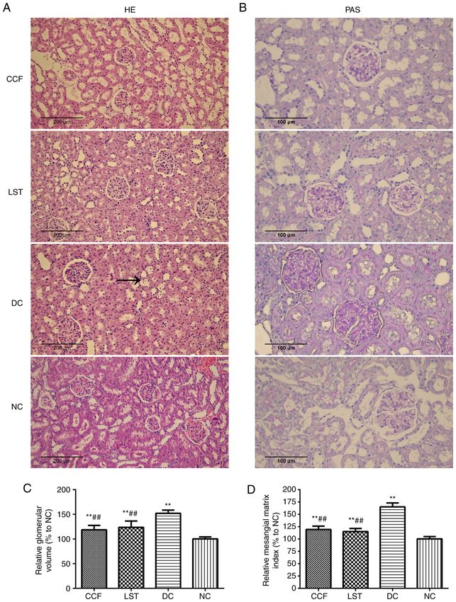
6                       ZHU et al: THE EFFECTS OF COMPOUND CENTELLA FORMULA IN A DIABETIC RAT MODEL

Figure 3. (A) Histopathological changes in the kidney were analysed by PAS staining (x400) and (B) H&E staining (x200) in four groups of Sprague‑Dawley
rats (n=7). (C) Relative glomerular volume and (D) relative mesangial matrix index were calculated in four groups (n=7). Data are expressed as the
mean±standard deviation. **P
EXPERIMENTAL AND THERAPEUTIC MEDICINE 22: 962, 2021                                                 7

                                                                               DC group compared with the NC group (DC vs. NC; P
8 ZHU et al: THE EFFECTS OF COMPOUND CENTELLA FORMULA IN A DIABETIC RAT MODEL Figure 5. The levels of (A) SOD and (B) MDA in renal tissues were measured by an assay kit (n=7). (C and D) The protein expression of NOX4 was analysed by Western blotting in four groups of Sprague‑Dawley rats (n=7). Data are expressed as the mean ± standard deviation. *P
EXPERIMENTAL AND THERAPEUTIC MEDICINE 22: 962, 2021                                                       9

Figure 6. The levels of (A) NF‑κ B and (B) TNF‑α were measured by ELISA
in four groups of Sprague‑Dawley rats (n=7). Data are expressed as the
mean ± standard deviation., **P
10                       ZHU et al: THE EFFECTS OF COMPOUND CENTELLA FORMULA IN A DIABETIC RAT MODEL

Figure 8. Protein expression and localisation of SIRT1 in renal tissues were observed by immunohistochemical staining in four groups of Sprague‑Dawley
rats (n=7). (A) Positive staining is shown in yellow. (B) The AOD results were calculated in four groups (n=7). Data are expressed as the mean ± standard
deviation. **P
EXPERIMENTAL AND THERAPEUTIC MEDICINE 22: 962, 2021                                                   11

Medical University. This article does not contain any studies             18. Zhu X, Chen Y, Chen Q, Yang H and Xie X: Astaxanthin
                                                                              promotes Nrf2/ARE signaling to alleviate renal fibronectin and
with human participants performed by any of the authors.                      collagen IV accumulation in diabetic rats. J Diabetes Res 2018:
                                                                              6730315, 2018.
Patient consent for publication                                           19. Artacho‑Perula E, Roldan‑Villalobos R, Salcedo‑Leal I and
                                                                              Vaamonde‑Lemos R: Stereological estimates of volume‑weighted
                                                                              mean glomerular volume in streptozotocin‑diabetic rats. Lab
Not applicable.                                                               Invest 68: 56‑61, 1993.
                                                                          20. Liu HF, Guo LQ, Huang YY, Chen K, Tao JL, Li SM and
                                                                              Chen XW: Thiazolidinedione attenuate proteinuria and
Competing interests                                                           glomerulosclerosis in Adriamycin‑induced nephropathy rats
                                                                              via slit diaphragm protection. Nephrology (Carlton) 15: 75‑83,
The authors declare that they have no competing interests.                    2010.
                                                                          21. Loeffler I and Wolf G: Pathophysiologie der diabetischen
                                                                              Nephropathie. Der Nephrologe 12: 391‑399, 2017.
References                                                                22. Karolina L, Hannes O, Risul A, Arvind P, Regina G, Taylor RF,
                                                                              Moosa M, Ann C, Karolina K and Larsson TE: Arterial klotho
 1. König A, Schwarzinger B, Stadlbauer V, Lanzerstorfer P,                   expression and FGF23 effects on vascular calcification and func‑
    Iken M, Schwarzinger C, Kolb P, Schwarzinger S, Mörwald K,                tion. Plos One 8: e60658, 2013.
    Brunner S, et al: Guava (Psidium guajava) fruit extract prepared      23. Martynyuk L, Martynyuk L, Ruzhytska O and Martynyuk O:
    by supercritical CO2 extraction inhibits intestinal glucose resorp‑       Effect of the herbal combination canephron N on diabetic
    tion in a double‑blind, randomized clinical study. Nutrients 11:          nephropathy in patients with diabetes mellitus: Results of a
    1512, 2019.                                                               comparative cohort study. J Altern Complement Med 20: 472‑478,
 2. Bakris GL, Hahr A, Khardori R, Koya D, Molitch M, Prischl FC,             2014.
    Schernthaner G and Thajudeen B: Managing diabetic nephropa‑           24. Tavafi M: Diabetic neph ropathy and antioxidants. J
    thies in clinical practice. Overview of diabetic nephropathy.             Nephropathology 2: 20‑27, 2013.
    10.1007/978‑3‑319‑08873‑0: 1‑21, 2017.                                25. Gosmanov AR, Wall BM and Gosmanova EO: Diagnosis and
 3. Varga ZV, Giricz Z, Liaudet L, Haskó G, Ferdinandy P                      treatment of diabetic kidney disease. Am J Med Sci 347: 406‑413,
    and Pacher P: Interplay of oxidative, nitrosative/nitrative               2014.
    stress, inflammation, cell death and autophagy in diabetic            26. Sego S: Pathophysiology of diabetic nephropathy. Nephrol Nurs
    cardiomyopathy. Biochim Biophys Acta 1852: 232‑242, 2014.                 J 34: 631‑633, 2008.
 4. Flyvbjerg A: The role of the complement system in diabetic            27. Magalhães DA, Kume WT, Cor reia FS, Queiroz TS,
    nephropathy. Nat Rev Nephrol 13: 311‑318, 2017.                           Allebrandt Neto EW, Santos MP, Kawashita NH and França SA:
 5. Choi JS, Kim J, Park J, Pyo S, Hong YK, Ku S and Kim MR:                  High‑fat diet and streptozotocin in the induction of type 2
    Blood glycemia‑modulating effects of melanian snail protein               diabetes mellitus: A new proposal. An Acad Bras Ciênc 91:
    hydrolysates in mice with type II diabetes. Int J Mol Med 39:             e20180314, 2019.
    1437‑1451, 2017.                                                      28. Guex CG, Reginato FZ, de Jesus PR, Brondani JC, Lopes GH
 6. Mizuno Y, Yamamotoya T, Nakatsu Y, Ueda K, Matsunaga Y,                   and Bauermann LF: Antidiabetic effects of Olea europaea L.
    Inoue MK, Sakoda H, Fujishiro M, Ono H, Kikuchi T, et al:                 leaves in diabetic rats induced by high‑fat diet and low‑dose
    Xanthine oxidase inhibitor febuxostat exerts an anti‑inflammatory         streptozotocin. J Ethnopharmacol 235: 1‑7, 2019.
    action and protects against diabetic nephropathy development in       29. Ahad A, Ganai AA, Mujeeb M and Siddiqui WA: Ellagic acid,
    KK‑Ay obese diabetic mice. Int J Mol Sci 20: 4680, 2019.                  an NF‑κ B inhibitor, ameliorates renal function in experimental
 7. Rajesh M, Mukhopadhyay P, Bátkai S, Patel V, Saito K,                     diabetic nephropathy. Chemico‑Biological Interactions 219:
    Matsumoto S, Kashiwaya Y, Horváth B, Mukhopadhyay B,                      64‑75, 2014.
    Becker L, et al: Cannabidiol attenuates cardiac dysfunction,          30. Ahad A, Ganai AA, Mujeeb M and Siddiqui WA: Chrysin, an
    oxidative stress, fibrosis, and inflammatory and cell death               anti‑inflammatory molecule, abrogates renal dysfunction in
    signaling pathways in diabetic cardiomyopathy. J Am Coll                  type 2 diabetic rats. Toxicol Appl Pharmacol 279: 1‑7, 2014.
    Cardiol 56: 2115‑2125, 2010.                                          31. Serdar M, Sertoglu E, Uyanik M, Tapan S, Bilgi C and Kurt I:
 8. Duecker R, Baer P, Eickmeier O, Strecker M, Kurz J, Schaible A,           Comparison of 8‑hydroxy‑2'‑deoxyguanosine (8‑OHdG) levels
    Henrich D, Zielen S and Schubert R: Oxidative stress‑driven               using mass spectrometer and urine albumin creatinine ratio as
    pulmonary inflammation and fibrosis in a mouse model of human             a predictor of development of diabetic nephropathy. Free Radic
    ataxia‑telangiectasia. Redox Biol 14: 645‑655, 2018.                      Res 46: 1291‑1295, 2012.
 9. Valacchi G, Virgili F, Cervellati C and Pecorelli A:                  32. Yang M, Jun L, Zhou X, Ding H, Xu J, Yang B, Sun B, Xiao D,
    OxInflammation: From subclinical condition to pathological                Yu J and Gong Q: Correlation analysis between serum vitamin D
    biomarker. Front Physiol 9: 858, 2018.                                    levels and lower extremity macrovascular complications in
10. Bonkowski M and Sinclair D: Slowing ageing by design: The rise            individuals with type 2 diabetes mellitus. J Diabetes Res 2019:
    of NAD+ and sirtuin‑activating compounds. Nat Rev Mol Cell                4251829, 2019.
    Biol 17: 679‑690, 2016.                                               33. Lampropoulou IT, Stangou M, Papagianni A, Didangelos T,
11. Guo R, Liu W, Liu B, Zhang B, Li W and Xu Y: SIRT1 suppresses             Iliadis F and Efstratiadis G: TNF‑ α and microalbuminuria
    cardiomyocyte apoptosis in diabetic cardiomyopathy: An insight            in patients with type 2 diabetes mellitus. J Diabetes Res 2014:
    into endoplasmic reticulum stress response mechanism. Int                 394206, 2014.
    J Cardiol 191: 36‑45, 2015.                                           34. Malikova J, Zdarilova A and Hlobilkova A: Effects of sanguina‑
12. Hao CM and Haase V: Sirtuins and their relevance to the kidney.           rine and chelerythrine on the cell cycle and apoptosis. Biomed
    J Am Soc Nephrol 21: 1620‑1627, 2010.                                     Pap Med Fac Univ Palacky Olomouc Czech Repub 150: 5‑12,
13. Wakino S, Hasegawa K and Itoh H: Sirtuin and metabolic kidney             2006.
    disease. Kidney Int 88: 691‑698, 2015.                                35. Liu T, Zhang L, Joo D and Sun SC: NF‑ κ B signaling in
14. Zhu Q, Zeng J, Li J, Chen X, Miao J, Jin Q and Chen H: Effects            inflammation. Signal Transduct Target Ther 2: 17023, 2017.
    of compound centella on oxidative stress and Keap1‑Nrf2‑ARE           36. Xu XY and Ye SW: Efficacy assessment of treating post‑stroke
    pathway expression in diabetic kidney disease rats. Evid Based            shoulder‑hand syndrome patients of yin deficiency yang hyperac‑
    Complement Alternat Med 2020: 9817932, 2020.                              tivity with blood stasis stagnation collaterals syndrome by yishen
15. Mengjie Z, Ziyang B, Liqiang Y, Danfeng G, Lin W and Jiao Z:              tongluo decoction. Zhongguo Zhong Xi Yi Jie He Za Zhi 34:
    Clinical Study on Modified Compound Jixuecao Tang for                     1069‑1073, 2014 (Article in Chinese).
    Chronic Glomerulonephritis and Chronic Kidney Disease at the          37. Kalantarinia K, Awad AS and Siragy HM: Urinary and renal
    Third Stage. Journal of New Chinese Medicine: 2019.                       interstitial concentrations of TNF‑α increase prior to the rise in
16. Spiers DE and Candas V: Relationship of skin surface area to              albuminuria in diabetic rats. Kidney Int 64: 1208‑1213, 2003.
    body mass in the immature rat: A reexamination. J Appl Physiol        38. Takebayashi K, Matsumoto S, Aso Y and Inukai T: Aldosterone
    Respir Environ Exerc Physiol 56: 240‑243, 1984.                           blockade attenuates urinary monocyte chemoattractant protein‑1
17. Ding Y, Zhang R, Zhang K, Lv X, Chen Y, Li A, Wang L, Zhang X             and oxidative stress in patients with type 2 diabetes complicated
    and Xia Q: Nischarin is differentially expressed in rat brain and         by diabetic nephropathy. J Clin Endocrinol Metab 91: 2214‑2217,
    regulates neuronal migration. PloS One 8: e54563, 2013.                   2006.
12 ZHU et al: THE EFFECTS OF COMPOUND CENTELLA FORMULA IN A DIABETIC RAT MODEL
39. Kowalczuk K and Stryjecka‑Zimmer M: The influence of oxida‑ 46. Elmarakby AA and Sullivan JC: Relationship between oxida‑
tive stress on the level of malondialdehyde (MDA) in different tive stress and inflammatory cytokines in diabetic nephropathy.
areas of the rabbit brain. Ann Univ Mariae Curie Sklodowska Cardiovasc Ther 30: 49‑59, 2012.
Med 57: 160‑164, 2002. 47. Webster BR, Lu Z, Sack MN and Scott I: The role of sirtuins in
40. Kaefer M, De Carvalho JA, Piva SJ, da Silva DB, Becker AM, modulating redox stressors. Free Radical Biol Med 52: 281‑290,
Sangoi MB, Almeida TC, Her mes CL, Coelho AC, 2011.
Tonello R, et al: Plasma malondialdehyde levels and risk factors 48. Yang X, Zhang B, Lu X, Yan M, Wen Y, Zhao T and Li P: Effects
for the development of chronic complications in type 2 diabetic of Tangshen Formula on urinary and plasma liver‑type fatty acid
patients on insulin therapy. Clin Lab 58: 973‑978, 2012. binding protein levels in patients with type 2 diabetic kidney
41. Li X, Cai W, Lee K, Liu B, Deng Y, Chen Y, Zhang X, He J and disease: Post‑hoc findings from a multi‑center, randomized,
Zhong Y: Puerarin attenuates diabetic kidney injury through the double‑blind, placebo‑controlled trial investigating the efficacy
suppression of NOX4 expression in podocytes. Sci Rep 7: 14603, and safety of Tangshen Formula in patients with type 2 diabetic
2017. kidney disease. BMC Complement Altern Med 16: 246, 2016.
42. Ribaldo PD, Souza DS, Biswas SK, Block K, Faria J, 49. Iskender H, Dokumacioglu E, Sen TM, Ince I, Kanbay Y and
Lopes de Faria JM and Lopes de Faria JB: Green tea (Camellia Saral S: The effect of hesperidin and quercetin on oxidative
sinensis) attenuates nephropathy by downregulating Nox4 stress, NF‑κ B and SIRT1 levels in a STZ‑induced experimental
NADPH oxidase in diabetic spontaneously hypertensive rats. diabetes model. Biomed Pharmacother 90: 500‑508, 2017.
J Nutr 139: 96‑100, 2009. 50. Huang K, Gao X and Wei W: The crosstalk between Sirt1 and
43. Peng Q, Liu F and Liang X: Superoxide dismutase and plant Keap1/Nrf2/ARE anti‑oxidative pathway forms a positive feed‑
resistance to the environmental stress. Heilongjiang Agricultural back loop to inhibit FN and TGF‑β1 expressions in rat glomerular
Science 1: 31‑34, 2002. mesangial cells. Exp Cell Res 361: 63‑72, 2017.
44. Samarghandian S, Borji A, Delkhosh M and Samini F: Safranal 51. Du YG, Zhang KN, Gao ZL, Dai F, Wu XX and Chai KF:
treatment improves hyperglycemia, hyperlipidemia and oxidative Tangshen formula improves inflammation in renal tissue of
stress in streptozotocin‑induced diabetic rats. J Pharm Pharm diabetic nephropathy through SIRT1/NF‑κ B pathway. Exp Ther
Sci 16: 352‑362, 2013. Med 15: 2156‑2164, 2018.
45. Tisato V, Gallo S, Melloni E, Celeghini C, Passaro A, Zauli G,
Secchiero P, Bergamini C, Trentini A, Bonaccorsi G, et al: This work is licensed under a Creative Commons
TRAIL and ceruloplasmin inverse correlation as a representative Attribution-NonCommercial-NoDerivatives 4.0
crosstalk between inflammation and oxidative stress. Mediators International (CC BY-NC-ND 4.0) License.
Inflamm 2018: 9629537, 2018.You can also read