The Effect of Chia Seed Extracts against Complete Freund's Adjuvant-Induced Rheumatoid Arthritis in Rats
←
→
Page content transcription
If your browser does not render page correctly, please read the page content below

Hindawi
Journal of Food Quality
Volume 2022, Article ID 3507674, 11 pages
https://doi.org/10.1155/2022/3507674

Research Article
The Effect of Chia Seed Extracts against Complete Freund’s
Adjuvant-Induced Rheumatoid Arthritis in Rats

          Huda Aljumayi ,1 Adel Aljumayi,2 Eman Algarni,1 Reham M. Algheshairy,3
          Hend F. Alharbi ,3 Wedad Azhar,4 Taqwa Bushnaq,1 and Alaa Qadhi4
          1
            Department of Food Science and Nutrition, College of Sciences, Taif University, P.O. Box 11099, Taif 21944, Saudi Arabia
          2
            King Abdulaziz-Medical City-NGHA WR, Jeddah, Saudi Arabia
          3
            Department of Food Science and Human Nutrition, College of Agricultural and Veterinary Medicine, Qassim University,
            Buraydah 51452, Saudi Arabia
          4
            Clinical Nutrition Department, College of Applied Medical Sciences, University of Umm Al-Qura, P.O. Box 7067,
            Makkah 21955, Saudi Arabia

          Correspondence should be addressed to Huda Aljumayi; huda.a@tu.edu.sa

          Received 30 April 2022; Revised 22 May 2022; Accepted 26 May 2022; Published 10 June 2022

          Academic Editor: Bilal Sadiq

          Copyright © 2022 Huda Aljumayi et al. This is an open access article distributed under the Creative Commons Attribution
          License, which permits unrestricted use, distribution, and reproduction in any medium, provided the original work is
          properly cited.
          Background and Objectives. It is known that the oxidation of chia seeds is minimal or absent, due to the presence of bioactive
          compounds, having a great potential in the foods and pharmacological industry. This investigation was done to estimate the
          nutrition values and anti-oxidant activity of chia seed extracts. Materials and Methods. The protective effects of chia seed extract
          against complete Freund’s adjuvant (CFA) rheumatoid arthritis in rats were investigated with 100, 200, 300, and 400 ppm/kg BW
          rat/day chia seeds aqueous extract for 4 weeks. Results. The results obtained revealed that chia seed extract contained high
          proportions of protein and total dietary fiber values of 21.35% and 27.24%, respectively. Moreover, the results reported that the
          mineral contents were the greatest in phosphorous, potassium, magnesium, calcium, and sodium (450.09, 410.46, 245.22, 218.69,
          and 120.34 mg/100 g, respectively). Meanwhile, iron, zinc, and copper were the lowest in chia seeds (9.26, 4.67, and 3.66 mg/100 g,
          respectively). Besides, vitamins C and E reported 1.65 and 0.82 mg/100 g, respectively. Chia seed extracts were effective in vitro for
          the bioactive components such as phenolics, flavonoids, and anti-oxidant activities. The biomarkers of complete blood picture,
          lipid profile, anti-oxidant enzymes, TNF-α, and IL-10 had been improved. Histopathological examination of the rat knee
          confirmed health amelioration, revealing that chia seed extract consumption can lower pathological changes in injured rheu-
          matoid arthritis rats. Conclusion. It could be seen that the chia seed extracts alleviated the harmful effect of rheumatoid arthritis
          CFA-induced rats.

1. Introduction                                                          agents that can ultimately converge in an overactive immune
                                                                         system are the basic idea of the etiology of rheumatoid
Rheumatoid arthritis (RA) is a disease of the articular                  arthritis [5]. Arthritis collectively points out to more
cartilage that results from a defect in the immune regulation            rheumatic illness described by inflammation, pain, and
that either directly or indirectly affects the physiology of the          stiffness in the musculoskeletal system [6].
cartilage cells [1]. It is also distinguished by inflammation                 Rheumatoid arthritis affects women being affected more
and injury to the bone joints, and if not treated, its com-              often than men; rheumatoid arthritis is also evaluated with a
plications increases [2, 3]. The etiology of rheumatoid ar-              high mortality rate [7, 8]. Oliveira-Alves et al. [9] suggested
thritis involves the infiltration of multiple inflammatory cells           that chia seeds could contribute to improving better the
and crosstalk with cytokines [4]. Genetic and environmental              consumers’ health. Therefore, phenolic compounds found in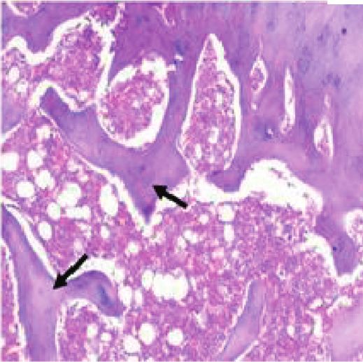
2                                                                                                     Journal of Food Quality

chia seeds, mainly caffeic acid and salvianolic acids, act as     2.6. Determination of Anti-Oxidant Assays (ABTS, DPPH, and
protective agents for illnesses, which may be caused by          FRAP). ABTS assay was evaluated according to the stabi-
oxidative overwork [10–12]. Likewise, bioactive ingredients      lization of the ABTS + radical cation [20]. The DPPH test
present in chia seeds are associated with lowering the risk of   was to assess the anti-oxidant capacity of the DPPH radical
chronic heart disease, liver influence, rheumatoid arthritis,     in ethanol. FRAP reagent ensured the anti-oxidant potential
plasma oxidative stress, and obesity-associated illness          by reducing iron (Fe2+) and iron (Fe3+) in chia seed extracts
[13, 14].                                                        [21]. The results were considered at Mmol TEACg−1.
    Despite the fact that various studies have been conducted
on rheumatoid arthritis prevention, much work has been
done using chia seeds as a preventive or remedial measure.
                                                                 3. Bioactive Components Determination
Therefore, the purpose of the current research work was to       3.1. Total Polyphenol Contents (TPC). Approximately
evaluate chia seed extract as a chemical analysis and anti-      100 μg/mL chia extracts were mixed thoroughly with the
oxidant compounds. In addition, estimate the biological          corresponding Folin reagent, and 1.0 mL 7% of Na2CO3
experiment with chia seed aqueous extract at different levels     solution was added and incubated for 90 min, detected at
to treat rheumatoid arthritis in rats’ chronic inflammatory       765 nm, and calculated as gallic acid equivalents (GAEs) mg
disorders.                                                       GAE/g DW [22].

2. Material and Methods
                                                                 3.2. Total Flavonoid Contents (TFC). Approximately 0.2 mL
2.1. Study Area. This research project was conducted from        of chia extracts and 0.2 mL of 30% ethanolic AlCl3·6H2O
September 2021 (Starting date) to January 2022 (Ending           were immediately added and incubated for 5 min. The ab-
date) according to the regulations and rules laid down by the    sorbance was detected at 430 nm and calculated as quercetin
committee of animals’ experimentation of Taif University,        equivalents mg QE/g DW [23].
Kingdom of Saudi Arabia.

                                                                 3.3. Total Tannin Contents (TTC). Approximately 1 mL of
2.2. Materials. Chia seeds (Salvia hispanica L.) were pur-       chia extracts and 5 mL of potassium iodide (2.5%) were
chased from the local market in Taif City, Saudi Arabia. Kits    mixed, closed, and placed in a ∼30°C water bath (10 min).
for all different parameters were obtained from Sigma-            The absorbance was assessed at 590 nm and calculated as
Aldrich Corp., MO. Also, complete Freund’s adjuvant (CFA)        tannic acid equivalents (TAEs) mg TAEs g/100 g DW
was purchased from Sigma, St. Louis, Mo. Male albino rats        [24].
(n � 36 rats), 150–160 g per each, were obtained from
Pharmacy Faculty at King Saud University and fed on the
basal diet.                                                      3.4. Biological Experimental. The experimental rats were fed
                                                                 on a basal diet for 7 days and randomly divided into 6
                                                                 groups, 6 rats for each. The first main group was fed on a
2.3. Preparation of Chia Seeds. Chia seeds were washed
                                                                 basal diet for four weeks, namely the control negative rats
under tap water to remove foreign particles, dried at
                                                                 group. The rest of the rats were injected with 100 µl of CFA
50–60°C, and milled to a fine powder. The powder was stored
                                                                 into the left hind knee joint to induce rheumatoid arthritis in
at 5°C until analysis.
                                                                 rats. After 7 days, secondary arthritis was induced by
                                                                 injecting 50 µL of CFA under the left hind knee joint
2.4. Determination of the Chemical Composition of Chia Seeds.    according to Narendhirakannan et al. [25] and then divided
Chemical compositions such as protein, fat, crude fiber, and      into 5 groups. A positive control group was also fed the basal
ash content were determined in the chia seeds according to       diet only, while the other 4 groups were fed basal diets and
Sami et al. [15, 16]. Minerals content as magnesium, sodium,     taken 100, 200, 300, and 400 ppm/kg BW rat/day chia
potassium, zinc, phosphorus, iron, calcium, and copper were      aqueous extract were taken orally by a stomach tube during
estimated in chia seeds according to Sami et al. [17].           the experimental period.
                                                                     After the end of the experiment, the blood was pulled
                                                                 withdrawn from different rat groups, centrifuged to obtain
2.5. Vitamin Analysis (HPLC). The vitamin C and E de-            serum, and kept at −20°C until analysis. Blood hemoglobin
terminations were detected according to the modified              (Hb), hematocrit (Ht), platelets, red blood cells (RBCs), and
method of Rokayya et al. [18]. Chromatographic analyses          white blood cells (WBCs) were estimated [26]. Lipid profiles
were detected for vitamin C by an Agilent HPLC system            such as triglycerides, total lipids, total cholesterol, high-
(2000 ECOM, Chrastany u Prahy, CZ 252 19, Czech) at              density lipoprotein (HDL), and low-density lipoproteins
254 nm with UV detection. Analytical column YMC-Triart           (LDL) were estimated according to Zollner and Kirsch [27].
C18 (150 × 4.6 mm) was used as the mobile phase of A/B 33/       Superoxidexide dismutase (SOD), nonenzyme glutathione
67; A: 0.1 M potassium acetate, distilled water 50:50, pH        (GSH), malondialdehyde (MDA), serum-selected cytokines
(4.9), and 1 mL/min for the flow rate at the ambient tem-         IL-10, and TNF-α were determined by Kandir and Keskin
perature [19].                                                   [28].
Journal of Food Quality                                                                                                                                                                                                                                            3

3.5. Histopathology Evaluations. The animals were sacrificed                                    40
(after four weeks following CFA injection), and the knee
                                                                                               35
joint was excised and kept in 10% buffered formalin then
sectioned and embedded in paraffin. Slides were stained with

                                                                    Chemical composition (%)
                                                                                               30
hematoxylin and eosin according to Laste et al. [29].
                                                                                               25

3.6. Statistical Analysis. All experimental data were applied                                  20
to the (ANOVA) test, and the results were shown as means                                       15
of standard deviation (±SD). Significant differences were
evaluated as (P ≤ 0.05).                                                                       10

                                                                                                5
4. Results
                                                                                                0
4.1. Chemical Composition. The chemical compositions and

                                                                                                        Crude protein

                                                                                                                          Crude fiber

                                                                                                                                                                                                                 Insoluble dietary fiber

                                                                                                                                                                                                                                           Soluble dietary fiber
                                                                                                                                        Total lipids

                                                                                                                                                       Ash content

                                                                                                                                                                     Total carbohydrates

                                                                                                                                                                                           Total dietary fiber
dietary fiber levels of chia seeds were determined and
published in (Figure 1). Chia seeds have elevated high
amounts of protein, total lipid, and crude fibers by 31.35%,
27.64%, and 10.96%, respectively. Moreover, the total dietary
fiber, insoluble and soluble dietary fibers detected were
27.24%, 18.98%, and 8.26%, respectively.
     Furthermore, the results of the mineral and vitamin          Figure 1: Chemical compositions and dietary fiber contents de-
contents in chia seeds were presented in Table 1. The results     termined in the chia seeds.
reported that the mineral contents were the greatest in
phosphorous, potassium, magnesium, calcium, and sodium
                                                                                               Table 1: Mineral and vitamin contents in chia seeds.
(450.09, 410.46, 245.22, 218.69, and 120.34 mg/100 g, re-
spectively). Meanwhile, iron, zinc, and copper were the                     Minerals and vitamins contents (mg/100 g)
lowest in chia seeds (9.26, 4.67, and 3.66 mg/100 g, re-          Magnesium                                        245.22 ± 2.47
spectively). Vitamins C and E in chia seeds contained 1.65        Sodium                                           120.34 ± 1.25
and 0.82 mg/100 g, respectively.                                  Potassium                                        410.46 ± 3.49
                                                                  Calcium                                          218.69 ± 1.28
                                                                  Phosphorus                                       450.09 ± 4.21
4.2. Anti-Oxidant Profile. The research findings on the anti-       Iron                                              9.26 ± 0.04
oxidant activity of chia seed extracts were reported in           Zinc                                              4.67 ± 0.01
Figure 2. Chia seed extract had high ABTS, DPPH, and              Copper                                            3.66 ± 0.02
FRAP activities by 3.21, 2.43, and 3.39 mmol TEAC/g DW,           Vitamin C                                         1.65 ± 0.01
respectively. Total phenolic acids, flavonoids, and total          Vitamin E                                         0.82 ± 0.01
tannins contents recorded 0.98 mg GAE/g DW, 0.23 mg QE/           Values are mean and SD ± (n � 3).
g DW, and 16.11 mg TAEs g/100 g DW, respectively
(Table 2).

4.3. Effect of Chia Seed Extract on Complete Blood Picture.                                          3
Data that are obtained from the research work and presented
                                                                            (Mmol TEACg-1 DW)
                                                                             Antioxidant capacity

in Table 3 showed that the hemoglobin was the highest in the
rat group that was fed on a basal diet as a control negative by
                                                                                                    2
12.7 g/dL. The positive control rats that induced rheumatoid
arthritis reported 9.8 g/dL, while the different groups rats’
induced rheumatoid arthritis that was fed on a basal diet and
taken orally with extracts at 100, 200, 300, and 400 ppm were                                       1
gradually increased to 11.2, 11.8, 12.3, and 12.5 g/dL, re-
spectively. Therefore, Omoigui et al. [30] confirmed that the
treatment with chia extract resulted in a hemoglobin in-                                            0
crease. The results observed that the hematocrit was lower in                                                           ABTS                                   DPPH                                                        FRAP
positive control (30.3%) compared with negative control                                         Figure 2: Anti-oxidant capacity in the chia seeds.
(38%), and it was increased in the group that was fed on chia
extract at 40 ppm by 37.8%. In addition, the red blood cells
increased at different extracts by 5.22, 5.84, 6.27, and 6.81 m/   control group by 10.14 cm compared with negative control
cm compared with negative control (4.45 m/cm), respec-            (5.67 cm), while the other groups that were taken 100, 200,
tively. White blood cells reported an increase in the positive    300, and 400 ppm were gradually decreased by 9.33, 8.53,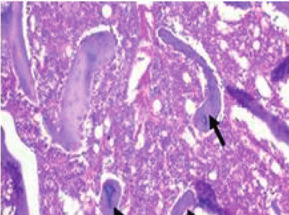
4                                                                                                                         Journal of Food Quality

                                                    Table 2: Anti-oxidant profile in chia seeds.
                                                                 Anti-oxidant activity
Total phenolic acids (mg GAE/g DW)                                                                                                    0.98 ± 0.04
Total flavonoid compounds (mg QE/g DW)                                                                                                 0.23 ± 0.01
Total tannin content (mg TAEs g/100 g DW)                                                                                             16.11 ± 0.15
Values are mean and SD ± (n � 3).

                                                          Table 3: Complete blood picture.
Groups                  Hemoglobin (g/dL)           Hematocrit (%)         Red blood cells (m/cm)          White blood cells (cm)   Platelets (cm)
Negative control            12.7 ± 0.8a               38.0 ± 2.4a                6.97 ± 0.30a                   5.67 ± 0.93e         753.3 ± 37.8a
Positive control           10.8 ± 1.03d               30.3 ± 3.1e                4.45 ± 0.43c                  10.14 ± 1.05a         388.3 ± 54.7e
Group, 100 ppm             11.2 ± 0.87c               32.5 ± 2.6d               5.22 ± 0.53b                   9.33 ± 2.85ab         465.3 ± 111d
Group, 200 ppm             11.8 ± 0.79b               34.4 ± 2.4c               5.84 ± 0.62b                    8.53 ± 1.47b         594.3 ± 67.3c
Group, 300 ppm             12.3 ± 0.94a              36.3 ± 2.76b                6.27 ± 0.73a                   7.64 ± 0.91c        649.2 ± 30.35b
Group, 400 ppm             12.5 ± 0.83a              37.8 ± 3.14ab               6.81 ± 0.68a                   6.17 ± 0.84d        743.8 ± 37.12a
Mean values in each raw having different superscript (a, b, c, and d) are significantly different at 0.05 levels.

7.64, and 6.17 cm, respectively. The platelets in positive                      chia seed extracts gave the gradually best results for GSH
control were 388.3 cm compared with negative control                            and SOD; especially, the groups 5 and 6 were taken orally at
753.3 cm, and it was gradually increased with chia extract to                   300 and 400 ppm levels that increased in GSH to 9.44 and
465.3, 594.3, 649.2, and 743.8 cm, respectively.                                10.50 m. mol/mg protein, as well as in SOD more increased
                                                                                to 11.91 and 13.98 m. mol/mg protein, respectively.
4.4. Effect of Chia Seed Extract on Lipid Profile. The results of
the total lipid of the chia seeds were presented in Table 4. The                4.6. Effect of Chia Seed Extract on Cytokines IL-10 and TNF-α.
results depicted that the negative control was 0.64 g/dL, and                   Results presented in Figure 4 revealed the effect of chia seed
a significant increase in the positive control rats group in-                    extract on cytokines. Figure 4 illustrated that the TNF-α in
duced rheumatoid arthritis by 1.42 g/dL. Moreover, chia                         the positive control group was the highest (48.53 pg/ML−1)
extracts at levels 100, 200, 300, and 400 ppm were 1.21, 1.00,                  compared with the negative healthy rat group (30.25 pg/
0.83, and 0.60 g/dL, respectively. Results of triglycerides,                    ML−1). Meanwhile, the best results were detected for the
total cholesterol, and LDL were 112.3, 86.3, and 25.0 mg/dL                     group that had 400 ppm/kg body weight/rat/day from chia
in the negative control, while in the positive control group                    extracts, which reported 32.15 pg/ML−1. These results were
increased to reach 245.7, 196.3, and 131.7 mg/dL, respec-                       confirmed by Shanahan and St [31] who found that when the
tively. Meanwhile, the other rat groups were gradually de-                      TNF-α value is lowered, the variety of inflammation is
creased to 212.0, 179.7, 146.4, and 113.3 mg/dL in                              decreased. From the same table, IL-10 acts as an anti-in-
triglycerides, as well as for total cholesterol the results in-                 flammatory cytokine that had the highest value of 25.86 pg/
dicated decreasing to 169.0, 142.3, 115.26, and 88.18 mg/dL,                    ML−1 in positive control rats, while the negative control
respectively; in addition, the LDL was decreased to 105.67,                     reported 13.28 pg/ML−1. Meanwhile, the best results were
79.3, 53.42 and 27.16 mg/dL, respectively. The HDL deter-                       detected for the group that had 400 ppm/kg body weight/rat/
mination reported a significant increase in serum rats’                          day from chia extracts that reported 15.12 pg/ML−1 IL-10
negative control to reach 73.7.3 mg/dL compared with                            can control the development of rheumatoid arthritis and
positive control (27.3 mg/dL). Moreover, the HDL was                            inhibit cytokine production, released by activated macro-
decreased in the other groups with chia extracts to reach                       phages [32].
38.0, 49.0, 61.28, and 72.73 mg/dL, respectively.
                                                                                4.7. Histopathology Evaluations. Microscopic examinations
4.5. Effect of Chia Seed Extract on Oxidative Stress.                            of joints for the negative control group were given in
Findings obtained from this study on the effect of chia seed                     Figures 5(a)–5(f ). The pictures in the plates revealed normal
extract on oxidative stress were presented in Figure 3. The                     histology of the joint and free from inflammatory or de-
lipid peroxidation as malondialdehyde (MDA), and the                            generative changes; it appeared to consist of two cartilages
activity of glutathione (GSH) and superoxide activity                           covered bone heads with synovial membrane lining the joint
(SOD) were determined in different rat groups’ induced                           capsule internally and histologically normal trabecular bone
rheumatoid arthritis at different levels 100, 200, 300, and                      of the epiphysis.
400 ppm taken orally, and the results are shown in Figure 3.                        On the other hand, microscopic examinations of
The results reported that the positive control rats were                        joints for the positive control group are given in
decreased to reach 3.16 and 4.39 m. mol/mg protein,                             Figures 5(a)–5(f ) and Figures 6(a)–6(e). There were
compared with negative control rats that increased to reach                     various alterations; the articular cartilage was thinned,
11.32 and 15.34 m. mol/mg protein, respectively. Rats with                      and the synovium and the joint capsule were greatly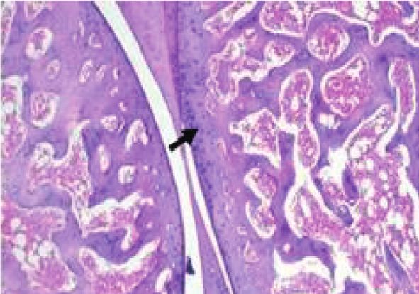
Journal of Food Quality 5
Table 4: Lipid profile.
Groups T. Lipid (g/dl) Triglycerides (mg/dl) T. cholesterol (mg/dl) HDL (mg/dl) LDL (mg/dl)
Negative control 0.64 ± 0.03e 112.3 ± 6.1e 86.3 ± 1.1e 73.7 ± 10.0a 25.0 ± 5.56e
Positive control 1.42 ± 0.17a 245.7 ± 27.9a 196.3 ± 6.5a 27.3 ± 17.2a 131.7 ± 20.2a
Group, 100 ppm 1.21 ± 0.13b 212.0 ± 30.0b 169.0 ± 7.0b 38.0 ± 5.3d 105.67 ± 10.0b
Group, 200 ppm 1.00 ± 0.06c 179.7 ± 9.07c 142.3 ± 3.5c 49.0 ± 3.0c 79.3 ± 6.03c
Group, 300 ppm 0.83 ± 0.07d 146.38 ± 8.35d 115.26 ± 4.12d 61.28 ± 4.29b 53.42 ± 3.17d
Group, 400 ppm 0.60 ± 0.04e 113.28 ± 6.58e 88.18 ± 2.31e 72.73 ± 4.78a 27.16 ± 1.56e
Mean values in each raw having different superscript (a, b, c, and d) are significantly different at 0.05 levels.
thickened due to expansion by inflammatory edema and
16
exudate. The trabecular bone forming the head was de-
creased in size and number.
Concerning the group fed on a basal diet and taken orally
(m.mol/mg/protein)
12 100 ppm/kg body weight/day from chia seed extract, the
Oxidative stress
microscopic examinations of joints are presented in
Figures 7(a)–7(e). The photographs in Figures 7(a)–7(e)
8 depicted that both articular surfaces and the synovium
were normal. The trabecular of the epiphysis was normal as
4
well.
Regarding the group fed on a basal diet and taken orally
200 ppm/kg body weight/day from chia seed extract, their
0 microscopic joint examinations are presented in
Negative control
Positive control
Group 100 ppm
Group 200 ppm
Group 300 ppm
Group 400 ppm
Figures 8(a)–8(e). As revealed, the joints exhibited mild
thinning in the articular cartilage covering the articular
surface. The synovium and the joint capsule appeared
normal, while the trabecular of the epiphysis was thinner
compared to the negative control group.
MDA
Lastly, the microscopic joint examinations of the rats fed
GSH on a basal diet and taken orally 300 ppm/kg body weight/day
SOD from chia seed extract are shown in Figures 9(a)–9(e). As
presented, the rats exhibited a good recovery, as the joint was
Figure 3: Oxidative stress.
free from degeneration.
The group fed on a basal diet and taken orally 400 ppm/
kg body weight/day from chia seed extract (Figure 10(a)–
10(c)) exhibited the best recovery with no inflammatory
50 lesions and appeared histologically normal.
40 5. Discussion
Cytokines (pg/mL-1)
30 The goal of this study was to characterize chia seeds that had
a high concentration of chemical components (protein, oil
content, dietary fiber, and total carbohydrates), minerals
20 content (phosphorous, potassium, magnesium, calcium, and
sodium), vitamins (E and C), and total dietary soluble and
10 insoluble dietary. These results were in agreement with
Ixtaina et al. [33] who reported that chia seed protein, fats,
0
carbohydrates, high dietary fiber, and ash range from 15to
25%, 30 to 33%, 26 to 41%, 18 to 30%, and 4.0 to 5.0%,
Negative control
Group 100 ppm
Group 200 ppm
Group 300 ppm
Group 400 ppm
Positive control
respectively. In addition, minerals, vitamins, and dry matter
were from 90% to 93%. The chia seeds have contented the
highest total dietary fiber ranging from 36% to 40%, which is
greater than grains and vegetables [34]. The insoluble dietary
fiber is associated with the gradual elevation in blood sugar
TNF-α
IL-10
levels after eating and lowering insulin resistance [35]. The
results were confirmed by De et al. [36]. Drusch and
Figure 4: Cytokines IL-10 and TNF-α. Mannino concluded that chia flour is rich in protein, with
6                                                                                                                Journal of Food Quality

                       (a)                                          (b)                                           (c)

                       (d)                                           (e)                                          (f )

Figure 5: (a) Negative control group showing normal cartilage (black arrow), normal synovium (black star), and bone (red star); (b) higher
magnification showing normal cartilage (black arrow); (c) normal synovial membrane and joint capsule (black arrow); (d) higher
magnification showing synovial membrane and joint capsule (black arrow); (e) magnification showing synovial membrane and joint capsule
(black arrows); and (f ) normal trabecular bone (arrows) of the head of the bone.

                        (a)                                         (b)                                         (c)

                                             (d)                                                (e)

Figure 6: (a) Positive control group, showing mild thinning in the cartilage coating the head of the bone (arrow); (b) showing inflamed joint
capsule and synovium (arrows); (c) higher magnification showing perivascular inflammatory cells infiltration (arrows) and edema in the
synovial membrane; (d) higher magnification showing inflammatory cells infiltration (arrows) and edema in the synovial membrane; and (e)
showing mild thinning in the trabecular bone (arrows) of the epiphyses.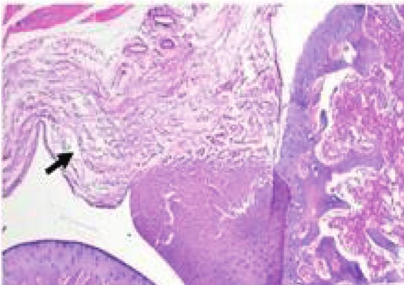
Journal of Food Quality                                                                                                                7

                       (a)                                        (b)                                          (c)

                                           (d)                                                (e)

Figure 7: (a) Group (100 ppm) showing normal articular cartilage, (b) normal articular cartilage (arrow) and synovium (star), (c) showing
normal synovium and joint capsule, (d) higher magnification showing normal synovium and joint capsule, and (e) showing normal bone of
the epiphysis.

                     (a)                                          (b)                                           (c)

                                          (d)                                                 (e)

Figure 8: (a) Group (200 ppm) showing mild thinning in the articular cartilage (arrow), normal synovial membrane, and joint capsule
(star); (b) mild thinning in the articular cartilage (arrow); (c) normal synovial member joint capsule; (d) synovial membrane and joint
capsule; and (e) higher in higher magnification, showing normal synovial membrane and joint capsule.

eight essential amino acids and high amounts of calcium,                    The anti-oxidant as total phenolic, total flavonoids, and
iron, ascorbic acid, omega [30], and anti-oxidants;                     tannic acid content, as well as anti-oxidant activity, had the
therefore, it can be called food for healthy skin, hair, and            highest in chia seeds. Sargi et al. [37] indicated that chia
nail.                                                                   seeds can inactive ABTS radicals and scavenge synthetic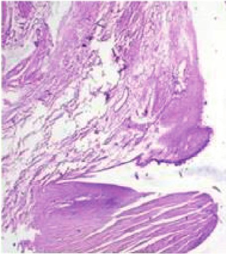
8                                                                                                               Journal of Food Quality

                           (a)                                     (b)                                         (c)

                                            (d)                                          (e)

Figure 9: Group (300 ppm): (a) normal articular cartilage (arrow) and synovium (star), (b) higher magnification and normal articular
cartilage, (c) higher magnification and normal synovium and joint capsule, (d) higher magnification and normal joint capsule, and (e)
normal trabecular bone (arrow).

                     (a)                                            (b)                                              (c)

Figure 10: Group (400 ppm): (a) normal articular cartilage (arrow) and synovium (star), (b) normal joint capsule, and (c) normal trabecular
bone (arrows) of the epiphysis.

DPPH radicals. Furthermore, Coelho and Salas-Mellado                      the immune response to assistance to the body in fighting
[38] validated the anti-oxidant efficacy of these findings.                  infection by producing antibodies that circulate in the
Chia’s high anti-oxidant activity can enhance the benefits for             bloodstream.
human health. Besides, the Brazilian Chia seeds had con-                      These results investigated that have shown that the
tented phenolic compounds by 0.97 and 0.99 mg GAE/g                       highest level of polyphenols in chia extract may play a great
DW33.                                                                     significant role in contributing to health and chronic heart
    Results from biological experiments, it could be found                disease [41]. The anti-inflammatory influence of treatment
that the chia seeds improved the complete blood picture,                  with chia extract is to adverse endothelial trouble by
lipid profile anti-oxidant enzymes, and cytokine in rat                    inhibiting LDL oxidation [42]. Also, the biologically active
groups with complete Freund’s adjuvant-induced rheuma-                    contents of chia extract such as natural anti-oxidants can act
toid arthritis. Therefore, Omoigui et al. [39] confirmed that              locally as a cytokine and defend the body against micro-
the treatment with chia extract resulted in a hemoglobin                  organisms [43]. Treating rats with chia extract reduced el-
increase. These results were in a similar trend to the previous           evated levels of triglycerides, LDL, and cholesterol in the
study of Anderson et al. [40], who reported the hemato-                   positive group. In addition, the treatment of chia extract
logical results of rheumatoid arthritis could due to activating           enhanced the lipid abnormalities that may be due to the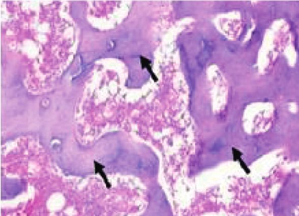
Journal of Food Quality                                                                                                                9

rheumatoid arthritis-like TG and TC values that returned to       Freund’s adjuvant-induced rheumatoid arthritis in rats. The
normal while HDL and LDL were significantly better [31].           formulation approach was used in this study to focus on the
    Hendawy et al. [32] observed that the activity of enzymes     pharmacological potential benefit of food extracts.
such as GSH was increased in the blood in animals that fed
with chia supplementation. Regarding the other enzymes, no        Data Availability
variations in their activity were shown. The results reported
that the MDA increased in the positive control group by           The data used to support the findings of this study are in-
4.23 m. mol/mg protein, while negative control reported           cluded within the article.
0.45 m. mol/mg protein. In addition, the orally fed groups of
100 and 200 ppm chia seed extracts reported 2.81 and              Conflicts of Interest
2.11 m. mol/mg protein. Meanwhile, the best results were for
groups 5 and 6 that reported the lowest values of 1.68 and        The authors declare that they have no conflicts of interest.
0.78 m. mol/mg protein, respectively. MDA is a product of
lipid peroxidation, whereas SOD is a function to remove
                                                                  References
reactive oxygen species (ROS). These factors can affect the
vitality of the body as when tissue damage, large ROS amounts      [1] T. Chia-Chun, C. Yi-Jen, C. Wei-An et al., “Dual role of
are produced and occur to oxidative stress (OS) [44].                  chondrocytes in rheumatoid arthritis: the chicken and the
    TNF-α is a pleiotropic cytokine as it plays a serious              egg,” International Journal of Molecular Sciences, vol. 21, no. 3,
function for each from acute and chronic inflammation by                pp. 1–15, 2020.
enhancing the adhesion of neutrophils and lymphocytes to           [2] I. B. McInnes, C. D. Buckley, and J. D. Isaacs, “Cytokines in
endothelial cells [45]. These results were confirmed by                 rheumatoid arthritis - shaping the immunological landscape,”
                                                                       Nature Reviews Rheumatology, vol. 12, no. 1, pp. 63–68, 2016.
Shanahan and St [46] who found that when the TNF-α
                                                                   [3] M. B. Alazzam, N. Tayyib, S. Z. Alshawwa, and M. Ahmed,
value is lowered, the variety of inflammation is decreased.             “Nursing care systematization with case-based reasoning and
From the same Table, IL-10 acts as an anti-inflammatory                 artificial intelligence,” Journal of Healthcare Engineering,
cytokine that had the highest value of 25.86 pg/ML−1 in                vol. 2022, Article ID 1959371, 9 pages, 2022.
positive control rats, while the negative control reported         [4] M. C. Moran-Moguel, S. Petarra-del Rio, E. E. Mayorquin-
13.28 pg/ML−1. Meanwhile, the best results were detected               Galvan, and M. G. Zavala-Cerna, “Rheumatoid arthritis and
for the group which had 400 ppm/kg body weight/rat/day                 miRNAs: a critical review through a functional view,” Journal
from chia extracts that reported 15.12 pg/ML−1 IL-10 can               of Immunology Research, vol. 2018, pp. 1–16, 2018.
control the development of rheumatoid arthritis and in-            [5] S. E. Gabriel and K. Michaud, “Epidemiological studies in
hibit cytokine production, released by activated macro-                incidence, prevalence, mortality, and comorbidity of the
phages [47, 48].                                                       rheumatic diseases,” Arthritis Research and Therapy, vol. 11,
                                                                       no. 3, pp. 229–245, 2009.
    This study reported that chia seeds have high values of
                                                                   [6] V. Y. Ixtaina, S. M. Nolasco, and M. C. Tomás, “Physical
bioactive compounds and effective anti-oxidant activities               properties of Chia (Salvia hispanica L.) seeds,” Industrial
besides the pharmacological effect against complete                     Crops and Products, vol. 28, no. 3, pp. 286–293, 2008.
Freund’s adjuvant-induced rheumatoid arthritis in rats. The        [7] M. B. Alazzam, W. T. Mohammad, M. B. Younis et al.,
formulation approach was used in this study to focus on the            “Studying the effects of cold plasma phosphorus using
pharmacological potential benefit of food extracts.                     physiological and digital image processing techniques,”
                                                                       Computational and Mathematical Methods in Medicine,
                                                                       vol. 2022, Article ID 8332737, 5 pages, 2022.
6. Conclusion                                                      [8] N. Nielsen, V. Pascal, A. Fasth et al., “Balance between ac-
The purpose of this study was to determine how chia seed               tivating NKG2D, DNAM-1, NKp44 and NKp46 and inhibi-
                                                                       tory CD94/NKG2A receptors determine natural killer
extract affected the development of bones and joints in an-
                                                                       degranulation towards rheumatoid arthritis synovial fibro-
imals. The researchers discovered that chia seed extract had a         blasts,” Immunology, vol. 142, no. 4, pp. 581–593, 2014.
significant favorable impact on the growth and development          [9] S. C. Oliveira-Alves, D. B. Vendramini-Costa, C. B. Betim
of rats’ bones. It was concluded that chia seeds have a high           Cazarin et al., “Characterization of phenolic compounds in
amount of nutrients, phenolic acids, and flavonoids that                chia (Salvia hispanica L.) seeds, fiber flour and oil,” Food
scavenge the free radicals in the blood. Therefore, when fed           Chemistry, vol. 232, no. 232, pp. 295–305, 2017.
the rats on a basal diet and taken orally at levels of 300 and    [10] S. Rokayya, C.-J. Li, Y. Zhao, Y. Li, and C.-H. Sun, “Cabbage
400 ppm chia seed extracts, it led to the best results for ox-         (Brassica oleracea L. var. capitata) phytochemicals with an-
idative stress, complete blood picture, lipids profile, and also        tioxidant and anti-inflammatory potential,” Asian Pacific
cytokines (TNF-α and IL-10) in rheumatoid arthritis in rats’           Journal of Cancer Prevention, vol. 14, no. 11, pp. 6657–6662,
chronic inflammatory disorders.                                         2013.
                                                                  [11] O. S. Ahmed, E. E. Omer, S. Z. Alshawwa, M. B. Alazzam, and
                                                                       R. A. Khan, “Approaches to federated computing for the
7. Significance Statement                                              protection of patient privacy and security using medical
                                                                       applications,” Applied Bionics and Biomechanics, vol. 2022,
This study reported that chia seeds have high values of                Article ID 1201339, 6 pages, 2022.
bioactive compounds and effective anti-oxidant activities          [12] G. H. Qiao, D. Wenxin, X. Zhigang, R. Sami, E. Khojah, and
besides the pharmacological effect against complete                     S. Amanullah, “Antioxidant and anti-inflammatory capacities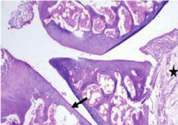
10                                                                                                                     Journal of Food Quality

       of pepper tissues,” Italian Journal of Food Science, vol. 32,               COVID-19 infection,” Clinical Laboratory, vol. 67, no. 06/
       no. 2, pp. 265–274, 2020.                                                   2021, pp. 1–11, 2021.
[13]   R. Ayerza, “The seed’s protein and oil content, fatty acid           [27]   D. Afrifa, K. Nsiah, A. C. Afriyie, and M. O. Moses, “Incidence
       composition, and growing cycle length of a single genotype of               of cardiovascular disease risk factors among football players in
       chia (salvia hispanica L.) as affected by environmental fac-                 ashanti region of Ghana,” International Journal of Social
       tors,” Journal of Oleo Science, vol. 58, no. 7, pp. 347–354, 2009.          Science and Humanities, vol. 2, no. 2, Article ID e98153, 2019.
[14]   U. Rahman, M. Nadeem, A. Khalique et al., “Nutritional and           [28]   S. Kandir and E. Keski̇n, “Tiroidektomize ratlarda serum IL-
       therapeutic perspectives of Chia (Salvia hispanica L.): a re-               1β, IL-6, IL-10 ve TNF-α seviyeleri,” Kafkas Universitesi
       view,” Journal of Food Science & Technology, vol. 53, no. 4,                Veteriner Fakultesi Dergisi, vol. 22, no. 2, pp. 297–300, 2015.
       pp. 1750–1758, 2016.                                                 [29]   G. Laste, I. C. Custódio de Souza, V. Souza dos Santos,
[15]   W. T. Mohammad, S. H. Mabrouk, R. M. A. E. Mostafa et al.,                  W. Caumo, and I. L. Torres, “Histopathological changes in
       “Artificial intelligence technique of synthesis and character-               three variations of wistar rat adjuvant-induced arthritis
       izations for measurement of optical particles in medical de-                model,” International Journal for Pharmaceutical Research
       vices,” Applied Bionics and Biomechanics, vol. 2022, Article ID             Scholars, vol. 3, no. I-2, pp. 780–790, 2014.
       9103551, 5 pages, 2022.                                              [30]   S. Drusch and S. Mannino, “Patent-based review on industrial
[16]   R. Sami, A. Elhakem, M. Alharbi, N. Benajiba, M. Almatrafi,                  approaches for the microencapsulation of oils rich in poly-
       and M. Helal, “Nutritional values of onion bulbs with some                  unsaturated fatty acids,” Trends in Food Science & Technology,
       essential structural parameters for packaging process,” Ap-                 vol. 20, no. 6-7, pp. 237–244, 2009.
       plied Sciences, vol. 11, no. 5, pp. 2317–2328, 2021.                 [31]   R. Parker, A. Huang, and B. Tesfamariam, “Influence of 3-
[17]   R. Sami, A. Elhakem, M. Alharbi et al., “In-vitro evaluation of             hydroxy-3-methylglutaryl-CoA (HMG-CoA) reductase in-
       the antioxidant and anti-inflammatory activity of volatile                   hibitors on endothelial nitric oxide synthase and the for-
       compounds and minerals in five different onion varieties,”                    mation of oxidants in the vasculature,” Atherosclerosis,
       Separations, vol. 8, no. 5, pp. 57–69, 2021.                                vol. 169, no. 1, pp. 19–29, 2003.
[18]   R. Sami, Y. Li, B. Qi et al., “HPLC analysis of water-soluble        [32]   A. Hendawy, “Effect of atorvastatin and vitamin D on
       vitamins (B2, B3, B6, B12, and C) and fat-soluble vitamins (E,              FreundÂ’s adjuvant-induced rheumatoid arthritis in rat,”
       K, D, A, andβ-carotene) of okra (abelmoschus esculentus),”                  Journal of Bioequivalence & Bioavailability, vol. 07, no. 02,
       Journal of Chemistry, vol. 2014, Article ID 831357, 6 pages,                pp. 090–094, 2015.
       2014.                                                                [33]   V. Y. Ixtaina, M. L. Martı́nez, V. Spotorno et al., “Charac-
[19]   H. M. Pinheiro-Sant’Ana, M. Guinazi, D. d. S. Oliveira,                     terization of chia seed oils obtained by pressing and solvent
       C. M. Della Lucia, B. d. L. Reis, and S. C. C. Brandão, “Method             extraction,” Journal of Food Composition and Analysis, vol. 24,
       for simultaneous analysis of eight vitamin E isomers in                     no. 2, pp. 166–174, 2011.
       various foods by high performance liquid chromatography              [34]   V.-O. Alfredo, R.-R. Gabriel, C.-G. Luis, and B.-A. David,
       and fluorescence detection,” Journal of Chromatography A,                    “Physicochemical properties of a fibrous fraction from chia
       vol. 1218, no. 47, pp. 8496–8502, 2011.                                     (Salvia hispanica L.),” Lebensmittel-Wissenschaft und -Tech-
[20]   A. H. Elhakem, M. M. Almatra, N. Benajiba, M. Y. Koko, and                  nologie- Food Science and Technology, vol. 42, no. 1,
       R. Sami, “Comparative analysis of bioactive compounds,                      pp. 168–173, 2009.
       antioxidant and anti-inflammatory activities of apple varie-          [35]   L. A. Muñoz, A. Cobos, O. Diaz, and J. M. Aguilera, “Chia
       ties,” Asian Journal of Plant Sciences, vol. 20, no. 1, pp. 61–66,          seeds: microstructure, mucilage extraction and hydration,”
       2020.                                                                       Journal of Food Engineering, vol. 108, no. 1, pp. 216–224, 2012.
[21]   R. Sami, A. Elhakem, M. Alharbi et al., “Evaluation of anti-         [36]   L. d. M. Cardoso, S. S. Pinheiro, L. L. da Silva et al.,
       oxidant activities, oxidation enzymes, and quality of nano-                 “Tocochromanols and carotenoids in sorghum (Sorghum
       coated button mushrooms (agaricus bisporus) during stor-                    bicolor L.): diversity and stability to the heat treatment,” Food
       age,” Coatings, vol. 11, no. 2, pp. 149–162, 2021.                          Chemistry, vol. 172, pp. 900–908, 2015.
[22]   R. Sami, A. Elhakem, M. Alharbi, N. Benajiba, M. Fikry, and          [37]   S. C. Sargi, B. C. Silva, H. M. C. Santos et al., “Antioxidant
       M. Helal, “The combined effect of coating treatments to nisin,               capacity and chemical composition in seeds rich in omega-3:
       nano-silica, and chitosan on oxidation processes of stored                  chia, flax, and perilla,” Food Science and Technology, vol. 33,
       button mushrooms at 4°C,” Scientific Reports, vol. 11, no. 1,                no. 3, pp. 541–548, 2013.
       pp. 6031–6047, 2021.                                                 [38]   M. S. Coelho and M. Salas-Mellado, “Chemical character-
[23]   C. B. Quettier-Deleu, J. Gressier, T. Vasseur et al., “Phenolic             ization of CHIA (salvia hispanica L.) for use in food prod-
       compounds and antioxidant activities of buckwheat (Fag-                     ucts,” Journal of Food and Nutrition Research, vol. 2, no. 5,
       opyrum esculentum moench) hulls and flour,” Journal of                       pp. 263–269, 2014.
       Ethnopharmacology, vol. 72, no. 1-2, pp. 35–42, 2000.                [39]   P. Porras-Loaiza, M. T. Jiménez-Munguı́a, M. E. Sosa-Mo-
[24]   F. Medini, H. Fellah, R. Ksouri, and C. Abdelly, “Total                     rales, E. Palou, A. López-Malo, and A. López-Malo, “Physical
       phenolic, flavonoid and tannin contents and antioxidant and                  properties, chemical characterization and fatty acid compo-
       antimicrobial activities of organic extracts of shoots of the               sition of Mexican chia (Salvia hispanicaL.) seeds,” Interna-
       plant Limonium delicatulum,” Journal of Taibah University                   tional Journal of Food Science and Technology, vol. 49, no. 2,
       for Science, vol. 8, no. 3, pp. 216–224, 2014.                              pp. 571–577, 2014.
[25]   R. T. Narendhirakannan, S. Subramanian, and                          [40]   S. Omoigui, A. Fadare, and C. Ogbechie, “Relief and reso-
       M. Kandaswamy, “Anti-inflammatory and lysosomal stability                    lution of fibromyalgia symptoms with low dose methotrexate
       actions of Cleome gynandra L. studied in adjuvant induced                   - the origin of pain is inflammation and the inflammatory
       arthritic rats,” Food and Chemical Toxicology, vol. 45, no. 6,              response,” Rheumatology: Current Research, vol. 04, no. 01,
       pp. 1001–1012, 2007.                                                        pp. 129–131, 2014.
[26]   A. Gharib, A. Askary, A. Hassan et al., “Profiling inflam-             [41]   J. Anderson, L. Caplan, J. Yazdany et al., “Rheumatoid ar-
       matory cytokines in a cohort study of Egyptian patients with                thritis disease activity measures: American College of
Journal of Food Quality 11
Rheumatology recommendations for use in clinical practice,”
Arthritis Care & Research, vol. 64, no. 5, pp. 640–647, 2012.
[42] C. Constantinou, A. Papas, A. Papas, and A. I. Constantinou,
“Vitamin E and cancer: an insight into the anticancer ac-
tivities of vitamin E isomers and analogs,” International
Journal of Cancer, vol. 123, no. 4, pp. 739–752, 2008.
[43] K. Nesaretnam, W. W. Yew, and M. B. Wahid, “Tocotrienols
and cancer: beyond antioxidant activity,” European Journal of
Lipid Science and Technology, vol. 109, no. 4, pp. 445–452,
2007.
[44] N. Kuksal, J. Chalker, and R. J. Mailloux, “Progress in un-
derstanding the molecular oxygen paradox - function of
mitochondrial reactive oxygen species in cell signaling,” Bi-
ological Chemistry, vol. 398, no. 11, pp. 1209–1227, 2017.
[45] S. Kant, J. Bajpai, V. Prakash et al., “Study of oxidative stress
biomarkers in chronic obstructive pulmonary disease and
their correlation with disease severity in north Indian pop-
ulation cohort,” Lung India, vol. 34, no. 4, pp. 324–329, 2017.
[46] J. C. Shanahan and E. W. St.Clair, “Tumor necrosis factor-α
blockade: a novel therapy for rheumatic disease,” Clinical
Immunology, vol. 103, no. 3, pp. 231–242, 2002.
[47] A. A. Hamad, M. L. Thivagar, M. B. Alazzam, F. Alassery,
F. Hajjej, and A. A. Shihab, “Applying dynamic systems to
social media by using controlling stability,” Computational
Intelligence and Neuroscience, vol. 2022, Article ID 4569879,
7 pages, 2022.
[48] Y. Yoshihara, H. Nakamura, K. H. Obata et al., “Matrix
metalloproteinases and tissue inhibitors of metalloproteinases
in synovial fluids from patients with rheumatoid arthritis or
osteoarthritis,” Annals of the Rheumatic Diseases, vol. 59,
no. 6, pp. 455–461, 2000.You can also read