Synergistic protective effect of Beta vulgaris with meso 2,3 dimercaptosuccinic acid against lead induced neurotoxicity in male rats
←
→
Page content transcription
If your browser does not render page correctly, please read the page content below
www.nature.com/scientificreports

                OPEN             Synergistic protective
                                 effect of Beta vulgaris
                                 with meso‑2,3‑dimercaptosuccinic
                                 acid against lead‑induced
                                 neurotoxicity in male rats
                                 Nadia Z. Shaban1*, Sara E. Abd El‑Kader1, Fayed A. K. Mogahed2,
                                 Mohamed A. L. El‑Kersh1 & Noha H. Habashy1

                                 Lead (Pb) toxicity is one of the most prevalent causes of human neurotoxicity. The available chelator
                                 drugs used now have many adverse effects. So, in this study, the protective role of Beta vulgaris
                                 juice (BVJ) on rat neurotoxicity induced by Pb was evaluated and the results were compared with the
                                 results of dimercaptosuccinic acid (DMSA, as used drug). Additionally, the synergistic effect of BVJ
                                 and DMSA against Pb-induced neurotoxicity was assessed. The study focused on the determination
                                 of the antioxidant, anti-inflammatory, and neurological potential of BVJ (alone, and with DMSA)
                                 towards lead-induced neurotoxicity. Also, the characterization of BVJ was studied. The results showed
                                 that BVJ contains considerable quantities of polyphenols, triterpenoids, and betalains which play
                                 an important role as antioxidants and anti-inflammatory. BVJ exhibited a protective effect against
                                 neurotoxicity via the reduction of Pb levels in blood and brain. Moreover, BVJ decreased the oxidative
                                 stress, inflammation, and cell death induced by Pb. Also, BVJ regulated the activities of acetylcholine
                                 esterase and monoamine oxidase-A which changed by Pb toxicity. BVJ and DMSA combination
                                 displayed a synergistic antineurotoxic effect (combination index ˂ 1). These results were in harmony
                                 with brain histopathology. Conclusion: BVJ has a powerful efficacy in the protection from brain toxicity
                                 via diminishing Pb in the brain and blood circulation, resulting in the prevention of the oxidative and
                                 inflammatory stress. Treatment with BVJ in combination with DMSA revealed a synergistic effect in
                                 the reduction of neurotoxicity induced by Pb. Also, the antioxidant and anti-inflammatory effects of
                                 the BVJ lead to the improvement of DMSA therapy.

                                 Abbreviations
                                 AchE	Acetylcholinesterase
                                 Asc	Ascorbic acid
                                 BV	      Beta vulgaris
                                 BSA	Bovine serum albumin
                                 BHT	Butylated hydroxytoluene
                                 BVJ	BV juice
                                 CI	Combination index
                                 CBC	Complete blood count
                                 DPPH	α,α-Diphenyl-β-picrylhydrazyl
                                 GA	Gallic acid
                                 GSH	Glutathione
                                 GPX	Glutathione peroxidase
                                 HCT	Hematocrit

                                 1
                                  Biochemistry Department, Faculty of Science, Alexandria University, Alexandria 21511, Egypt. 2Department of
                                 Nucleic Acid Research, Genetic Engineering and Biotechnology Research Institute, City of Scientific Research and
                                 Technological Applications (SRTA-City), New Borg El‑Arab, Alexandria 21934, Egypt. *email: nshaban2001@
                                 yahoo.co.uk

Scientific Reports |   (2021) 11:252                | https://doi.org/10.1038/s41598-020-80669-4                                               1

                                                                                                                                          Vol.:(0123456789)
www.nature.com/scientificreports/
Hb Hemoglobin
HPLC High-performance liquid chromatography
IL-6 Interleukin-6
MDA Malondialdehyde
MCHC Mean corpuscular Hb concentration
DMSA Meso-2,3-dimercaptosuccinic acid
MAO-A Monoamine oxidase A
NO Nitric oxide
QR Quercetin
ROS Reactive oxygen species
RBCs Red blood cells
SOD Superoxide dismutase
TBA Thiobarbituric acid
TBARS Thiobarbituric acid reactive substances
TAC Total antioxidant capacity
UA Ursolic acid
Heavy metal are classified into essential and nonessential m etals1. Essential metals including manganese, cobalt,
iron, and zinc, are present in trace amount in the body and have pivotal roles as cofactors, coenzymes, and/
or mediators. In contrast, if human or any organism exposed to a high amount of these metals, they become
poisonous1,2. While, environmental contamination and exposure to numerous nonessential heavy metals which
have no known benefit for human, animal and aquatic organisms p hysiology3–5, such as lead (Pb), arsenic
cadmium, and mercury, is a dangerous growing universal p roblem4,5, especially in some developing countries
including Egypt6.
Pb is the oldest toxic heavy metal that exists in several occupational and environmental sources as soils, rocks,
water, and the aquatic e nvironment4,7,8. Pb can introduce the body via three main routes: inhalation, ingestion,
and dermal contact and subsequently distributed between red blood cells (RBCs) and soft and mineral tissues16.
Inorganic Pb is not metabolized by living systems, finally leading to its accumulation up to toxic levels12. Pb
toxicity poses risks to the very young, compromising development due to early life exposure, leading to lifelong
physical, behavioral, and intellectual impairments9,10. Individuals with an elevated body load of heavy metals
are more procumbent to diseases such as neurotoxicity, diabetes, cardiovascular diseases, infertility, risk of renal
damage, and cancer9,13. Because, Pb and its compounds interfere with the functions of different organs and
systems such as, the nervous system, the hematopoietic system, liver, and kidney9. Pb intoxication may cause
blood vessels and tissue inflammation, making more calcium to be dragged to the area as a buffer and resulting
in stiffening of the arterial walls with progressive blockage of the arteries and o steoporosis14,15. Since calcium
is a critical ion in neuronal function, including cell growth and differentiation, neurotransmitter release, and
intracellular biochemical cascades17. Thus, it has been showed that the neurotoxicity induced by Pb is arised
because of its ability to replace some essential metals in the brain especially calcium ion (Ca2+)18,19. This will
disrupt various biological processes including ionic conduction, metal transport system, cell adhesion, inter- and
intracellular signaling, apoptosis, and others16,20. Moreover, Pb has high affinity to important functional groups
including carboxyl, amino, and especially sulfhydryl groups and thus it can interfere with several biomolecules
and enzymes8. These includethe molecules that are involved in the antioxidant defense system such as reduced
glutathione (GSH), glutathione peroxidase (GPx), catalase, and superoxide dismutase (SOD). As a result, the
antioxidant system hemostasis will be disrupted leading to the generation of reactive oxygen species (ROS)
and induction of oxidative stress (OS)4,11, resulting in several neurodegenerative diseases such as Alzheimer,
Parkinson, and S chizophrenia19.
Acute and chronic Pb intoxications can be effectively treated in many cases by using chelation t herapies21
such as Edetate calcium disodium, Dimercaprol, 2,3-dimercaptopropanol, 2,3-dimercapto-1-propane sulfonic
acid, and meso-2,3-dimercaptosuccinic acid (DMSA). Chelating agents convert Pb ions with different mecha-
nisms into a chemically and biochemically inert form that can be excreted in u rine22. For example, the calcium
in Edetate calcium disodium can be displaced by divalent and trivalent metals, especially lead, to form stable
soluble complexes which can then be excreted in u rine8,21. Otherwise, lead and some other heavy metals act
by chemically reacting with neighboring thiol residues on metabolic enzymes, creating a chelate complex that
inhibits the affected enzyme’s activity. Dimercaprol, DMSA or any chelating agentes containing thiol group,
competes with the thiol groups for binding the Pb ion which is then excreted in the u rine22.
Unfortunately, most of these chelators have drawbacks and cause renal failures, tetany, hypocalcemia, hypo-
tension, bone marrow depression, prolonged bleeding time, convulsions, as well as respiratory arrest23. The
DMSA is a less toxic and most effective chelating drugin decreasing Pb levels of blood and tissues via the binding
Pb with its sulfhydryl groups, forming complex which excreted in u rine24. In addition, it has a higher LD50 (more
21
than 3 g/kg in mice and rats) as compared to other chelators . However, DMSA cannot cross the cell membrane
and the blood–brain b arrier25. Also, it may cause gastrointestinal anxiety, skin irritation, mild neutropenia,
and transient elevation in liver enzymes. Furthermore, it chelates essential metals such as calcium and i ron26,27.
The previous studies revealed that the supplementation of antioxidants and mineral-rich nutrients with the
chelating agents has confirmed to be a better treatment s tyle26–28. Numerous edible plants are known to possess
antioxidant properties, as they are rich in phytoantioxidants, namely phenolic compounds, flavonoids, etc.1,26,29.
These natural antioxidants may improve metal mobilization from the body, reduce the dose of potential toxic
chelators, and restrict the redistribution of toxic metal from one organ to a nother30.
Scientific Reports | (2021) 11:252 | https://doi.org/10.1038/s41598-020-80669-4 2
Vol:.(1234567890)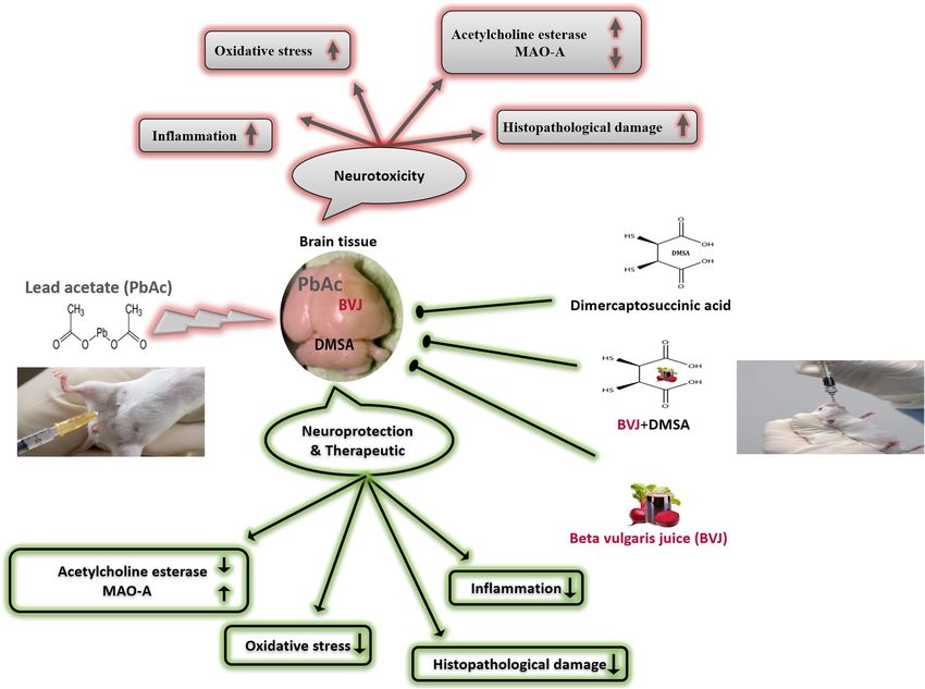
www.nature.com/scientificreports/
Phytochemical constituents Content
Total betalains (mg g−1 BVJ) 5.73 ± 0.04
Betacyanins (mg g−1 BVJ) 3.68 ± 0.07
Betaxanthins (mg g−1 BVJ) 2.04 ± 0.05
Triterpenoids (µg UA eq g−1 BVJ) 25.16 ± 0.722
Flavonols (µg QR Eq g−1 BVJ) 606.40 ± 0.00
Total phenolics (mg GA Eq. g−1 BVJ) 17.26 ± 0.825
Total flavonoids (mg QR Eq. g−1 BVJ) 1.47 ± 0.00
HPLC analysis of phenolic compounds (µg g−1 BVJ)
Pyrogallol 44.095 ± 0.739
p-Hydroxy benzoic acid 191.900 ± 1.572
Chlorogenic acid 10.860 ± 0.548
Caffeic acid 20.915 ± 0.680
Vanillin 8.193 ± 0.380
p-Coumaric acid 6.878 ± 0.201
Salicylic acid 476.725 ± 2.783
Myricetin 3979.998 ± 110.604
Cinnamic acid 3.276 ± 0.146
Quercetin 881.732 ± 4.585
Naringenin 212.253 ± 1.582
Catechol ND
Kaempferol ND
Table 1. Phytochemical content and phenolics HPLC analysis of Beta vulgaris juice (BVJ). The results are
presented as mean ± SE (n = 3). QR quercetin, UA ursolic acid, GA gallic acid, Eq equivalent, ND not detected.
Beetroot (Beta vulgaris L., BV) is a high-nutrient vegetable that belongs to the Chenopodiaceae family. It is
usually consumed as a food and used as a natural food colorant (E162) and a medicinal plant in Europe. The BV
is a great source of antioxidant compounds and it is a nitrate-rich plant contains the water-soluble nitrogenous
pigments named betalains “red betacyanins and the yellow betaxanthins"31. Also, it is a good source of polyphe-
nols, minerals, and vitamins. Recent studies have proved that beet root ingestion shows beneficial physiological
effects and improves several pathologies, such as hypertension, atherosclerosis, type 2 diabetes, cardiovascular
diseases, hepatotoxicity and d ementia26,31. Therefore, in this study, we used BVJ alone and with DMSA in the
treatment of neurotoxicity induced by Pb in male rats. Where BVJ may reduce the OS and apoptosis induced by
Pb, maintain the levels of the essential minerals in the rat body, may improve DMSA therapy, and may enhance
Pb elimination. Likewise, BVJ may ameliorate the hemoglobin level, resulting in the improvement of the circula-
tory system and the cerebrovascular blood flow, which in turn prevent cognitive disorders. The study focused,
for the first time, on the determination of the possible synergistic antineurotoxicity, antioxidant, antiapoptotic
and anti-inflammatory actions of the treatment with (BVJ and DMSA together) against Pb toxicity. Moreover,
the chemical composition and antioxidant potential of the BVJ were evaluated for a better explanation of its
probable antineurotoxicity role.
Results
Characterization of BVJ. The present results showed that BVJ contains various phytochemical constitu-
ents with different concentrations as shown in (Table 1). Where, the phenolics, betacyanins, betalains, betaxan-
thins, flavonoids, flavonols, and triterpenoids are present in a large amounts. Additionally, the high-performance
liquid chromatography (HPLC) analysis of BVJ revealed the presence of many phenolic and flavonoid com-
pounds identified against known phenolic standards by comparing their corresponding retention times (Fig. 1I
and Table 1). Where, Myricetin, Quercetin, Naringenin, Pyrogallol, Caffeic acid and Chlorogenic acid exist in
great amounts while Catechol and Kaempferol are not present (Table 1).
In vitro antioxidant properties of BVJ alone and with DMSA. The results demonstrated the potent
scavenging ability of BVJ to α,α-diphenyl-β-picrylhydrazyl (DPPH), Nitric oxide (NO), superoxide anion
(O2–۰), and hydroxyl (OH−۰) radicals (Fig. 1II–V). The scavenging ability of BVJ to NO radical was significantly
(p ˂ 0.05) superior (lower IC50 value) than that of ascorbic acid (Asc) but was significantly inferior (higher IC50
value) than those of DPPH and OH‾۰radicals. Additionally, the same potency was observed to O2–۰ radical. As
well, DMSA had scavenging power similar to Asc for DPPH, O H‾۰, while its ability to scavenge NO
2–۰, and O
was significantly (p ˂ 0.05) lower than it. Concerning the synergistic analysis, the combination of BVJ and DMSA
(BVJ + DMSA) showed a synergistic (combination index "CI" ˂ 1) scavenging effect for DPPH, NO, and O2–۰. In
H‾· (Table 2). The scavenging power of BVJ
contrast, it exhibited an antagonistic (CI > 1) scavenging effect on O
and DMSA in combination for the NO and O2−۰ radicals was similar to Asc. While The scavenging power of this
combination for DPPH and OH‾ radicals was lower than the Asc.
Scientific Reports | (2021) 11:252 | https://doi.org/10.1038/s41598-020-80669-4 3
Vol.:(0123456789)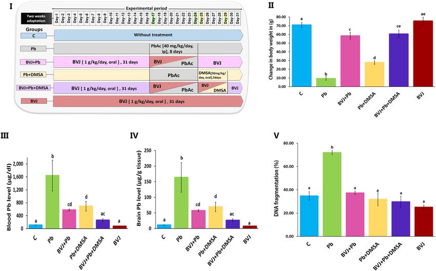
www.nature.com/scientificreports/
Figure 1. HPLC analysis and in vitro antioxidant activities of Beta vulgaris juice (BVJ), dimercaptosuccinic acid
(DMSA), and their combination (BVJ + DMSA) compared to ascorbic acid (Asc). Where (I): HPLC analysis of
BVJ, (II): superoxide radical (O·‾) scavenging activitiy, (III): hydroxyl radical (OH−۰) scavenging activity, (IV):
α,α-diphenyl-β-picrylhydrazyl (DPPH) scavenging activity, and (V) nitric oxide (NO), scavenging activity.
The results are shown as mean ± SE (n = 3). Different letters for the same parameter are significantly different at
p < 0.05.
Groups C Pb BVJ + Pb Pb + DMSA BVJ + Pb + DMSA BVJ
Hb (g/dL) 14.03 ± 0.58ad 9.26 ± 0.58b 13.90 ± 1.73ad 10.84 ± 0.54bc 12.30 ± 0.31 cd 15.77 ± 0.42a
ac b a c ac
RBCs (miL/Cmm) 6.23 ± 0.34 4.31 ± 0.26 6.85 ± 0.72 5.57 ± 0.28 6.21 ± 0.26 7.11 ± 0.31a
ac b ac bc abc
HCT (%) 36.51 ± 0.82 26.64 ± 2.23 35.54 ± 3.93 30.78 ± 1.18 32.88 ± 0.68 39.50 ± 1.04a
a a a a a
MCV (fL) 58.48 ± 3.17 56.37 ± 4.48 51.94 ± 1.02 55.62 ± 1.91 53.20 ± 1.22 55.88 ± 1.27a
a ab b b b
MCH (Pg) 23.73 ± 0.71 21.09 ± 0.74 20.12 ± 0.47 19.48 ± 0.79 19.83 ± 0.46 22.23 ± 0.51a
ab ab ab b ab
MCHC (g/dL) 39.44 ± 1.64 36.94 ± 0.85 38.84 ± 0.56 35.28 ± 1.878 37.35 ± 0.25 39.87 ± 0.36a
Platelet/Cmm 342.00 ± 66.00a 219.00 ± 34.00a 183.00 ± 10.00a 590.00 ± 90.00b 314.00 ± 114.00 a 371.00 ± 103.00 a
WBCs/Cmm 14.32 ± 2.57ab 27.95 ± 4.38b 10.34 ± 3.02a 26.56 ± 5.16b 16.85 ± 1.39ab 9.23 ± 1.33a
Calcium (mg/dL) 9.82 ± 0.31a 8.99 ± 0.19a 9.52 ± 0.14a 9.57 ± 0.63a 9.60 ± 0.09a 9.82 ± 0.29a
Phosphorus (mg/dL) 7.51 ± 0.09a 7.78 ± 0.31a 7.72 ± 0.22a 8.08 ± 0.37a 7.52 ± 0.15a 7.61 ± 0.092a
Sodium (mEq/L) 150.67 ± 2.02a 144.75 ± 2.54a 145.40 ± 2.80a 147.50 ± 3.37a 152.25 ± 1.49a 153.60 ± 2.50a
Potassium (mM) 7.97 ± 1.47a 7.14 ± 0.32a 7.71 ± 0.09a 6.60 ± 0.33a 7.69 ± 0.16a 7.26 ± 0.37a
Table 2. The changes in the hematological parameters and levels of serum electrolytes in all the studied
groups. Hb hemoglobin, RBCs red blood cells, HCT hematocrit "volume of RBCs in blood", MCV mean
corpuscular volume "average volume of RBCs", MCH mean corpuscular Hb "average mass of Hb/RBCs",
MCHC mean corpuscular Hb concentration "concentration of Hb in a given volume of RBCs", WBCs white
blood cells. Results are expressed as mean ± SE (n = 5). Different letters are significantly different for the same
row at P < 0.05.
The protective effects of BVJ alone and with DMSA on rat neurotoxicity induced by Pb. Re-
duction of Pb level and the improvement of rat body weight. As shown in Fig. 2III, IV, the injection of rats with
Pb acetate (Pb group) resulted in a significant (p ˂ 0.05) elevation of Pb levels in the blood and brain compared
to the control group. In addition, the change in the body weight of rats after 31 days was greatly reduced than the
control (Fig. 2II). Administration of BVJ before, during, and after the injection of Pb acetate (BVJ + Pb group)
Scientific Reports | (2021) 11:252 | https://doi.org/10.1038/s41598-020-80669-4 4
Vol:.(1234567890)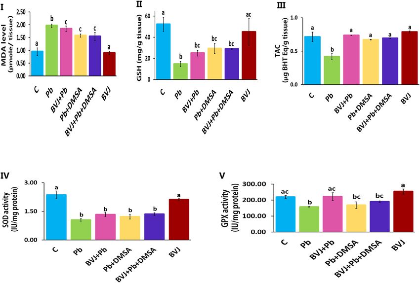
www.nature.com/scientificreports/

                                 Figure 2.  The ameliorating effects of Beta vulgaris juice (BVJ), dimercaptosuccinic acid (DMSA), and their
                                 combination (BVJ + DMSA) on the blood and brain Pb and DNA fragmentation in Pb-intoxicated rats. Where
                                 (I): An illustration of the current experimental design, (II): the change in the body weight of rats in all the
                                 studied groups, (III): the blood in all the studied groups, (IV): the brain Pb levels in all the studied groups, and
                                 (V) the % of DNA fragmentation level in all the studied groups. The results are shown as mean ± SE (n = 8).
                                 Different letters for the same parameter are significantly different at p < 0.05.C control,; ip intraperitoneal.

                                 obviously reduced Pb levels in both the blood and brain. Similarly, treatment with DMSA after Pb injection
                                 (Pb + DMSA group) decreased Pb levels in both the blood and brain. Likewise, the change in body weight was
                                 significantly (p ˂ 0.05) improved in rats of (BVJ + Pb) group more than rats in (Pb + DMSA) group as compared
                                 to the Pb group. Treatment of rats with BVJ and DMSA together (BVJ + Pb + DMSA group) revealed a highly
                                 synergistic depleting effect (CI ˂ 1) of the Pb level in blood and brain (Table 3). Also, these treatments caused the
                                 synergistic elevation in the body weight change as compared with Pb group. Otherwise, the administration of
                                 BVJ alone to healthy rats caused non-significant changes in either the Pb level or the body weight.

                                 Reduction of DNA fragmentation (DNAF) induced by Pb toxicity. The current findings revealed that Pb injec-
                                 tion increased significantly (p ˂ 0.05) the DNAF level in rat brain tissue (Fig. 2V). However, the treatments with
                                 BVJ, DMSA, or their combination after Pb injection, significantly (p ˂ 0.05) reduced DNAF induced by Pb as
                                 compared with the Pb group. Where, the DNAF level became near to the control level in case of BVJ treatment,
                                 while it became lower than the control level after treatment with DMSA, or (BVJ and DMSA). The CI study
                                 revealed synergistic (CI ˂ 1) protective effect of the investigated combination of BVJ and DMSA against the
                                 elevation in DNAF induced by Pb toxicity (Table 3). BVJ administration to healthy rats caused decreased DNAF
                                 level as compared to the control group.

                                 Improvement the changes in hematological parameters and serum electrolytes induced by Pb toxicity. The results
                                 disclosed that Pb administration affects the blood picture by decreasing the levels of hemoglobin (Hb), red blood
                                 cells (RBCs), and hematocrit (HCT) as well as elevating white blood cells (WBCs) (Table 2). Treatment with
                                 BVJ before, during, and after Pb injection improved the blood picture which changed by Pb toxicity. In contrast,
                                 treatment with DMSA after Pb injection was unable to improve the blood picture as in BVJ treatment.
                                     Hence, the levels of Hb, RBCs, HCT, and mean corpuscular Hb concentration (MCHC) were significantly
                                 (p ˂ 0.05) decreased, while the levels of platelets and WBCs were significantly (p ˂ 0.05) increased. On the other
                                 hand, treatment with BVJ and DMSA together after Pb injection improved the blood criteria. The results also
                                 revealed that BVJ administration to the healthy rats did not change the blood picture (Table 2). Likewise, the
                                 results demonstrated that there were non-significant changes in the electrolyte concentrations in all studied
                                 groups (Table 2).

Scientific Reports |   (2021) 11:252 |                https://doi.org/10.1038/s41598-020-80669-4                                                        5

                                                                                                                                                Vol.:(0123456789)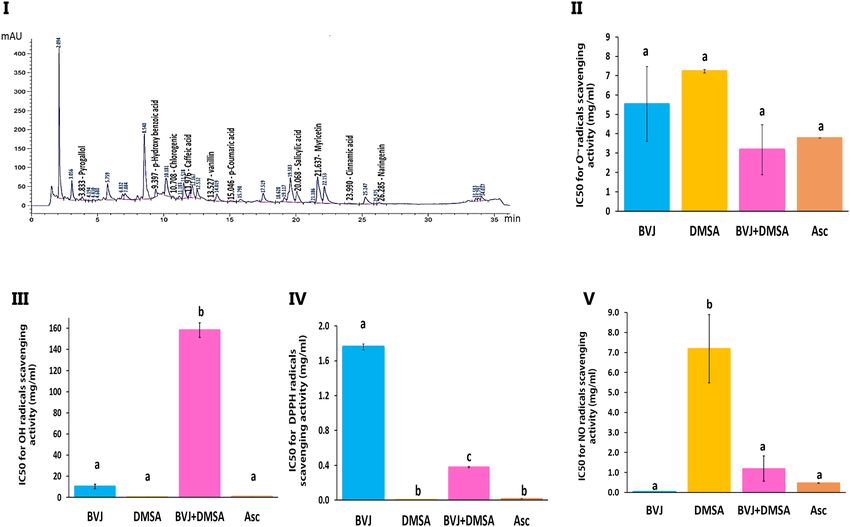
www.nature.com/scientificreports/
Effect CI Parameters
In vitro antioxidant models
Synergistic 0.430 ± 0.000 DPPH (mg/mL)
Synergistic 0.477 ± 0.134 NO (mg/mL)
Antagonistic 31.578 ± 7.523 Hydroxyl radical (mg/mL)
Synergistic 0.397 ± 0.272 Superoxide radical (mg/mL)
Change in body weight
Synergistic 0.412 ± 0.041 Body weight (g)
Lead level
Synergistic 0.037 ± 0.012 Blood lead level (µg/dL)
Synergistic 0.090 ± 0.030 Brain lead level (µg/g tissue)
Serum electrolytes
Synergistic 0.856 ± 0.036 Phosphorus (mg/dL)
Synergistic 0.941 ± 0.027 Sodium (mEq/L)
Synergistic 0.975 ± 0.017 Calcium (mg/dL)
Synergistic 0.795 ± 0.087 Potassium (mmol/L)
Oxidative stress parameters
Additive 1.000 ± 0.037 TAC (µg BHT Eq/g tissue)
Synergistic 0.429 ± 0.044 Lipid peroxidation (pmol/g tissue)
Synergistic 0.563 ± 0.062 GSH (mg/g tissue)
Synergistic 0.860 ± 0.054 SOD (IU/mg protein)
Synergistic 0.820 ± 0.078 GPX (IU/mg protein)
Inflammatory parameters
Synergistic 0.382 ± 0.077 IL-6 (pg/g tissue)
Synergistic 0.715 ± 0.067 NO (mmol/g tissue)
Neurotransmitters-associated enzymes
Synergistic 0.563 ± 0.161 MAO-A (ng/mg protein)
Synergistic 0.477 ± 0.047 AChE (pmol/mg protein)
DNA fragmentation
Synergistic 0.876 ± 0.234 DNA fragmentation (%)
Table 3. The Combination index (CI)# values for the studied parameters on the mixture of Beta vulgaris
juice (BVJ) and meso-2,3-dimercaptosuccinic acid (DMSA). # CI value of < 1 shows synergistic effect; > 1
shows antagonistic effect; = 1 shows additive effect. TACtotal antioxidant capacity, DPPH α, α-diphenyl-β-
picrylhydrazyl, NO nitric oxide, GSH Reduced Glutathione, SOD superoxide dismutase, GPX glutathione
peroxidase, IL-6 Interleukin-6, MAO-A monoamine oxidase-A, AChE acetylcholine esterase.
BVJ reduced the OS induced by Pb in rat brain. The injection of Pb into rats dramatically increased the level of
lipid peroxidation (Fig. 3I) and decreased the level of GSH and total antioxidant capacity (TAC) (Fig. 3II, III,
respectively), and the activity of SOD and GPx (Fig. 3IV, V, respectively). Treatment with BVJ before, during, and
after Pb injection showed significant (p ˂ 0.05) suppression in lipid peroxidation level and substantial elevation
as well as normalization of TAC level and GPx activity relative to the Pb group. Also, this treatment increased
GSH level (significantly) (p ˂ 0.05) and SOD activity (non-significantly) as compared to the Pb group. While the
administration of DMSA after Pb injection was significantly (p ˂ 0.05) lowered the lipid peroxidation induced
by Pb in brain tissue, but its level was still higher than the control. Additionally, this treatment improved signifi-
cantly the TAC and GSH levels and GPx activity and also, SOD activity (non-significantly) as compared to the
Pb group where some parameters became near to their controls.
Similarly, treatment with BVJ and DMSA together influenced on the antioxidant indices. Regarding the CI
value, the combination of BVJ and DMSA had synergistic (CI ˂ 1) action on the OS parameters, except for TAC
that showed an additive (CI = 1) effect (Table 3). On the other hand, the healthy rats administered with BVJ alone
for 31 days reported no significant changes in the OS indices in the brain tissue.
BVJ reduced the inflammation induced by Pb in rat brain. The findings reported that Pb injection did not influ-
ence on the NO level of brain tissue (Fig. 4I). Likewise, treatment with either, (BVJ and DMSA together) or
DMSA after Pb injection showed no significant changes in the brain NO level. Also, administration of healthy
rats with BVJ after Pb injection appeared no significant change in the brain NO level.
Otherwise, the interleukin (IL)-6 level was significantly (p ˂ 0.05) elevated in rat brain tissues which injected
with Pb (Fig. 4II). Treatment with BVJ, (BVJ and DMSA together), or DMSA after Pb injection dramatically
inhibited the elevation in IL-6 comparable to the Pb group, but the level stilled higher than the control. The CI
analysis demonstrated the synergistic (CI ˂ 1) effects of the combination between BVJ and DMSA on depleting
Scientific Reports | (2021) 11:252 | https://doi.org/10.1038/s41598-020-80669-4 6
Vol:.(1234567890)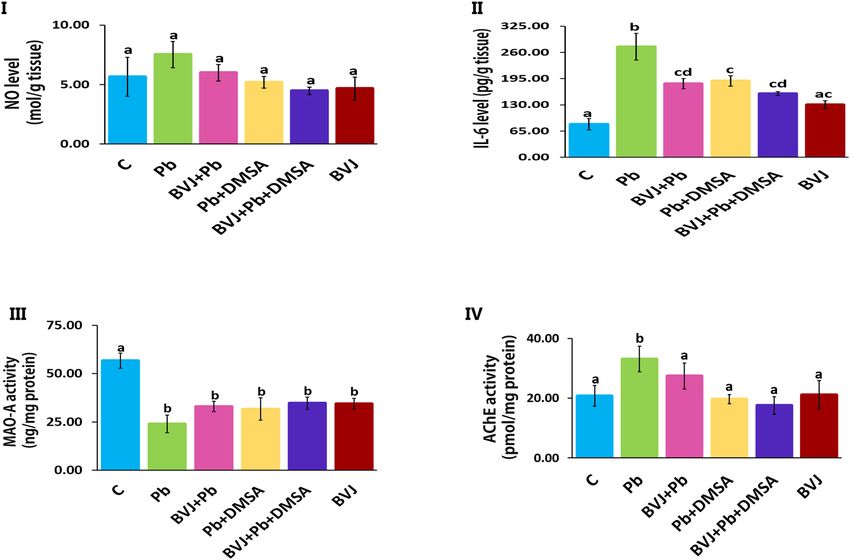
www.nature.com/scientificreports/

                                 Figure 3.  The alleviating influences of Beta vulgaris juice (BVJ), dimercaptosuccinic acid (DMSA), and their
                                 combination (BVJ + DMSA) on Pb-induced oxidative stress in the brain tissue of Pb-intoxicated rats. Where (I):
                                 lipid peroxidation level in the brain tissue of all the studied groups, (II): reduced glutathione (GSH) level in the
                                 brain tissue of all the studied groups, (III): total antioxidant capacity (TAC) in the brain tissue of all the studied
                                 groups, (IV): the activity of superoxide dismutase (SOD) in the brain tissue of all the studied groups, and (V)
                                 glutathione peroxidase (GPX) in the brain tissue of all the studied groups. The results are shown as mean ± SE
                                 (n = 8). Different letters for the same parameter are significantly different at p < 0.05.C control.

                                 the levels of both NO and IL-6 in the brain tissue (Table 3). Further, the administration of BVJ to healthy rats
                                 caused a slight elevation in IL-6 level as compared with the control group.

                                 BVJ improved the disturbance in the activities of monoamine oxidase‑A and acetylcholine esterase in rat brain. The
                                 results showed that Pb intoxication caused a drastic depletion of the monoamine oxidase A (MAO-A) activity
                                 with a significant elevation in acetylcholine esterase (AchE) activity as compared to the control (Fig. 4III, IV).
                                 However, the treatments with BVJ, (BVJ and DMSA together) or DMSA did not significantly protect the deple-
                                 tion of MAO-A activity comparable to the Pb group. In contrast, these treatments significantly prevented the
                                 unusual increase of AChE activity by Pb and preserved its activity close to the control. The CI values of treatment
                                 with BVJ and DMSA together for the activities of MAO-A and AChE, were less than one (Table 3). Therefore,
                                 BVJ displayed a synergistic protective effect on both enzymes. Also, the intake of BVJ alone to the healthy rats
                                 was able to dramatically decrease the activity of MAO-A, but did not influence on AChE activity as compared
                                 to the control.

                                 Histopathological results. In this study three areas of the brain were chosen for histopathological study,
                                 cerebellum (Fig. 5I), cerebral cortex (Fig. 5II), and hippocampus (Fig. 6). The brain sections taken from the
                                 control rats (C group) displayed normal cerebellum, cerebral cortex, and hippocampus structures. The light
                                 photomicrographs showed the proper cerebellar architecture with three cortical layers "granular, Purkinje, and
                                 molecular" and a white matter layer. The cerebral cortex revealed typical pyramidal nerve cells, glial cells, and
                                 blood vessels. Similarly, the hippocampus showed normal pyramidal nerve cells.
                                      The microscopic examination of the brain sections of rats injected with Pb (Pb group) has detected severe
                                 damage in the three brain regions. The cerebellum showed serious destruction of Purkinje cells, neuronal necro-
                                 sis, serious molecular degeneration, as well as serious white matter spongiosis with vacuolization. Multiple altera-
                                 tions were detected in the cerebral cortex such as vacuolization of pyramidal cells, congestion of blood vessels,
                                 and focal reactive gliosis. Moreover, degenerated neurons, microgliosis, neuronal necrosis, swollen of some
                                 degenerate axons, pyknosis of glial cells, severe spongiosis with vacuolation, and with many other disturbances

Scientific Reports |   (2021) 11:252 |                https://doi.org/10.1038/s41598-020-80669-4                                                      7

                                                                                                                                                 Vol.:(0123456789)
www.nature.com/scientificreports/
Figure 4. The alleviating effects of Beta vulgaris juice (BVJ), dimercaptosuccinic acid (DMSA), and their
combination (BVJ + DMSA) on Pb-induced inflammation and disturbance to the neurotransmitters-associated
enzymes in brain tissue of Pb-intoxicated rats. Where (I): nitric oxide (NO) level in the brain tissue of all the
studied groups, (II): interleukin (IL)-6 level in the brain tissue of all the studied groups, (III): the activity
of monoamine oxidase (MAO)-A in the brain tissue of all the studied groups, and (IV): the activity of
acetylcholine esterase (AChE) in the brain tissue of all the studied groups. The results are shown as mean ± SE
(n = 8). Different letters for the same parameter are significantly different at p < 0.05. C control.
were detected in this brain area. The hippocampus revealed extreme necrosis and neuronal loss with pyramidal
cell vacuolation, serious vacuolation, and neuronal pyknosis.
Otherwise, treatment with BVJ before, during and after Pb injection (BVJ + Pb group) maintained normal
cerebellar architecture, except for a small loss of Purkinje cells. The cerebral cortex showed few vacuolated
pyramidal nerve cells and congested blood vessels with normal glial cells, while the hippocampus disclosed few
neuronal loss and neuronal pyknosis.
Also, the results showed that the treatment with DMSA after Pb injection (Pb + DMSA group) restored most
of Pb toxicity in the three investigated brain areas. In the cerebellum, only moderate disturbance in Purkinje
cells, molecular layer and white matter were noted. The microscopic observation of the cerebral cortex showed
moderate vacuolation of pyramidal neurons, congestion of the blood vessels, neuronal necrosis, apoptotic bod-
ies, and pyknosis of glial cells with other damage. The hippocampus demonstrated moderate necrosis, neuronal
loss with vacuolization of pyramidal nerve cells, and neuronal pyknosis.
The administration of the combination of BVJ and DMSA together (BVJ + Pb + DMSA group) after Pb injec-
tion reported a slight loss of Purkinje cells with mild neuronal necrosis in the cerebellar sections. However,
there were few vacuolated pyramidal nerve cells with normal glial cells and blood vessels in the cerebral cortex.
Moreover, the results observed normal glial cells, few vacuolated pyramidal cells with mild necrosis, some neu-
ronal pyknosis, and mild congestion of blood vessels in the hippocampus.
Otherwise, the administration of BVJ alone for healthy rats for 31 days showed normal cerebellar architec-
ture with normal cortical layers and white matter. Also, it observed normal cerebral cortex and hippocampus
architectures in the brain sections of these rats.
Results summary. All results obtained in this study were summarized in Fig. 7.
Discussion
The brain is the most critical organ for lead toxicity9. Both of the PNS and the CNS are affected by lead toxicity19
but, the mechanisms of lead neurotoxicity are complex and still not clearly u nderstood17,32. However, in adults, the
PNS is more affected, while in children, the CNS is more a ffected33. It is well identified that Pb-induced neuronal
Scientific Reports | (2021) 11:252 | https://doi.org/10.1038/s41598-020-80669-4 8
Vol:.(1234567890)www.nature.com/scientificreports/
Figure 5. Representative photomicrographs of the cerebellum and the cerebral cortex in the brain of control
(C) and Pb-intoxicated rats with and without administration of Beta vulgaris juice (BVJ), dimercaptosuccinic
acid (DMSA), or their combination (BVJ + DMSA). Where (I): photomicrographs of the cerebellum, and (II):
photomicrographs of the cerebral cortex. ab apoptotic bodies, BV blood vessel, CBV congested blood vessel,
D degeneration, G glial layer, gc glial cells, H hemorrhage, L.pc loss of Purkinje cells, M molecular layer, Mi
microgliosis, NA necrotic area, n. C neuronal chromatolysis, pc Purkinje cells, W white matter layer, n.N
neuronal necrosis, O osseous metaplasia, P pyknosis, Py pyramidal cells, R reactive gliosis, S spongiosis, sda
swollen degenerate axons, V vacuolization, VPy vacuolized pyramidal cells, × 40 magnification of the image.
injury is correlated with oxidative challenge, neuroinflammation, and the enhancement of programmed cell
death, which may play an important role in the development of the neurodegenerative diseases. The Pb capability
to substitute for the calcium ions, allowing its passage across the blood–brain barrier and its accumulation in
the brain via calcium-ATPase pumps19. Where the increasing in Pb intake may lead to the reduction of calcium
steady state34. Consequently, both the peripheral and central nervous systems are d isrupted34. This substitution
may results in many neurological disorders including mental retardation, brain damage, behavioral problems,
and may lead to several diseases like Alzheimer, Parkinson, and Schizophrenia35.
The histopathological results of this study (Figs. 5, 6) revealed extreme damage in the cerebellum, cerebral
cortex, and hippocampus parts of the brain, which confirm the serious damage induced by Pb in the b rain36.
The histopathological results are in harmony with the biochemical outcomes. Where, the data showed a sig-
nificant elevation in MDA level with significant reduction in GSH level and the activities of SOD and GPx in
in rat brain homogenate after Pb administration when compared to the control (Fig. 3). This indicates that
lead induced OS in brain tissues, therefore, Pb administration led in turn to a drastic decrease in TAC of brain
tissue (Fig. 3). OS is a balance loss of ROS production and elimination in tissues and cellular components. In
the cells, MDA is one of the eventual products of peroxidation of polyunsaturated fatty acids. A rise in the free
radicals, such as O2–۰, and hydrogen peroxide (H2O2) radicals, causes the overproduction of MDA37 MDA is
well recognized as an indicator of OS and antioxidant status and an elevation in its level is a substantial index
eroxidation37. In the body, SOD initiates the defense action versus O
of lipid p 2–۰ radicals since it is believed as
the most effective antioxidant. GPx provides a second way of protection against organic hydroperoxides and
inorganic (and ROOH and H2O2 respectively). Where GPx, in the incidence of GSH, stimulates the reduction
of these hydroperoxides. GSH works as a nucleophilic scavenger of many compounds through chemical and
enzymatic mechanisms. In this study, the depletion of GSH may be related to the increased demand of GSH
for lipid hydroperoxide metabolism to terminate free radical reactions. While GSH depletion could contribute
eroxidation37. The peroxidation of lipid contents of the membrane leads to a lack of
to the activation of lipid p
Scientific Reports | (2021) 11:252 | https://doi.org/10.1038/s41598-020-80669-4 9
Vol.:(0123456789)www.nature.com/scientificreports/
Figure 6. Representative photomicrographs of the hippocampus in the brain of control (C) and Pb-intoxicated
rats with and without administration of Beta vulgaris juice (BVJ), dimercaptosuccinic acid (DMSA), or their
combination (BVJ + DMSA). ab apoptotic bodies, CBV congested blood vessel, gl granular layer, N necrosis,
nL neuronal loss, P pyknosis, Py pyramidal cells, V vacuolization, VPy vacuolized pyramidal cells, × 40
magnification of the image.
cell perfection, an increment in membrane permeability, and alteration of C a2+ homeostasis that donate to cell
death. In addition, the lipid peroxidation caused the destruction of mitochondrial through the formation of the
permeability transition pore, and priming activation of a poptosis38. Therefore, our results revealed an increase
in DNAF after Pb administration (Fig. 2V). The fundamental mechanism underlying Pb-induced oxidative
damage to membranes is linked with changes in its fatty acid composition39. Where Pb induces the elongation
of arachidonic acid which might be accountable for the increased lipid p eroxidation39. The second mechanism
for Pb-induced OS is the inhibition of the antioxidant defense systems of cells. Pb and other metals such as Cd
and Hg have a high affinity for sulfhydryl (SH) groups. Mercaptides are created with the SH group of cysteine,
which are less stable c omplexes8. So, Pb alters the antioxidant activities by inhibiting functional SH groups in
several enzymes such as δ-aminolevulinic acid dehydrase, SOD, catalase, and GPx. In addition, GPx, CAT, and
SOD are possible targets for lead toxicity because these enzymes depend on different essential trace elements
for proper molecular structure and a ctivity39. Inhibition the δ-aminolevulinic acid d ehydrase40 leading to the
accumulation of δ-aminolevulinic acid. δ-aminolevulinic acid resembles and competes with γ-aminobutyric
acid, a neurotransmitter in the hypothalamus, cortex, and other nervous system tissues. This can stimulate
γ-aminobutyric acid receptors and induce n eurotoxicity8 through generating H 2O2 and O 2•− radicals as well as
•− 41 41
binding to oxyHb producing O H radicals resulting in the induction of the O S . 4,5-dioxovaleric acid, (the
final oxidation product of δ-aminolevulinic acid) is an effective alkylating agent and play an important role in
DNA damage39. Moreover, lead accumulation in tissues has reported inducing oxidative DNA damage, through
strand break38. Moreover, other studies suggested that Pb induces alteration in gene e xpression35 and it appears
to react with zinc-binding sites on an important DNA-linked protein, human p rotamine35.
Additionally, the present results showed that Pb administration caused significant reductions in the rat body
weight and the levels of Hb, RBCs, and HCT with a significant increase in WBCs as compared the control
(Table 2). However, the levels of minerals were non-significantly changed after Pb administration (Table 2).
The reduction in Hb may be related to inhibition of its biosynthesis through the inhibition of δ-aminolevulinic
acid synthase, δ-ALA dehydratase and ferrochelatase by P b40. Additionally, Pb can bind with RBCs and inhib-
42
its pyruvate kinase "keeps cellular energy homeostasis" and pyrimidine 5′-nucleotidase (essential for RBCs
Scientific Reports | (2021) 11:252 | https://doi.org/10.1038/s41598-020-80669-4 10
Vol:.(1234567890)www.nature.com/scientificreports/
Figure 7. Graphical abstract.
maturation)43. The inhibition of these enzymes destabilizes the RBCs membrane and decreases its fluidity leading
to a shortage of the erythrocyte life span, hemolytic anemia, and ROS g eneration42,43. High levels of ROS lead
to oxidation of the cellular biomolecules, especially, the biomembrane lipids causing lipid peroxidation44. These
results are agreed with the previous s tudies18,26,32,36,45.
Inflammation is considered as a beneficial process under normal condition via controlling the innate response
to biological or physical triggers such as trauma, infection and other pathogens that may disrupt homeostasis
and cause d iseases46. Cytokines include interleukins, chemokines and other signalling molecules which can be
classified into pro and anti-inflammatory m ediators46,47. These mediators are working together in balance to
perform the overall effect of the physiologically inflammatory response for repairing cells and tissue injuries.
Nevertheless, the disturbance of cytokines’ balance leading to inappropriate activation of inflammatory pro-
cesses that can cause excessive cell and tissue damage ultimately leading to many pathological conditions46. Our
results revealed that Pb administration caused a significant elevation in the level of interleukin-6 (IL-6) in the
brain tissues compared to the control group (Fig. 4II). This may be due to increasing its expression in response
to Pb as suggested by the previous studies48. IL-6 is one of the potent pro-inflammatory cytokines that recruit
neutrophil influx and induce prostaglandin synthesis, activation, and proliferation of immune cells. This may
have a dangerous effect on the differentiation and growth of n eurons49.
MAO is an intracellular flavin-containing enzyme that catalyzes the oxidative deamination of monoamine
neurotransmitters like norepinephrine and epinephrine in the brain and peripheral t issues50. MAO is local-
ized on the outer membrane of mitochondria regulating the extracellular concentration of monoamines50. In
addition, AChE is the key enzyme in the nervous system responsible for the degradation of acetylcholine “an
important neurotansmitor” during neurotransmission, which is necessary for learning and memory51. Lead
toxicity is known to decrease M AO52 and AChE activities in the crude homogenate of the rat b rain5,53. It is
thought that the inhibition of AChE and MAO activities under the effect of Pb is caused by the binding of met-
als to the functional groups of proteins like imidazole, sulfhydryl, and c arboxyl8. Our results exhibited that Pb
administration significantly decreased MAO-A activity but significantly increased AChE activity. The activation
of AChE activity by Pb toxicity is in agreement with Ghareep et al. (2010) and others54–56. Till now the causes
of the activation of AChE by Pb are not known. It may be due to the effect of Pb on the gene expression of this
enzyme. So many studies must be carried out in this field. The previous studies revealed that Pb inhibits MAO
activity and this inhibition is dose and its duration dependent. The change in MAO activity leads to various
Scientific Reports | (2021) 11:252 | https://doi.org/10.1038/s41598-020-80669-4 11
Vol.:(0123456789)www.nature.com/scientificreports/
mental and neurodegenerative disorders52. Also, activation of AchE may lead to alteration in the exploratory
behavior and locomotor activity of rats54.
However, it is known that Pb intoxication can be effectively treated using chelation therapies such as DMSA.
Previous studies showed that DMSA has some drawbacks including, CNS convulsions, essential metal loss, hepa-
totoxicity, nephrotoxicity, headache, nausea, hypertension, respiratory arrest, skin rashes, thrombocytopenia,
and neutropenia25,26. Therefore, in the present study, the protective role of BVJ on rat neurotoxicity induced by
Pb was investigated and the results were compared with those resulted from the treatment with DMSA. Besides,
the possible synergistic antineurotoxicity action of the combination between BVJ and DMSA was determined.
Additionally, the phytochemical components of BVJ and its characterization were identified to be used in the
discussion of the results of the biological role of BVJ against Pb-induced brain toxicity.
The present results showed that BVJ contains various phytochemical constituents with different concentra-
tions as shown in (Table 1). Where, the phenolics, betacyanins, betalains, betaxanthins, flavonoids, and flavonols
are present in a large amount. Additionally, HPLC analysis revealed the presence of many phenolic acids (Table 1).
Since, Myricetin, Quercetin, Naringenin, Pyrogallol, Caffeic acid and Chlorogenic acid are present in great
amounts (Table 1). Also BVJ contains carotenoids and vitamins31, especially vitamin C 26. All these compounds
have antioxidant activities and protective role for cellular components against OS and can improve the clinical
outcomes for several d iseases31. Owed to these constituents, BVJ observed antiradical activity against DPPH, NO,
O-·, and O H‾· radicals as shown in Fig. 1. The present results were in harmony with some previous s tudies26,57.
Also, BVJ contains minerals such as iron and zinc31. It has been demonstrated that dietary supplement or foods
rich in calcium, zinc, magnesium, and phosphate may decrease the absorption of Pb58.
Likewise, the current data showed that DMSA has antioxidant efficiency against the tested free radicals
(Fig. 1) and this may be related to its structure that contains –SH groups "dithiol compound"59. DMSA revealed
the higher potency than BVJ toward DPPH and O H‾· radicals, while BVJ showed better affect against NO radi-
cal. Moreover, the results showed that the combination between DMSA and BVJ had more efficacy (synergistic
action, CI ˂ 1) than that of DMSA or BVJ in a separate form, except for the O H-۰ radical (Table 3). In general,
these results demonstrated that the combination between DMSA and BVJ will increase the therapeutic potential
against Pb toxicity.
On the other hand, the present results revealed that the level of Pb in blood and brain was significantly
reduced in rats treated with BVJ before, during, and after Pb administration as compared to the Pb group, and
thus leading to the normalization of the blood parameters (Table 2). This indicates that the active gradients of
BVJ such as polyphenols, phenolic acids and fl avonoids60, and b etalains26,61 may act as metal chelators1. These
results agree with previous studies which showed that BVJ prevented silver pathological effects and restored
renal functions levels to the control values via interacting with silver ions facilitating their clearance62. Besides it
showed hepatoprotective activity aganit pb toxicity by reducing blood and liver Pb level and reducing the O S26.
Also, the activity of AChE was not being elevated, but the activity of MAO-A was non-significantly elevated
relative to the Pb group (Fig. 4III, IV). The efficiency of BVJ as a chelator and its effect on the neurotransmitter
enzymes was similar to DMSA that used here as a standard chelator for Pb. However, DMSA revealed a slight
improvement on the blood parameters and no effect on the tested minerals (Table 2).
Furthermore, the current results elucidated an attenuation of brain damage induced by Pb in rats treated with
BVJ before, during and after Pb administration as shown from the results of brain histopathology. Where, slight
damage appears in the morphology of the three studied regions of the brain in rats treated with BVJ before, dur-
ing and after Pb administration. In addition, BVJ treatment decreased lipid peroxidation and apoptosis induced
by Pb since the levels of MDA, NO, and DNAF were decreased with an elevation in GSH level, and the activities
of GPx, and SOD as compared with the Pb group. This indicates that BVJ has antioxidant, radical-scavenging
properties and anti-apoptotic activity which reduce the programmed cell death. This conclusion was confirmed
by our results which showed that the total antioxidant activity of BVJ is very high. The antioxidant and antia-
poptotic effects of BVJ are related to the effects of its bioactive ingredients which are mentioned before (Table 1).
Several studies showed that the antioxidative characteristics of plant polyphenols may arise from their reactiv-
ity as an electron or hydrogen donors, their ability to stabilize unpaired electrons, and their ability to end Fenton
reactions63. In general, the polyphenolic compounds can prevent oxidative damage as a result of their ability to
scavenge ROS. Also, they improve GSH level and the activities of SOD and GPx64. Moreover, treatment with
BVJ before, during and after Pb administration diminished the inflammation in brain tissues induced by Pb, as
obvious from the reduction of IL-6 level as compared with the Pb group (Fig. 4II). This indicates that BVJ has
anti-inflammatory activity which may be due to the effect of its bioactive compounds (Table 1). These results
agree with the previous studies which showed that BVJ has anti-inflammatory a ctivity20,61.
Otherwise, the results showed that treatment with DMSA after Pb intoxication reduced the Pb level in brain
tissues and blood. Also, the results showed that DMSA significantly reduced lipid peroxidation level and DNAF
induced by Pb toxicity and restored the TAC of the brain tissue, but had no effect on the level of GSH and the
activity of GPX and SOD (Fig. 3). This indicates that thiol groups in DMSA play an important role as chelating
agent for removing Pb besides its role as antioxidant. These results are in accordance with the previous findings
of Ercal et al.59. In addition, treatment with DMSA reduced the IL-6 level as compared with Pb group. Also, after
this treatment the histopathological findings showing moderate damage in the morphology of the three studied
regions of the brain (Figs. 5, 6).
In general, the present study not only proved the protective effect of BVJ against Pb toxicity, but also the safety
of its administration for healthy rats for 31 days on rat’s brain tissue. This was confirmed by the biochemical
and histopathological outcomes. Only, the activity of MAO-A was significantly decreased as compared to the
control. This may be owed to the presence of quercetin (Table 1; Fig. 1I) orits derivativesin BVJ constituents.
These flavonoid compounds can act as MAO inhibitors and used in the therapy of depression, anxiety, and
neurodegenerative disorders65.
Scientific Reports | (2021) 11:252 | https://doi.org/10.1038/s41598-020-80669-4 12
Vol:.(1234567890)www.nature.com/scientificreports/
On the other hand, we also evaluated the protective effect of the combination between BVJ and DMSA against
Pb-induced neurotoxicity. From the value of CI (Table 3), this combination exhibited synergistic action (CI ˂ 1)
towards all the studied parameters except for the TAC which showed additive effect (CI = 1). As previously pub-
lished, DMSA is an effective Pb chelator, but various side effects are associated with its t herapy66. In contrast,
BVJ is safe and effective in protecting from Pb toxicity the combination of both BVJ and DMSA enhanced their
efficacy leading to the improvement of DMSA therapy. Therefore, this study suggested the use of BVJ with DMSA
during the treatment of lead poisoning, while BVJ can be used alone for protection.
In summary, our findings clearly revealed that BVJ has a vigorous efficiency in the protection from Pb-induced
toxicity in brain tissue by reducing blood and brain Pb and preventing oxidative and inflammatory stress. The
combination of both BVJ and DMSA revealed synergistic antioxidant and anti-inflammatory, antiradical and
chelating potentials against neurotoxicity induced by Pb. Therefore, BVJ is a promising extract in protection from
Pb toxicity and its combination with DMSA exerted a potent therapeutic effect for this damage.
Materials and methods
Chemicals. Gallic acid, catechin, quercetin, ursolic acid, Butylated hydroxytoluene, DMSA, thiobarbitu-
ricacid (TBA), Folin–Ciocalteau reagent, reduced GSH, DPPH, 2,2′-azino-bis(3-ethylbenzothiazoline-6-sul-
phonic acid) (ABTS) and bovine serum albumin (BSA) were purchased from Sigma-Aldrich (St. Louis, MO,
USA). Lead acetate was obtained from ISO-CHEM, France. Potassium, sodium, and phosphorous colorimetric
kits were obtained from Spectrum, Egyptian Company for Biotechnology (S.A.E), Egypt. Calcium colorimetric
kit was purchased from Biosystems (Barcelona, Spain). Rat interleukin (IL)-6 ELISA kit was obtained from Ray
Biotech (Norcross, USA). The MAO-A ELISA kit was supplied from FIVE photon Biochemicals (California,
USA). Amplite colorimetric AchE assay ELISA kit was purchased from AAT Bioquest, Inc. (California, US).
Other chemicals were obtained with a high grade.
Juice preparation. The BV plant roots (NCBI: txid 3555) were obtained from the Local market in Alexan-
dria, Egypt, during September, then the roots were washed and peeled. The BVJ was prepared using a household
dry juice extractor then the extract was dried using the Lyophilizer (Telstar, Terrassa, Spain) to obtain the pow-
dered juice (BVJ, yield 13 g/100 mL BVJ), which stored at − 20 °C until used.
Quantification of BVJ phytochemicals content. Some of the phytochemical compounds in BVJ were
determined, including betalains, phenolics (flavonoids and flavonols), and triterpenoids. Total betalains in BVJ
was determined as the sum of betacyanins and betaxanthins concentrations according to the method of Stintzing
et al.67. The diluted solution (1:3 wt/v) of BVJ was measured at three different wavelengths, 536 nm, 485 nm, and
650 nm (impurities), then the concentrations of betacyanins or betaxanthins were calculated using the following
formula:
A × DF × M.W × 1000
betacyanins or betaxanthins (mg/L) = ,
ǫ×i
where, A = A536–A650 (for betacyanins) or A485–A650 (for betaxanthins), DF: dilution factor, M.W: molecular
weight of betacyanins or betaxanthins (550 and 336 g/mol, respectively), ǫ : molar extinction coefficient for
betacyanins or betaxanthins (60,000 and 48,000 L/mol cm, respectively), i: path length (cm).
Total phenolics in mg equivalents of GA/g BVJ were quantified using Folin–Ciocalteu reagent, which was
reduced by BVJ phenols producing a blue color solution with a maximum absorbance at 750 nm68. Total flavo-
noids (mg catechin equivalents/g BVJ) were measured using 10% A lCl3 and 5% N aNO2 solutions69 While total
flavonols (mg QR equivalents/g BVJ) were estimated using 2% AlCl3 and 50 g/L sodium acetate solutions70.
The triterpenoids (µg UA equivalents/g BVJ) were determined colorimetrically using vanillin color r eaction71.
Investigation of the phytochemical compositions of BVJ using HPLC. Phytochemical compo-
nents of BVJ were identified and quantified by agilent 1260 infinity HPLC series (Agilent Technologies, Palo
odification72. Briefly, 20 µL of BVJ was separated on a Kine-
Alto, CA, USA) as indicated previously with slight m
tex EVO C18 column (100 mm × 4.6 mm) using a ternary linear elution gradient. The elution was performed
using 0.2% H
3PO4, methanol, and acetonitrile and the detection was done at 284 nm. Pure standards were run
in the same chromatographic conditions to match the retention items.
In vitro antioxidant activities of BVJ, DMSA and their combination. The antioxidant activities
of BVJ, DMSA, and their combination (BVJ + DMSA, 20:1) were evaluated using various methods, including,
DPPH, NO, O 2−۰and OH‾·radicals scavenging activities. The antioxidant activities were compared between BVJ,
DMSA, and (BVJ + DMSA) using Asc as a standard antioxidant. The I C50 value (50% inhibitory concentration)
for each radical was determined using the % of inhibition values [(AControl − ASample/AControl) × 100] at different
studied samples (BVJ), (DMSA), (BVJ + DMSA) or (Asc) concentrations72.
Different concentrations (0.062–2 mg/mL) of each studied sample and Asc were prepared for using in each
experiment. DPPH free radical scavenging activity was determined by incubating the DPPH radicals with the
studied samples or Asc, separately for 30 min then the absorbance of the non-scavenged radical was read at
490 nm73. NO scavenging activity was measured using the Griess reaction to produce a bright-reddish-purple
colored azo dye following the method of Marcocci et al.74. In this reaction, sodium nitroprusside and Griess rea-
gent (1% sulfanilamide, 2% phosphoric acid and 0.1% naphthylethylenediamine dihydrochloride) were used. The
O2−۰ scavenging activity was measured as described previously75 in the presence of pyrogallol and the absorbance
Scientific Reports | (2021) 11:252 | https://doi.org/10.1038/s41598-020-80669-4 13
Vol.:(0123456789)www.nature.com/scientificreports/
H‾· radicals cavenging activity was meas-
of the reaction mixture was measured at 320 nm every 30 s for 5 min. O
ured at 510 nm using salicylic acid a ssay76.
Animals and treatments. All animal experiments in this study were carried out in correspondence with
the Alexandria University Guidelines for Care and Use of Laboratory Animals. The institutional animal care and
use committee of Alexandria University (AlexU-IACUC) approved the protocol of this study (AlexU-IACUC
protocol no. AU 04 20 05 16 3 02).
Seventy-two male four week-old Albino rats and weighing (90–100 g) were obtained from Theodor Bilharz
Research Institute, Giza, Egypt. Animals were kept in stainless steel wire bottom cages at about 30 °C with a 12 h
light–dark cycle and allowed free access to a standard commercial diet and tap water. Rats were acclimatized
under these conventional conditions for 2 weeks, and then they were randomly classified into six groups (twelve
rats in each) as illustrated in Fig. 2I. The PbAc dose (40 mg/kg body weight "bw") was chosen and given intra-
peritoneally (ip)according to previous study77. The BVJ dose (“1 g BVJ dissolved in distilled water/kg bw = 8 mL
juice/kg bw, where 1 g of BVJ contains 17.26 mg galic acid equivalent as total phenolic compounds) was chosen
and given orally using oral cavage according to the previous study78. Also, the dose of DMSA “50 mg/kg bw”
was chosen and given orally according to the previous studies79. The C group: control rats without any treat-
ment; Pb group: rats were (ip) injected with lead acetate (PbAc) in a dose of [40 mg/kg body weight (bw)/mL
distilled deionized water "dd.H2O"], for a consecutive 8 days; (BVJ + Pb) group: rats were administered with BVJ
before, during and after Pb injection, where rats were administered orally (using oral cavage) with BVJ [1 g/kg
bw/8 mL dd.H2O], daily for 31 days (the experiment period), while PbAc (as in Pb group) was given for 8 days
from 17th–24th day; (Pb + DMSA) group: rats were firstly injected with PbAc as described above then treated
with chelating drug DMSA (50 mg/kg bw/mL dd.H2O), for a consecutive 5 days; (BVJ + Pb + DMSA) group:
rats were treated BVJ and DMSA in combination, where rats were administered with BVJ before, during and
after PbAc injection (i.e. for 31 days as described before in BVJ + Pb), also the rats were treated with DMSA for
5 days as in mentioned before; and the BVJ group: rats were administered with BVJ only with the same dose
mentioned before for 31 days. At the end of the experimental period (day 31), the animals were sacrificed under
carbon dioxide euthanasia following the euthanasia guidelines in the Guide for the Care and Use of Laboratory
Animals. Then, the blood was collected by cardiac puncture and brain tissues were removed immediately. The
heparinized blood was used in the examination of the complete blood count (CBC) and blood Pb level. In addi-
tion, serum samples were obtained by centrifugation of the non-heparinized blood for 15 min at 6000 rpm for
the electrolytes quantification. The brain tissues were washed with cold saline solution (0.9% NaCl) and small
portions were fixed in 10% formalin for histopathological examination. The remaining brain tissue was kept at
− 80 °C until used in the biochemical analyses.
Analysis of hematological indices, Pb level and electrolytes. The hematological indices of rats in
each studied group were assessed using CBC Analyzer (Nihon Kohden, Celttac, Japan). The Pb concentrations in
both blood and brain samples were determined using atomic absorption spectrometry (Varian, model spectr AA
240, Mulgrave, Australia) after their digestion with HNO3/H2O280. While the electrolytes calcium, phosphorus,
sodium and potassium were quantified using the specific kits.
Determination of DNAF. The level of DNAF was determined spectrophotometrically as indicated previ-
odifications81. In brief, 25 mg of brain tissue was homogenized in phosphate buffer 1 M, pH 7.0 then
ously with m
250 µL of DNA lysis buffer "TTE" (1 M Tris-HCI pH 8, 0.2% Triton X-100, and 0.5 M EDTA), and 10 µL protein-
ase K were added and vortexed. Then, the homogenate was centrifuged at 15,000 rpm (4 °C) for 10 min. Where,
0.5 mL of TTE solution was added to the pellets and 50 µL ice-cold NaCl (5 M) was mixed vigorously with the
supernatant before DNA precipitation using isopropanol. The DNA was washed with ethanol (70%), dissolved
in deionized water-RNase solution, and finally incubated for two days at 37 °C. The absorbance (A) was recorded
using a nanodrop spectrophotometer at 260 nm, then DNA fragmentation (%) was measured according to the
formula: [Asupernatant/(Asupernatant + Apellet)] × 100.
Determination of OS markers. The level of MDA (the most abundant aldehyde in lipid peroxidation)
and the antioxidant markers including GSH level, and the activities of SOD and GPx besides total antioxidant
capacity were assessed in brain homogenate to evaluate the oxidative stress. The brain tissue from each stud-
ied group was homogenized in 5% TCA, 3 mM EDTA for GSH analysis and in phosphate buffer (0.1 M, pH
7.0) for analysis of the remaining parameters. The MDA level was measured colorimetrically as thiobarbituric
acid reactive substances "TBARS"82. This method depends on the reaction between MDA with TBA and meas-
ure the resulting pink chromogen colour at 532 nm. The GSH was determined spectrophotometrically using
5,5′-dithiobis-(2-nitrobenzoic acid)83. This chromogen was reduced by the sulfhydryl group (-SH) of GSH yield-
ing a yellow-colored product that was recorded at 412 nm. The GPx activity was determined colorimetrically
using cumene hydroperoxide and GSH as s ubstrates84. The activity of Cu/Zn SOD was assessed via spontaneous
auto-oxidation of pyrogallol and the change in absorbance during 2 min was measured at 420 nm85. The unit
of enzyme activity is expressed to the amount of enzyme which suppresses 50% of the auto-oxidation rate of
20 mM pyrogallol under the standard conditions. Total protein content in the brain homogenate samples was
quantified by the Lowery method using BSA as a standard86 for calculating the specific activity (unit activity/
mg of protein) of the antioxidant enzymes. The TAC of the brain tissue homogenates in each studied group was
determined by the ABTS+ radical cation-decolorization assay87. The reduction of the ABTS+ radical to ABTS
by the antioxidants in the homogenate samples was detected by the disappearance of the blue-green color and
recorded at 734 nm.ABTS+ radical was prepared before mixing with the tissue homogenates by incubating 7 mM
Scientific Reports | (2021) 11:252 | https://doi.org/10.1038/s41598-020-80669-4 14
Vol:.(1234567890)You can also read