Supplement to May/June 2021 - Cardiovascular Systems, Inc.
←
→
Page content transcription
If your browser does not render page correctly, please read the page content below
Supplement to                                 May/June 2021

Optimizing Outcomes
in Complex PCI With
Orbital Atherectomy
Insights into the
contemporary practice
and use of the
Diamondback® System.

Funding for this supplement provided by CSI
EN-6842.A
Optimizing Outcomes
   in Complex PCI With
   Orbital Atherectomy
    Contents
   3	INTRODUCTION
      By Jeffrey W. Chambers, MD

   5	ORBITAL ATHERECTOMY IN PCI FOR CALCIFIC
      DISEASE: WHAT HAVE WE LEARNED AND WHERE ARE
      WE HEADED?
      A data review on the role of orbital atherectomy in PCI for
      calcific coronary disease.
      By Kathleen E. Kearney, MD

   9	IMAGING AND TREATING THE COMPLEX PATIENT
       Optimizing outcomes in complex PCI with orbital
       atherectomy.
       By Evan Shlofmitz, DO, and Richard Shlofmitz, MD

   13	STREAMLINING CARE WITH ORBITAL ATHERECTOMY
       IN COMPLEX PCI
       Facilitating evidence-based adjunctive therapy for PCI
       procedures in calcified lesions
       By Adhir Shroff, MD, MPH

Funding for this supplement provided by CSI
OPTIMIZING OUTCOMES IN COMPLEX PCI WITH OA
                                                                     Funding for this supplement provided by CSI

Introduction
By Jeffrey W. Chambers, MD

                  Calcified coronary arteries remain one of the most difficult
                  types of lesions to treat. The disease states that are predictive of
                  coronary artery calcification, including advanced age, diabetes,
                  smoking, chronic kidney disease, hypertension, hyperlipidemia,
                  gender, ethnicity, and body mass index, are on the rise.1,2 Currently,
                  calcification is found in an estimated 40% to 70% of imaged lesions3
                  and severe calcification is present in 6% to 20% of coronary artery
disease (CAD) patients.4,5 These rates are expected to increase over the next 10
years.
   It is challenging to achieve optimal results in severely calcified lesions with
percutaneous coronary intervention (PCI), and it is difficult to completely
dilate calcified plaques because the process may lead to underexpansion or
malaposition.2,6,7 Calcified occlusions are prone to dissection during balloon
angioplasty or predilitation.8 Delivering a stent to the desired location and calcium
may result in stent distortion and insufficient drug penetration.2,9,10 Patients
are often referred to bypass surgery due to the potential for adverse outcomes
(both short and long term) related to treating severe coronary calcification.5,11-13
Fortunately, the availability and optimization of coronary atherectomy has allowed
many patients to be treated successfully.
   In 2013, the Diamondback 360® Orbital Atherectomy System (OAS)
(Cardiovascular Systems, Inc.) was approved by the United States Food and Drug
Administration for the treatment of de novo, severely calcified coronary artery
lesions. In nearly a decade of clinical use, refinements in technique, including
integration of imaging strategies along with a growing body of evidence and
continuous innovation of the device platform, have enhanced outcomes with
orbital atherectomy.14
   The OAS uses a unique dual mechanism of action that combines differential
sanding and pulsatile forces to sand intimal lesions and fracture medial calcium,
thereby allowing optimal stent expansion.11,14 A single Diamondback® device treats
vessels from 2.5 to 4.0 mm in diameter. Differential sanding protects soft tissue,
while continuous flow of blood and saline during treatment reduces the risk of slow
flow and no reflow events. Because of this, a single Diamondback device can safely
treat a broad range of lesions with concentric, eccentric, and nodular calcium.
   Diamondback utilization and therapy has evolved from early experience and
clinical trials. The ORBIT I and ORBIT II clinical trials, as well as 11 major studies,
including real-world multicenter studies enrolling approximately 1,000 patients,15,16
have constantly demonstrated the long-term safety and efficacy of coronary OAS

                                                          Funding for this supplement provided by CSI
                        VOL. 15, NO. 3 MAY/JUNE 2021 SUPPLEMENT TO CARDIAC INTERVENTIONS TODAY 3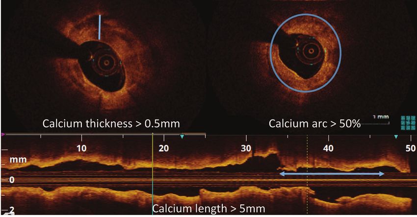
OPTIMIZING OUTCOMES IN COMPLEX PCI WITH OA
Funding for this supplement provided by CSI

                                                                                                                                8. Fitzgerald PJ, Ports TA, Yock PG. Contribution of localized calcium deposits to dissection after angioplasty. An
    for the treatment of severely calcified coronary lesions prior                                                              observational study using intravascular ultrasound. Circulation. 1992;86:64-70. doi: 10.1161/01.cir.86.1.64
    to stent delivery, with low procedural complication rates                                                                   9. Gilutz H, Weinstein JM, Ilia R. Repeated balloon rupture during coronary stenting due to a calcified lesion:
                                                                                                                                an intravascular ultrasound study. Cathet Cardiovasc Intervent. 2000;50:212-214. doi: 10.1002/(sici)1522-
    and low rates of revascularization through 3 years.11,14,15,17,18                                                           726x(200006)50:23.0.co;2-t
      The evidence supporting orbital atherectomy continues                                                                     10. Meraj PM, Shlofmitz E, Kaplan B, et al. Clinical outcomes of atherectomy prior to percutaneous coronary
                                                                                                                                intervention: a comparison of outcomes following rotational versus orbital atherectomy (COAP-PCI study). J Interv
    to expand. The prospective, randomized, multicenter                                                                         Cardiol. 2018;31:478-485.
    ECLIPSE trial (NCT03108456) is currently enrolling. ECLIPSE                                                                 11. Chambers JW, Feldman RL, Himmelstein SI, et al. Pivotal trial to evaluate the safety and efficacy of the orbital
                                                                                                                                atherectomy system in treating de novo, severely calcified coronary lesions (ORBIT II). JACC Cardiovasc Interv.
    will evaluate vessel preparation with Diamondback                                                                           2014;7:510-518. doi: 10.1016/j.jcin.2014.01.158
    compared to conventional balloon angioplasty technique                                                                      12. Kaan K, Hakan S. Coronary artery bypass surgery. In: Coronary Artery Disease – Assessment, Surgery, Prevention.
                                                                                                                                IntechOpen; November 18, 2015. doi: 10.5772/61404. Accessed May 7, 2021. https://www.intechopen.com/books/
    prior to drug-eluting stent implantation in severely calcified                                                              coronary-artery-disease-assessment-surgery-prevention/coronary-artery-bypass-surgery
    coronary artery lesions. Approximately 2,000 patients                                                                       13. Harvard Health Publishing. Should you have stenting or bypass surgery? Accessed April 30, 2021. https://www.
                                                                                                                                health.harvard.edu/heart-health/should-you-have-stenting-or-bypass-surgery
    with severely calcified coronary lesions will be enrolled at                                                                14. Shlofmitz E, Martinsen BJ, Lee M, et al. Orbital atherectomy for the treatment of severely calcified
    approximately 150 sites in the United States.                                                                               coronary lesions: evidence, technique, and best practices. Expert Rev Med Devices. 2017:14:867-879. doi:
                                                                                                                                10.1080/17434440.2017.1384695
      This series of articles provides insight into the                                                                         15. Lee M, Genereux P, Shlofmitz R, et al. Orbital atherectomy for treating de novo, severely calcified coronary lesions:
    contemporary practice and use of the Diamondback®                                                                           3-year results of the pivotal ORBIT II trial. Cardiovasc Revasc Med. 2017;18:261-264. doi: 10.1016/j.carrev.2017.01.011
                                                                                                                                16. Vinardell J, et al. Orbital atherectomy for treating de novo severely calcified coronary lesions: a tertiary center
    system. n                                                                                                                   experience. J Am Coll Cardiol. 2020;76(17):B71.
                                                                                                                                17. Chambers JW, Diage T. Evaluation of the Diamondback 360 coronary orbital atherectomy system for treating de
                                                                                                                                novo, severly calcified lesions. Expert Rev Med Devices. 2014;11:457-466. doi: 10.1586/17434440.2014.929493
                                                                                                                                18. Parikh K, Chandra P, Choksi N, et al. Safety and feasibility of orbital atherectomy for the treatment of calcified
    1. Shaikh K, Nakanishi R, Kim N, Budoff MJ. Coronary artery calcification and ethnicity. J Cardiovasc Comput Tomogr.
                                                                                                                                coronary lesions: the ORBIT I trial. Catheter Cardiovasc Interv. 2013;81:1134-1139. doi:10.1002/ccd.24700
    2019;13:353-359. doi: 10.1016/j.jcct.2018.10.002
    2. Cavusoglu E, Kini AS, Marmur JD, Sharma SK. Current status of rotational atherectomy. Catheter Cardiovasc Interv.
    2004;62:485-498. doi: 10.1002/ccd.20081
    3. Mintz GS, Popma JJ, Pichard AD, et al. Patterns of calcification in coronary artery disease. A statistical analysis of
    intravascular ultrasound and coronary angiography in 1155 lesions. Circulation. 1995;91:1959-1965. doi: 10.1161/01.
    cir.91.7.1959
    4. Bourantas CV, Zhang YJ, Garg S, et al. Prognostic implications of coronary calcification in patients with obstructive
    coronary artery disease treated by percutaneous coronary intervention: a patient-level pooled analysis of 7
    contemporary stent trials. Heart. 2014;100:1158-1164. doi: 10.1136/heartjnl-2013-305180
    5. Genereux P, Madhavan MV, Mintz GS, et al. Ischemic outcomes after coronary intervention of calcified vessels in                 Jeffrey W. Chambers, MD
    acute coronary syndromes. Pooled analysis from the HORIZONS-AMI (Harmonizing Outcomes With Revascularization                       Metropolitan Heart & Vascular Institute
    and Stents in Acute Myocardial Infarction) and ACUITY (Acute Catheterization and Urgent Intervention Triage Strategy)
    TRIALS. J Am Coll Cardiol. 2014;63:1845-1854. doi: 10.1016/j.jacc.2014.01.034                                                      Coon Rapids, Michigan
    6. Mosseri M, Satler LF, Pichard AD, Waksman R. Impact of vessel calcification on outcomes after coronary stenting.                j.chambers@mhvi.com
    Cardiovasc Revasc Med. 2005;6:147-153. doi: 10.1016/j.carrev.2005.08.008
    7. Moussa I, Di Mario C, Moses J, et al. Coronary stenting after rotational atherectomy in calcified and complex lesions.
    Angiographic and clinical follow-up results. Circulation. 1997;96:128-136. doi: 10.1161/01.cir.96.1.128

    4 SUPPLEMENT TO CARDIAC INTERVENTIONS TODAY MAY/JUNE 2021 VOL. 15, NO. 3
OPTIMIZING OUTCOMES IN COMPLEX PCI WITH OA
                                                                                           Funding for this supplement provided by CSI

Orbital Atherectomy in PCI for Calcific
Disease: What Have We Learned and
Where Are We Headed?
A data review on the role of orbital atherectomy in PCI for calcific coronary disease.
By Kathleen E. Kearney, MD

                                                                is associated with a major adverse cardiac event (MACE)
                  Kathleen E. Kearney, MD                       rate of approximately 30% in less than 1 year.6 This is
                  Assistant Professor                           why adequate vessel preparation is so critical. In a pooled
                  University of Washington Medical              analysis from randomized trials using contemporary drug-
                  Center                                        eluting stents (DESs), moderate-to-severe calcium was a
                  Seattle, Washington                           major predictor of target lesion failure between 30 days
                  kakearney@cardiology.washington.edu           to 1 year, observed at a rate of 2.1%.7 Although the rate
                                                                of probable or definite stent thrombosis at 1 year was
                                                                fortunately only 0.6% in the same pooled analysis, other

A
                                                                studies have implicated severe calcification as a significant
        dvances in stent technology and cardiology              risk factor,8 likely linked to stent underexpansion.
        training have led to broader adoption of                Limitations in practice for calcium management are
        percutaneous coronary intervention (PCI)                numerous; among them are operator training and
        in increasingly complex patients,1 yet calcific         experience with atherectomy and concerns about time
        disease continues to hamper outcomes.2                  and cost. Lack of definitive data is also cited in the face
Atherectomy is now widely available regionally, but             of these other concerns for those who have not adopted
practice heterogeneity and variability in the access to         atherectomy in their practice.
and utilization of technology dedicated toward vessel
preparation in calcified coronary arterial disease (CAD)        ATHERECTOMY: DATA, TRIALS, AND TRIBULATIONS
persist. This article reviews the available data to guide our      The constant conundrum facing the interventional
learning curve on orbital atherectomy (OA) as it applies        cardiologist regarding device selection is a balance of
to this space, identifies gaps in current knowledge, and        risks and benefits of applying a technology. Of course,
suggests future studies that may impact practice patterns.      device utilization is impacted by operator training in best
                                                                practices, but case selection, complication management,
IMPLICATIONS OF CALCIFIC DISEASE IN OUTCOMES                    and practice environment all color that risk-benefit
OF PCI                                                          assessment. Additionally, our practice patterns emphasize
  Advances in stent design and operator experience              the short-term outcomes for the patient, and a lack
have reduced in-stent complications, with definite              of disease-based registries or consistent definitions in
or probable stent thrombosis in less than 1% of                 disease characteristics such as calcium burden make the
the non–acute coronary syndrome population at                   application of data more complicated than the surface
2 years3 and in approximately 1% of all cases in the            layer of results. In the case of atherectomy, successful
Medicare population,4 yet 10% of PCI in the National            stent implantation may be possible without additional
Cardiovascular Data Registry was performed for in-stent         calcium modification, but the question remains: do
restenosis (ISR).5 ISR can be challenging to manage and         we improve long-term patient outcomes in cases of

                                              VOL. 15, NO. 3 MAY/JUNE 2021 SUPPLEMENT TO CARDIAC INTERVENTIONS TODAY 5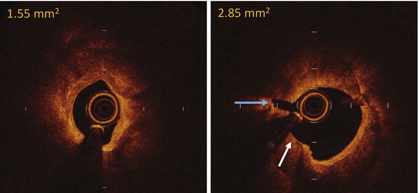
OPTIMIZING OUTCOMES IN COMPLEX PCI WITH OA
Funding for this supplement provided by CSI

                                     TABLE 1. DATA OVERVIEW OF STUDIES EVALUATING CORONARY ATHERECTOMY
                                                                                                        Slow Flow/
     Study                   Year                    N               Dissection (%)   Perforation (%)                     30-Day TVR (%)
                                                                                                        No Reflow (%)
     ORBIT II9               2014                    443             3.4*             1.8               0.9               1.4
     Lee et al10             2016                    458             0.9              0.7               0.7               0.0
     COAP-PCI    11          2018                    273 OAS         1.3 *            0.4               –                 –
     Koifman et al12         2018                    67              7.5              –                 –                 –
     Chambers et al13        2018                    78              –                –                 1.3               1.3
     Desai et al14           2018                    40              0.0              2.5               2.5               0.0
     Whitbeck et al15        2018                    70              0.0†             1.4               1.4               Only acute (up
                                                                                                                          to discharge)
                                                                                                                          MACE rates were
                                                                                                                          reported
     Okamoto et al16         2019                    184             1.6              1.6               2.2               –
     COAST17                 2020                    100             2.0*             2.0               2.0               1.0
     *Type C-F significant or severe dissections.

     †There was no severe dissection, but 4.3% type A dissections.

     Abbreviation: TVR, target vessel revascularization.

    calcific disease with atherectomy? Unfortunately, these                   power to detect clinical events in stable ischemic heart
    questions may never be fully answered in randomized                       disease patients and the current DES platforms.
    trials as those who stand to gain the most from device                       OA is the more recent addition to the market
    therapies are often not enrolled when the operator does                   (Diamondback 360® Coronary Orbital System,
    not see equipoise, and crossover to the intervention arm                  Cardiovascular Systems, Inc.), and is thus building on a
    clouds results. Despite these limitations, several trials                 different mechanism of action, using centrifugal forces
    have identified the relative efficacy and safety profile of               and orbital motion of the burr to fracture calcium
    atherectomy use, and this article focuses on the recent                   and perform differential sanding.21-23 ORBIT I was the
    data exhibiting clinical outcomes after OA (Table 1).9-17                 introductory study and first evaluated 50 elective PCI
       Rotational atherectomy (RA) was early to the market                    patients in 2008 across nine operators and two sites,
    and used in the first studies evaluating atherectomy                      in lesions ≤ 25 mm in length with mild-to-severe
    as an adjunct to PCI in calcific CAD. The ROTAXUS                         calcium to establish baseline safety and efficacy data.24
    trial randomized patients to DES implantation with or                     Procedural success, defined as ≤ 20% residual stenosis
    without the aid of RA but failed to show a clinical benefit               after stent placement, was 97%, with 2 minor and 1
    with regard to early restenosis or clinical outcomes                      major dissections noted without clinical consequence
    at 2 years.18,19 A more recent follow-up trial that                       and one perforation after stent placement. Of note,
    randomized 200 patients to RA versus cutting/scoring                      only 6 patients underwent angioplasty after OA prior
    balloon as vessel preparation demonstrated improved                       to stent placement, while some did have angioplasty
    procedural success with RA, but again clinical events                     and intravascular ultrasound (IVUS) performed prior
    were not significantly different nor were they powered                    to OA. Still, in-hospital MACE was low, including only
    for detection in this analysis.20 Notably, there was 16%                  6% (2 patients) and 12% at 6 months. This led the way
    crossover, and while patients with severe calcification                   for ORBIT II, evaluating 443 consecutive patients with
    were included, the core laboratory found that 25% of                      severely calcified coronary lesions across 49 sites.9 Of
    cases fit criteria for moderate calcification. These early                note, 11% of patients in ORBIT II received bare-metal
    trials are important in emphasizing key characteristics                   stents. Severe calcification was defined as fluoroscopic
    for interventional trials—challenges with anatomic                        visualization without cardiac motion on both sites
    definitions, crossover to the interventional strategy, and                of the vessel, length > 15 mm, or ≥ 270° arc on IVUS

    6 SUPPLEMENT TO CARDIAC INTERVENTIONS TODAY MAY/JUNE 2021 VOL. 15, NO. 3
OPTIMIZING OUTCOMES IN COMPLEX PCI WITH OA
Funding for this supplement provided by CSI
A B
APPLYING DATA TO THE REAL WORLD: DOES IT
WORK WHERE IT REALLY COUNTS?
Given the confines of the trial setting and the often
lower overall risk profile of patients, subsequent
registries shed insight into understanding outcome data
in the broader population with real-world use. Lee et
al published a study of 458 consecutive patients with
C D severely calcified CAD who underwent OA-assisted
PCI.10 This retrospective review of 458 consecutive
patients showed low rates of 30-day MACE (1.7%), with
0.9% stent thrombosis, 1.1% MI, 0% TVR, and 1.3% all-
cause mortality, indicating significant overlap in these
presenting events. Perforation, dissection, and no reflow
were all < 1% each, indicating an acceptable safety profile
in real practice, although generalizable in the context
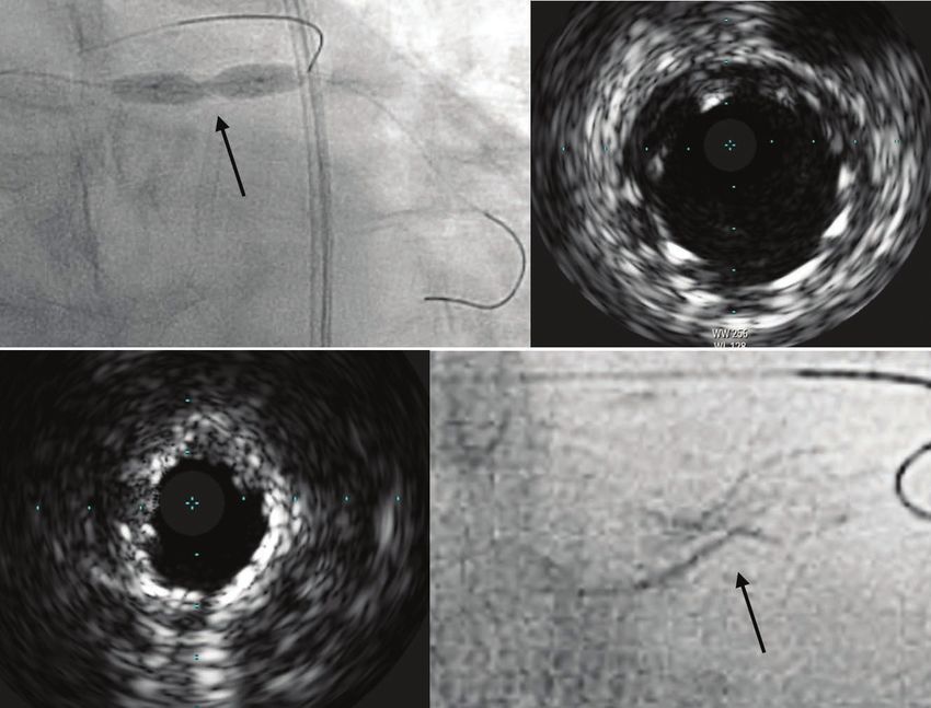
Figure 1. A 70-year-old man with IVUS-guided sizing for
where operators are likely highly trained in device
a 5.0-mm stent based on the proximal vessel. There was
utilization and managing complications in complex PCI.
poor expansion (arrow) despite high-pressure inflation with
Meraj et al performed a prospective registry to evaluate
a 5.0‑mm noncompliant balloon catheter (A). IVUS of the
outcomes related to PCI using OA versus RA in 907
proximal stent showed adequate apposition and expansion (B).
patients across five tertiary care hospitals.11 OA was
IVUS corresponding to the waist demonstrated a diameter of
associated with lower rates of the primary endpoint of
2.4 mm (C). Cineography of the stent demonstrated a severely
in-hospital MI (primary endpoint of 6.7% vs 13.8% in RA)
underexpanded section (arrow) (D).
and similar procedural safety outcomes in the 546 cases
compared after propensity score matching. A recent
cross-sectional imaging. The efficacy endpoint of stent meta-analysis of seven retrospective studies comparing
implantation with < 50% residual stenosis after stent rates of MI and vascular complications also noted a
implantation and freedom from in-hospital MACE was stronger association of periprocedural MI after RA
met in 88.9% of participants, with successful stent delivery versus OA but a lower risk of dissection or perforation.26
and < 50% stenosis in 97.7% of cases and low rates of Although these data are subject to selection bias based on
in-hospital Q-wave myocardial infarction (MI) (0.7%), angiographic features and operator preferences despite
cardiac death (0.2%), and target vessel revascularization propensity matching, they do support future study
(TVR) (0.7%). Follow-up at 3 years was completed in 360 regarding the best use for OA in treating calcified CAD.
(81.3%) patients, demonstrating a cumulative event rate
of MACE of 23.5%, cardiac death of 6.7%, MI of 11.2%, and Imaging Versus Angiographic Classification of
TVR of 10.2%. Target lesion revascularization at 3 years Calcification
was 7.8%, as compared with 13.8% and 16.7% in the The definition of significant calcification by angiography
ROTAXUS trial in the RA and control treatment arms, and variable definitions used in studies to date are
respectively.9,18,19,25 In contrast with current practice for significant limitations of the current data. In ORBIT II,
many operators, the minority of lesions were treated with calcification burden was defined by IVUS in only 8% of
angioplasty after OA prior to stent placement (still only cases, with the remaining patients included on the basis
up to 42% in ORBIT II), whereas 52% had postdilatation of angiographic criteria. A substudy evaluating IVUS in
after stent placement.9 In total, these data indicate OA ORBIT II found that there was a reduction in the number
may improve management of severely calcific disease with of stents used in those with IVUS; 3-year MACE rates were
an acceptable safety profile in a patient population that not statistically different but were higher in the no-IVUS
has been poorly represented in trials but are yet limited cohort (24.2% vs 14.3% in the IVUS group; P = .26).27 As
by lack of a control arm. Recognizing that multiple facets this substudy was limited to 35 patients who underwent
of PCI have changed over time and other patient selection IVUS prior to OA, this may favor lesions that were more
factors differ across studies, these data are encouraging in amenable to imaging prior to OA. However, taken in
that calcific CAD can and should be treated in patients the context of contemporary data supporting IVUS as a
with an indication for PCI. tool to improve PCI outcomes,28 it is likely that coupling
VOL. 15, NO. 3 MAY/JUNE 2021 SUPPLEMENT TO CARDIAC INTERVENTIONS TODAY 7
OPTIMIZING OUTCOMES IN COMPLEX PCI WITH OA
Funding for this supplement provided by CSI

    intracoronary imaging with atherectomy would further           further guide use of the full complement of tools aimed
    improve PCI outcomes in treating calcified lesions.            at treating calcific CAD. n

    LOOKING AHEAD: WHAT QUESTIONS REMAIN?                          1. Kataruka A, Maynard CC, Kearney KE, et al. Temporal trends in percutaneous coronary intervention and coronary
                                                                   artery bypass grafting: insights from the Washington Cardiac Care Outcomes Assessment Program. J Am Heart Assoc.
       The current data have established a platform for OA         2020;9:e015317.
                                                                   2. Généreux P, Madhavan MV, Mintz GS, et al. Ischemic outcomes after coronary intervention of calcified vessels in
    in treating calcified CAD but are limited in terms of          acute coronary syndromes. Pooled analysis from the HORIZONS-AMI (Harmonizing Outcomes With Revascularization
    patient selection and how that applies to the operator         and Stents in Acute Myocardial Infarction) and ACUITY (Acute Catheterization and Urgent Intervention Triage Strategy)
                                                                   TRIALS. J Am Coll Cardiol. 2014;63:1845-1854.
    making a rapid decision that has real consequences to          3. Chau KH, Kirtane AJ, Easterwood RM, et al. Stent thrombosis risk over time on the basis of clinical presentation and
    the patient: should atherectomy be used in this patient?       platelet reactivity: analysis from ADAPT-DES. JACC Cardiovasc Interv. 2021;14:417-427.
                                                                   4. Dhruva SS, Parzynski CS, Gamble GM, et al. Attribution of adverse events following coronary stent placement
    Frequently, this is not realized until a poor stent result     identified using administrative claims data. J Am Heart Assoc. 2020;9:e013606.
    is recognized and is much more challenging to recover          5. Moussa ID, Mohananey D, Saucedo J, et al. Trends and outcomes of restenosis after coronary stent implantation in
                                                                   the United States. J Am Coll Cardiol. 2020;76:1521-1531.
    (Figure 1). The evaluation of treatment strategies for         6. Radke PW, Kaiser A, Frost C, Sigwart U. Outcome after treatment of coronary in-stent restenosis: results from a
    severe calcific coronary arteries (OA vs angioplasty           systematic review using meta-analysis techniques. Eur Heart J. 2003;24:266-273.
                                                                   7. Konigstein M, Madhavan MV, Ben-Yehuda O, et al. Incidence and predictors of target lesion failure in patients
    technique) prior to implantation of DES in the ECLIPSE         undergoing contemporary DES implantation-Individual patient data pooled analysis from 6 randomized controlled
    trial will aid in answering these questions. Currently         trials. Am Heart J. 2019;213:105-111.
                                                                   8. Huisman J, van der Heijden LC, Kok MM, et al. Impact of severe lesion calcification on clinical outcome of patients
    enrolling with a target of 2,000 patients, this randomized     with stable angina, treated with newer generation permanent polymer-coated drug-eluting stents: a patient-level
    trial is comparing vessel preparation with OA and balloon      pooled analysis from TWENTE and DUTCH PEERS (TWENTE II). Am Heart J. 2016;175:121-129.
                                                                   9. Chambers JW, Feldman RL, Himmelstein SI, et al. Pivotal trial to evaluate the safety and efficacy of the orbital
    pre-dilatation to that with conventional and/or specialty      atherectomy system in treating de novo, severely calcified coronary lesions (ORBIT II). JACC Cardiovasc Interv.
    balloon preparation, with a primary outcome of target          2014;7:510-518.
                                                                   10. Lee MS, Shlofmitz E, Kaplan B, et al. Real-world multicenter registry of patients with severe coronary artery
    vessel failure at 1 year (composite of cardiac death, target   calcification undergoing orbital atherectomy. J Interv Cardiol. 2016;29:357-362.
    vessel–related MI, or ischemia-driven revascularization).      11. Meraj PM, Shlofmitz E, Kaplan B, et al. Clinical outcomes of atherectomy prior to percutaneous coronary
                                                                   intervention: a comparison of outcomes following rotational versus orbital atherectomy (COAP-PCI study). J Interv
    An imaging cohort using optical coherence tomography           Cardiol. 2018;31:478-485.
    in 500 patients will also assess minimal stent area as         12. Koifman E, Garcia-Garcia HM, Kuku KO, et al. Comparison of the efficacy and safety of orbital and rotational
                                                                   atherectomy in calcified narrowings in patients who underwent percutaneous coronary intervention. Am J Cardiol.
    another primary endpoint, as well as secondary outcomes        2018;121(8):934-939.
    of procedural and strategy success. Importantly, the study     13. Chambers JW, Warner C, Cortez J, et al. Outcomes after atherectomy treatment of severely calcified coronary
                                                                   bifurcation lesions: a single center experience. Cardiovasc Revasc Med. 2019;20:569-572.
    population is expanded to include ACS patients provided        14. Desai R, Mirza O, Martinsen BJ, Kumar G. Plaque modification of severely calcified coronary lesions via orbital
    they are stabilized > 48 hours after ST-segment elevation      atherectomy: single-center observations from a complex Veterans Affairs cohort. Health Sci Rep. 2018;1:e99.
                                                                   15. Whitbeck MG, Dewar J, Behrens AN, et al. Acute outcomes after coronary orbital atherectomy at a single
    MI and excludes patients with severe heart failure             center without on-site surgical backup: an experience in diabetics versus non-diabetics. Cardiovasc Revasc Med.
    symptoms or left ventricular ejection fraction < 25%.          201819(6S):12-15.
                                                                   16. Okamoto N, Ueda H, Bhatheja S, et al. Procedural and one-year outcomes of patients treated with orbital and
       The ECLIPSE trial is well positioned to inform whether      rotational atherectomy with mechanistic insights from optical coherence tomography. EuroIntervention. 2019;14:1760-
    the practice of using OA or "vessel preparation with           1767.
                                                                   17. Redfors B, Sharma SK, Saito S, et al. Novel micro crown orbital atherectomy for severe lesion calcification: Coronary
    balloon angioplasty only" provides the best outcomes.          Orbital Atherectomy System Study (COAST). Circ Cardiovasc Interv. 2020;13:e008993.
    This study far outpaces the aforementioned studies             18. Abdel-Wahab M, Richardt G, Joachim Büttner H, et al. High-speed rotational atherectomy before paclitaxel-eluting
                                                                   stent implantation in complex calcified coronary lesions: the randomized ROTAXUS (Rotational Atherectomy Prior to
    evaluating RA and OA in terms of size; the inclusion of an     Taxus Stent Treatment for Complex Native Coronary Artery Disease) trial. JACC Cardiovasc Interv. 2013;6:10-19.
    imaging cohort, evaluating crossover to the alternative        19. de Waha S, Allali A, Büttner HJ, et al. Rotational atherectomy before paclitaxel-eluting stent implantation in
                                                                   complex calcified coronary lesions: two-year clinical outcome of the randomized ROTAXUS trial. Catheter Cardiovasc
    strategy, and use of current-generation DESs will lend         Interv. 2016;87:691-700.
    further insight as to how the vessel preparation strategy      20. Abdel-Wahab M, Toelg R, Byrne RA, et al. High-speed rotational atherectomy versus modified balloons prior to
                                                                   drug-eluting stent implantation in severely calcified coronary lesions. Circ Cardiovasc Interv. 2018;11:e007415.
    affects clinical and procedural outcomes.                      21. Kini AS, Vengrenyuk Y, Pena J, et al. Optical coherence tomography assessment of the mechanistic effects of
                                                                   rotational and orbital atherectomy in severely calcified coronary lesions. Catheter Cardiovasc Interv. 2015;86:1024-1032.
    CONCLUSION                                                     22. Shlofmitz E, Martinsen BJ, Lee M, et al. Orbital atherectomy for the treatment of severely calcified coronary lesions:
                                                                   evidence, technique, and best practices. Expert Rev Med Devices. 2017:14:867-879.
       Early data evaluating the safety and efficacy of OA are     23. Yamamoto MH, Maehara A, Kim SS, et al. Effect of orbital atherectomy in calcified coronary artery lesions as
                                                                   assessed by optical coherence tomography. Catheter Cardiovasc Interv. 2019;93:1211-1218.
    promising. Although patient selection and best practices       24. Bhatt P, Parikh P, Patel A, et al. Orbital atherectomy system in treating calcified coronary lesions: 3-year follow-up
    for technique remain paramount for improving clinical          in first human use study (ORBIT I trial). Cardiovasc Revasc Med. 2014;15:204-208.
                                                                   25. Lee M, Genereux P, Shlofmitz R, et al. Orbital atherectomy for treating de novo, severely calcified coronary lesions:
    outcomes, many cases should not be undertaken without          3-year results of the pivotal ORBIT II trial. Cardiovasc Revasc Med. 2017;18:261-264.
    additional calcium modification and vessel preparation,        26. Doshi R, Thakkar S, Patel K, et al. Short term outcomes of rotational atherectomy versus orbital atherectomy in
                                                                   patients undergoing complex percutaneous coronary intervention: a systematic review and meta-analysis [published
    and training in these tools is imperative for the modern       online ahead of print January 18, 2021]. Scand Cardiovasc J. https://doi: 10.1080/14017431.2021.1875139.
    interventional cardiologist. Studies using better-defined      27. Shlofmitz E, Martinsen B, Lee M, et al. Utilizing intravascular ultrasound imaging prior to treatment of severely
                                                                   calcified coronary lesions with orbital atherectomy: an ORBIT II sub-analysis. J Interv Cardiol. 2017;30:570-576.
    classification schemes based on intracoronary imaging to       28. Gao XF, Ge Z, Kong XQ, et al; ULTIMATE Investigators. 3-year outcomes of the ULTIMATE trial comparing
    define calcific burden and assess procedural outcomes          intravascular ultrasound versus angiography-guided drug-eluting stent implantation. JACC Cardiovasc Interv.
                                                                   2021;14:247-257.
    will better showcase the risks and benefits of OA and

    8 SUPPLEMENT TO CARDIAC INTERVENTIONS TODAY MAY/JUNE 2021 VOL. 15, NO. 3
OPTIMIZING OUTCOMES IN COMPLEX PCI WITH OA
                                                                                        Funding for this supplement provided by CSI

Imaging and Treating the Complex Patient
Optimizing outcomes in complex PCI with orbital atherectomy.
By Evan Shlofmitz, DO, and Richard Shlofmitz, MD

                                                              nodules. Traditionally, CAC was classified based on its
                  Evan Shlofmitz, DO                          angiographic appearance, with calcium visualization
                  Director of Intravascular Imaging           on both sides of the lumen prior to contrast injection
                  St. Francis Hospital–The Heart Center       without motion considered to be severe calcification.5
                  Roslyn, New York                            We now know that there are inherent limitations
                  Evan.Shlofmitz@CHSLI.org                    to angiographic assessment of coronary calcium.
                                                              Angiographic recognition of calcification does not guide
                                                              optimal treatment strategies and is simply a prompt
                  Richard Shlofmitz, MD                       for the need for further investigation with intravascular
                  Chairman of Cardiology                      imaging to define the calcium morphology. An angiogram
                  St. Francis Hospital–The Heart Center       cannot distinguish between deep wall calcification,
                  Roslyn, New York                            superficial calcification, and calcified nodules. Treatment
                                                              of CAC should be determined based on this calcium
                                                              morphology, which can only be characterized with
                                                              intravascular imaging.

W
                                                                In the LightLab Study, OCT findings led to changes in
            hether from a randomized controlled trial,        angiographic-based decisions in 88% of lesions, with a
            observational registry, or meta-analysis,         change in need for vessel preparation observed in 28% of
            data have consistently demonstrated the           cases.6 It is widely known that CAC is underrecognized
            benefit of intravascular imaging on clinical      with angiography.5 Beyond recognition of calcium and
            outcomes.1-3 The mechanism by which               understanding of its morphology, baseline intravascular
outcomes are improved is largely related to improved          imaging allows for precise selection of the optimal
stent expansion when intravascular imaging is utilized.1      stent diameter and length.7 After stent implantation,
Coronary artery calcification (CAC) is the predominant        intravascular imaging can exclude significant edge
barrier to adequate stent expansion when stent sizing         dissection and severe malapposition while ensuring that
has been appropriate. Atherectomy has historically been       adequate stent expansion has been attained.
indicated for undilatable and uncrossable lesions, but this
indication falls short of the complete role of atherectomy    CHARACTERIZATION OF CALCIUM
in modern percutaneous coronary intervention (PCI).4            On baseline intravascular imaging, predominant
In the presence of heavy calcification, lesion preparation    calcium morphology should be characterized between
should be used not just for successful stent delivery, but    concentric calcification, eccentric calcification, and
also importantly to facilitate adequate stent expansion via   calcified nodules.8 The St. Francis Calcium-OCT Score,
plaque modification.                                          commonly known as the “rule of 5s,” guides when
                                                              orbital atherectomy should be considered (Figure 1). In
INTRAVASCULAR IMAGING                                         the presence of CAC > 5 mm in length, an arc > 50%
  Although intravascular ultrasound is an important           of a cross-section with a thickness > 0.5 mm indicates
intravascular imaging modality, in most cases where           heavy calcification that is at increased risk for stent
evaluation of coronary calcification is required, we prefer   underexpansion without adequate lesion preparation.9
optical coherence tomography (OCT) due to its unique          Because conventional balloon-based technology is often
ability to readily assess important prognostic factors        inadequate to create calcium fracture when calcium
including calcium thickness and recognition of calcified      thickness exceeds 0.5 mm, adjunctive therapies are

                                              VOL. 15, NO. 3 MAY/JUNE 2021 SUPPLEMENT TO CARDIAC INTERVENTIONS TODAY 9
OPTIMIZING OUTCOMES IN COMPLEX PCI WITH OA
Funding for this supplement provided by CSI

      A                                                                    B

      C

   Figure 1. The St. Francis Calcium-OCT Score. Representative OCT demonstrating CAC with thickness > 0.5 mm (A), an arc of
   calcium > 50% of the cross-section (B), and length > 5 mm (C). In the presence of these three features, lesion preparation should be
   considered.

    needed.10 Plaque modification with calcium fracture can be         Calcified Nodules
    achieved with lesion preparation with orbital atherectomy            A calcium nodule is defined as an eruptive
    (Figure 2).                                                        accumulation of nodular calcification protruding into the
                                                                       lumen.17 Calcium nodules are often underrecognized, as
    ORBITAL ATHERECTOMY                                                they cannot be appreciated by angiography. However,
       Orbital atherectomy (Diamondback 360® Coronary                  calcified nodules are not an uncommon entity, and
    Orbital Atherectomy System, Cardiovascular Systems,                are seen in as many as 6% of cases and over 48% of
    Inc.) utilizes a 1.25-mm diamond-coated crown that                 calcified lesions.18,19 It is important to detect calcium
    orbits at either 80,000 rpm (low speed) or 120,000 rpm             nodules prior to stent implantation because they do not
    (high speed) in a bidirectional fashion to modify                  behave similarly to severe concentric CAC. Treatment
    calcified plaque.11 The unique dual mechanism of                   of calcified nodules with other adjunctive lesion
    action utilizes differential sanding and pulsatile force           preparation modalities, including rotational atherectomy,
    that safely ablates superficial calcification while                intravascular lithotripsy, and specialty balloons is not ideal
    creating focused fractures in the calcified plaque, which          due to the eccentric protruding nature of these nodules.
    enables expansion with stent implantation.12,13 During             Orbital atherectomy uniquely allows for significant plaque
    superficial calcium sanding, the small crown size permits          modification of calcified nodules with debulking of the
    continuous flow during ablation, with creation of                  nodule with lumen enlargement (Figure 3).
    particles < 2 µm in size.12 Yamamoto et al demonstrated
    that orbital atherectomy is associated with greater                CONCLUSION
    calcium modification in lesions with larger lumen area               Intravascular imaging not only guides when orbital
    as compared with rotational atherectomy, with calcium              atherectomy is needed but also demonstrates where
    fracture behind the stent attained in 82% of cases.14              orbital atherectomy should be applied in a vessel. Orbital
    This is an important concept, as the universal orbital             atherectomy should be performed until the operator
    atherectomy crown size can be used to treat calcified              appreciates a change in tactile resistance and no longer
    plaque in a wide range of coronary vessel sizes, including         hears audible pitch variation during treatment of the
    the left main coronary artery.15,16                                target region. After orbital atherectomy, intravascular

    10 SUPPLEMENT TO CARDIAC INTERVENTIONS TODAY MAY/JUNE 2021 VOL. 15, NO. 3
OPTIMIZING OUTCOMES IN COMPLEX PCI WITH OA
Funding for this supplement provided by CSI
A B
Figure 2. Baseline OCT with severe calcification and a lumen area of 1.55 mm2 (A). OCT after lesion preparation with orbital
atherectomy demonstrating the dual mechanism of action with both smooth concentric ablation (white arrow) and calcium fracture
(blue arrow), with an enlarged lumen of 2.85 mm2 (B).
A B
Figure 3. Baseline OCT demonstrating a protruding calcified nodule (*) (A). OCT after lesion preparation with orbital atherectomy
demonstrates reduction in the nodule size with enlargement of the lumen area (B).
imaging should be performed. By documenting fracture preparation can enhance the likelihood of procedural
prior to stent implantation with intravascular imaging, success. Maximizing stent expansion with implantation
one can have a higher level of confidence they will can help to minimize future restenosis. Inadequate
achieve adequate stent expansion. lesion preparation for de novo calcified lesions prior to
When treating calcium with PCI, adequate lesion stent implantation represents a lost opportunity, as any
VOL. 15, NO. 3 MAY/JUNE 2021 SUPPLEMENT TO CARDIAC INTERVENTIONS TODAY 11OPTIMIZING OUTCOMES IN COMPLEX PCI WITH OA
Funding for this supplement provided by CSI
future treatment of in-stent restenosis is associated with
outcomes worse than with de novo disease.20 De novo
calcification should be addressed with lesion preparation
upfront when indicated and guided by intravascular
imaging. Results from the large-scale, multicenter,
randomized controlled ECLIPSE trial (NCT03108456) and
its prespecified OCT substudy will provide substantial
insights on the impact of orbital atherectomy and
optimal techniques and lesion selection. n
1. Zhang J, Gao X, Kan J, et al. Intravascular ultrasound versus angiography-guided drug-eluting stent implantation: the
ULTIMATE trial. J Am Coll Cardiol. 2018;72:3126-3137. doi: 10.1016/j.jacc.2018.09.013
2. Shlofmitz E, Torguson R, Zhang C, et al. Impact of intravascular ultrasound on outcomes following PErcutaneous
Coronary InterventioN in Complex Lesions (iOPEN Complex). Am Heart J. 2020;221:74-83. doi: 10.1016/j.
ahj.2019.12.008
3. Kuku KO, Ekanem E, Azizi V, et al. Optical coherence tomography-guided percutaneous coronary intervention
compared with other imaging guidance: a meta-analysis. Int J Cardiovasc Imaging. 2018;34:503-513. doi: 10.1007/
s10554-017-1272-2
4. Levine GN, Bates ER, Blankenship JC, et al. 2011 ACCF/AHA/SCAI guideline for percutaneous coronary intervention.
A report of the American College of Cardiology Foundation/American Heart Association Task Force on Practice
Guidelines and the Society for Cardiovascular Angiography and Interventions. J Am Coll Cardiol. 2011;58:e44-122. doi:
10.1016/j.jacc.2011.08.007
5. Mintz GS, Popma JJ, Pichard AD, et al. Patterns of calcification in coronary artery disease. A statistical analysis of
intravascular ultrasound and coronary angiography in 1155 lesions. Circulation. 1995;91:1959-65. doi: 10.1161/01.
cir.91.7.1959
6. Bezerra H. Analysis of changes in decision-making process during optical coherence tomography-guided
percutaneous coronary interventions: insights from the LightLab Initiative. Presented at the PCR e-Course of the
European Association of Percutaneous Cardiovascular Interventions (EAPCI); June 25-27, 2020; virtual presentation.
7. Shlofmitz E, Shlofmitz RA, Galougahi KK, et al. Algorithmic approach for optical coherence tomography-guided
stent implantation during percutaneous coronary intervention. Interv Cardiol Clin. 2018;7:329-344. doi: 10.1016/j.
iccl.2018.03.001
8. Shlofmitz E, Ali ZA, Maehara A, et al. Intravascular imaging-guided percutaneous coronary intervention: a
universal approach for optimization of stent implantation. Circ Cardiovasc Interv. 2020;13:e008686. doi: 10.1161/
CIRCINTERVENTIONS.120.008686
9. Fujino A, Mintz GS, Matsumura M, et al. A new optical coherence tomography-based calcium scoring system to
predict stent underexpansion. EuroIntervention. 2018;13:e2182-e2189. doi: 10.4244/EIJ-D-17-00962
10. Fujino A, Mintz GS, Lee T, et al. Predictors of calcium fracture derived from balloon angioplasty and its effect
on stent expansion assessed by optical coherence tomography. JACC Cardiovasc Interv. 2018;11:1015-1017. doi:
10.1016/j.jcin.2018.02.004
11. Shlofmitz E, Martinsen BJ, Lee M, et al. Orbital atherectomy for the treatment of severely calcified
coronary lesions: evidence, technique, and best practices. Expert Rev Med Devices. 2017;14:867-879. doi:
10.1080/17434440.2017.1384695
12. Shlofmitz E, Shlofmitz R, Lee MS. Orbital atherectomy: a comprehensive review. Interv Cardiol Clin. 2019;8:161-
171. doi: 10.1016/j.iccl.2018.11.006
13. Yamamoto MH, Maehara A, Kim SS, et al. Effect of orbital atherectomy in calcified coronary artery lesions as
assessed by optical coherence tomography. Catheter Cardiovasc Interv. 2019;93:1211-1218. doi: 10.1002/ccd.27902
14. Yamamoto MH, Maehara A, Karimi Galougahi K, et al. Mechanisms of orbital versus rotational atherectomy
plaque modification in severely calcified lesions assessed by optical coherence tomography. JACC Cardiovasc Interv.
2017;10:2584-2586. doi: 10.1016/j.jcin.2017.09.031
15. Lee MS, Shlofmitz E, Park KW, et al. Orbital atherectomy of severely calcified unprotected left main coronary artery
disease: one-year outcomes. J Invasive Cardiol. 2018;30:270-274.
16. Lee MS, Shlofmitz E, Shlofmitz R. Outcomes of orbital atherectomy in severely calcified small (2.5 mm) coronary
artery vessels. J Invasive Cardiol. 2018;30:310-314.
17. Lee T, Mintz GS, Matsumura M, et al. Prevalence, predictors, and clinical presentation of a calcified nodule
as assessed by optical coherence tomography. JACC Cardiovasc Imaging. 2017;10:883-891. doi: 10.1016/j.
jcmg.2017.05.013
18. Yamamoto MH, Maehara A, Song L, et al. Optical coherence tomography assessment of morphological
characteristics in suspected coronary artery disease, but angiographically nonobstructive lesions. Cardiovasc Revasc
Med. 2019;20:475-479. doi: 10.1016/j.carrev.2018.07.011
19. Morofuji T, Kuramitsu S, Shinozaki T, et al. Clinical impact of calcified nodule in patients with heavily calcified
lesions requiring rotational atherectomy. Catheter Cardiovasc Interv. 2021;97:10-19. doi: 10.1002/ccd.28896
20. Shlofmitz E, Iantorno M, Waksman R. Restenosis of drug-eluting stents: a new classification system based on
disease mechanism to guide treatment and state-of-the-art review. Circ Cardiovasc Interv. 2019;12:e007023. doi:
10.1161/CIRCINTERVENTIONS.118.007023
12 SUPPLEMENT TO CARDIAC INTERVENTIONS TODAY MAY/JUNE 2021 VOL. 15, NO. 3OPTIMIZING OUTCOMES IN COMPLEX PCI WITH OA
Funding for this supplement provided by CSI
Streamlining Care With
Orbital Atherectomy in Complex PCI
Facilitating evidence-based adjunctive therapy for PCI procedures in calcified lesions
By Adhir Shroff, MD, MPH
Coronary Orbital Atherectomy System (OAS)
Adhir Shroff, MD, MPH (Cardiovascular Systems Inc.) into this program.
University of Illinois – Chicago The Diamondback 360® is a useful tool to adjunctively
Chicago, Illinois treat severely calcified coronary artery stenoses. It offers
ashroff@uic.edu; @ARS_MD2004 several clinical advantages to streamline complex PCI:
• A single Diamondback® device can treat lesions from
2.5 mm to 4.0 mm in diameter, as well as long, diffuse
lesions and multivessel disease
I
• Diamondback is 6-F guide catheter compatible for
n the current health care environment, providers and lesions as large as 4.0 mm, making it ideal for radial
patients are looking for opportunities to improve access procedures
efficiency and minimize time in the hospital while • Diamondback allows continuous blood flow during
improving safety. Radial approaches, intravascular treatment, minimizing the risk of slow flow and no
imaging, appropriate device selection, and same- reflow that can increase periprocedural complications
day discharge after outpatient percutaneous coronary and prevent same-day discharge
intervention (PCI) procedures can help achieve these goals. • The need for temporary pacemaker placement is
Calcification can impede stent delivery and deployment extremely low with Diamondback, eliminating the
and increase procedural risk.1,2 Atherectomy has improved need for additional access sites
clinicians’ ability to treat calcified lesions. In 2015, the Due to the unique mechanism of action of the
Centers for Medicare & Medicaid Services assigned Diamondback 360 system, there are other potential
unique codes with incremental reimbursement for PCI advantages that can improve efficiency:
with atherectomy. This enabled physicians to incorporate • The ViperWire Advance® Coronary Guide Wire with
imaging and hemodynamic assessment alongside Flex Tip (Cardiovascular Systems Inc.) has excellent
atherectomy to optimize procedural outcomes with less deliverability and can be used to directly wire lesions
financial concern. prior to treatment with Diamondback to avoid wire
With technical, pharmacologic, and PCI-related exchange
advancements, many atherectomy patients are suitable • Given its one-size-fits-all design, inventory
for same-day discharge, enabling patients to recover at management may be simplified
home and avoid hospital-related infections; hospitals can • Diamondback’s electric-powered drive train requires
also maintain capacity while reducing costs of routine less setup
observation. In 2018, the Society for Cardiovascular Our institution has successfully incorporated
Angiography and Interventions updated its length of stay Diamondback atherectomy into our outpatient same-day
guidelines.3 Focus shifted from proscriptive criteria to a discharge PCI program.
more patient-focused approach, including consideration We share two patient cases using Diamondback with
of same-day discharge for stable patients undergoing the new, nitinol ViperWire® with Flex Tip Guide Wire to
successful procedures as part of a structured program. We treat heavily calcified lesions via a radial approach, safely
were able to successfully incorporate Diamondback 360® discharging them on the same day of their procedure.
VOL. 15, NO. 3 MAY/JUNE 2021 SUPPLEMENT TO CARDIAC INTERVENTIONS TODAY 13OPTIMIZING OUTCOMES IN COMPLEX PCI WITH OA
Funding for this supplement provided by CSI
A B C D E F
Figure 1. Focal, calcified mid right coronary artery lesion (A). Predilatation with a 1.5-mm balloon (B). Final result after OA and a 3.5-
X 23-mm DES deployment (C). Diffuse, calcified left anterior descending artery lesion (D). OA performed at 80K rpm (E). Final result
after deployment of a 2.5- X 23-mm and a 3- X 12-mm DES (F).
CASE PATIENT 1 crossing the lesion with a workhorse wire, we exchanged
A 68-year-old woman presented for diagnostic coronary that for a ViperWire with Flex Tip and performed three
angiography after experiencing chest heaviness during passes of OA at 80 kRPM (Figure 1E). During treatment, the
yard work for several weeks. She was diabetic and had patient became transiently hypotensive but responded to a
hyperlipidemia. Despite the focality of the extremely tight bolus injection of Neosynephrine®. (Interestingly, the patient
mid-right coronary artery lesion (Figure 1A), due to the admitted to using an erectile dysfunction medication on
extent of calcification we used an orbital-first approach the morning of the procedure.) We predilated the lesion
to streamline treatment. Initial attempts to deliver the with a 2.5-mm balloon, then deployed a 2.5- X 23-mm DES
Diamondback® System were unsuccessful, so a 1.5-mm distally and a 3.0- X 12-mm DES proximally in this long,
compliant balloon was used to facilitate access (Figure 1B). tapered lesion, and we postdilated to 3.25 mm. Final results
We performed atherectomy at 80 kRPM for two passes, showed optimal stent expansion with normal flow into the
administering an aminophylline infusion (250 mg distal vessel and side branch (Figure 1F). The radial band
intravenously over 5 minutes) just prior to beginning orbital was removed and hemostasis was confirmed. Recovery was
atherectomy (OA). No bradycardia or heart block was uneventful and the patient was discharged after 6 hours of
observed during atherectomy. We performed wire exchanges observation. He was contacted the following morning and
with a 2.5-mm over-the-wire balloon, which we then reported no adverse events overnight.
used for predilation. Finally, we deployed a 3.5- X 23-mm
drug-eluting stent (DES) at 18 atm, noting uniform stent CONCLUSION
expansion with an optimal angiographic result (Figure 1C). OA with the Diamondback System can facilitate
The sheath was removed, and a hemostasis band provided evidence-based4 adjunctive therapy for PCI procedures in
site compression. The patient was observed for 6 hours and calcified lesions. Carefully selected patients may be suitable
was discharged to home at 5 PM. We contacted her the next for a shortened length of stay as part of a structured
morning and she had an uneventful night. program. With current outpatient reimbursement
programs, clinicians can utilize appropriate therapies to
CASE PATIENT 2 achieve optimal clinical results with fiscal responsibility. n
A 72-year-old man was referred for coronary angiography
1. Fitzgerald PJ, Ports TA, Yock PG. Contribution of localized calcium deposits to dissection after angioplasty.
after anterior wall motion abnormality was observed on stress An observational study using intravascular ultrasound. Circulation. 1992;86:64-70.
echocardiography. He had hypertension and hyperlipidemia 2. Cavusoglu E, Kini AS, Marmur JD, Sharma SK. Current status of rotational atherectomy. Catheter Cardiovasc Interv.
2004;62:485-498.
treated with medications. We confirmed the significance of 3. Seto AH, Shroff A, Abu-Fadel M, et al. Length of stay following percutaneous coronary intervention: an expert
the left anterior descending artery lesion with fractional flow consensus document update from the society for cardiovascular angiography and interventions. Catheter Cardiovasc
Interv. 2018;92:717-731.
reserve of 0.72 (at baseline prior to hyperemia). An angiogram 4. Généreux P, Lee AC, Kim CY, et al. Orbital atherectomy for treating de novo severely calcified coronary narrowing
identified a long, diffuse, severe calcification (Figure 1D). After (1-year results from the pivotal ORBIT II trial). Am J Cardiol. 2015;115:1685-1690.
14 SUPPLEMENT TO CARDIAC INTERVENTIONS TODAY MAY/JUNE 2021 VOL. 15, NO. 3OPTIMIZING OUTCOMES IN COMPLEX PCI WITH OA
Funding for this supplement provided by CSI
Drs. J. Chambers, K. Kearney, E. Shlfomitz, R. Shlofmitz, and A. Shroff are consultants of CSI.
Indication: The Diamondback 360® Coronary Orbital Atherectomy System (OAS) is a percutaneous orbital atherectomy system indicated to facilitate stent delivery in patients with coronary artery disease (CAD) who are acceptable candidates for PTCA
or stenting due to de novo, severely calcified coronary artery lesions. Contraindications: The OAS is contraindicated when the ViperWire Advance® Coronary guide wire cannot pass across the coronary lesion or the target lesion is within a bypass graft
or stent. The OAS is contraindicated when the patient is not an appropriate candidate for bypass surgery, angioplasty, or atherectomy therapy, or has angiographic evidence of thrombus, or has only one open vessel, or has angiographic evidence of
significant dissection at the treatment site and for women who are pregnant or children. Warnings/Precautions: Performing treatment in excessively tortuous vessels or bifurcations may result in vessel damage; The OAS was only evaluated in severely
calcified lesions, a temporary pacing lead may be necessary when treating lesions in the right coronary and circumflex arteries; On-site surgical back-up should be included as a clinical consideration; Use in patients with an ejection fraction (EF ) of less
than 25% has not been evaluated. See the instructions for use before performing Diamondback 360 coronary orbital atherectomy procedures for detailed information regarding the procedure, indications, contraindications, warnings, precautions, and
potential adverse events. Caution: Federal law (USA) restricts this device to sale by, or on the order of, a physician.
CSI, Diamondback, Diamondback 360, ViperWire and ViperWire Advance are registered trademarks of Cardiovascular Systems, Inc. ©2021 Cardiovascular Systems, Inc. All rights reserved. EN-6842.A 0521
VOL. 15, NO. 3 MAY/JUNE 2021 SUPPLEMENT TO CARDIAC INTERVENTIONS TODAY 15You can also read