Structural Study of the Hydration of Lipid Membranes Upon Interaction With Mesoporous Supports Prepared by Standard Methods and/or X-Ray Irradiation
←
→
Page content transcription
If your browser does not render page correctly, please read the page content below
ORIGINAL RESEARCH
                                                                                                                                                   published: 05 August 2021
                                                                                                                                             doi: 10.3389/fmats.2021.686353

                                               Structural Study of the Hydration of
                                               Lipid Membranes Upon Interaction
                                               With Mesoporous Supports Prepared
                                               by Standard Methods and/or
                                               X-Ray Irradiation
                                               Benedetta Marmiroli 1*, Barbara Sartori 1, Adriana R. Kyvik 2,3, Imma Ratera 2,3 and
                                               Heinz Amenitsch 1*
                                               1
                                                 Institute of Inorganic Chemistry, Graz University of Technology, Graz, Austria, 2Institute of Materials Science of Barcelona
                                               (ICMAB-CSIC), Campus UAB, Bellaterra, Spain, 3Networking Research Center on Bioengineering, Biomaterials and
                                               Nanomedicine (CIBER-BBN), Campus UAB, Bellaterra, Spain

                                               Mesoporous materials feature ordered tailored structures with uniform pore sizes and
                                               highly accessible surface areas, making them an ideal host for functional organic molecules
                           Edited by:          or nanoparticles for analytical and sensing applications. Moreover, as their porosity could
                    P. Davide Cozzoli,
            University of Salento, Italy       be employed to deliver fluids, they could be suitable materials for nanofluidic devices. As a
                        Reviewed by:           first step in this direction, we present a study of the hydration of 1-palmitoyl-2-oleoyl-sn-glycero-
                    Angela Agostiano,          3-phosphocholine (POPC) model lipid membranes on solid mesoporous support. POPC was
    University of Bari Aldo Moro, Italy
                                               selected as it changes the structure upon hydration at room temperature. Mesoporous films
                     Piotr Warszynski,
Jerzy Haber Institute of Catalysis and         were prepared using two different templating agents, Pluronic P123 (PEO–PPO–PEO triblock
    Surface Chemistry (PAN), Poland            copolymer where PEO is polyethylene oxide and PPO is polypropylene oxide) and Brij 58
                  *Correspondence:             (C16H33(EO)20OH where EO is ethylene oxide), both following the conventional route and
                 Benedetta Marmiroli
        benedetta.marmiroli@tugraz.at
                                               by X-ray irradiation via deep X-ray lithography technique and subsequent development. The
                    Heinz Amenitsch            same samples were additionally functionalized with a self-assembly monolayer (SAM) of
           heinz.amenitsch@tugraz.at
                                               (3-aminopropyl)triethoxysilane. For every film, the contact angle was measured. A time
                                               resolved structural study was conducted using in situ grazing incidence small-angle X-ray
                    Specialty section:
         This article was submitted to         scattering while increasing the external humidity (RH), from 15 to 75% in a specially designed
    Colloidal Materials and Interfaces,        chamber. The measurements evidenced that the lipid membrane hydration on mesoporous
               a section of the journal
                  Frontiers in Materials       films occurs at a lower humidity value with respect to POPC deposited on silicon substrates,
          Received: 26 March 2021              demonstrating the possibility of using porosity to convey water from below. A different level of
           Accepted: 02 July 2021              hydration was reached by using the mesoporous thin film prepared with conventional methods
         Published: 05 August 2021
                                               or the irradiated ones, or by functionalizing the film using the SAM strategy, meaning that the
                             Citation:
                                               hydration can be partially selectively tuned. Therefore, mesoporous films can be employed as
     Marmiroli B, Sartori B, Kyvik AR,
     Ratera I and Amenitsch H (2021)           “interactive” sample holders with specimens deposited on them. Moreover, thanks to the
 Structural Study of the Hydration of          possibility of patterning the films using deep X-ray lithography, devices for biological studies of
   Lipid Membranes Upon Interaction
With Mesoporous Supports Prepared              increasing complexity by selectively functionalizing the mesopores with biofunctional SAMs
      by Standard Methods and/or X-            could be designed and fabricated.
                       Ray Irradiation.
              Front. Mater. 8:686353.          Keywords: deep X-ray lithography, mesoporous silica thin films, lipid membrane hydration, small-angle X-ray
     doi: 10.3389/fmats.2021.686353            scattering, contact angle, molecular surface functionalization

Frontiers in Materials | www.frontiersin.org                                            1                                            August 2021 | Volume 8 | Article 686353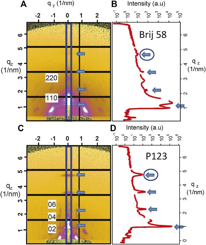
Marmiroli et al.                                                                                        Lipid Hydration Through Mesoporous Supports

INTRODUCTION                                                              lipid bilayers are widely studied, as they can be employed
                                                                          to mimic cell membranes, making them very attractive in
The ability to manipulate small amounts of fluids, in particular in        fields such as drug delivery and biosensing. As mesoporous
the field of medicine and biology, where some samples are                  silica films are subject to water filling and condensation as a
available in small quantities and/or very expensive, has                  function of relative humidity (RH) (Dourdain and Gibaud,
boosted the development of both microfluidics and, more                    2005), this effect could be exploited to deliver water through
recently, nanofluidics (Xu, 2018; Scheler et al., 2019; Craig              the pores.
et al., 2020). A further improvement of micro/nanofluidic                      In this work, we present our study of the hydration of lipid
systems for handling and detecting (bio)samples would consist             membranes supported on mesoporous silica films and their
in the insertion of materials presenting desired functionalities in       comparison with the ones deposited on silicon.
chosen areas of the devices. Mesoporous thin films are materials               Among the lipid membranes, 1-palmitoyl-2-oleoyl-sn-
synthesized through a bottom-up process that can be integrated            glycero-3-phosphocholine (POPC) was chosen, as its structure
into microdevices using top-down fabrication techniques. They             is changing as a function of humidity in three different lamellar
are composed of an inorganic compound and a templating agent              phases (Katsaras et al., 1993). In order to follow the evolution of
that controls the structure morphology such as the pore network           the structure with increasing RH, grazing incidence small-angle
or pore size during self-assembly. The films, usually produced by          X-ray scattering (GISAXS) measurements were conducted, as this
dip or spin coating of the precursor solution and subsequent              technique allows for in situ observation of structural features
thermal treatment, present a thickness around 100–500 nm and              typically between 1 and 100 nm and has already been widely
include uniform and connected pores of 2–50 nm (Grosso et al.,            employed to examine POPC under different conditions among
2004; Innocenzi and Malfatti, 2013). Among mesoporous                     which RH changes (Pabst et al., 2000; Amenitsch et al., 2004;
materials, our attention was focused on mesoporous silica,                Rappolt et al., 2004). GISAXS had also been previously applied to
which is biocompatible and can be further functionalized                  investigate the structure and the porous properties of mesoporous
using molecular SAMs by a silane coupling chemistry (Ketteler             films, in particular the contraction of pores in mesoporous films
et al., 2008; Seras-Franzoso et al., 2012; Sánchez et al., 2013;          due to capillary condensation with the rise of humidity (Gibaud
Tatkiewicz et al., 2013). It is known that silica is hydrolyzed in        et al., 2004; Grosso et al., 2004; Dourdain et al., 2005; Sharifi et al.,
water, and it undergoes degradation with time. However, a recent          2014).
study demonstrated that dissolution kinetics depend on the                    Here, mesoporous silica films were prepared using two
accessible surface area, the surrounding media composition,               surfactants, Brij 58 and P123, resulting in different pore
the residual surfactant molecules inside the pores, and their             dimensions and lattice arrangement. After sol–gel preparation
functionalization (Bindini et al., 2020).                                 and deposition on the silicon substrate, the following protocols
    Adding the possibility of micropatterning thin films, it would         were applied: 1) thermal treatment with the standard preparation
allow for the fabrication of microdevices, for example, for               route, 2) irradiation and development via DXRL, and 3) both
biosensing or biocatalysis (Magner, 2013; Lei et al., 2020).              treatments. The effect of film functionalization using molecular
    Among the different lithographic techniques, deep X-ray               SAMs with subsequent increase of hydrophobicity was evaluated.
lithography (DXRL) was shown to be an effective top-down                  The films were characterized by both measuring the contact angle
method to pattern sol–gel materials (Marmiroli and                        (CA) and using GISAXS. Then, their structural change with
Amenitsch, 2012; Innocenzi et al., 2014). DXRL is based on                increasing humidity was investigated using in situ GISAXS in
the exposure of materials to synchrotron high-energy X-rays               a custom-made humidity chamber. After the deposition of POPC,
(2–30 keV), leading to chemical and structural changes. If                in situ GISAXS measurements were repeated in order to check the
the irradiation is conducted through a suitable mask,                     hydration of POPC. The results were compared with the ones
microstructures can be obtained with high aspect ratio                    obtained by depositing POPC on a silicon wafer.
(ratio between height and minimum lateral dimension)
and 200 nm lateral resolution (Tormen et al., 2013). In
previous research, mesoporous silica films were patterned                  EXPERIMENTAL DETAILS
using DXRL where the X-rays simultaneously increase the
polycondensation of the exposed silica while partially                    Preparation of Mesoporous Silica Films
removing the surfactant (Falcaro et al., 2008; Falcaro                    Tetraethyl orthosilicate (TEOS), ethanol (EtOH) 99.8%, and HCl
et al., 2009; Innocenzi et al., 2011).                                    were purchased from Carl Roth GmbH + Co. KG (Karlsruhe,
    The possibility of employing mesoporous silica films in                                                            ®
                                                                          Germany); Brij 58 and Pluronic P123 were purchased from
micro/nanofluidic devices is still under investigation. They               Merck KGaA (Darmstadt, Germany). All reagents were used as
were already used as support for lipid membranes, increasing              supplied.
their mechanical stability and leaving space under the                       Two kinds of mesoporous silica films were synthesized, based
membrane, for example, for transmembrane proteins. Lipid                  on the different templating surfactants.
membranes could therefore be studied with characterization                   The first batch of samples was prepared using Brij 58, adapting
techniques such as AFM, quartz crystal balance, and                       the recipe proposed by Fuertes et al. (2007) as follows.
electrochemical impedance spectroscopy (Claesson et al.,                     TEOS (2.1 g), ethanol (6 g), water, and concentrated HCl were
2010; Claesson et al., 2011; Zhou et al., 2020). Synthetic                mixed at room temperature for 1 h. Brij 58 (0.56 g) was dissolved

Frontiers in Materials | www.frontiersin.org                          2                                      August 2021 | Volume 8 | Article 686353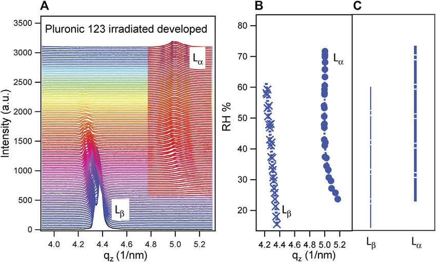
Marmiroli et al. Lipid Hydration Through Mesoporous Supports
TABLE 1 | List of samples with different treatments subject to investigation.
Surfactant Irradiation + development Thermal treatment Functionalization
Brij 58 x
Brij 58 x x
Brij 58 x
Brij 58 x x
Brij 58 x x
Brij 58 x x x
P123 x
P123 x x
P123 x
P123 x x
P123 x x
P123 x x x
in 5 g of ethanol. After 1 h, the two solutions were stirred together Thermal Treatment
for 1 h at room temperature. The pH was checked to be lower This is the standard method to both remove the surfactant (P123
than 3 before spin coating. The final molar composition of the or Brij 58) and crosslink the silica of the precursor (TEOS). It was
solution was 1 TEOS:0.05 Brij 58:5.2 H2O:24 EtOH:0.28 HCl. performed in an oven (Carbolite Oven, with Eurotherm 2416
For the second batch, Pluronic 123 (P123) was employed, control) at different temperatures.
according to the recipe proposed by Yan et al. (2007) as follows. For Brij 58, a temperature ramp was set to achieve
TEOS (1.75 g), ethanol (1 g), and 1.25 ml of 0.0025 M HCl were 350 °C in 1 h, and the value was subsequently kept
mixed and stirred for 1 h at room temperature. 0.584 g of P123 was constant for 2 h.
dissolved in 26.8 g of ethanol at room temperature with vigorous stirring. For P123, a temperature of 450°C was reached in 1 h, and then
After 1 h, the surfactant solution was added to silica and mixed for 1.5 h. it was maintained constant for 30 min.
1.93 ml of acidified water was added to the solution and stirred for
15 min. The pH was checked and adjusted to 2.5 with concentrated HCl. X-Ray Irradiation
In the following, the mesoporous silica films will be identified DXRL is based on the fact that some materials (defined as
by the surfactants Brij 58 and P123. resists) undergo a dissolution rate change in a specific solvent
Both film batches were processed via spin coating on silicon (called developer) when exposed to X-rays. Resists are exposed
wafers (Laurell WS-650_MZ-23 NPPB, Laurell Technologies to synchrotron radiation in a controlled way (Becker et al.,
Corporation, United States), for 1 min at 800 rpm. 1986; Romanato et al., 2006). If an X-ray mask, composed of an
After spin coating, the samples were subject to three different absorbing material (gold) and an X-ray transparent supporting
treatments: membrane (thin layer of graphite, or SiNx or TiO or SU8...)
is inserted between X-rays and the sample, then it is possible
1) Thermal treatment. to obtain micropatterning with good lateral resolution
2) X-ray irradiation and development. (200 nm) and comparatively high resist thickness with
3) X-ray irradiation and development followed by thermal respect to all other lithographic techniques. In the case of
treatment. mesoporous silica films, silica condensation is induced
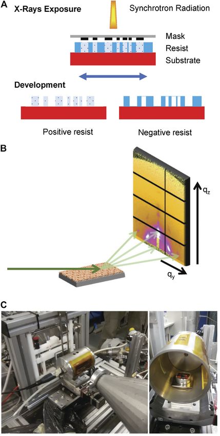
together with the breakage of the surfactant. Therefore,
After every kind of processing, the films were first mesoporous silica is a negative resist. The scheme of the
characterized and then further functionalized with molecular process is shown in Figure 1A.
SAMs in order to change their hydrophilicity. The samples were irradiated at the DXRL beamline of the
The list of the different mesoporous films studied in this work Elettra synchrotron radiation source (Pérennès et al., 2001;
is shown in Table 1. Pérennès and Pantenburg, 2001) using a Dex02 Jenoptik
CA and GISAXS measurements were performed on each film. scanner (Jenoptik AG, Jena, Germany). The storage ring was
The samples were selected in order to study the following: working at 2 GeV with a current of 310 mA. The irradiation
occurred through a white beam of energy range 2–20 KeV and
1) The effect of different pore arrangement and dimensions. peak around 6 KeV. Brij 58 samples received an energy per unit
2) The effect of irradiation compared to standard thermal treatment. surface (dose) of 68 J/cm2, while the P123 ones got 272 J/cm2. The
3) The effect of functionalization of the pores. dose had been previously selected by performing an irradiation
series with increasing energy to determine the minimum
After CA and GISAXS measurements, a lipid membrane was irradiation dose for which the structure was retained after
deposited on each type of mesoporous film, and GISAXS was development (Steinberg et al., 2021).
performed again. In the following, a more detailed description of The films were exposed immediately after spin coating and,
every procedure is given. just after exposure, developed to remove the templating agent, as
Frontiers in Materials | www.frontiersin.org 3 August 2021 | Volume 8 | Article 686353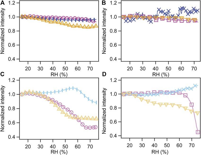
Marmiroli et al.                                                                                            Lipid Hydration Through Mesoporous Supports

                                                                                     In both cases, after development, the films were rinsed in
                                                                                  ethanol and dried with nitrogen. Some samples were
                                                                                  thermally treated following the procedures explained earlier,
                                                                                  in order to assure that all surfactant had been completely
                                                                                  removed, as already evidenced for Brij 58 by Steinberg et al.
                                                                                  (2021).

                                                                                  Self-Assembly Monolayer Functionalization of the
                                                                                  Mesoporous Films
                                                                                  The mesoporous silica thin films were functionalized with
                                                                                  (3-aminopropyl)triethoxysilane (APTES) (Merck KGaA,
                                                                                  Darmstadt, Germany), commonly used for surface silanization.
                                                                                  It was chosen as amino terminal functional groups can be
                                                                                  employed as linkers to further functionalize the films with bio-
                                                                                  macromolecules. A post-grafting functionalization was selected
                                                                                  because the number of available functional groups is higher than
                                                                                  the one obtained when the precursors are co-condensed during
                                                                                  the mesoporous material synthesis (Calvo et al., 2009). The
                                                                                  scheme of the first mesoporous layers in films functionalized
                                                                                  using SAMs via post-grafting is shown in Supplementary
                                                                                  Figure S1.
                                                                                     The functionalization protocol consisted in the following
                                                                                  steps:

                                                                                  1) Cleaning of the substrates by immersion for 5 min in acetone,
                                                                                     5 min in chloroform, and 5 min in isopropanol (all from
                                                                                     Merck KGaA, Darmstadt, Germany), then rinsing in Milli-
                                                                                     Q water, and drying with nitrogen.
                                                                                  2) Plasma treatment with O2 at a power of 20 W for 10 min to
                                                                                     eliminate residues.
                                                                                  3) Immersion in dry toluene solution (Merck KGaA, Darmstadt,
                                                                                     Germany) containing 1 mM APTES overnight at 80°C under
                                                                                     inert atmosphere.
                                                                                  4) Rinsing in toluene and ethanol.

                                                                                  Deposition of 1-Palmitoyl-2-oleoyl-sn-glycero-3-
                                                                                  phosphocholine Lipid Membrane
                                                                                  POPC (Avanti Polar Lipids, Alabaster, AL, United States) was
                                                                                  dissolved (10% w/w) in isopropyl alcohol (Rotisolv, >99.95%,
                                                                                  Carl Roth GmbH + Co. KG). A suitable volume was dispensed
                                                                                  on the mesoporous thin film with a micropipette and let
                                                                                  undisturbed until dry (Tristram-Nagle et al., 1993; Claesson
  FIGURE 1 | (A) Scheme of the DXRL process, showing the irradiation of
                                                                                  et al., 2010). Once dry, the samples were stored at room
  both positive and negative resists with synchrotron X-rays. (B) Graphic         temperature in vacuum for at least 16 h, to completely
  representation of the GISAXS experiment. X-rays hit the sample at grazing       evaporate the residual solvent, and removed from the vacuum
  angle, and the scattered photons are collected at the detector. (C)             chamber just before the measurement.
  Humidity sample chamber mounted at the Austrian SAXS beamline at Elettra.

                                                                                  Contact Angle Measurements
                                                                                  The CA was determined using a Drop Shape Analyzer 100
aging of the sol–gel would prevent the removal of the surfactant                  (KRÜSS GmbH, Hamburg, Germany) through the drop shape
after irradiation.                                                                analysis method. A 2 µL drop of ultra-pure water was placed on
   For Brij 58, the developer solution consisted of ethylene glycol:              each mesoporous film to obtain the CA between the substrate and
acetone:ethanol:water  1:1:1:1 (v/v) and the samples were                        the liquid. All measurements were performed in the same
immersed for 10 min.                                                              conditions. For each substrate, three trials were conducted,
   For P123, the solution was composed of ethylene glycol:acetone:                with two fits each. The CA of each substrate was obtained by
ethanol  4:2:1 (v/v). The development time was 30 min.                           calculating the average. Measurements of non-functionalized and

Frontiers in Materials | www.frontiersin.org                                  4                                  August 2021 | Volume 8 | Article 686353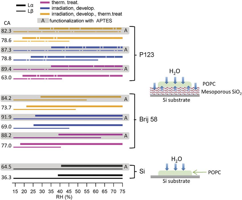
Marmiroli et al.                                                                                                             Lipid Hydration Through Mesoporous Supports

                                                                                               angle of 0.4°. The angular scale of the detector was calibrated with
                                                                                               silver behenate. After every measurement, the detector image was
                                                                                               processed using SAXSDOG, a software program for automatic
                                                                                               data reduction developed at the Austrian SAXS beamline (Burian
                                                                                               et al., 2020). In the present case, the diffraction patterns of vertical
                                                                                               cut qz in the out-of-plane direction were considered for data
                                                                                               analysis selecting the region (10 pixels) evidenced in Figure 2A.
                                                                                               The detector distance was defined to have qz in the range
                                                                                               0.31–6.8°nm−1.
                                                                                                   The experiments were conducted at controlled relative
                                                                                               humidity (RH), defined as the ratio of the water vapor
                                                                                               pressure to the saturation vapor pressure at a given
                                                                                               temperature. The chamber is described by Sharifi et al. (2014)
                                                                                               and shown in Figure 1C. It is composed of a metal cylinder
                                                                                               equipped with two Kapton windows (13 µm thickness) to let the
                                                                                               incident and the scattered beam pass through. Humidity is
                                                                                               controlled by mixing dry (95% RH)
                                                                                               air produced with a supersonic humidifier in a mixer chamber
                                                                                               and then flowing the mixture in the cell. A humidity sensor inside
                                                                                               the chamber in connection with a proportional integral derivative
                                                                                               (PID) controller is used to set the requested RH. The temperature
                                                                                               was constant at 25 ± 1°C.
                                                                                                   All samples listed in Table 1 were measured at RH  15% to
                                                                                               determine the unit cell parameters (shown in Supplementary
                                                                                               Figure S1). Then, the scattering pattern was collected while
                                                                                               increasing the relative humidity from 15 to 75%, at a rate of
  FIGURE 2 | (A) GISAXS image and (B) I–qz patterns corresponding to                           0.5%/min. The exposure time was set to 10 s every minute.
  the vertical cut evidenced by the rectangle for mesoporous silica prepared                       The specimens were then covered with POPC films following
  with Brij 58 and calcined after POPC deposition. The distorted (110)-oriented
                                                                                               the procedure in Deposition of 1-Palmitoyl-2-oleoyl-sn-glycero-3-
  body centered cubic structure is evident and partially indexed along the
  qz direction. (C) Image at the detector and (D) I–qz patterns corresponding to
                                                                                               phosphocholine Lipid Membrane and underwent the same RH
  the vertical cut evidenced by the rectangle for mesoporous silica prepared                   ramp while acquiring images for 3 s every minute to avoid
  with P123 and calcined after POPC deposition. The distorted hexagonal                        radiation damage of the lipid membrane.
  structure is evident and indexed along the qz direction. The arrows indicate the                 In this way, we could decouple the behavior of the mesoporous
  contribution of POPC, and the circled ones define the peaks that were used
                                                                                               material from the one of the deposited POPC.
  for the determination of the POPC phase transition.
                                                                                                   After completing the measurement, POPC was removed from
                                                                                               the mesoporous samples by washing in isopropyl alcohol and
                                                                                               dried in a vacuum chamber for at least 24 h. Then, SAM
SAM-functionalized            silicon     wafers      were      performed            for       functionalization with APTES was performed as described in
comparison.                                                                                    Self-Assembly Monolayer Functionalization of the Mesoporous
                                                                                               Films. The same GISAXS experiments were conducted as for
                                                                                               not functionalized samples to determine the unit cell parameters
Grazing Incidence Small-Angle X-Ray                                                            and the effect of humidity increase on functionalized mesoporous
Scattering Measurement                                                                         films alone. Afterward, the samples were dried in vacuum.
In GISAXS technique, a monochromatic X-ray beam hits the                                       Subsequently, POPC was deposited and the humidity was
sample at a low angle. A 2D detector measures the scattered                                    increased again to examine the lipid membrane hydration.
photons’ intensity in the directions parallel (qy) and
perpendicular (qz) to the sample surface (Renaud et al., 2009).
From the 2D image on the reciprocal space, it is possible to                                   RESULTS AND DISCUSSION
determine both the dimension and arrangement of the pores of
the mesoporous materials (Grosso et al., 2004) and the structure                               Contact Angle
of the lipid membrane (Amenitsch et al., 2004; Rappolt et al.,                                 As a first approach to investigate the hydrophilicity/
2004). The scheme of the GISAXS experiment is shown in                                         hydrophobicity of the prepared surfaces, the contact angle was
Figure 1B.                                                                                     determined. The results for each sample are given in Table 2.
   GISAXS measurements were performed at the Austrian SAXS                                         Although the standard deviation is quite high, we found that
beamline at Elettra (Amenitsch et al., 1997). The wavelength was                               all samples without functionalization are hydrophilic, following
λ  0.154 nm, and 2D GISAXS patterns were collected with a                                     the classical definition, as their contact angle is less than 90° (Law,
Pilatus3 1M detector (Dectris, Baden, Switzerland) at a grazing                                2014). As expected, functionalization increases hydrophobicity,

Frontiers in Materials | www.frontiersin.org                                               5                                      August 2021 | Volume 8 | Article 686353
Marmiroli et al. Lipid Hydration Through Mesoporous Supports
TABLE 2 | Contact angle (CA) of mesoporous materials prepared with two surfactants and subject to different treatments.
Sample description CA (st dev) CA (st dev) with APTES
Silicon 36.3° (1.42) 64.5° (0.89)
Brij 58 thermal treatment 77° (3.39) 88.23° (1.43)
Brij 58 irradiation and development 69° (3) 91.87° (0.46)
Brij 58 irradiation, development, and thermal treatment 73.7° (7.1) 84.267° (3.78)
P123 thermal treatment 70.6° (4.7) 89.4° (0.57)
P123 irradiation and development 78.8° (1.12) 87.3° (1.42)
P123 irradiation, development, and thermal treatment 78.6° (2.28) 82.3° (4.69)
as it replaces some of the -SiOH surface sites with more The influence of the increase of RH (%) on the mesoporous
hydrophobic groups like –(CH2)3NH2 of the APTES molecules silica structure was evidenced using the following procedure:
(Calvo et al., 2009).
1. The intensity of peak (110) for mesoporous materials prepared
with Brij 58, and of peak (200) for the ones with P123, was
Grazing Incidence Small-Angle X-Ray directly calculated from the detector image integrating the 2D
Scattering Measurement peak area.
The representative detector images for mesoporous materials are 2. The intensity change due to the increase of RH was normalized
shown in Figure 2A for Brij 58 and Figure 2C for P123 calcined to the initial value of intensity for the dry sample at RH 15%.
films, respectively. The indexing of the peaks is limited to the In this way, the behavior of the different mesoporous materials
most noticeable spots along the vertical cut, to evidence the could be compared.
position of the mesoporous silica peaks.
The diffraction patterns (I–qz) of the out-of-plane cuts are The normalized intensity changes versus RH for all samples
presented in Figures 2B,D. The arrows indicate the contribution with their corresponding treatment (thermally treated samples,
which is unequivocally related to the POPC lipid membrane. The irradiated and developed, irradiated/developed/thermally
circle highlights the peak that has been considered to demonstrate treated) are shown in Figure 3. In detail, Figures 3(A) and (C)
the phase transition of POPC upon humidity change. The refer to the mesoporous silica prepared with the Brij 58
diffraction peaks in Figure 2A demonstrate the orthorhombic surfactant, respectively, without and with functionalization;
Fmmm structure of the Brij 58 templated silica mesopores derived Figures 3(B) and (D) describe the behavior of samples with
from the distorted (110)-oriented body centered cubic Im3m to surfactant P123 in the absence and presence of APTES SAMs.
which the indexing is referred (Crepaldi et al., 2003; Voss et al., Mesoporous silica films prepared with both surfactants
2014). The peaks in Figure 2C show the hexagonal arrangement without further functionalization had similar behavior in spite
of the pores in the P123 templated mesoporous film (Sharifi et al., of different dimensions and arrangement of the pores. This is in
2014). line with the research conducted by Ceratti et al. (2015), who
found that there is not direct correlation between the mesopores’
Mesoporous Silica Films With and Without dimension or arrangement and their capillary filling rate.
Self-Assembly Monolayer Functionalization For both kinds of structures, only the film condensed regime
The unit cells considered for the calculation of the structure was present in the RH range 15–75%, meaning that water
parameters are shown in Supplementary Figure S2, and the unit molecules form a thin film on the pore walls (Huber, 2015). A
cell parameters for every kind of sample are reported in hint of the onset of capillary condensation at an RH>70% could
Supplementary Table S1. In the case of mesoporous materials be observed for thermally treated samples (both with and without
prepared using the Brij 58 surfactant, the Fmmm cells were irradiation and development). As far as mesoporous silica
around b 6 nm in the direction parallel to the substrate and prepared with P123 and thermally treated is concerned, the
a 3.8 nm in the perpendicular direction, indicating a strong measurement agreed well with the results presented by Sharifi
shrinkage of the original mesoporous structure upon both et al. (2014) and Ganser et al. (2016) for a thermally treated
thermal treatment and irradiation. The samples subject to mesoporous material prepared with a similar recipe.
both irradiation and thermal treatment presented the highest The behavior of not functionalized films prepared by
contraction of the original structure (a 2.8 nm). irradiation and development, particularly those templated by
Similar considerations can be drawn for the p6m structure of the P123 surfactant, shows a different trend with nearly no
the mesoporous materials prepared with surfactant P123. In change with RH. On the contrary, the functionalization with
this case, the unit cell parameters were bigger: b 13–14 nm, APTES SAMs had an influence on the performance of the
a 14–15 nm. These data are consistent with the findings mesoporous films with RH, and it affected differently the
presented by Ganser et al. (2016) relative to mesoporous structures derived from the two surfactants.
materials prepared with the same surfactant and a similar Functionalized samples prepared using Brij 58 and
thermal treatment. The effect of both irradiation and thermal undergoing a thermal treatment (with and without irradiation)
treatment leads to a further distortion and a 10.1 nm. presented a capillary condensation starting at RH 30–35%. The
Frontiers in Materials | www.frontiersin.org 6 August 2021 | Volume 8 | Article 686353Marmiroli et al. Lipid Hydration Through Mesoporous Supports
FIGURE 3 | Normalized intensity of peak (110) of the cubic phase (Brij 58) and of peak (200) of the hexagonal phase (P123) as a function of RH. (A) Mesoporous
silica prepared with surfactant Brij 58. (B) Mesoporous silica with the P123 templating agent. (C) Mesoporous silica with Brij 58 functionalized with APTES. (D)
Mesoporous silica with P123 functionalized with APTES. Pink curves represent thermally treated samples (circles, Brij 58; squares, P123), blue ones represent irradiated
and developed ones (vertical bar markers, Brij 58; x markers, P123), and yellow ones represent irradiated, developed, and thermally treated ones (triangles with tip
up, Brij 58; triangles with tip down, P123).
irradiated film had a drop of intensity at a higher RH (around Mesoporous Silica Films With and Without
60%) indicating an onset of capillary condensation, even if the Functionalization Plus
complete filling of the pores does not occur. 1-Palmitoyl-2-oleoyl-sn-glycero-3-phosphocholine
As far as the functionalized films obtained employing P123 are It is known from the literature that, at 20°C, POPC presents three
concerned, only the thermally treated one without irradiation different lamellar phases depending on RH (%) (Katsaras et al.,
displayed a clearly defined capillary condensation starting at 1993):
around RH 60%. The irradiated film presented the same
behavior as the not functionalized one, suggesting that Lδ at RH 0–15% with d-spacing 5.47 nm at RH 0%
development after irradiation did not result in a full pore Lβ at RH 15–50% with d-spacing 5.74 nm at RH 40%
opening. The films subject to irradiation, development, and Lα at RH 65–100% with d-spacing 5.12 nm at RH 100%
thermal treatment exhibited a hydration mechanism which is
more difficult to interpret but still showed that water is entering at The measurements described in this work were conducted at
least partially the pores. 25°C, but both the behavior and the d-spacing were similar. To
The behavior of mesoporous materials thermally treated confirm it, POPC was deposited on bare Si with and without
and functionalized with APTES was opposite to the one functionalization with the procedure described in Deposition of
described by Khalil et al. (2020) for functionalized mesoporous POPC Lipid Membrane, and an in situ GISAXS experiment was
materials, where the onset of capillary condensation with RH performed at increasing RH. Then, mesoporous samples with
increases with hydrophobicity. However, both the used POPC were measured and compared to silicon ones. The I–qz
(1,1,2H,2H-perfluorooctyldimethylchlorosilane) molecule, patterns for each humidity ramp have been plotted together at
which is much longer and more hydrophobic than APTES, increasing RH. An example is shown in Figure 4A for the sample
and the functionalization procedure were different. We prepared using surfactant P123, irradiated and developed. The
believe that, in our case, the functionalization produced a selected q-range is the one relative to the fourth order of the
dense layer of amino terminal groups inside the pores that led POPC lamellar phase, and it was chosen to avoid the influence of
to a steric reduction of their dimension and thus to the the signal of mesoporous silica dominating in the lower q-range
capillary filling for lower values of RH (see Supplementary (see also Figure 2). The lipid membrane undergoes a structural
Figure S1). transition from the Lβ phase, present at lower RH, to the Lα phase.
Frontiers in Materials | www.frontiersin.org 7 August 2021 | Volume 8 | Article 686353Marmiroli et al. Lipid Hydration Through Mesoporous Supports
FIGURE 4 | (A) Out-of-plane pattern (I–qz) at increasing RH for POPC deposited on a thin mesoporous silica film prepared with surfactant P123, irradiated and
developed. The two phases Lβ and Lα are shown, evidencing the structural transition of POPC upon increasing relative humidity. The red curves, superimposed to the
measured ones in correspondence with the Lα phase, represent the Lorentzian fitting of the peaks of the lamellar phase. (B) qz resulting from the Lorentzian fitting of the
Lβ phase (left) and Lα phase (right) as a function of RH (%). (C) Scheme of the RH (%) range of existence of the Lβ phase (left) and Lα phase (right).
In the current case, both phases were coexisting for a certain RH The lipid membrane hydration was evaluated mainly by
range. The presence of two peaks in every phase, particularly considering the RH value at which the humid phase Lα
evident in Lβ, was due to a partial disorder of the deposited POPC. appeared. In fact, the disappearing of Lβ (dry phase) was more
All peaks were fitted with a Lorentzian function (as shown for Lα), difficult to interpret, as POPC had been deposited via drop
as Lorentzian distribution describes well the peak shape of casting and its thickness in the different samples was not
smectic liquid crystalline systems (Rappolt, 2010). The uniform, and the phase transition is related to water diffusion
resulting qz for both phases as a function of RH (%) for the and to water availability across the thickness.
sample prepared with P123, irradiated and developed, is shown in The following considerations can be drawn:
Figure 4B. The RH (%) ranges of existence of the two phases are
schematized in Figure 4C. 1) Mesoporous materials conveyed water to POPC through the
The d-spacing of the lipid membrane phases and its evolution pores, as in all cases the Lα phase (the hydrated one) occurred
with the increase of RH was calculated using the following at a value of RH which was lower with respect to POPC
formula for lamellar systems (Mangold, 1995): deposited on bare Si. With Si, the onset of Lα started at RH
around 40%, while with mesoporous materials, it occurred at
l about 20%.
d 2π 2) It was confirmed that different surfactants do not lead to a
q
drastic change of the behavior. P123 and Brij 58 mesoporous
where l is the reflection order. In the present case, l 4. materials displayed a similar performance even if both the
A summary of the results is shown in Supplementary Figure pore arrangement and the pore dimension were different
S3. The determined values agree with the ones reported in the (Ceratti et al., 2015).
literature. 3) In general, thermally treated samples conveyed more water
A summary of the RH ranges for Lβ and Lα phases for all than only irradiated ones. This is attributed to the fact that the
samples is shown in Figure 5. The lines are correspondent to developer solution was not able to efficiently open the pores,
different substrates: the dashed ones are relative to the films as already inferred from the lack of capillary condensation.
prepared with the P123 surfactant (top) and the continuous ones This was most evident for the irradiated film prepared with
represent the mesoporous films prepared using Brij 58 (middle). surfactant P123. It looks like the pores are sterically blocked as
Thicker lines represent Lα phases, and thinner ones represent Lβ. they still contain the surfactant. Anyway, such films can still
The shadowed areas with the letter A depict the APTES- transfer water to the POPC membrane. One hypothesis,
functionalized samples. Colors refer to diverse treatments. The following Steinberg et al. (2021), was that pores are
corresponding CA value is reported beside every sample. Two partially open and water flows at the interface between
schemes showing the hydration of the lipid membrane with and them and the lipid membrane. Water could also enter the
without mesoporous film are also shown. interface between the silica and the surfactant or inside the
Frontiers in Materials | www.frontiersin.org 8 August 2021 | Volume 8 | Article 686353Marmiroli et al. Lipid Hydration Through Mesoporous Supports FIGURE 5 | Summary of the RH range of Lα and Lβ phases for all samples and sketch of the hydration pathway. Black lines represent POPC deposited on silicon substrates (bottom panel), continuous lines show the mesoporous films prepared using the Brij 58 surfactant (middle panel) and dashed lines show the films prepared with P123 (top panel). Thicker lines correspond to Lα phases, and thinner ones correspond to Lβ. The shadowed areas with the letter A indicate the samples functionalized with APTES. The pink colors (bottom lines) represent thermally treated samples, the blue ones (middle lines) represent irradiated and developed ones, and the yellow ones (top lines) represent irradiated, developed, and thermally treated ones. The CA value is reported beside the corresponding sample. Schemes of the hydration pathway of the lipid membrane deposited on Si and mesoporous thin films are, respectively, shown beside the corresponding substrates. partially unconsolidated oxide. This could also explain why When the lipid membrane is deposited on a hydrophilic there was no visible change in the SAXS pattern of surface (like silicon with a thin layer of native oxide), there is mesoporous silica with RH, as water is not replacing air in usually a hydration layer less than 2 nm thick that prevents the empty pores but the pores still contain surfactant; therefore, lipid from interacting strongly with the substrate (Kim et al., there is not electron density contrast. 2001). In this case, we speak of the continuous pore-spanning 4) The functionalization of Si with APTES did not lead to a big membrane. Functionalization with APTES favors the adsorption change of the hydration of POPC, even if the CA increased of the lipid membrane on the substrate, as the amino groups give considerably. a positive net charge to the substrate surface. This increases the 5) APTES increased hydrophobicity of all mesoporous materials electrostatic interaction with the negatively charged phosphate of (as confirmed by the CA). This affected the hydration of the the zwitterionic POPC headgroups, leading to a hybrid pore- lipid membrane (evidenced by the onset of phase Lα). Lipid spanning membrane (Leonenko et al., 2000; Bhattacharya et al., membranes supported on the APTES-functionalized 2011). In this latter case, the fluidity of the resulting supported mesoporous material exhibited a phase transition at RH lipid membrane is not affected (Miyashita et al., 2018; Sun et al., values around 30%, lower than those at which it occurred 2020). As the sample prepared by drop casting presents thick when the lipid was deposited on bare silicon, but still at RH layers, the structure and the hydration behavior of the lipid 5–10% higher than the one observed when POPC was drop membrane depend only on the water content in the casted on the not functionalized material. The disappearing of environment. This justifies why the behavior of POPC the dry phase Lβ occurred at higher values for APTES-treated deposited on silicon substrates with and without samples than the untreated ones. In some case, it did not even functionalization is comparable. For mesoporous substrates, disappear. This partially contradicts the fact that capillary the amount of water delivered through the pores (in the liquid condensation, and therefore pore filling, occurs at a lower RH or vapor form) influences the onset of the humid phase (Lα) of (%) for mesoporous materials functionalized with APTES, POPC. The results show that the APTES-functionalized samples compared with the not functionalized ones. convey less water to the lipid membrane when compared to the Frontiers in Materials | www.frontiersin.org 9 August 2021 | Volume 8 | Article 686353
Marmiroli et al.                                                                                       Lipid Hydration Through Mesoporous Supports

not functionalized ones, and this is in line with increased                 arrangement of the pores in the silica film, does not have a
hydrophobicity of the surface and the pores.                                profound influence on POPC behavior.
    From these measurements, it could be concluded that                        The possibility of patterning the films, and a suitable surface
mesoporous films have an active effect on the hydration of                   functionalization of the mesopores with SAMs of different
supported POPC. The shift of the onset of Lα phase of POPC                  molecules with specific binding terminal groups could open
occurs as the water hydrates the lipid membrane also through the            the use of mesoporous films as “interactive” supports for
pores of the mesoporous materials. This is caused by the osmotic            biological studies of increasing complexity, such as protein/
pressure due to the different water activity (chemical potential) in        peptide incorporation for signaling. Thus, samples could be
the lipid membrane and the surrounding environment (Milhaud,                handled and modified from below and be investigated with
2004). The hydration source can be at both the liquid and vapor             different techniques from above.
phases. In the present case, there is vapor phase outside the                  The next step will be the investigation of the possibility of
mesopores due to an increase of RH (%). Inside the pores, the               conveying liquids through the pores at constant RH by means of
mechanism is more complex. The main contribution is from the                an appropriate reservoir. Studies of water delivery are already
vapor phase, as the anticipated onset of Lα is decoupled from the           being conducted by our group using IR and GISAXS techniques.
capillary condensation of water inside the pores. In the range of
RH (%) where Lα appears, mesoporous films are in the condensed
regime, where the molecules form a thin film of water on the pore            DATA AVAILABILITY STATEMENT
walls. The addition of APTES reduces the amount of water
entering the pores and consequently the shift of Lα onset. As               The raw data supporting the conclusions of this article will be
the capillary filling rate and condensation regime in the                    made available by the authors, without undue reservation.
mesoporous films are not in direct correlation with the pore
dimension and arrangement, as demonstrated by Ceratti et al.
(2015), the effect of surfactant on the film preparation does not            AUTHOR CONTRIBUTIONS
have big influence. Hydration can be partially tuned by playing
with film treatment and surface functionalization.                           BS participated in the synthesis and functionalization and in the
    The present results refer to POPC membranes deposited on                GISAXS studies. IR and AK defined the surface functionalization
the films by drop casting. Preliminary experiments on POPC                   protocol and performed CA measurements. HA and BM directed
deposited by dip coating showed the same behavior. However,                 the project. HA participated in SAXS measurements. BM
further studies are required to investigate the effect of the POPC          conducted the X-ray irradiation of the films and participated
deposition method and of the resulting lipid membrane thickness             in GISAXS measurements. All authors contributed to the
on the hydration of supported lipid membranes on mesoporous                 manuscript writing process.
silica substrates.

                                                                            ACKNOWLEDGMENTS
CONCLUSION
                                                                            This research was partly supported by the European
In the present work, we studied the hydration with RH increase of           Commission under grant agreement 654360 NFFA-EUROPE.
POPC lipid membranes supported on silicon and mesoporous                    This work includes part of the scientific activities of the internal
silica films prepared with different surfactants, subject to diverse         CERIC-ERIC project renewals. The authors are thankful to C.
thermal treatment, and with and without surface                             Morello and L. Sancin for their technical support. IR is grateful
functionalization with APTES. The contact angle was                         to the support of the Spanish Ministry of Economy and
measured for every film prior to lipid membrane deposition.                  Competitiveness      (MINECO)          (PID2019-105622RBI00)
In situ GISAXS experiments were conducted in a humidity                     through the Severo Ochoa Programme for Centres of
chamber on POPC deposited both on mesoporous materials                      Excellence in R&D (CEX 2019-000917-S), Instituto de Salud
and on bare silicon wafers with and without surface treatment               Carlos III through the Networking Research Center on
with APTES. The hydration was examined by detecting the onset               Bioengineering, Biomaterials, and Nanomedicine (CIBER-
of the Lα phase of POPC (humid phase). It was found that the                BBN), Generalitat de Catalunya (SGR-918), and Fundacio
water uptake of the mesopores during RH increase leads to a                 Marato de TV3 (No. 201812).
hydration of the supported lipid membrane at a considerably
lower RH with respect to the membrane deposited on bare silicon.
The water uptake and the subsequent interaction with the lipid              SUPPLEMENTARY MATERIAL
membrane can be partially tuned by using thermal treatment, to
increase water adsorption and therefore hydration, and by                   The Supplementary Material for this article can be found online at:
functionalizing the surface with APTES for the opposite effect.             https://www.frontiersin.org/articles/10.3389/fmats.2021.686353/
The surfactant choice, leading to different dimensions and                  full#supplementary-material

Frontiers in Materials | www.frontiersin.org                           10                                    August 2021 | Volume 8 | Article 686353
Marmiroli et al. Lipid Hydration Through Mesoporous Supports
REFERENCES Ganser, C., Fritz-Popovski, G., Morak, R., Sharifi, P., Marmiroli, B., Sartori, B., et al.
(2016). Cantilever Bending Based on Humidity-Actuated Mesoporous Silica/
silicon Bilayers. Beilstein J. Nanotechnol. 7, 637–644. doi:10.3762/bjnano.7.56
Amenitsch, H., Bernstorff, S., Kriechbaum, M., Lombardo, D., Mio, H., Rappolt, Gibaud, A., Dourdain, S., Gang, O., and Ocko, B. M. (2004). In Situgrazing
M., et al. (1997). Performance and First Results of the ELETTRA High-Flux Incidence Small-Angle X-ray Scattering Real-Time Monitoring of the Role
Beamline for Small-Angle X-ray Scattering. J. Appl. Cryst. 30, 872–876. of Humidity during the Structural Formation of Templated Silica Thin Films.
doi:10.1107/S0021889897001593 Phys. Rev. B 70, 1–4. doi:10.1103/PhysRevB.70.161403
Amenitsch, H., Rappolt, M., Teixeira, C. V., Majerowicz, M., and Laggner, P. Grosso, D., Cagnol, F., Soler-Illia, G. J. D. A. A., Crepaldi, E. L., Amenitsch, H.,
(2004). In Situ sensing of Salinity in Oriented Lipid Multilayers by Surface Brunet-Bruneau, A., et al. (2004). Fundamentals of Mesostructuring through
X-ray Scattering. Langmuir 20, 4621–4628. doi:10.1021/la036319p Evaporation-Induced Self-Assembly. Adv. Funct. Mater. 14, 309–322.
Becker, E. W., Ehrfeld, W., Hagmann, P., Maner, A., and Münchmeyer, D. (1986). doi:10.1002/adfm.200305036
Fabrication of Microstructures with High Aspect Ratios and Great Structural Huber, P. (2015). Soft Matter in Hard Confinement: Phase Transition
Heights by Synchrotron Radiation Lithography, Galvanoforming, and Plastic Thermodynamics, Structure, Texture, Diffusion and Flow in Nanoporous
Moulding (LIGA Process). Microelectron. Eng. 4, 35–56. doi:10.1016/0167- media. J. Phys. Condens. Matter 27, 103102. doi:10.1088/0953-8984/27/10/103102
9317(86)90004-3 Innocenzi, P., and Malfatti, L. (2013). Mesoporous Thin Films: Properties and
Bhattacharya, J., Kisner, A., Offenhäusser, A., and Wolfrum, B. (2011). Applications. Chem. Soc. Rev. 42, 4198–4216. doi:10.1039/c3cs35377j
Microfluidic Anodization of Aluminum Films for the Fabrication of Innocenzi, P., Malfatti, L., Kidchob, T., Costacurta, S., Falcaro, P., Marmiroli, B.,
Nanoporous Lipid Bilayer Support Structures. Beilstein J. Nanotechnol. 2, et al. (2011). Densification of Sol-Gel Silica Thin Films Induced by Hard X-Rays
104–109. doi:10.3762/bjnano.2.12 Generated by Synchrotron Radiation. J. Synchrotron Radiat. 18, 280–286.
Bindini, E., Chehadi, Z., Faustini, M., Albouy, P.-A., Grosso, D., Cattoni, A., et al. doi:10.1107/S0909049510051666
(2020). Following In Situ the Degradation of Mesoporous Silica in Biorelevant Innocenzi, P., Malfatti, L., Marmiroli, B., and Falcaro, P. (2014). Hard X-Rays and
Conditions: At Last, a Good Comprehension of the Structure Influence. ACS Soft-Matter: Processing of Sol-Gel Films from a Top Down Route. J. Sol-gel Sci.
Appl. Mater. Inter. 12, 13598–13612. doi:10.1021/acsami.9b19956 Technol. 70, 236–244. doi:10.1007/s10971-013-3227-y
Burian, M., Meisenbichler, C., Naumenko, D., and Amenitsch, H. (2020). Katsaras, J., Jeffrey, K. R., Yang, D. S. C., and Epand, R. M. (1993). Direct Evidence for
SAXSDOG: Open Software for Real-Time Azimuthal Integration of 2D the Partial Dehydration of Phosphatidylethanolamine Bilayers on Approaching
Scattering Images. 1–17. Available at: http://arxiv.org/abs/2007.02022 the Hexagonal Phase. Biochemistry 32, 10700–10707. doi:10.1021/bi00091a021
(Accessed July 4, 2020). Ketteler, G., Ashby, P., Mun, B. S., Ratera, I., Bluhm, H., Kasemo, B., et al. (2008). In
Calvo, A., Joselevich, M., Soler-Illia, G. J. A. A., and Williams, F. J. (2009). Chemical Situphotoelectron Spectroscopy Study of Water Adsorption on Model
Reactivity of Amino-Functionalized Mesoporous Silica Thin Films Obtained by Biomaterial Surfaces. J. Phys. Condens. Matter 20, 184024. doi:10.1088/0953-
Co-condensation and post-grafting Routes. Microporous Mesoporous Mater. 8984/20/18/184024
121, 67–72. doi:10.1016/j.micromeso.2009.01.005 Khalil, A., Zimmermann, M., Bell, A. K., Kunz, U., Hardt, S., Kleebe, H.-J., et al.
Ceratti, D. R., Faustini, M., Sinturel, C., Vayer, M., Dahirel, V., Jardat, M., et al. (2020). Insights into the Interplay of Wetting and Transport in Mesoporous
(2015). Critical Effect of Pore Characteristics on Capillary Infiltration in Silica Films. J. Colloid Interf. Sci. 560, 369–378. doi:10.1016/
Mesoporous Films. Nanoscale 7, 5371–5382. doi:10.1039/C4NR03021D j.jcis.2019.09.093
Claesson, M., Cho, N.-J., Frank, C. W., and Andersson, M. (2010). Vesicle Kim, J., Kim, G., and Cremer, P. S. (2001). Investigations of Water Structure at the
Adsorption on Mesoporous Silica and Titania. Langmuir 26, 16630–16633. Solid/liquid Interface in the Presence of Supported Lipid Bilayers by Vibrational
doi:10.1021/la102719w Sum Frequency Spectroscopy. Langmuir 17, 7255–7260. doi:10.1021/la0017274
Claesson, M., Frost, R., Svedhem, S., and Andersson, M. (2011). Pore Spanning Law, K.-Y. (2014). Definitions for Hydrophilicity, Hydrophobicity, and
Lipid Bilayers on Mesoporous Silica Having Varying Pore Size. Langmuir 27, Superhydrophobicity: Getting the Basics Right. J. Phys. Chem. Lett. 5,
8974–8982. doi:10.1021/la201411b 686–688. doi:10.1021/jz402762h
Craig, M., Jenner, A. L., Namgung, B., Lee, L. P., and Goldman, A. (2020). Lei, Q., Guo, J., Noureddine, A., Wang, A., Wuttke, S., Brinker, C. J., et al. (2020).
Engineering in Medicine to Address the Challenge of Cancer Drug Resistance: Sol-Gel-Based Advanced Porous Silica Materials for Biomedical Applications.
From Micro- and Nanotechnologies to Computational and Mathematical Adv. Funct. Mater. 30, 1–28. doi:10.1002/adfm.201909539
Modeling. Chem. Rev. 121, 3352–3389. doi:10.1021/acs.chemrev.0c00356 Leonenko, Z. V., Carnini, A., and Cramb, D. T. (2000). Supported Planar Bilayer
Crepaldi, E. L., Soler-Illia, G. J. d. A. A., Grosso, D., Cagnol, F., Ribot, F., and Formation by Vesicle Fusion: The Interaction of Phospholipid Vesicles with
Sanchez, C. (2003). Controlled Formation of Highly Organized Mesoporous Surfaces and the Effect of Gramicidin on Bilayer Properties Using Atomic Force
Titania Thin Films: From Mesostructured Hybrids to Mesoporous Microscopy. Biochim. Biophys. Acta (Bba) - Biomembr. 1509, 131–147.
Nanoanatase TiO2. J. Am. Chem. Soc. 125, 9770–9786. doi:10.1021/ja030070g doi:10.1016/S0005-2736(00)00288-1
Dourdain, S., and Gibaud, A. (2005). On the Capillary Condensation of Water in Magner, E. (2013). Immobilisation of Enzymes on Mesoporous Silicate Materials.
Mesoporous Silica Films Measured by X-ray Reflectivity. Appl. Phys. Lett. 87, Chem. Soc. Rev. 42, 6213–6222. doi:10.1039/c2cs35450k
223105-3. doi:10.1063/1.2136412 Mangold, H. K. (1995). The Lipid Handbook. Second Edition, F. D. Gunstone,
Dourdain, S., Bardeau, J.-F., Colas, M., Smarsly, B., Mehdi, A., Ocko, B. M., et al. J. L. Harwood, and F. B. Padley London: Chapman & HallLipid/Fett 97, 315–316.
(2005). Determination by X-ray Reflectivity and Small Angle X-ray Scattering 1994Preis: £ 255. – (ISBN 0 412 43320 6). doi:10.1002/lipi.19950970720
of the Porous Properties of Mesoporous Silica Thin Films. Appl. Phys. Lett. 86, Marmiroli, B., and Amenitsch, H. (2012). X-ray Lithography and Small-Angle
113108-3. doi:10.1063/1.1887821 X-ray Scattering: A Combination of Techniques Merging Biology and Materials
Falcaro, P., Costacurta, S., Malfatti, L., Takahashi, M., Kidchob, T., Casula, M. F., Science. Eur. Biophys. J. 41, 851–861. doi:10.1007/s00249-012-0843-3
et al. (2008). Fabrication of Mesoporous Functionalized Arrays by Integrating Milhaud, J. (2004). New Insights into Water-Phospholipid Model Membrane
Deep X-ray Lithography with Dip-Pen Writing. Adv. Mater. 20, 1864–1869. Interactions. Biochim. Biophys. Acta (Bba) - Biomembr. 1663, 19–51.
doi:10.1002/adma.200702795 doi:10.1016/j.bbamem.2004.02.003
Falcaro, P., Malfatti, L., Vaccari, L., Amenitsch, H., Marmiroli, B., Grenci, G., et al. Miyashita, W., Saeki, D., and Matsuyama, H. (2018). Formation of Supported Lipid
(2009). Fabrication of Advanced Functional Devices Combining Soft Chemistry Bilayers on Porous Polymeric Substrates Induced by Hydrophobic Interaction.
with X-ray Lithography in One Step. Adv. Mater. 21, 4932–4936. doi:10.1002/ Colloids Surf. A: Physicochem. Eng. Aspects 538, 297–303. doi:10.1016/
adma.200901561 j.colsurfa.2017.11.006
Fuertes, M. C., López-Alcaraz, F. J., Marchi, M. C., Troiani, H. E., Luca, V., Míguez, Pabst, G., Rappolt, M., Amenitsch, H., and Laggner, P. (2000). Structural
H., et al. (2007). Photonic Crystals from Ordered Mesoporous Thin-Film Information from Multilamellar Liposomes at Full Hydration: Fullq-Range
Functional Building Blocks. Adv. Funct. Mater. 17, 1247–1254. doi:10.1002/ Fitting with High Quality X-ray Data. Phys. Rev. E 62, 4000–4009. doi:10.1103/
adfm.200601190 PhysRevE.62.4000
Frontiers in Materials | www.frontiersin.org 11 August 2021 | Volume 8 | Article 686353Marmiroli et al. Lipid Hydration Through Mesoporous Supports
Pérennès, F., De Bona, F., and Pantenburg, F. J. (2001). Deep X-Ray Lithography Tatkiewicz, W. I., Seras-Franzoso, J., Garcia-Fruitos, E., Vazquez, E., Ventosa, N.,
Beamline at ELETTRA. Nucl. Instr. Methods Phys. Res. 467–468, 1274–1278. Ratera, I., et al. (2013). 2D Engineering of Protein-Based Nanoparticles for Cell
doi:10.1016/S0168-9002(01)00632-5 Guidance. Tech. Proc. 2013 NSTI Nanotechnol. Conf. Expo. Nsti-nanotech 2013
Pérennès, F., and Pantenburg, F. J. (2001). Adhesion Improvement in the Deep 3, 229–231.
X-ray Lithography Process Using a central Beam-Stop. Nucl. Instr. Methods Tormen, M., Grenci, G., Marmiroli, B., and Romanato, F. (2013). X-ray
Phys. Res. Section B: Beam Interact.Mater. Atoms 174, 317–323. doi:10.1016/ Lithography: Fundamentals and Applications. Hoboken, NJ: John Wiley &
S0168-583X(00)00588-7 Sons, Inc., 1–86. doi:10.1002/9781118622582.ch1
Rappolt, M., Amenitsch, H., Strancar, J., Teixeira, C. V., Kriechbaum, M., Pabst, G., Tristram-Nagle, S., Zhang, R., Suter, R. M., Worthington, C. R., Sun, W. J., and
et al. (2004). Phospholipid Mesophases at Solid Interfaces: In-Situ X-ray Nagle, J. F. (1993). Measurement of Chain Tilt Angle in Fully Hydrated Bilayers
Diffraction and Spin-Label Studies. Adv. Colloid Interf. Sci. 111, 63–77. of Gel Phase Lecithins. Biophysical J. 64, 1097–1109. doi:10.1016/S0006-
doi:10.1016/j.cis.2004.07.004 3495(93)81475-9
Rappolt, M. (2010). Bilayer Thickness Estimations with “Poor” Diffraction Data. Voss, G. J. B., Chavez Panduro, E. A., Midttveit, A., Fløystad, J. B., Høydalsvik, K.,
J. Appl. Phys. 107, 084701. doi:10.1063/1.3393600 Gibaud, A., et al. (2014). Mesostructured Alumina as Powders and Thin Films.
Renaud, G., Lazzari, R., and Leroy, F. (2009). Probing Surface and Interface J. Mater. Chem. A. 2, 9727–9735. doi:10.1039/c4ta00604f
Morphology with Grazing Incidence Small Angle X-Ray Scattering. Surf. Sci. Xu, Y. (2018). Nanofluidics: A New Arena for Materials Science. Adv. Mater. 30,
Rep. 64, 255–380. doi:10.1016/j.surfrep.2009.07.002 1702419. doi:10.1002/adma.201702419
Romanato, F., Businaro, L., Tormen, M., Perennes, F., Matteucci, M., Marmiroli, B., Yan, M., Henderson, M. J., and Gibaud, A. (2007). Grating Induced Micelle
et al. (2006). Fabrication of 3D Micro and Nanostructures for MEMS and Alignment of Mesostructured Silica Films. Appl. Phys. Lett. 91, 023104–023105.
MOEMS: An Approach Based on Combined Lithographies. J. Phys. Conf. Ser. doi:10.1063/1.2755722
34, 904–911. doi:10.1088/1742-6596/34/1/150 Zhou, S., Guilfoil, E., He, Y., Nagpure, S., Islam, S. Z., Khan, M. A., et al. (2020).
Sánchez, G., Curiel, D., Ratera, I., Tárraga, A., Veciana, J., and Molina, P. (2013). Nanoconfinement Effects on Redox Probe Transport in Lipid Assemblies on
Modified Mesoporous Silica Nanoparticles as a Reusable, Selective and in Mesoporous Silica Thin Films. Adv. Mater. Inter. 7, 1901787–1901789.
Chromogenic Sensor for Mercury(ii) Recognition. Dalton Trans. 42, doi:10.1002/admi.201901787
6318–6326. doi:10.1039/c2dt32243a
Scheler, O., Postek, W., and Garstecki, P. (2019). Recent Developments of Conflict of Interest: The authors declare that the research was conducted in the
Microfluidics as a Tool for Biotechnology and Microbiology. Curr. Opin. absence of any commercial or financial relationships that could be construed as a
Biotechnol. 55, 60–67. doi:10.1016/j.copbio.2018.08.004 potential conflict of interest.
Seras-Franzoso, J., Díez-Gil, C., Vazquez, E., García-Fruitós, E., Cubarsi, R., Ratera,
I., et al. (2012). Bioadhesiveness and Efficient Mechanotransduction Stimuli Publisher’s Note: All claims expressed in this article are solely those of the authors
Synergistically provided by Bacterial Inclusion Bodies as Scaffolds for Tissue and do not necessarily represent those of their affiliated organizations, or those of
Engineering. Nanomedicine 7, 79–93. doi:10.2217/nnm.11.83 the publisher, the editors and the reviewers. Any product that may be evaluated in
Sharifi, P., Marmiroli, B., Sartori, B., Cacho-Nerin, F., Keckes, J., Amenitsch, H., et al. this article, or claim that may be made by its manufacturer, is not guaranteed or
(2014). Humidity-driven Deformation of Ordered Mesoporous Silica Films. endorsed by the publisher.
Bioinspired, Biomimetic Nanobiomater. 3, 183–190. doi:10.1680/bbn.14.00017
Steinberg, P. Y., Lionello, D. F., Medone Acosta, D. E., Zalduendo, M. M., Amenitsch, Copyright © 2021 Marmiroli, Sartori, Kyvik, Ratera and Amenitsch. This is an open-
H., Granja, L. P., et al. (2021). Structural and Mechanical Properties of Silica access article distributed under the terms of the Creative Commons Attribution
Mesoporous Films Synthesized Using Deep X-Rays: Implications in the License (CC BY). The use, distribution or reproduction in other forums is permitted,
Construction of Devices. Front. Mater. 8, 1–13. doi:10.3389/fmats.2021.628245 provided the original author(s) and the copyright owner(s) are credited and that the
Sun, Y., Zang, X., Sun, Y., Wang, L., and Gao, Z. (2020). Lipid Membranes original publication in this journal is cited, in accordance with accepted academic
Supported by Planar Porous Substrates. Chem. Phys. Lipids 228, 104893. practice. No use, distribution or reproduction is permitted which does not comply
doi:10.1016/j.chemphyslip.2020.104893 with these terms.
Frontiers in Materials | www.frontiersin.org 12 August 2021 | Volume 8 | Article 686353You can also read