Statistical estimation of deltoid subcutaneous fat pad thickness: implications for needle length for vaccination
←
→
Page content transcription
If your browser does not render page correctly, please read the page content below
www.nature.com/scientificreports
OPEN Statistical estimation of deltoid
subcutaneous fat pad thickness:
implications for needle length
for vaccination
Ronnie Sebro
Current US Centers for Disease Control and Prevention intramuscular injection needle length
guidelines for injection fo the deltoid muscle are based on weight and gender. The aims of this
study are (1) to evaluate whether other biometric data (age, gender, height, weight and body
mass index (BMI)) are better predictors of the thickness of the deltoid subcutaneous fat pad
(DSFP) than weight and gender and (2) to evaluate the performance of the CDC weight-based
needle length guidelines. This was a retrospective single center cohort study of 386 patients who
underwent surveillance PET/CT between 01/01/2020 and 04/01/2021. Patient age, gender, height,
weight, BMI and CT measurements of the DSFP were evaluated. DSFP was positively correlated
with weight and BMI in men (r = 0.67, P < 0.001; r = 0.74, P < 0.001) and women (r = 0.69, P < 0.001;
r = 0.75, P < 0.001) respectively. DSFP was negatively correlated with age in women (r = − 0.19,
P = 0.013). Age and BMI were better predictors of DSFP than weight. The best model to predict
the DSFP is: DSFPinmm = 6.267199−2.711159∗Male +0.228407∗BMI +0.012295∗BMI 2 −0.294211∗BMI ∗Male
−0.110063 ∗ Age + 0.085589 ∗ Age ∗ Male A 1-inch needle is expected to reach the deltoid in 85.3% of
women less than 200 pounds, and 98.6% of men less than 260 pounds. This rate differed between
genders (P < 0.001, odds ratio (OR) 0.08, 95% CI (0.02, 0.29)). A 1.5-inch needle is expected to reach
the deltoid in 76.7% of women greater than 200 pounds, and 75.0% of men greater than 260 pounds.
Current CDC deltoid intramuscular injection needle length guidelines result in women and obese
individuals being more likely to receive subcutaneous injections. Age and BMI based guidelines for
needle length selection are more accurate.
Vaccines are one of the most effect tools available for preventing infectious diseases. Vaccine administration
requires appropriate technique, including appropriate selection of needle length to administer the vaccine in the
intended location. Intramuscular vaccines including Hepatitis A, Hepatitis B, Rabies, Influenza, Pneumococcal,
Diptheria toxoid and Coronavirus-19 (COVID-19), are to be administered intramuscularly to have maximum
efficacy, and are usually within the deltoid muscle1,2. Inappropriate (too short) needle length has been shown
to be associated with subcutaneous administration of the vaccine injectate3–8. Subcutaneous administration
of injectate results in decreased vaccine e fficacy1,2 and has been shown to lead to decreased seropositivity and
decreased antibody titers compared to intramuscular injections3–5,9.
Several studies have tried to predict the optimal needle length for deltoid intramuscular i njections10,11. The
optimal needle length required for a deltoid intramuscular injection required estimation of the distance from the
skin overlying the deltoid muscle to the deltoid muscle, which is the deltoid subcutaneous fat pad (DSFP) thick-
ness and the distance from the skin overlying the deltoid to the h umerus10,11. A study by Poland et al. evaluated
the thickness of the deltoid subcutaneous fat pad (DSFP) in 220 healthy healthcare workers using ultrasound
and showed that the DSFP varied by gender and weight, with heavier individuals having a thicker DSFP10. This
study was the basis for the current United States Centers for Disease Control and Prevention (CDC) needle
length guidelines for deltoid intramuscular injections. Current CDC guidelines recommend a needle length of
1 inch for women weighing less than 200 pounds, and for men weighing less than 260 pounds; and recommend
a needle length of 1.5 inches for women weighing 200 pounds or more, and for men weighing 260 pounds or
more10. The current CDC guidelines are based on gender and weight only. This means that the recommended
1
Department of Radiology, Mayo Clinic, 4500 San Pablo Rd S, Jacksonville, FL 32224, USA. 2Center for Augmented
Intelligence, Mayo Clinic, 4500 San Pablo Rd S, Jacksonville, FL 32224, USA. email: Sebro.ronnie@mayo.edu
Scientific Reports | (2022) 12:1069 | https://doi.org/10.1038/s41598-022-05020-5 1
Vol.:(0123456789)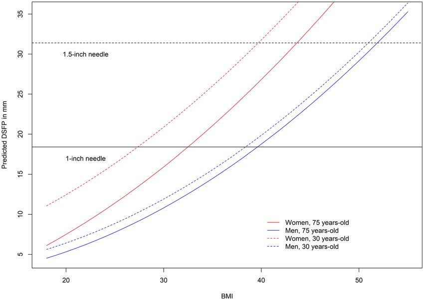
www.nature.com/scientificreports/
needle length for a 5-foot 4-inch -tall (1.626 m) man who weighs 259 pounds (117.7 kg) with a body mass index
(BMI) of 44.5 kg/m2 is the same as that for a 6-foot 7-inch-tall (2.01 m) man who weighs 259 pounds (117.7 kg)
with a BMI of 29.2 kg/m2. Similarly, the CDC recommended needle length for a 5-foot-tall (1.524 m) woman
who weighs 199 pounds (90.5 kg) with a BMI of 38.9 kg/m2 is the same as that for a 6-foot-tall (1.829 m) woman
who weighs 199 pounds (90.5 kg) with a BMI of 27.0 kg/m2. In each of these examples, one individual is morbidly
obese and the other is slightly overweight.
We hypothesized that the CDC guidelines may be inadequate for clinical practice as they rely on weight,
rather than on BMI. The goal of this study is to use statistical modeling using patient clinical and demographic
factors (age, gender, height, weight and BMI) to predict DSFP thickness and to better predict the optimal needle
length required to achieve an intramuscular deltoid injection.
Methods
The study experimental protocol was reviewed and approved by the local Institutional Review Board at the Mayo
Clinic, Florida (IRB approval # 21-003745), and the need for signed informed consent from each patient was
waived. All methods were carried out in accordance with the Health Insurance Portability and Accountability
Act of 1996 (HIPAA) guidelines and regulations.
Prior reports have measured the DSFP using ultrasound and tried to avoid compression of the deltoid tissues
by using “light” probe pressure or used a "stand-off pad" to increase the near field resolution of the skin line10,11.
It is unclear how much pressure is “light” pressure and how much this “light” pressure could distort the subcu-
taneous fat, since by nature fat is soft and easily compressible. Ultrasound by nature of its compressive technique
often distorts/compresses the subcutaneous t issues10,11, so ultrasound was not chosen as the modality to obtain
measurements for this study. Magnetic resonance imaging (MRIs) of the shoulder were considered, however,
MRIs of the shoulder utilize a shoulder coil. This shoulder coil also potentially compresses the subcutaneous
tissues, and so MRIs were not chosen as the modality to obtain measurements. Chest computed tomography
(CTs) were considered as these studies often include the shoulder, however, the arms are generally held above the
head to minimize artifacts and radiation dose to the patient, so chest CTs were not utilized. The CT component
of whole-body positron emission tomography/computed tomography (PET/CTs) often includes the shoulder in
the anatomic position and generally have no mass effect on the subcutaneous tissues because the patient is lying
supine, so this modality was chosen to obtain measurements.
PET/CT technique. The CT component of PET/CTs of 1000 consecutive patients imaged at a tertiary care
academic center between 01/01/2020 and 04/01/2021 were retrospectively reviewed. PET/CT scans were per-
formed using a Siemens Biograph PET/CT scanner with a 128-slice CT scanner (Siemens Healthineers, Mal-
vern, PA, USA) using a slice thickness of 4 mm, field of view (FOV) of 500 mm, pitch of 0.8, 120 kVp, and tilt of
0°. 2-Deoxy-2-[18F] fluoroglucose (18F-FDG) is a standard radiotracer used for PET/CT imaging and 18F-FDG
was injected intravenously into patients 60 min prior to each PET/CT scan. Measurements were taken to the
nearest 0.1 mm using the Visage 7 Picture Archiving and Communications Systems (PACS) (San Diego, CA,
USA) measurement tool. Patients were excluded if their arms were positioned over the head or if they had a
shoulder arthroplasty (n = 614). Clinical and demographic variables including age, gender, height, weight and
BMI were recorded at the time of the PET/CT scan.
DSFP measurement technique. A fellowship-trained musculoskeletal radiologist with over 12 years of
experience evaluated scans and made measurements on the left arm in all but two patients who had intramuscu-
lar lipomas in the deltoid. The left arm was chosen for measurements because it is the common non-dominant
arm and usually chosen for deltoid injections. The distance from the skin overlying the deltoid to the deltoid
muscle, d (equivalent to the DSFP); and the distance from the skin overlying the deltoid to the humerus, D, were
measured at exactly 5 cm inferior to the acromion in the mid anterior–posterior shoulder at 90° perpendicular
to the skin measured in the coronal plane, following the CDC guidelines12 (Fig. 1). Two millimeters of the needle
was assumed to be above the skin to facilitate retrieval of the needle in the event of a fractured needle. A previous
manuscript noted that an injection would be considered intramuscular if the needle would penetrate the deltoid
muscle by at least 5 mm10, so this threshold was used.
Statistical analyses. T-tests were used to compare mean weights and mean DSFP thickness for men and
women. Pearson’s correlation coefficients were used to evaluate the correlations between DSFP thickness and
weight, BMI, age and height for men and women. Receiver operator characteristic (ROC) curves were used to
evaluate the predictive performance of age, height, weight, and BMI to predict whether injections were intra-
muscular versus subcutaneous using the CDC current guidelines. DeLong’s test was used to compare ROC
curves. Next, logistic regression was used to predict the odds of an inadvertent subcutaneous model using the
CDC guidelines, comparing patients above the CDC weight threshold (200 pounds for women, 260 pounds for
men) compared to patients below the CDC weight threshold.
Predictive statistical modeling was used to estimate a patient’s DSFP based on age, gender, height, weight,
and BMI using generalized linear models. The Shapiro-Wilks test was performed to confirm the distribution
of DSFP was not significantly different from a normal distribution. First, univariate linear regression models to
predict DSFP using age, height, weight and BMI as covariates were created. Age (P < 0.001), height (P < 0.001),
weight (P < 0.001) and BMI(P < 0.001) were significant univariate predictors of DSFP.
We built the best multivariable model to predict DSFP using age, gender, height, and weight (model 1).
Variables with P-values < 0.20 in the univariate analysis were included in the first multivariable model, and
we removed terms with P-values ≥ 0.05 in this multivariable model using stepwise backward regression. We
Scientific Reports | (2022) 12:1069 | https://doi.org/10.1038/s41598-022-05020-5 2
Vol:.(1234567890)www.nature.com/scientificreports/
Figure 1. (A) Coronal CT image of a 77-year-old man with history of head and neck squamous cell carcinoma,
weight of 74 kg, height of 1.70 m, demonstrating the deltoid subcutaneous fat pad (DSFP) measurement
technique. Orange arrow identifies the region exactly 5.0 cm inferior to the acromion. Yellow line is
perpendicular to the orange line and identifies the estimated injection site. 90° angle at the estimated injection
site in gray was used to identify the trajectory of the needle. Green arrow identifies the distance from the skin
surface to the deltoid muscle measured perpendicular to the skin in the coronal plane (note arrow is jittered to
show blue arrow along the same path) (DSFP = d = 11.2 mm). Blue arrow identifies the distance from the skin
surface to the humerus measured perpendicular to the skin in the coronal plane (D = 40.3 mm). (B) Axial CT
image of the same man demonstrating identification of the correct coronal plane to measure the DSFP. Blue
lines identify the anterior and posterior borders of the shoulder 5.0 cm inferior to the acromion. The orange
arrow measures the distance between the anterior and posterior borders of the shoulder. The green arrow
measures the half-way distance between the anterior and posterior borders of the shoulder. The yellow arrow
was used to identify the coronal plane to take measurements of the deltoid subcutaneous fat pad.
subsequently included all squared terms for each remaining variable to the multivariable model. Variables which
were not statistically significant (P ≥ 0.05) were removed from the model using stepwise backward regression.
Next, all pairwise interaction terms between the remaining variables in the multivariable model were added to
the multivariable model. Variables which were not statistically significant (P ≥ 0.05) were removed from the model
using stepwise backward regression. The final model 1 contained the variables age, gender, weight, age*gender,
age*weight and gender*weight (Adjusted R-squared = 0.565, Akaike Information Criterion (AIC) = 2289.8). Full
details are shown in Supplementary material 1.
We built the best multivariable model to predict DSFP using age, gender, height, and BMI (model 2). Variables
with P-values < 0.20 in the univariate analysis were included in the first multivariable model, and we removed
terms with P-values ≥ 0.05 in this multivariable model using stepwise backward regression. We subsequently
included all squared terms for each remaining variable to the multivariable model. Variables which were not
statistically significant (P ≥ 0.05) were removed from the model using stepwise backward regression. Next, all
pairwise interaction terms between the remaining variables in the multivariable model were added to the multi-
variable model. Variables which were not statistically significant (P ≥ 0.05) were removed from the model using
stepwise backward regression. The final model 2 included the following variables: age, gender, BMI, BMI-squared,
gender*BMI interaction term, age and age*gender interaction term (Adjusted R-squared = 0.648, AIC = 2204.1).
Model 2 was compared to model 1 using the Akaike Information Criterion (AIC). Model 2 was superior to model
1 and was used for predictive modeling.
The best model to predict the DSFP is:
DSFP in mm = 6.267199 − 2.711159 ∗ Male + 0.228407 ∗ BMI + 0.012295 ∗ BMI 2
(1)
− 0.294211 ∗ BMI ∗ Male − 0.110063 ∗ Age + 0.085589 ∗ Age ∗ Male
Model 2 (Eq. 1) was used to create plots of the predicted thickness of the DSFP in men and women aged
35 and 75 years with varying BMIs (Figure iv). Statistical analyses were conducted using Rv4.04. All tests were
two-sided and P-values less than 0.05 were considered statistically significant.
Scientific Reports | (2022) 12:1069 | https://doi.org/10.1038/s41598-022-05020-5 3
Vol.:(0123456789)www.nature.com/scientificreports/
Variable All Male (N = 219) Female (N = 167) P-value
Mean age in years (range) 67.5 (19–93) 68.7 (21–90) 65.9 (19–93) 0.027†
Race/ethnicity
American Indian/Alaska Native (AINA) 1 (0.3%) 0 (0.0%) 1 (0.6%) 0.388§
Asian 7 (1.8%) 3 (1.4%) 4 (2.4%)
Black 28 (7.3%) 13 (5.9%) 15 (9.0%)
Hispanic 10 (2.6%) 7 (3.2%) 3 (1.8%)
Native Hawaiian/Pacific Islander (NHPI) 1 (0.3%) 0 (0.0%) 1 (0.6%)
Other 5 (1.3%) 4 (1.8%) 1 (0.6%)
White 334 (86.5%) 192 (87.7%) 142 (85.0%)
History of cancer 374 (96.9%) 213 (97.3%) 161 (96.4%) 0.769§
Mean height in m (SD) 1.71 (0.1) 1.77 (0.07) 1.63 (0.07) < 0.001†
Mean weight in kg (SD)* 80.9 (18.6) 86.4 (17.5) 73.7 (17.5) < 0.001†
Mean BMI in kg/m2 (SD)* 27.6 (5.8) 27.4 (5.3) 27.8 (6.4) 0.484†
Mean deltoid subcutaneous fat pad, d in mm (SD) 12.2 (7.2) 9.6 (4.9) 15.5 (8.3) < 0.001†
Mean deltoid thickness from skin to bone, D in mm (SD) 37.2 (12.4) 35.6 (10.8) 39.4 (14.1) 0.004†
Table 1. Patient clinical and demographic characteristics. *One man and one woman did not have recorded
weights at imaging. † By 2-sample t-test with unequal variances. § By Fisher’s exact test.
Results
386 patients with a median age of 68 years (range 19 to 93 years old) were evaluated. The other 614 patients were
excluded because their arms were positioned over the head during imaging. Most patients (96.9%) underwent
PET/CT for surveillance of malignancy. Patients’ demographics are shown in Table 1. Although men were on
average heavier than women (P < 0.001, 95% CI (9.2, 16.3) kg), women had thicker DSFPs than men (P < 0.001,
95% CI (4.4, 7.3) mm). DSFP was positively correlated with weight in men (r = 0.67, P < 0.001) and women
(r = 0.69, P < 0.001); and with BMI in men (r = 0.74, P < 0.001) and women (r = 0.75, P < 0.001). DSFP was nega-
tively correlated with age in women (r = − 0.19, P = 0.013) and borderline negatively correlated with age in men
(r = − 0.13, P = 0.054). We found no correlation between DSFP and height in either men (r = − 0.04, P = 0.601)
or women (r = − 0.03, P = 0.691).
Our results show that a 1-inch needle is expected to reach the deltoid in 85.3% (116/136) of women less than
200 pounds, and 98.6% (207/210) of men less than 260 pounds. This rate differed between genders (P < 0.001,
odds ratio (OR) 0.08, 95% CI (0.02, 0.29)). A 1.5-inch needle is expected to reach the deltoid in 76.7% (23/30)
of women greater than 200 pounds, and 75.0% (6/8) of men greater than 260 pounds (Fig. 2). The odds of inad-
vertent subcutaneous injection in men greater than 260 pounds and women greater than 200 pounds (above
the CDC weight thresholds) are over 4 times higher than those below the threshold (OR = 4.33, P = 0.002)
when using the CDC-recommended needle lengths. BMI was the best predictor of DSFP in men but was not
significantly better than weight as a predictor in women (Fig. 3, Supplementary Fig. 1). For women, the area
under the receiver-operator curve (ROC) curve (AUC) for BMI (0.767) to predict an intramuscular injection
was significantly better than the AUC for height (AUC = 0.527, P < 0.001) and the AUC for age (AUC = 0.562,
P < 0.001) but was not significantly different from the AUC for weight (AUC = 0.750, P = 0.210). For men, we
found that the AUC for BMI (AUC = 0.832) to predict an intramuscular injection was significantly better than
the AUC for weight (AUC = 0.777, P = 0.001), the AUC for height (AUC = 0.546, P < 0.001) and the AUC for age
(AUC = 0.612, P < 0.001).
The best model to predict the DSFP is:
DSFP in mm = 6.267199 − 2.711159 ∗ Male + 0.228407 ∗ BMI + 0.012295 ∗ BMI 2
(2)
− 0.294211 ∗ BMI ∗ Male − 0.110063 ∗ Age + 0.085589 ∗ Age ∗ Male
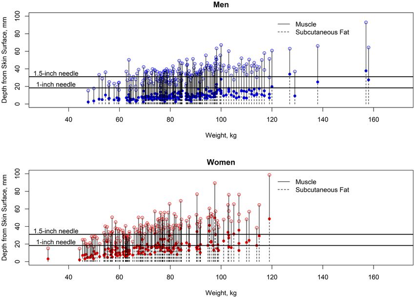
We used this model to predict the DSFP thickness for males (aged 30 and 75 years) and females (aged 30 and
75 years) with varying BMI (Fig. 4).
Discussion
In this study, we show that both weight and BMI were strongly correlated with DSFP in men and women. Despite
weighing less on average than men, women tended to have thicker DSFPs than men. In our predictive modeling,
we found that age, gender and BMI were more predictive of DSFP than the best model using age, gender and
weight.
A deltoid intramuscular injection has been defined as an injection with penetration of the muscle by 5 mm
or more, with 2 mm of needle superficial to the skin to aid in needle retrieval in the event of an accidental needle
break10. Based on this, we conclude that the DFSP can be predicted using the following equation:
DSFP in mm = 6.267199 − 2.711159 ∗ Male + 0.228407 ∗ BMI + 0.012295 ∗ BMI 2
− 0.294211 ∗ BMI ∗ Male − 0.110063 ∗ Age + 0.085589 ∗ Age ∗ Male
Scientific Reports | (2022) 12:1069 | https://doi.org/10.1038/s41598-022-05020-5 4
Vol:.(1234567890)www.nature.com/scientificreports/
Figure 2. Plots of the deltoid subcutaneous fat and muscle thickness in men and women versus weight in
kg. Weight in kilograms for each male (top) and female (bottom) individual, with CT measurements of depth
from skin surface (0 mm) to bone. The skin-to-muscle distance (which defines the deltoid subcutanous fat pad
thickness) is represented by dotted lines ending in a solid circle, while the muscle-to-bone distances (which
defined muscle thickness) is represented by solid lines ending in an open square. Bone is represented by the
area above the open circles. Blue filled circles – Distance from skin surface to the deltoid muscle surface in men.
Blue unfilled circles – Distance from the skin surface to the humerus in men. Red filled circles – Distance from
skin surface to the deltoid muscle surface in women. Red unfilled circles – Distance from the skin surface to the
humerus in women. Horizontal black lines represent the effective maximum penetration depth of 1-inch and
1.5-inch needles.
The predicted DSFP can then be used to find the appropriate needle length where the appropriate needle
length is predicted DSFP + 7 mm. Therefore, if the predicted DSFP < 18.4 mm, then a 1-inch (25.4 mm) needle
would be appropriate. Similarly, if the predicted DSFP > 18.4 mm but < 31.1 mm, then a 1.5-inch (38.1 mm) nee-
dle would be appropriate. Because of this, a 1-inch needle may be too short to reliably get to the deltoid muscle
in women, particularly in heavier women and a 1.5-inch needle may be too short to achieve an intramuscular
injection in women greater than 200 pounds, and men greater than 260 pounds. Age and BMI together are better
than weight for predicting DSFP thickness. DSFP decreases with age faster in women.
More recently new vaccines have been developed for COVID-19 in the wake of the COVID-19 pandemic.
Currently, there is a global effort to vaccinate individuals with these new COVID-19 vaccines. This effort requires
appropriate needle length selection for vaccination administration. Our results suggest that women, and in
particular younger and obese women would be more likely to have inadvertent subcutaneous injections and be
at increased risk for vaccine failures. Recent CDC Morbidity and Mortality Weekly Report (MMWR) data show
that 63% of COVID-19 vaccine failures have been in w omen13, however, the relative proportions of males to
females in the vaccinated population is unknown.
While this study and other studies have shown associations between the DSFP and weight and BMI10,11,
there are a few differences between this study and previously published reports. The prior report by Poland
et al. investigated healthcare workers aged 18–59 years, weight range of 59–118 kg and BMI of 17.1–49.9 kg/m2
whereas this study investigated patients aged 19–93 years, weight range of 31.9–158 kg and BMI of 13.3–55.9 kg/
m2. This study has individuals over a greater age range, weight range and BMI range than the study by Poland
et al.10. Similarly, Cook et al.11 conclude that a 1-inch needle (25.4 mm) is an appropriate length for men of all
BMI, whereas this study showed that this needle length was appropriate for 96.8% of men of all BMI (211/218)
and only appropriate for 37.5% (3/8) men who weighed more than 260 pounds. This is likely because our study
had larger weight and BMI ranges than these prior studies. Poland et al. suggest that a 5/8 inch, 1-inch (25.4 mm)
and 1.5-inch (38.1 mm) needle is appropriate for women less than 60 kg, 60–90 kg and greater than 90 kg
respectively, which contradicts the finding by Cook et al. who suggested that a 1-inch needle is appropriate for
Scientific Reports | (2022) 12:1069 | https://doi.org/10.1038/s41598-022-05020-5 5
Vol.:(0123456789)www.nature.com/scientificreports/
Figure 3. Receiver operator curves evaluating weight, BMI, height and age to predict intramuscular injections
using the CDC guidelines (1-inch needle for females less than 200 pounds; 1.5-inch needle for females greater
than 200 pounds; 1-inch needle for males less than 260 pounds; 1.5-inch needle for males greater than 260
pounds).
Scientific Reports | (2022) 12:1069 | https://doi.org/10.1038/s41598-022-05020-5 6
Vol:.(1234567890)www.nature.com/scientificreports/
Figure 4. Predictive modeling of DSFP thickness in mm by BMI for individuals from Eq. 1. Red solid line
represents the predicted DSFP for women aged 75 years. Red broken line represents the predicted DSFP for
women aged 30 years. Blue solid line represents the predicted DSFP for men aged 75 years. Blue broken line
represents the predicted DSFP for men aged 30 years.
all women with BMIwww.nature.com/scientificreports/
Received: 27 September 2021; Accepted: 17 December 2021
References
1. Ng, J. Y. Inadvertent subcutaneous injection of COVID-19 vaccine. Postgrad. Med. J. https://doi.org/10.1136/postgradmedj-2021-
139870 (2021) (Epub ahead of print).
2. Zuckerman, J. N. The importance of injecting vaccines into muscle. Different patients need different needle sizes. BMJ 321(7271),
1237–1238. https://doi.org/10.1136/bmj.321.7271.1237 (2000).
3. McLean, A. A., Guess, H. A. & Scolnick, E. M. Suboptimal response to hepatitis B vaccine given by injection into the buttock.
Morb. Mortal. Wkly. Rep. 34, 105–113 (1985).
4. Lindsay, K. L., Herbert, D. A. & Gitnick, G. L. Hepatitis B vaccine: Low postvaccination immunity in hospital personnel given
gluteal injections. Hepatology 5, 1088–1090 (1985).
5. Cockshott, W. P., Thompson, G. T., Howlett, L. J. & Seeley, E. T. Intramuscular or intralipomatous injections?. N. Engl. J. Med.
307(6), 356–358. https://doi.org/10.1056/NEJM198208053070607 (1982).
6. Dayananda, L., Belaval, V. V., Raina, A. & Chandana, R. Intended intramuscular gluteal injections: Are they truly intramuscular?.
J. Postgrad. Med. 60(2), 175–178. https://doi.org/10.4103/0022-3859.132334 (2014).
7. Burbridge, B. E. Computed tomographic measurement of gluteal subcutaneous fat thickness in reference to failure of gluteal
intramuscular injections. Can. Assoc. Radiol. J. 58(2), 72–75 (2007).
8. Chan, V. O. et al. Intramuscular injections into the buttocks: Are they truly intramuscular?. Eur. J. Radiol. 58(3), 480–484. https://
doi.org/10.1016/j.ejrad.2006.01.008 (2006) (Epub 2006 Feb 21).
9. Shaw, F. E. Jr. et al. Effect of anatomic injection site, age and smoking on the immune response to hepatitis B vaccination. Vaccine
7(5), 425–430. https://doi.org/10.1016/0264-410x(89)90157-6 (1989).
10. Poland, G. A. et al. Determination of deltoid fat pad thickness. Implications for needle length in adult immunization. JAMA
277(21), 1709–1711 (1997).
11. Cook, I. F., Williamson, M. & Pond, D. Definition of needle length required for intramuscular deltoid injection in elderly adults:
an ultrasonographic study. Vaccine 24(7), 937–940. https://doi.org/10.1016/j.vaccine.2005.08.098 (2006) (Epub 2005 Sep 13).
12. https://www.cdc.gov/vaccines/hcp/admin/downloads/vaccine-administration-needle-length.pdf. Accessed April 4, 2021.
13. COVID-19 Vaccine Breakthrough Infections Reported to CDC—United States, January 1–April 30, 2021. Morb. Mortal. Wkly.
Rep. 70, 792–793 (2021). https://doi.org/10.15585/mmwr.mm7021e3 external icon.
14. Palmer, A. K. & Kirkland, J. L. Aging and adipose tissue: potential interventions for diabetes and regenerative medicine. Exp.
Gerontol. 86, 97–105. https://doi.org/10.1016/j.exger.2016.02.013 (2016).
15. Kompaniyets, L., Goodman, A. B., Belay, B., et al. Body Mass Index and Risk for COVID-19–related hospitalization, intensive care
unit admission, invasive mechanical ventilation, and death—United States, March–December 2020. Morb. Mortal. Wkly. Rep. 70,
355–361 (2021). https://doi.org/10.15585/mmwr.mm7010e4 external icon.
Acknowledgements
RS was supported by R21 NIH/NIMH MH093415.
Author contributions
Concept and design: Sebro. Acquisition, analysis, or interpretation of data: All authors. Drafting of the manu-
script: Sebro. Critical revision of the manuscript for important intellectual content: All authors. Statistical analy-
sis: Sebro (PhD Biostatistics Harvard University) Administrative, technical, or material support: Sebro.
Funding
RS has received funding from the National Institutes of Health (NIH) Grant # R21 NIH/NIMH MH093415.
Competing interests
The authors declare no competing interests.
Additional information
Supplementary Information The online version contains supplementary material available at https://doi.org/
10.1038/s41598-022-05020-5.
Correspondence and requests for materials should be addressed to R.S.
Reprints and permissions information is available at www.nature.com/reprints.
Publisher’s note Springer Nature remains neutral with regard to jurisdictional claims in published maps and
institutional affiliations.
Open Access This article is licensed under a Creative Commons Attribution 4.0 International
License, which permits use, sharing, adaptation, distribution and reproduction in any medium or
format, as long as you give appropriate credit to the original author(s) and the source, provide a link to the
Creative Commons licence, and indicate if changes were made. The images or other third party material in this
article are included in the article’s Creative Commons licence, unless indicated otherwise in a credit line to the
material. If material is not included in the article’s Creative Commons licence and your intended use is not
permitted by statutory regulation or exceeds the permitted use, you will need to obtain permission directly from
the copyright holder. To view a copy of this licence, visit http://creativecommons.org/licenses/by/4.0/.
© The Author(s) 2022
Scientific Reports | (2022) 12:1069 | https://doi.org/10.1038/s41598-022-05020-5 8
Vol:.(1234567890)You can also read