Spontaneous ultraweak photon emission imaging of oxidative metabolic processes in human skin: effect of molecular oxygen and antioxidant defense ...
←
→
Page content transcription
If your browser does not render page correctly, please read the page content below

Spontaneous ultraweak photon emission
                 imaging of oxidative metabolic processes
                 in human skin: effect of molecular oxygen
                 and antioxidant defense system

                 Anshu Rastogi
                 Pavel Pospı́šil

Downloaded From: https://www.spiedigitallibrary.org/journals/Journal-of-Biomedical-Optics on 19 Feb 2021
Terms of Use: https://www.spiedigitallibrary.org/terms-of-use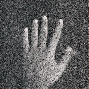
Journal of Biomedical Optics 16(9), 096005 (September 2011)

                Spontaneous ultraweak photon emission imaging of
                oxidative metabolic processes in human skin: effect of
                molecular oxygen and antioxidant defense system
                Anshu Rastogi and Pavel Pospı́šil
                Palacký University, Department of Biophysics, Centre of the Region Haná for Biotechnological and Agricultural
                Research, Faculty of Science, Šlechtitelů 11, 783 71 Olomouc, Czech Republic

                               Abstract. All living organisms emit spontaneous ultraweak photon emission as a result of cellular metabolic
                               processes. In this study, the involvement of reactive oxygen species (ROS) formed as the byproduct of oxidative
                               metabolic processes in spontaneous ultraweak photon emission was studied in human hand skin. The effect
                               of molecular oxygen and ROS scavengers on spontaneous ultraweak photon emission from human skin was
                               monitored using a highly sensitive photomultiplier tube and charged coupled device camera. When spontaneous
                               ultraweak photon emission was measured under anaerobic conditions, the photon emission was decreased,
                               whereas under hyperaerobic condition the enhancement in photon emission was observed. Spontaneous ultraweak
                               photon emission measured after topical application of glutathione, α-tocopherol, ascorbate, and coenzyme Q10
                               was observed to be decreased. These results reveal that ROS formed during the cellular metabolic processes
                               in the epidermal cells play a significant role in the spontaneous ultraweak photon emission. It is proposed that
                               spontaneous ultraweak photon emission can be used as a noninvasive tool for the temporal and spatial monitoring
                               of the oxidative metabolic processes and intrinsic antioxidant system in human skin. C 2011 Society of Photo-Optical
                               Instrumentation Engineers (SPIE). [DOI: 10.1117/1.3616135]

                               Keywords: antioxidant; molecular oxygen; reactive oxygen species; ultraweak photon emission.
                               Paper 11193R received Apr. 15, 2011; revised manuscript received Jul. 1, 2011; accepted for publication Jul. 7, 2011; published online
                               Sep. 1, 2011.

                1 Introduction                                                                         tochondria is reduced to O2 .− .9 Apart from the mitochondria,
                Metabolic processes are the fundamental chemical reactions oc-                         O2 .− is formed in the cytoplasm of skin keratinocyte cells by a
                curring inside the living cells. It is known that metabolic pro-                       reduction of molecular oxygen catalyzed by heme-flavoprotein
                cesses (e.g., cellular respiration, oxidative burst) result in the                     NADPH oxidase (NOX). 10
                generation of reactive oxygen species (ROS) known to play an                               Various types of nonenzymatic and enzymatic antioxi-
                important role in the defense against infection, cell signaling,                       dant systems are engaged to eliminate the cellular oxidative
                apoptosis, and aging.1, 2 However, due to the high positive redox                      damage.11 Nonenzymatic antioxidant systems in human skin
                potential, ROS have a capability to oxidize various cellular com-                      include ascorbate, α-tocopherol, carotenoids, gluthatione, and
                ponents such as lipids, proteins, and nucleic acids.2–5 To prevent                     coenzyme Q10, whereas enzymatic scavenging is maintained by
                the oxidative damage of cellular components, the cells devel-                          enzymes such as the superoxide dismutase (SOD) family of en-
                oped their own defense system for the protection against ROS.                          zymes (copper-zinc SOD, manganese SOD) and the peroxidase
                Under certain circumstances, when the formation of ROS ex-                             family of enzymes (glutathione peroxidases and catalase).11–13
                ceeds the capacity of an antioxidant system, the dangerous ROS                         When the formation of ROS overwhelms the skin antioxidant
                are insufficiently scavenged. The imbalance between formation                          capacity, the subsequent oxidation of lipids, proteins, and nu-
                and scavenging of ROS brings about the damage of cellular                              cleic acids leads to lipid peroxidation, protein degradation, and
                membrane known to lead to the death of the cell.                                       nucleic acid damage,14 which cause the aging of the skin.
                    In human skin, superoxide anion radical (O2 .− ) is formed                             The oxidation of lipids, proteins, and nucleic acids by ROS is
                by one-electron reduction of molecular oxygen catalyzed by                             accompanied by the formation of electronically excited species
                various mitochondrial and cytoplasmic enzymes. In the mito-                            such as triplet carbonyls 3 (C=O)* and singlet oxygen (1 O2 ).15, 16
                chondrial membrane, the leakage of electron from complexes                             The light emission of the electronically excited species in the
                I and III within the electron transport chain leads to the for-                        spectral range from 200 to 800 nm is known as weak chemilu-
                mation of O2 .− .6, 7 More recent evidence reveals that complex                        minescence often called ultraweak photon emission.17–21 Typi-
                II in the mitochondrial membrane also contributes to the over-                         cally, the intensity of photon emission is less than approximately
                all O2 .− production in human skin cells.8 Under physiological                         10 − 15 W/cm2 on the surface of human skin. It is known that
                conditions, up to 5% of molecular oxygen consumed by mi-                               different wavelengths of light have different penetration depths
                                                                                                       in human skin.22 Due to its ultraweak nature and emission in
                                                                                                       the visible range of the spectrum, it is possible to detect the
                Address all correspondence to: Pavel Pospı́šil, Palacký University, Centre of the
                Region Haná for Biotechnological and Agricultural Research, Department of
                Biophysics, Faculty of Science, Šlechtitelů 11, 783 71 Olomouc, Czech Republic;
                Tel.: + 420 585634174; Fax: + 420 585225737; E-mail: pospip@prfnw.upol.cz.             1083-3668/2011/16(9)/096005/7/$25.00
                                                                                                                                            C 2011 SPIE

                Journal of Biomedical Optics                                                     096005-1                                      September 2011 r Vol. 16(9)

Downloaded From: https://www.spiedigitallibrary.org/journals/Journal-of-Biomedical-Optics on 19 Feb 2021
Terms of Use: https://www.spiedigitallibrary.org/terms-of-use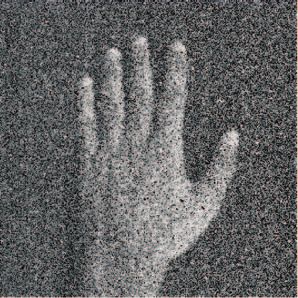
Rastogi and Pospı́šil: Spontaneous ultraweak photon emission imaging...

                 ultraweak photon emission only from the 3 mm layer below
                 the body surface.23 It indicates that the oxidative metabolic pro-
                 cesses in the skin play a significant role in the overall photon
                 emission from the human body.
                     Apart from ultraweak photon emission, other noninvasive
                 techniques such as attenuated total reflection-Flourier transform
                 infrared spectroscopy (ATR-FTIR), resonance Raman spec-
                 troscopy, and confocal Raman microscopy can be used for the
                 measurement of oxidative stress in human skin. For the non-
                 invasive detection of lipid composition from stratum corneum,
                 ATR-FTIR can be used.24 On the other hand, resonance Ra-
                 man spectroscopy can noninvasively detect the antioxidant level
                 of human skin,25, 26 whereas confocal Raman microscopy can
                 be used for noninvasive depth-resolved assessment of spe-
                                                                                                    Fig. 1 Experimental setup for the detection of two-dimensional spon-
                 cific antioxidants.27, 28 The ultraweak photon emission or weak                    taneous ultraweak photon emission under aerobic, anaerobic, and hy-
                 chemiluminescence is the most frequent noninvasive technique                       peraerobic conditions. The gas chamber is distanced 500 cm from the
                 used for the measurement of oxidative stress because of its direct                 CCD window.
                 correlation with the oxidative metabolic processes.29–35
                     The effect of scavengers on ultraweak photon emission was                      The person was adapted to the dark for 20 min prior to the
                 studied in human skin exposed to various types of physical                         measurement. The measurement was performed between 11 and
                 (ultraviolet and ultrasonic radiation) and chemical (hydrogen                      15 h of the day during the same season to avoid any effect of
                 peroxide, ozone, cigarette smoke) stress factors.30–33 It has been                 diurnal and seasonal change.36
                 demonstrated that topical application of ascorbic acid prior to
                 exposure of human skin to ultraviolet radiation (UV) signifi-
                 cantly reduced UV-induced ultraweak photon emission.33 More                        2.2 Aerobic, Hyperaerobic, and Anaerobic
                 recently, a topical application of antioxidant system (tocopheryl                      Conditions
                 acetate, ferulic acid, and rutin) on human skin has been shown to                  A special gas chamber designed for the detection of spontaneous
                 significantly decrease UV-induced ultraweak photon emission.29                     ultraweak photon emission from the dorsal side of the hand was
                 Hagens et al. (2008) demonstrated that exposure of human skin                      used to study the effect of anaerobic and hyperaerobic condi-
                 to ozone and cigarette smoke caused an enhancement in ultra-                       tions on ultraweak photon emission. The front side of the gas
                 weak photon emission, whereas topical application of antiox-                       chamber was made of a glass plate, which allowed the light to
                 idant (ascorbate and α-glucosylrutin) to human skin reduced                        penetrate in the visible range of the spectra. The gas chamber
                 ultraweak photon emission.31                                                       was hermetically sealed from the surrounding atmosphere. Prior
                     Whereas the application of ultraweak photon emission for the                   to the detection of photon emission, a dark-adapted hand was
                 detection of the oxidative metabolic processes in human skin ex-                   placed inside the chamber faced to the PMT or CCD window at
                 posed to physical and chemical stress factors is widely used, the                  a distance of 10 and 50 cm, respectively (Fig. 1). Nitrogen and
                 application of spontaneous ultraweak photon emission for the                       oxygen gas was continuously supplied to create the anaerobic
                 detection of oxidative processes in the skin the related to the nor-               and hyperaerobic condition, respectively, whereas the aerobic
                 mal metabolic processes, i.e., without the use of any exogenous                    condition was maintained at the atmospheric air.
                 stress factor has received less attention. As far as we know, no
                 experimental evidence has been provided on the correlation of
                 spontaneous ultraweak photon emission and oxidative metabolic                      2.3 Topical Application of Scavengers
                 processes in human skin. The lack of experimental data on the                      Glutathione, ascorbate, α-tocopherol, and coenzyme Q10 were
                 involvement of ROS produced during the normal metabolic pro-                       used in 5 mM concentration for the study. Scavengers were
                 cesses in spontaneous ultraweak photon emission was due to the                     applied using a micropipette and gloves covering the left hand.
                 insufficient abundance sensitivity of the detection systems in the                 For a one-dimensional measurement, a fixed volume of 300
                 past. However, the recent development of a highly sensitive de-                    μL of scavengers was used for each measurement and equally
                 vice like the photomultiplier tube (PMT) and charged coupled                       distributed to the fixed area of the right hand with the help of the
                 device (CCD) camera has made it possible to detect the sponta-                     left hand by the subject himself (5 min prior to the measurement)
                 neous ultraweak photon emission from human skin.30, 34, 35 Our                     in the dark; whereas for a two-dimensional measurement, 1000
                 study, to the best of our knowledge for the first time, shows                      μL of scavengers were applied on the dorsal side of the hand.
                 spontaneous ultraweak photon emission as the tool for the mea-                     In the control sample, the same volume of water or ethanol was
                 surement of oxidative and aging processes in human skin.                           applied. The person was adapted to the dark for 20 min including
                                                                                                    a 5 min time interval after the application of scavengers.
                 2 Material and Method
                 2.1 Human                                                                          2.4 One-Dimensional Ultraweak Photon Emission
                 Measurements were done on a healthy man of age 25 to 27                            A photon counting unit C9744 (Hamamatsu Photonics
                 years and each measurement was repeated at least three times.                      K.K., Iwata City, Japan) and photomultiplier tube R7518P
                 The dorsal side of the right hand was used for all measurements.                   (Hamamatsu Photonics K.K., Iwata City, Japan) were used to

                 Journal of Biomedical Optics                                              096005-2                                       September 2011 r Vol. 16(9)

Downloaded From: https://www.spiedigitallibrary.org/journals/Journal-of-Biomedical-Optics on 19 Feb 2021
Terms of Use: https://www.spiedigitallibrary.org/terms-of-use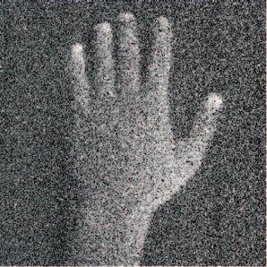
Rastogi and Pospı́šil: Spontaneous ultraweak photon emission imaging...

                detect the spontaneous ultraweak photon emission from human
                                                                                                                    (a)
                hand skin. The sensitivity of the instrument was within the range
                of 185 to 730 nm. To reduce thermal electrons, PMT was cooled
                to − 30◦ C using a thermoelectric cooler C9143 (Hamamatsu
                Photonics K.K., Iwata City, Japan). A data equation unit and a
                computer were placed in an operational dark room. The screen
                and light emitting diodes were shielded by a black curtain.
                Inside the operational dark room, an experimental dark room
                (3 × 2 × 2 m) was built to avoid any effect of the lost photons
                on PMT. The whole interior in the experimental dark room was
                painted black and the door was protected from incoming light
                by a black curtain.

                2.5 Two-Dimensional Ultraweak Photon Emission
                For two-dimensional imaging of spontaneous ultraweak pho-                                           (b)
                ton emission, a highly sensitive CCD camera VersArray 1300B
                (Princeton instruments, Trenton, New Jersey) was used. The
                CCD camera was equipped with a 50-mm focal distance lens
                with an f-number of 1.2 (F mount Nikkor 50-mm, f:1.2, Nikon,
                Tokyo, Japan) to enhance the light collecting efficiency. The
                spectral sensitivity of the CCD camera was within the range of
                200 to 1000 nm. Due to the lenses used, the spectral sensitivity
                of CCD camera was restricted to the visible range of the spec-
                tra. To reduce thermal electrons, the CCD camera was cooled
                to − 110 ◦ C using liquid nitrogen. The camera was placed in
                the experimental dark room and controlled by the computer in
                the operational dark room. The data correction was made by
                subtracting the background noise before every measurement.                         Fig. 2 (a) Dark count rate of the photomultiplier tube and (b) one-
                The measurement was done in the image format of 1340×1300                          dimensional spontaneous ultraweak photon emission from the dorsal
                                                                                                   side of the hand. Before the measurement the subject was adapted to
                pixels. To improve the image quality the binning mode with                         complete darkness for 20 min.
                the binning factor 4 was applied, which reduces the number of
                pixels by 4. CCD camera parameters were as follows: scan rate,
                100 kHz; gain, 3; accumulation time, 30 min.                                       conditions. To control gas environment around the human hand,
                                                                                                   the human hand was placed in a gas chamber. Under aerobic
                3 Results                                                                          conditions, the spontaneous ultraweak photon emission from the
                3.1 One-Dimensional Spontaneous Ultraweak                                          dorsal side of the hand was 2 counts s–1 (Fig. 3). The slight de-
                    Photon Emission from the Human Hand                                            crease in the spontaneous ultraweak photon emission observed
                                                                                                   in the gas chamber, compared to that observed without the gas
                One-dimensional spontaneous ultraweak photon emission from                         chamber [Fig. 2(b)], is due to the photon absorption by the glass
                the human hand was measured using a highly sensitive PMT.                          plate in the glass chamber. When the air in the gas chamber was
                Figure 2(a) shows that the dark count of PMT was adjusted to 2                     replaced with nitrogen gas, spontaneous ultraweak photon emis-
                counts s–1 . When the dorsal side of the hand was placed below                     sion was suppressed to 1.25 counts s–1 (by 40% compared to
                the PMT window, the count rate of 4.5 counts s–1 was observed                      aerobic conditions). The supply of molecular oxygen in the gas
                [Fig. 2(b)]. After subtraction of the dark count, spontaneous ul-                  chamber results in the enhancement in spontaneous ultraweak
                traweak photon emission from the dorsal side of the hand was                       photon emission to 3 counts s–1 (by 50% compared to aerobic
                determined as 2.5 counts s–1 . When the exogenous environmen-                      conditions). These results reveal that the molecular oxygen is
                tal conditions in the experimental dark room were unchanged,                       involved in spontaneous ultraweak photon emission from the
                a spontaneous ultraweak photon emission persisted on the time                      epidermal cells in human skin.
                scale of several hours. These observations reveal that sponta-
                neous ultraweak photon emission connected to the oxidative
                metabolic processes is an intrinsic property of the epidermal                      3.3 Effect of Molecular Oxygen on
                cells in human skin.                                                                   Two-Dimensional Spontaneous Ultraweak
                                                                                                       Photon Emission
                3.2 Effect of Molecular Oxygen on                                                  To monitor spatial distribution of photon emission from the
                    One-Dimensional Spontaneous Ultraweak                                          human hand, two-dimensional spontaneous ultraweak photon
                    Photon Emission                                                                emission was measured using CCD camera. Figure 4 shows the
                To study the involvement of molecular oxygen in spontaneous                        photograph of the dorsal side of the hand and its corresponding
                ultraweak photon emission from human skin, the photon emis-                        photon emission images measured under aerobic, anaerobic, and
                sion was measured under aerobic, anaerobic, and hyperaerobic                       hyperaerobic conditions. To quantify the differences in sponta-

                Journal of Biomedical Optics                                              096005-3                                       September 2011 r Vol. 16(9)

Downloaded From: https://www.spiedigitallibrary.org/journals/Journal-of-Biomedical-Optics on 19 Feb 2021
Terms of Use: https://www.spiedigitallibrary.org/terms-of-use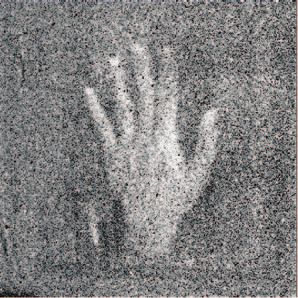
Rastogi and Pospı́šil: Spontaneous ultraweak photon emission imaging...

                                                                                                    Fig. 5 Effect of ROS scavengers on one-dimensional spontaneous ul-
                                                                                                    traweak photon emission from the dorsal side of the hand. Prior to
                                                                                                    the measurement the hydrophilic (ascorbate and gluthatione) and the
                 Fig. 3 Effect of molecular oxygen on one-dimensional spontaneous                   lipophilic (α-tocopherol and coenzyme Q10) ROS scavengers at the
                 ultraweak photon emission from the dorsal side of the hand. Aerobic                concentration of 5 mM were topically applied on the the dorsal side
                 condition were established by the supply of the atmospheric air into the           of the hand. Spontaneous ultraweak photon emission from the human
                 gas chamber. Anaerobic conditions were maintained by establishing a                hand was measured for 15 min. Each bar represents the mean value
                 continuous flow of nitrogen gas into the gas chamber, whereas molec-               of at least three independent measurements ± SD made at the same
                 ular oxygen was continuously supplied into the gas chamber to create               diurnal time of three different days.
                 the hyperaerobic conditions. Spontaneous ultraweak photon emission
                 from human hand was measured for 15 min. Each bar represents the
                 mean value of at least three independent measurements ± SD made                    decrease in the photon emission to 12 counts, whereas the supply
                 at the same diurnal time of three different days.                                  of molecular oxygen in the gas chamber resulted in the enhance-
                                                                                                    ment in photon emission to 25 counts. These observations show
                 neous ultraweak photon emission from the human hand under
                                                                                                    that molecular oxygen is involved in the two-dimensional spon-
                 various concentrations of molecular oxygen, the spatial profile
                                                                                                    taneous ultraweak photon emission, which provides valuable
                 of photon emission in the middle strip of the image was used.
                                                                                                    information on the spatial distribution of electronically excited
                 Under aerobic conditions, the spatial profile of photon emis-
                                                                                                    species formed during the oxidative metabolic processes in the
                 sion shows the maximum photon emission of 17 counts. The
                                                                                                    epidermal cells of the skin.
                 replacement of air by nitrogen gas in the gas chamber caused a

                                                                                                    3.4 Effect of Reactive Oxygen Species Scavengers
                                                                                                        on One-Dimensional Spontaneous Ultraweak
                                                                                                        Photon Emission
                                                                                                    To study the involvement of ROS in ultraweak photon emission,
                                                                                                    the effect of various ROS scavengers on spontaneous ultraweak
                                                                                                    photon emission from the human hand was measured. When hy-
                                                                                                    drophilic (sodium ascorbate and glutathione) and lipophilic (α-
                                                                                                    tocopherol and coenzyme Q10) ROS scavengers were topically
                                                                                                    applied on human skin, the decrease in spontaneous ultraweak
                                                                                                    photon emission to 1.6 counts s–1 (by 30% compared to control)
                                                                                                    was observed (Fig. 5). To confirm that the decrease in photon
                                                                                                    emission is not a pure effect of the solvent, the effect of water
                                                                                                    and ethanol on photon emission from human skin was tested.
                                                                                                    There was no measurable difference observed when only dis-
                                                                                                    tilled water or ethanol was topically applied on the human hand
                                                                                                    (data not shown). These results indicate that ROS are involved in
                                                                                                    spontaneous ultraweak photon emission from human skin. The
                                                                                                    observation that suppression of spontaneous ultraweak photon
                                                                                                    emission with the topical application of hydrophilic or lipophilic
                                                                                                    scavengers was found to be approximately the same, indicates
                                                                                                    that the solubility of scavenger does not considerably affect the
                                                                                                    scavenging property of topically applied scavengers.

                 Fig. 4 Effect of molecular oxygen on two-dimensional spontaneous                   3.5 Effect of Reactive Oxygen Species Scavengers
                 ultraweak photon emission from the dorsal side of the hand. The bottom                 on Two-Dimensional Spontaneous Ultraweak
                 panel shows the spatial profile of photon emission in the middle strip
                                                                                                        Photon Emission
                 of the image. Y axis denotes the number of counts accumulated after
                 30 min, whereas the X axis denotes the pixel of the image. The other               In further study, the effect of ROS scavengers on two-
                 experimental conditions were the same as in Fig. 3.                                dimensional spontaneous ultraweak photon emission from the

                 Journal of Biomedical Optics                                              096005-4                                       September 2011 r Vol. 16(9)

Downloaded From: https://www.spiedigitallibrary.org/journals/Journal-of-Biomedical-Optics on 19 Feb 2021
Terms of Use: https://www.spiedigitallibrary.org/terms-of-use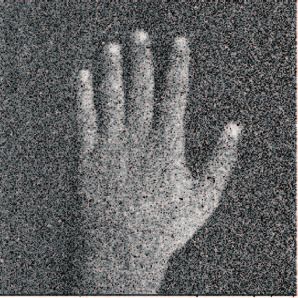
Rastogi and Pospı́šil: Spontaneous ultraweak photon emission imaging...
dorsal side of the hand has no effect on spontaneous ultraweak
photon emission, indicates that the decrease in photon emission
is solely caused by the scavenging (data not shown). These ob-
servations reveal that ROS are involved in the two-dimensional
spontaneous ultraweak photon emission, monitoring the spatial
distribution of ROS in the oxidative metabolic processes in the
epidermal cells of the skin.
4 Discussion
In this study, the effect of molecular oxygen and various ROS
scavengers on spontaneous ultraweak emission from human skin
was studied using highly sensitive PMT and CCD camera. The
direct evidence is provided on the involvement of molecular
oxygen and ROS in the spontaneous ultraweak emission from
human skin. Based on the observation that removal of molecu-
lar oxygen and topical application of O2 .− scavengers on human
skin decreased ultraweak photon emission, it is concluded that
O2 .− formed during oxidative metabolic processes plays a cru-
cial role in the spontaneous ultraweak photon emission from
human skin. The direct contact of human skin with atmospheric
molecular oxygen and the reduction of molecular oxygen by
heme-contaning enzymes in the mitochondrial membrane or
cytoplasm are discussed as a possible mechanism responsible
for the involvement of O2 .− in spontaneous ultraweak photon
emission from human skin.
It is well accepted that skin as the outermost layer of the body
is in direct contact with the atmospheric gases, such as molecu-
lar nitrogen and potentially deleterious molecular oxygen. The
exchange of molecular oxygen between epidermal cell and the
surrounding atmosphere proceeds via cutaneous respiration.37
At normal rest and ordinary atmospheric conditions, the rate of
cutaneous respiration in a human is 1 to 1.5% of pulmonary
respiration.38 As the epidermal cells in the stratum corneum ab-
sorb the molecular oxygen directly from the atmosphere, the
probability of ROS formation by respiration process is higher in
the stratum corneum in comparison to other layers of the skin.
The data presented in this study shows that molecular oxygen is
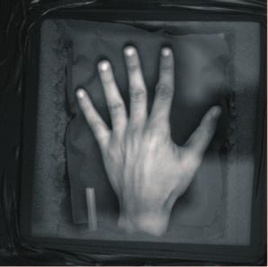
Fig. 6 Effect of ROS scavengers on two-dimensional spontaneous ul-
traweak photon emission from the dorsal side of the hand. The bottom
involved in spontaneous ultraweak photon emission from human
panel shows the spatial profile of photon emission in the middle strip skin (Figs. 3 and 4).
of the image. The Y axis denotes the number of counts accumulated It is well known that epidermal cells are continuously re-
after 30 min, whereas the X axis denotes the pixel of the image. The newed with a high turnover rate utilizing reservoirs of stem
other experimental conditions were the same as in Fig. 5. cells located in the basal layer.39 It has been observed that un-
der in vitro conditions, the mitochondrial activity increases in
the epithelial cell with high proliferation.40 It seems likely that
human hand was measured using a CCD camera. Figure 6 shows enhancement in the mitochondrial activity increases the proba-
the photograph of the human hand and corresponding photon bility of electron leakage to molecular oxygen and production of
emission images measured on the dorsal side of the hand without O2 .− . In addition to the mitochondrial electron transport chain,
scavengers (control) and after topical application of hydrophilic the skin karatinocytes cell contains a special cytoplasmic en-
(sodium ascorbate and glutathione) and lipophilic (α-tocopherol zyme, i.e., heme-flavoprotein NOX known to reduce molecular
and coenzyme Q10) ROS scavengers. In the control sample, the oxygen to O2 .− .10
spatial profile of photon emission shows the maximum photon To prevent a deleterious effect of O2 .− formed during
emission of 30 counts. The spontaneous ultraweak photon emis- the normal metabolic processes in the epidermal cells, it has
sion is higher compared to the photon emission observed in the developed effective nonenzymatic and enzymatic antioxidant
gas chamber (Fig. 4) due to the lack of photon absorption by systems.11 Nonenzymatic antioxidant system in human skin in-
the glass plate in the gas chamber. The topical application of cludes hydrophilic (ascorbate and gluthatione) and lipophilic (α-
hydrophilic (sodium ascorbate and glutathione) and lipophilic tocopherol and coenzyme Q10) antioxidants.13 The observation
(α-tocopherol and coenzyme Q10) ROS scavengers caused a that the topical application of both hydrophilic and lipophilic
decrease in the photon emission to about 20 counts. The ob- O2 .− scavengers on human skin decreased the photon emis-
servation that a topical application of water and ethanol on the sion, reveals that O2 .− is involved in spontaneous ultraweak
Journal of Biomedical Optics 096005-5 September 2011 r Vol. 16(9)
Downloaded From: https://www.spiedigitallibrary.org/journals/Journal-of-Biomedical-Optics on 19 Feb 2021
Terms of Use: https://www.spiedigitallibrary.org/terms-of-use
Rastogi and Pospı́šil: Spontaneous ultraweak photon emission imaging...

                 photon emission from human skin (Figs. 5 and 6). Ascorbate,                        References
                 glutathione, α-tocopherol, and coenzyme Q10 were reported to                        1. V. J. Thannickal and B. L. Fanburg, “Reactive oxygen species in cell
                 penetrate in the skin with the following efficiency: 100% stra-                        signaling,” Am. J. Physiol. Lung Cell Mol. Physiol. 279, L1005–L1028
                 tum corneum >20% viable layer of epidermis >2% dermis.41–43                            (2000), http://www.ncbi.nlm.nih.gov/pubmed/11076791.
                 It has been previously established that these are predomi-                          2. B. Halliwell and J. M. C. Gutteridge, in Free Radicals in Biology and
                 nantly reduced antioxidants that effectively eliminate O2 .− by                        Medicine, 4th ed., Oxford University, London (2007).
                                                                                                     3. J. P. Spencer, A. Jenner, K. Chimel, O. I. Aruoma, C. E. Cross, R. Wu,
                 its reduction to hydrogen peroxide (H2 O2 ).44, 45 The oxida-                          and B. Halliwell, “DNA strand breakage and base modification induced
                 tion of hydrophilic ascorbate and gluthatione by O2 .− results                         by hydrogen peroxide treatment of human respiratory tract epithelial
                 in the formation of monodehydroascorbate and glutathione                               cells,” FEBS Lett. 374, 233–236 (1995).
                 disulfide, respectively, whereas the oxidation of lipophilic α-                     4. J. Krutmann, “Ultraviolet A radiation-induced biological effects in hu-
                                                                                                        man skin: relevance for photoaging and photodermatosis,” J. Dermatol.
                 tocopherol and coenzyme Q10 by O2 .− forms α-tocopheryl and
                                                                                                        Sci. 23, S22–S26 (2000).
                 ubisemiquinone, respectively.46, 47                                                 5. M. Kunisada, H. Kumimoto, K. Ishizaki, K. Sakumi, Y. Nakabeppu, and
                     To maintain continuous scavenging of O2 .− by reduced scav-                        C. Nishigori, “Narrow-band UVB induces more carcinogenic skin tu-
                 engers, re-reduction of oxidized antioxidants has to be main-                          mors than broad-band uvb through the formation of cyclobutane pyrim-
                 tained. The re-reduction of an oxidized antioxidant proceeds                           idine dimer,” J. Invest. Dermatol. 127, 2865–2871 (2007).
                                                                                                     6. G. Lenaz, “Role of mitochondria in oxidative stress and ageing,”
                 either by nonenzymatic or enzymatic reactions. In the nonen-                           Biochim. Biophys. Acta 1366, 53–67 (1998).
                 zymatic reaction, self-reaction of monodehydroascorbate forms                       7. S. Raha and B. H. Robinson, “Mitochondria, oxygen free radicals,
                 ascorbate and dehydroascorbate, whereas α-tocopherol is re-                            disease and ageing,” TIBS 25, 502–508 (2000).
                 covered by the reduction of α-tocopheryl by ascorbate.46, 48 It                     8. G. R. Aitken, J. R. Henderson, S.-C. Chang, C. J. McNeil, and
                 has been previously reported that the epidermal cells contain                          M. A. Birch-Machin, “Direct monitoring of UV-induced free radical
                                                                                                        generation in HaCaT keratinocytes,” Clin. Exp. Dermatol. 32, 722–727
                 a higher level of ascorbate and α-tocopherol compared to the                           (2007).
                 dermal cells of the skin.11, 49 In the enzymatic reaction, the re-                  9. M. A. Birch-Machin and H. Swalwell, “How mitochondria record the
                 reduction of oxidized antioxidants is catalyzed by various types                       effects of UV exposure and oxidative stress using human skin as a model
                 of NADPH-reductase.2 The reduction of glutathione disulfide                            tissue,” Mutagenesis 25, 101–107 (2010).
                                                                                                    10. W. Chamulitrat, W. Stremmel, T. Kawahara, K. Rokutan, H. Fujii,
                 to glutathione is catalyzed by NADPH-glutathione reductase,50
                                                                                                        K. Wingler, H. H. H. W. Schmidt, and R. Schmidt, “A constitutive
                 whereas NADPH quinine reductase converts ubisemiquinone                                NADPH oxidase-like system containing gp91phox homologs in human
                 to ubiquinol.41 It has been previously reported that the activ-                        keratinocytes,” J. Invest. Dermatol. 122, 1000–1009 (2004).
                 ity of NADPH-glutathione reductase11 and NADPH quinine                             11. Y. Shindo, E. Witt, D. Han, W. Epstein, and L. Packer, “Enzymic and
                 reductase47 in the epidermis cells is several times higher com-                        non-enzymic antioxidents in epidermis and dermis of human skin,” J.
                                                                                                        Invest. Dermatol. 102, 122–124 (1994).
                 pared to the dermis cells. Enzymatic scavenging is maintained
                                                                                                    12. B. Allemann and L. Baumann, “Antioxidents used in skin care for-
                 by the peroxidase family of enzymes such as glutathione per-                           mulations,” Skin Therapy Letter 13, 5–9 (2008), http://www.ncbi.
                 oxidases and catalase.49 Glutathione peroxidase catalyzes de-                          nlm.nih.gov/pubmed/18839043.
                 composition of H2 O2 in the water and molecular oxygen using                       13. H. Masaki, “Role of antioxidents in skin: anti-ageing effects,” J. Der-
                 glutathione as a substrate, whereas catalase catalyzes the de-                         matol. Sci. 58, 85–90 (2010).
                                                                                                    14. S. Briganti and M. Picardo, “Antioxidant activity, lipid peroxidation
                 composition of H2 O2 in the water and molecular oxygen. It has                         and skin diseases. What’s new,” J. Eur. Acad. Dermatol. Venereol. 17,
                 been previously shown that catalase activity is several orders                         663–669 (2003).
                 higher in the epidermal cells when compared to the dermal cells                    15. N. Duran and E. Cadenas, “The role of singlet oxygen and triplet
                 of the skin.11, 51                                                                     carbonyls in biological systems,” Rev. Chem. Intermed. 8, 147–187
                     In this study, the ultraweak photon emission is demonstrated                       (1987).
                                                                                                    16. G. Cilento and I. L. Brunetti, “Triplet carbonyls: from photophysics to
                 as a nondestructive technique for the monitoring of oxidative                          biochemistry,” J. Mol. Struct. 324, 45–48 (1994).
                 metabolic processes in the epidermal cells of the skin. It is                      17. M. Kobayashi, M. Usa, and H. Inaba, “Highly sensitive detection
                 proposed here that ultraweak photon emission can serve as a                            and spectral analysis of ultraweak photon emission from living sam-
                 reliable and relevant method for the noninvasive determination                         ples of humans origin for the measurement of biomedical informa-
                                                                                                        tion,” Trans. Soc. Instrument. Control Eng. E-1, 214–220 (2001)
                 of antioxidant capacity in human skin. The application of ultra-
                                                                                                        http://srv01.sice.or.jp/˜e-trans/papers/E1-27.pdf.
                 weak photon emission in the clinical studies opens the area for                    18. C. Kageyama, K. Kato, H. Iyozumi, H. Inagaki, A. Yamaguchi,
                 the use of ultraweak photon emission as a highly sensitive and                         K. Furuse, and K. Baba, “Photon emissions from rice cells elicited
                 fast diagnostic tool for the early detection of various chronic                        by N-acetylchitooligosaccharide are generated through phospholipid
                 skin diseases and skin aging processes in the medicine and the                         signaling in close association with the production of reactive oxygen
                                                                                                        species,” Plant. Physiol. Biochem. 44, 901–909 (2006).
                 cosmetic industry.
                                                                                                    19. B. S. Cheun, S. H. Yi, K. Y. Baik, J. K. Lim, J. S. Yoo, H. W. Shin, and
                                                                                                        K. S. Soh, “Biophoton emission of MDCK cell with hydrogen peroxide
                                                                                                        and 60 Hz AC magnetic field,” J. Environ. Bio. 28, 735–740 (2007),
                 Acknowledgments                                                                        http://www.ncbi.nlm.nih.gov/pubmed?term=18405105%20.
                 This work was supported by the Centre of the region Haná                          20. R. V. Wijk, E. P. A. V. Wijk, F. A. C. Wiegant, and J. Evis, “Free
                                                                                                        radicals and low-level photon emission in human pathogenesis: state
                 for biotechnological and agricultural research (Grant No.                              of the art,” Indian J. Exp. Biol. 46, 273–309 (2008), http://www.ncbi.
                 CZ.1.05/2.1.00/01.0007), the Ministry of Education, Youth and                          nlm.nih.gov/pubmed?term=18697612%5Buid%5D.
                 Sports of Czech Republic (Grant No. MSM6198959215), and                            21. A. Rastogi and P. Pospı́šil, “Effect of exogenous hydrogen peroxide on
                 the student project PrF_2010_050 of the Palacký University. We                        biophoton emission from radish root cells,” Plant. Physiol. Biochem.
                                                                                                        48, 117–123 (2010).
                 thank to Ankush Prasad for his help with data collection and Dr.
                                                                                                    22. K. Lieboda, D. Fabler, W. D. Schmidt, T. Kühn, and U. Wollina, “In vivo
                 Pavel Krchňák for technical assistance with respect to PMT and                       spectroscopy in dermatology: methods and new fields of application,”
                 CCD camera measurements.                                                               J. Eur. Acad. Dermatol. Venereol. 14, 1–4 (2000).

                 Journal of Biomedical Optics                                              096005-6                                            September 2011 r Vol. 16(9)

Downloaded From: https://www.spiedigitallibrary.org/journals/Journal-of-Biomedical-Optics on 19 Feb 2021
Terms of Use: https://www.spiedigitallibrary.org/terms-of-use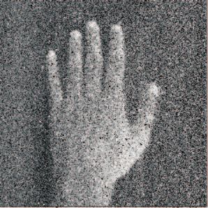
Rastogi and Pospı́šil: Spontaneous ultraweak photon emission imaging...

                23. M. Takeda, M. Kobayashi, M. Takayama, S. Suzuki, T. Ishida, K.                         pp. 4.1–4.11, Wiley-Blackwell, John Wiley & Sons Ltd, Chichester,
                    Ohnuki, T. Moriya, and N. Ohuchi, “Biophoton detection as a novel                      UK (2010).
                    technique for cancer imaging,” Cancer Sci. 95, 656–661 (2004).                 38.     H. Takiwaki, “Measurement of transcutaneous oxygen tension,” in
                24. L. Brancaleon, M. P. Bamberg, T. Sakamki, and N. Kollias, “Attenuated                  Handbook of Non-Invasive Methods and the Skin, J. Serup and G. B. E.
                    total reflection-Fourier transform infrared spectroscopy as a possible                 Jamec, Eds., pp. 185–195, Chemical Rubber, Boca Raton (1995).
                    method to investigate biophysical parameters of stratum corneum in             39.     E. Reefman, P. C. Limburg, C. G. M. Kallenberg, and M. Bijl, “Apopto-
                    vivo,” J. Invest. Dermatol. 116, 380–386 (2001).                                       sis in human skin: role in pathogenesis of various diseases and relevance
                25. M. E. Darvin, W. Sterry, and J. Lademann, “Resonance Raman spec-                       for therapy,” Ann. N.Y. Acad. Sci. 1051, 52–63 (2005).
                    troscopy as an effective tool for the determination of antioxidative sta-      40.     K. Takeda, S. Akagi, S. Takahashi, A. Onishi, H. Hanada, and C. A.
                    bility of cosmetic formulation,” J. Biophoton. 3, 82–88 (2010).                        Pinkert, “Mitochondrial activity in response to serum strvation in bo-
                26. J. Lademann, S. Schanzer, M. Meinke, W. Sterry, and M. E. Darvin,                      vaine (Bos taurus) cell culture,” Cloning and Stem Cells 4, 223–229
                    “Interaction between carotenoids and free radicals in human skin,” Skin                (2002), http://www.ncbi.nlm.nih.gov/pubmed/12398803.
                    Pharmacol. Physiol. 24, 238–244 (2011).                                        41.     U. Hoppe, J. Bergemann, W. Diembeck, J. Ennen, S. Gohla, I. Harris, J.
                27. J. W. Fluhr, P. Caspers, J. A. V. D. Pol, H. Richter, W. Sterry, J. Lade-              Jocob, J. Kielholz, W. Mei, D. Pollet, D. Schachtschabel, G. Sauermann,
                    mann, and M. E. Darvin, “Kinetics of carotenoid distribution in human                  V. Schreiner, F. Stab, and F. Steckel, “Coenzyme Q10, a cutaneous
                    skin in vivo after exogenous stress: disinfectant and wIRA-induced                     antioxidant and energizer,” BioFactors 9, 371–378 (1999).
                    carotenoid depletion recovers from outside to inside,” J. Biomed. Opt.         42.     R. M. W. Moison and G. M. J. B. V. Henegouwen, “Topical antioxi-
                    16, 035002-1–035002-7 (2011).                                                          dant vitamins C and E prevent UVB-radiation-induced peroxidation of
                28. M. E. Darvin, J. W. Fluhr, P. Caspers, A. V. D. Pool, H. Richter,                      eicosapentaenoic acid in pig skin,” Radiat. Res. 157, 402–409 (2002).
                    A. Patzelt, W. Sterry, and J. Lademann, “In vivo distribution of               43.     J. M. Rijnkels, R. M. W. Moison, E. Podda, and G. M. J. B. V.
                    carotenoids in different anatomical locations of human skin: compara-                  Henegouwen, “Photoprotection by antioxidants against UVB-radiation-
                    tive assessment with two different Raman spectroscopy methods” Exp.                    induced damage in pig skin organ culture,” Radiat. Res. 159, 210–217
                    Dermatol. 18, 1060–1063 (2009).                                                        (2003).
                29. A. Jain, I. Rieger, M. Rohr, and A. Schrader, “Antioxidant efficieny on        44.     A. Nandi and I. B. Chatterjee, “Scavenging of superoxide radical by
                    human skin in vivo investigated by UVA-induced chemiluminescence                       ascorbic acid,” J. Biosci. 11, 435–441 (1987).
                    decay analysis via induced chemiluminescence human skin,” Skin Phar-           45.     E. Damiani, P. Astolfi, P. Carloni, P. Stipa, and L. Greci, “Antioxidents:
                    macol. Physiol. 23, 266–272 (2010).                                                    how they work,” in Oxidants in Biology, G. Valacchi and P. A. Davis,
                30. A. Rastogi and P. Pospı́šil, “Ultra-weak photon emission as a non-                    Eds. pp. 251–266, Springer Science, New York (2008).
                    invasive tool for monitoring of oxidative processes in the epidermal           46.     L. Packer, S. U. Weber, and G. Rimbach, “Molecular aspect
                    cells of human skin: comparative study on the dorsal and the palm side                 of α-tocoptrienol antioxidant action and cell signaling,” J. Nutr.
                    of the hand,” Skin Res. Technol. 16, 365–370 (2010).                                   131, 369S–373S (2001), http://www.ncbi.nlm.nih.gov/pubmed?term=
                31. R. Hagens, F. Khabiri, V. Schreiner, H. Wenck, K.-P. Wittern, H.-J.                    11160563%5Buid%5D.
                    Duchstein, and W. Mei, “Non-invasive monitoring of oxidative skin              47.     M. Takada, T. Yuzuriha, and C. Yamato, “Redox levels of intra-
                    stress by ultraweak photon emission measurement. II: biological val-                   venously administered [14 C] Coenzyme Q10 and Coenzyme Q10-
                    idation on ultraviolet stressed skin,” Skin Res. Technol. 14, 112–120                  reducing activity in subcellular fractions of guinea pig liver,” J.
                    (2008).                                                                                Nutr. Sci. Vitaminol. (Tokyo) 31, 147–155 (1985), http://www.ncbi.
                32. H. Kim, S. Ahn, and J. Kim, “Biophoton emission induced by ultrasonic                  nlm.nih.gov/pubmed?term=3928843%5Buid%5D.
                    irradiation,” IFMBE Proceedings 14(8), 1277–1280 (2007).                       48.     G. Banhegyi, L. Braun, M. Csala, P. Ferenc, and M. Jozsef, “Ascorbate
                33. H. Ou-Yang, G. Stamatas, C. Saliouand, and N. Kollias, “A Chemilu-                     metabolism and its regulation in animals,” Free Radic. Biol. Med. 23,
                    minescence study of UVA-induced oxidative stress in human skin in                      793–803 (1997).
                    vivo” J. Invest. Dermatol. 122, 1020–1029 (2004).                              49.     M. Lopez-Torres, J. J. Thiele, Y. Shindo, D. Han, and L. Packer, “Topi-
                34. R. V. Wijk, M. Kobayashi, and E. P. A. V. Wijk, “Anatomic character-                   cal application of α-tocopherol modulates the antioxidant network and
                    ization of human ultra-weak photon emission with a moveable photo-                     diminishes ultraviolet-induced oxidative damage in murine skin,” Br. J.
                    multiplier and CCD imaging,” J. Photochem. Photobiol. B 83, 69–76                      Dermatol. 138, 207–215 (1998).
                    (2006).                                                                        50.     O. Ortolani, A. Conti, A. R. De Gaudio, E. Moraldi, Q. Cantini,
                35. M. Kobayashi, D. Kikuchi, and H. Okamura, “Imaging of ultraweak                        and G. Novelli, “The effect of glutathione and N-acetylcysteine on
                    spontaneous photon emission from human body displaying diurnal                         lipoperoxidative damage in patients with early septic shock,” Am.
                    rhythm,” PLoS ONE 4(7), e6256 (2009).                                                  J. Respir. Crit. Care. Med. 161, 1907–1911 (2000) http://www.ncbi.
                36. H. H. Jung, J. M. Yang, W. M. Woo, C. Choi, J. S. Yang, and K.                         nlm.nih.gov/pubmed?term=10852765.
                    S. Soh, “Years-long biophoton measurements: normalized frequency               51.     G. E. Rhie, M. H. Shin, J. Y. Seo, W. W. Choi, K. H. Cho, K. H. Kim,
                    count analysis and seasonal dependency,” J. Photochem. Photobiol., B                   K. C. Park, H. C. Eun, and J. H. Chung, “Aging- and photoageing-
                    78, 149–154 (2005).                                                                    dependent changes of enzymic and nonenzymic antioxidents in the
                37. C. B. Archer, “Functions of the skin,” in Rook’s Textbook of Der-                      epidermis and dermis of human skin in vivo,” J. Invest. Dermatol. 117,
                    matology, T. Burns, S. Breathnach, N. Cox, and C. Griffiths, Eds.,                     1212–1217 (2001).

                Journal of Biomedical Optics                                               096005-7                                                September 2011 r Vol. 16(9)

Downloaded From: https://www.spiedigitallibrary.org/journals/Journal-of-Biomedical-Optics on 19 Feb 2021
Terms of Use: https://www.spiedigitallibrary.org/terms-of-use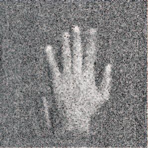
You can also read