Specific Upregulation of TRPC1 and TRPC5 Channels by Mineralocorticoid Pathway in Adult Rat Ventricular Cardiomyocytes - MDPI
←
→
Page content transcription
If your browser does not render page correctly, please read the page content below
cells
Communication
Specific Upregulation of TRPC1 and TRPC5 Channels
by Mineralocorticoid Pathway in Adult Rat
Ventricular Cardiomyocytes
Fiona Bartoli 1 , Soraya Moradi Bachiller 1 , Fabrice Antigny 2 , Kaveen Bedouet 1 ,
Pascale Gerbaud 1 , Jessica Sabourin 1, *,† and Jean-Pierre Benitah 1, *,†
 1    Inserm, UMR-S 1180, Signalisation et Physiopathologie Cardiovasculaire, Université Paris-Saclay,
      92296 Châtenay-Malabry, France; F.Bartoli@leeds.ac.uk (F.B.); soraya_22mb@hotmail.com (S.M.B.);
      kaveen.bedouet@gmail.com (K.B.); pascale.gerbaud@u-psud.fr (P.G.)
 2    Inserm, UMR-S 999, Centre Chirurgical Marie Lannelongue, Université Paris-Saclay,
      92350 Le Plessis-Robinson, France; fabrice.antigny@u-psud.fr
 *    Correspondence: jessica.sabourin@u-psud.fr (J.S.); jean-pierre.benitah@inserm.fr (J.-P.B.);
      Tel.: +33-146-83-52-49 (J.S.); +33-146-83-57-66 (J.-P.B.); Fax: +33-146-83-54-75 (J.S. & J.-P.B.)
 †    These authors contributed equally to this work.

 Received: 26 November 2019; Accepted: 22 December 2019; Published: 23 December 2019

 Abstract: Whereas cardiac TRPC (transient receptor potential canonical) channels and the associated
 store-operated Ca2+ entry (SOCE) are abnormally elevated during cardiac hypertrophy and heart
 failure, the mechanism of this upregulation is not fully elucidated but might be related to the
 activation of the mineralocorticoid pathway. Using a combination of biochemical, Ca2+ imaging,
 and electrophysiological techniques, we determined the effect of 24-h aldosterone treatment on
 the TRPCs/Orai-dependent SOCE in adult rat ventricular cardiomyocytes (ARVMs). The 24-h
 aldosterone treatment (from 100 nM to 1 µM) enhanced depletion-induced Ca2+ entry in ARVMs,
 as assessed by a faster reduction of Fura-2 fluorescence decay upon the addition of Mn2+ and
 increased Fluo-4/AM fluorescence following Ca2+ store depletion. These effects were prevented
 by co-treatment with a specific mineralocorticoid receptor (MR) antagonist, RU-28318, and they are
 associated with the enhanced depletion-induced N-[4-[3,5-Bis(trifluoromethyl)-1H-pyrazol-1-yl]phenyl]-
 4-methyl-1,2,3-thiadiazole-5-carboxamide (BTP2)-sensitive macroscopic current recorded by patch-clamp
 experiments. Molecular screening by qRT-PCR and Western blot showed a specific upregulation of
 TRPC1, TRPC5, and STIM1 expression at the messenger RNA (mRNA) and protein levels upon 24-h
 aldosterone treatment of ARVMs, corroborated by immunostaining. Our study provides evidence
 that the mineralocorticoid pathway specifically promotes TRPC1/TRPC5-mediated SOCE in adult
 rat cardiomyocytes.

 Keywords: aldosterone; TRPC channels; STIM1; adult rat ventricular cardiomyocytes; SOCE; ISOC

1. Introduction
       So far, the physiological impact of store-operated Ca2+ entry (SOCE) in adult cardiomyocytes is
still under debate, notably due to low-to-moderate expression of their molecular constituents including
the molecular complex of STIM1 with Orai1 channels but also with different isoforms of transient
receptor potential canonical channels (TRPCs; notably 1, 4, and 5) [1–5]. However, there is now
strong evidence revealing a role for SOCE in developmental and pathologic cardiac growth, in which
Ca2+ handling regulation through Ca2+ -permeable channels is a cornerstone [6–12]. Indeed, hardly
detectable in healthy adult cardiac myocytes [13,14], SOCE is present in embryonic and neonatal cardiac
myocytes [15] and re-emerges during cardiac hypertrophy and heart failure [1,3,4,16]. While it was

Cells 2020, 9, 47; doi:10.3390/cells9010047                                          www.mdpi.com/journal/cells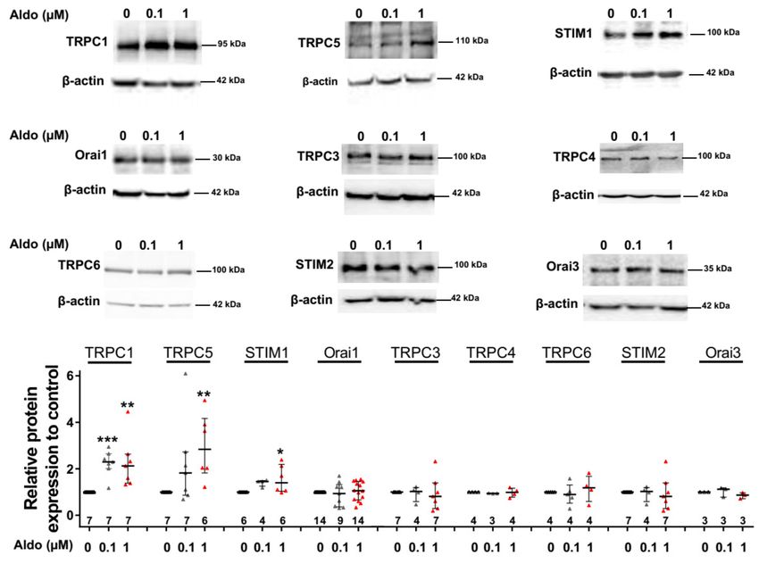
Cells 2020, 9, 47                                                                                    2 of 14

proposed that the upregulation of cardiac SOCE during pathological growth is part of the fetal gene
program [4,17], neuroendocrine factors, which underpin the pathogenesis of heart failure and trigger
Ca2+ -dependent processes leading to cell growth and cardiac hypertrophy, might be involved. Notably,
in addition to being an Na+ -retaining and kalituretic hormone that acts on the kidney, the adrenal steroid
aldosterone and its classical biological effector, the mineralocorticoid receptor (MR), exert deleterious
effects on cardiac function, independently of their action on blood pressure [18]. Indeed, cardiomyocytes
express the MR, a ligand-dependent transcription factor interacting with the glucocorticoid-responsive
elements of target genes, whose activity might control Ca2+ homeostasis on the target cells [19–21]. Prior
studies suggested that stimulation of neonatal rat ventricular myocytes (NRVMs) or human embryonic
stem-cell-derived cardiomyocytes with angiotensin II, phenylephrine, endothelin-1, or aldosterone
evoked an exacerbated SOCE, correlating with increased expression of Orai1, TRPC1, TRPC4, and/or
TRPC5 [11,12,22–26]. Arguably, primary cultures of NRVMs constitute a reliable in vitro model to
study time-related myocardial cell biology but may not accurately reflect the matured adult state. Here,
we showed in adult rat ventricular cardiomyocytes that the aldosterone-enhanced SOCE is associated
with specific upregulation of TRPC1/C5, pointing out the necessity to corroborate data obtained in
NRVMs in a mature system.

2. Material and Methods
    All data and materials supporting the findings of this study are available within the article or
from the corresponding authors upon reasonable request.
    All experiments were carried out according to the ethical principles laid down by the French
Ministry of Agriculture (agreement D-92–019-01) and were performed in accordance with the guidelines
from Directive 2010/63/EU of the European Parliament on the protection of animals. Animal studies
were approved by the local Ethics Committee (CREEA Ile-de-France Sud).

2.1. Chemicals
     Nifedipine (NIF) and caffeine (caf) were obtained from Sigma-Aldrich. The RU-28318, thapsigargin
(Tg) and the KB-R7943 were obtained from TOCRIS Bioscience (Bristol, UK). Fluo-4/AM and Fura-2/AM
were obtained from Thermo Fisher Scientific (Waltham, MA, USA).

2.2. Ventricular Myocyte Isolation
      Adult rat ventricular cardiomyocytes (ARVMs) were isolated from rat hearts using a standard
enzymatic perfusion method and maintained for 24 h at 37 ◦ C in a serum-free Tyrode solution (in mM:
130 NaCl, 5.4 KCl, 0.4 NaH2 PO4 , 0.5 MgCl2 , 25 HEPES, 22 d-glucose, 1 CaCl2 ; pH 7.4 with NaOH) with
or without aldosterone and with or without RU28318, as previously described [19]. Only rod-shaped
cells, quiescent when unstimulated and excitable, were used. In comparison to freshly isolated ARVMs,
no major alterations in cellular excitability and size were noted [19–21,27].

2.3. Measurement of Cytosolic Ca2+ Changes
      After washing, the ARVMs were incubated at room temperature in a solution containing (in mM)
135 NaCl, 5 KCl, 1.8 CaCl2 , 1 MgCl2 , 10 HEPES, 10 d-glucose, pH 7.4 with NaOH, supplemented either
with 5 µM Fluo-4/AM (for 30 min) or with 1 µM Fura-2/AM (for 15 min). Both fluorescence probes
were solved in DMSO plus 20% pluronic acid in stock solutions. The SOCE was measured as described
previously [14]. The Fura-2 340/380 ratios were calibrated using the equation [Ca2+ ]I = KD ·β·(R −
Rmin )/(Rmax − R), where R is the fluorescence ratio recorded at the two excitation wavelengths (F340
and F380 ), KD represents the dissociation constant, Rmin and Rmax are the fluorescence ratios under
Ca2+ -free and Ca2+ -saturating conditions, and β = F340 , zero Ca2+ /F380 , saturating Ca2+ .
      For in situ measurements of Rmin and Rmax , Fura-2-loaded ARVMs were firstly exposed to
Ca -free solution with 10 mM caf + 5 µM Tg for 5 min, to empty Sarcoplasmic Reticulum
   2+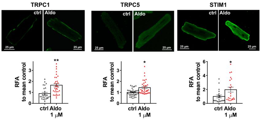
Cells 2020, 9, 47                                                                                3 of 14

(SR) Ca2+ stores.      The bath solution was then switched to K+ buffer (in mM: 10 NaCl,
130 KCl, 1 MgCl2 , 10 HEPES, pH: 7.2, ionic strength: 0.142, 37 ◦ C) containing 10 mM Ethylene
glycol-bis(2-aminoethylether)-N,N,N0 ,N0 -tetraacetic acid (EGTA). To obtain Rmin , the cells were
incubated for 30 to 45 min with 20 µM non-fluorescent Ca2+ ionophore, ionomycin, and 10 µM carbonyl
cyanide p-(trifluoromethoxy)-phenylhydrazone in K+ -buffer Ca2+ -free solution, and measurements
were taken at both wavelengths after the fluorescence reached stable values. Then, Rmax was obtained
by saturating the indicator with 10 mM or 20 mM CaCl2 in the presence of 10 mM EGTA.
     To calculate the KD , a dose response (0, 1.0, 2.0, 3.0, 4.0, 5.0, 6.0, 7.0, 8.0, 9.0, and 10.0 mM
CaCl2 /EGTA (free Ca2+ ranging from 0 to 37 µM)) was performed in K+ buffer. As a double-log plot,
the Ca2+ response of the indicator was linear with the x-intercept equal to the log of the apparent KD
of the indicator. We calculated Rmin = 0.66, Rmax = 1.84, and β = 1.35 with KD = 377 nM. Of note,
aldosterone treatment did not change any parameters.

2.4. Measurement of Cation Influx Using Mn2+ -Quenching of Fura-2 Fluorescence Quenching
     The Mn2+ influx was measured on Fura-2/AM loaded ARVMs as described previously [11]. Fura-2
was excited at the isosbestic wavelength, 360 nm, and emission fluorescence was monitored at 510 nm.
After SR depletion (see below), a 500 µM MnCl2 solution was perfused in the presence of NIF and
KB-R7943, and the initial linear slope of the Fura-2 fluorescence’s decrease was measured, reflecting
the SOC channel activity. The quenching rate of fluorescence intensity (F360 ) was estimated using
linear regression of the initial decaying phase (slope, ∆F/dt) just after Mn2+ addition, expressed as
a percentage decrease in F360 per min (the maximal F360 signal obtained before Mn2+ was set at 100%
to correct for differences in the cell size and/or fluorophore loading). Photobleaching was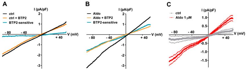
Cells 2020, 9, 47                                                                                        4 of 14

             Table 1. List of primary antibodies. TRPC—transient receptor potential canonical channel.

                      Protein        Host             Dilution     Code                 Source
                      TRPC1         Mouse              1/200     SC-133076           Santa Cruz
                      TRPC3         Rabbit             1/200      ACC-016             Alomone
                      TRPC4         Rabbit             1/200      ACC-018             Alomone
                                                                                   UC Davis/NIH
                      TRPC5         Mouse              1/200       73-104
                                                                                  NeuroMab Facility
                      TRPC6         Rabbit             1/200     ACC-017              Alomone
                       Orai1        Rabbit             1/200      O8264                Sigma
                       Orai3        Rabbit             1/200     ACC-065              Alomone
                       STIM1        Rabbit             1/200      S6197                Sigma
                       STIM2        Rabbit             1/200     ACC-064              Alomone
                    β-actin HRP     Mouse             1/30,000   SC-47778            Santa Cruz

2.7. Immunostaining
      ARVMs were fixed and permeabilized with 2% paraformaldehyde (PFA)/0.5% Triton X-100 as
previously described [14]. Images were acquired using a Leica TCS SP5 confocal microscope, keeping
all the acquisition parameters the same at all times. For each cell, we selected a single plane at the
depth where the antibody detection was optimal. After background subtraction, as semi-quantitative
analysis, the total cell fluorescence signal (integrated density) of the region of interest (ROI, by circling
the cells) was normalized to the ROI area of each cell using ImageJ software (1.52d, National Institutes
of Health, Bethesda, MD, USA).

2.8. Quantitative Real-Time PCR
     The RNA extraction and RT-qPCR quantification were performed as previously described [11],
using the primers listed in Table 2. Briefly, total RNA from ARVMs was isolated using the TRIzol
procedure according to the manufacturer’s instructions. For complementary DNA (cDNA) synthesis,
1 µg of total RNA was reverse-transcribed using the iScript cDNA synthesis kit (Bio-Rad, Hercules, CA,
USA) according to the manufacturer’s instructions. Then, cDNA was used as a template for real-time
PCR with SYBR Green Supermix. Quantitative determination of the different messenger RNA (mRNA)
expression levels was performed with a CFX96 Touch™ Real-Time PCR Detection System with either
gene-specific primers or primers for endogenous controls. The mRNA levels were normalized to
housekeeping genes and were expressed as a fold change of that determined in control (ctrl) ARVMs
for each isolation.

                                     Table 2. List of primers used for RT-qPCR.

        Gene                      Forward (50 –30 )                               Reverse (50 –30 )
       RPL32              GCT GCT GAT GTG CAA CAA A                 GGG ATT GGT GAC TCT GAT GG
      YWHAZ             AGA CGG AAG GTG CTG AGA AA                  GAA GCA TTG GGG ATC AAG AA
        TBP              AAA GAC CAT TGC ACT TCG TG                  GCT CCT GTG CAC ACC ATT TT
       STIM1          TCT CTG AGT TGG AGG ATG AGT AGA             CAA TAT AGG GGA GCA GAG GTA AGA
       TRPC1            TTC CAA AGA GCA GAA GGA CTG                  AGG TGC CAA TGA ACG AGT G
       TRPC5             TGA GTG GAA GTT TGC GAG AA                 TGG GAC AGA AGG TGT TGT TG

2.9. Statistical Analysis
     The number of cells (n) or animals (N) studied per experiment is indicated. Statistical analyses,
described in the figure legends, were performed using GraphPad Prism 6.0 software (GraphPad
Software, San Diego, CA, USA). The variance assumption was automatically tested. A value of p < 0.05
was considered significant.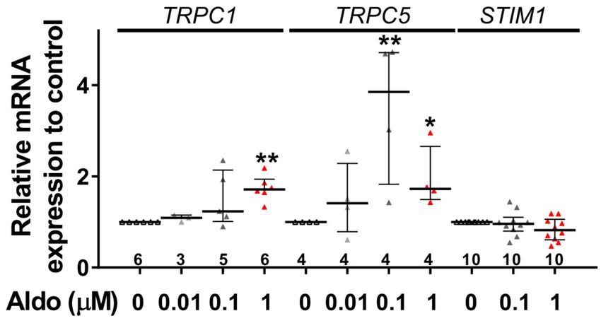
Cells 2020, 9, 47 5 of 14
3. Results
3.1. Aldosterone Increases Store-Operated Ca2+ Entry (SOCE) via MR Activation in Adult Cardiomyocytes
We firstly examined the SOCE activity in quiescent rod-shaped Fluo-4/AM-loaded ARVMs, which
were treated with or without aldosterone (Aldo) for 24 h. Of interest, 24-h aldosterone treatment did
not induce cellular hypertrophy as assessed by the similar membrane capacitance of isolated ARVMs
treated 24 h with or without 1 µM aldosterone (123 ± 6 vs. 120 ± 6 pF, for 23 control cells vs. 23 Aldo
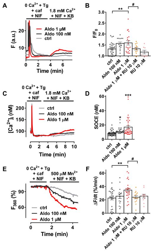
treated cells, respectively). As exemplified in Figure 1A, after SR depletion in a Ca2+ -free solution
containing thapsigargin (a SERCA inhibitor, Tg 5 µM), caffeine (an RyR2 activator, caf 10 mM), and
nifedipine (an L-type Ca2+ channel blocker, NIF 10 µM), the subsequent addition of 1.8 mM Ca2+ in
the presence of NIF and KB-R7943 (an Na+ –Ca2+ exchanger inhibitor, KB 5 µM) induced a moderate
increase of fluorescence in 69% of ctrl ARVMs (gray trace), while a stronger response was observed in
a dose-dependent manner in all aldosterone-treated cells (100 nM, dark-gray trace and 1 µM, red trace).
On average (Figure 1B), we denoted a 1.4-fold increase in Ca2+ entry in 1 µM aldosterone-treated
ARVMs, which was prevented in co-treated ARVMs with a selective mineralocorticoid receptor (MR)
antagonist RU-28318 (RU at 10 µM). Similar results were obtained using the ratiometric Fura-2/AM
fluorescent probe (Figure 1C,D). To confirm those results, we used Mn2+ -quenching microfluorimetry
in the presence of major cardiac Ca2+ entry pathway inhibitors (NIF and KB). Figure 1E shows
representative Fura-2/AM fluorescence traces after SR depletion of ARVMs treated with or without
aldosterone for 24 h. The quenching rates, proportional to Mn2+ entry, were increased up to 1.5-fold
after aldosterone incubation; the effect was blunted in ARVMs co-treated with 10 µM RU-28318
(Figure 1F). These data confirmed an increased cation entry after aldosterone treatment, which was
dependent on MR activation.
To further explore the nature of the SOCE modulated by the aldosterone pathway, we recorded the
store-dependent currents using the patch-clamp technique in whole-cell configuration, using solutions
to limit Ca2+ and K+ currents by including inhibitors (NIF and KB) and Cs+ to block K+ channels. After
SR Ca2+ store depletion (see above), the SOC currents (ISOC ) were elicited by a standard ramp protocol
before and after the perfusion of the non-selective SOC channel blocker BTP2 (5 µM). As shown in
Figure 2A–C, the small BTP2-sensitive currents with a linear current density–voltage relationship and
a reversal potential around 0 mV were recorded in ARVMs incubated without aldosterone. The ISOC
was increased in aldosterone-treated ARVMs (Figure 2C). The feature of this aldosterone-enhanced ISOC
induced by store depletion, i.e., its reversal potential around 0 mV and sensitivity to BTP2, suggested
the activation of non-selective cationic channels carried by TRPCs.
3.2. Aldosterone Increases TRPC1, TRPC5, and STIM1 Protein Expression in Adult Cardiomyocytes
To assess the molecular nature of the aldosterone-increased SOCE, we investigated the effect of
aldosterone treatment on the expression of different SOCE machinery actors, i.e., TRPCs and Orai1
channels and STIM proteins in ARVMs after 24 h of incubation. As assessed by Western blots (Figure 3),
we observed an increased protein expression of TRPC1, TRPC5, and STIM1, without any changes in
Orai1, TRPC3, TRPC4, TRPC6, STIM2, and Orai3 protein levels. Uncropped blots are presented in
Figure S1 (Supplementary Materials). The increases in TRPC1, TRPC5 and STIM1 protein expressions
were further tested by immunostaining of treated isolated cardiomyocytes (Figure 4). The fluorescence
signal of these three proteins appeared more intense in 1 µM Aldo-treated cells than in ctrl cells and
revealed a localization of TRPC1 and STIM1 at the surface membrane, while TRPC5 appeared to
be present at the striated area, as previously reported [2,28–30]. At mRNA levels, using RT-qPCR,
we observed an increased mRNA expression of TRPC1 and TRPC5 induced by 100 nM and 1 µM
aldosterone treatment, whereas lower doses (10 nM) had no effect. STIM1 mRNA levels remained
unchanged after 24 h of aldosterone treatment, no matter the concentration used (Figure 5).Cells 2020, 9, 47 6 of 14
Figure 1. A 24-h aldosterone treatment increases store-operated Ca2+ entry (SOCE) in adult
rat ventricular cardiomyocytes (ARVMs). (A) Representative traces of fluorescence variation in
Fluo-4/AM-loaded ARVMs pre-incubated in the absence (control (ctrl), gray trace) and in the presence
of aldosterone (Aldo, 100 nM, dark-gray trace or 1 µM, red trace). Cells were exposed to 5 µM
thapsigargin (Tg) plus 10 mM caffeine (caf) in the presence of 10 µM nifedipine (NIF) in Ca2+ -free
medium to deplete the SR and then to Ca2+ -containing solution in the presence of 5 µM of KB-R7943
and NIF to evaluate the SOCE. (B) Averaged amplitude of SOCE (F/F0 ) in ARVMs incubated for
24 h in the absence (ctrl, white symbols) or in the presence of aldosterone (100 nM, gray symbols
or 1 µM, red symbols) without or with 10 µM of the selective MR antagonist RU-28318 (RU, orange
symbols in aldosterone condition and pink symbols in ctrl condition). Average results of 8–32 cells
from three isolations. (C) Representative traces of [Ca2+ ]i variation (in nM) in Fura-2/AM-loaded
ARVMs incubated for 24 h in the absence (ctrl, gray trace) or in the presence of aldosterone (100 nM,
dark-gray trace; 1 µM, red trace). Fura-2/AM fluorescence 340/380 ratios were converted to [Ca2+ ]i as
described in the methods. (D) Quantitative assessment of SOCE (in nM) from untreated (ctrl, white
symbols) and 100 nM (gray symbols) or 1 µM (red symbols) aldosterone-treated cells. Average results
of 24–48 cells from four isolations. (E) Representative traces of Mn2+ -induced Fura-2/AM fluorescence
decay in ARVMs incubated in the absence of aldosterone (ctrl) are shown in gray traces, and those
in the presence of aldosterone 100 nM or 1 µM are shown in dark-gray and red traces, respectively.
(F) Bar graph of the initial slope of the Mn2+ -induced decrease of Fura-2/AM fluorescence fitted by
linear regression and averaged (∆F/dt, %/min) for ARVMs incubated for 24 h in the absence (ctrl, white
symbols) or in the presence of aldosterone (100 nM, dark-gray symbols or 1 µM, red symbols) withoutCells 2020, 9, 47 7 of 14
or with 10 µM RU (orange symbols in aldosterone condition and pink symbols in ctrl condition).
Average results of 11–25 cells from three isolations. Results are presented with scatter plots with mean
± standard error of the mean (SEM). Statistical significance was evaluated using one-way ANOVA
followed by post hoc Fisher least significant difference (LSD) test for multiple comparisons. * p < 0.05,
** p < 0.01, *** p < 0.001 vs. ctrl; # p < 0.05 vs. aldosterone.
Figure 2. A 24-h aldosterone treatment increases SOC currents (ISOC ) in ARVMs. (A,B) Representative
current–voltage (I–V) relationships elicited by ramp voltage-clamp protocol in ARVMs incubated for
24 h without (A) or with aldosterone 1 µM (B), following SR Ca2+ depletion (black traces) and then in
the presence of 5 µM BTP2 (orange traces). BTP2-sensitive (blue traces) represents the difference current.
(C) Scatter plots of mean ± SEM of the BTP2-sensitive I–V relationships recorded in ARVMs incubated
for 24 h without (gray trace, −0.19 ± 0.19 pA/pF at −90 mV and 0.30 ± 0.17 pA/pF at +60 mV, n = 7) or
with Aldo 1 µM (red trace, −1.51 ± 0.20 pA/pF at −90 mV (p < 0.01) and 0.90 ± 0.15 pA/pF at +60 mV
(p < 0.05), n = 9) from three cell isolations. Statistical significance was evaluated using Student’s t-test.
Figure 3. A 24-h aldosterone treatment increases transient receptor potential canonical channel 1
(TRPC1), TRPC5, and STIM1 protein expression in ARVMs. Representative Western blot (top panels)
and pooled data (bottom bar graph) of TRPC1, TRPC5, STIM1, Orai1, TRPC3, TRPC4, TRPC6, STIM2,
and Orai3 in ARVMs incubated for 24 h in the absence or in the presence of aldosterone (100 nM or
1 µM). Protein levels were normalized by β-actin and are expressed as fold changes of that determined
in untreated ARVMs for each cell isolation. Results are presented as scatter plots with median and
interquartile range. Statistical significance was evaluated using one-way ANOVA (Kruskal–Wallis)
followed by post hoc Dunn’s test for multiple comparisons. * p < 0.05, ** p < 0.01, *** p < 0.001 vs. ctrl.Cells 2020, 9, 47 8 of 14
Figure 4. Cellular localization of TRPC1, TRPC5, and STIM1 in ARVMs. Representative
immunofluorescences images (top) and quantification of TRPC1, TRPC5, and STIM1 in ARVMs
incubated for 24 h in the absence or in the presence of aldosterone 1 µM. Results are expressed as
relative fluorescence/cellular area (RFA). Average results of 20–39 cells from three isolations. Results
are presented as scatter plots with mean ± SEM. Statistical significance was evaluated using Student’s
t-test. * p < 0.05, ** p < 0.01 vs. ctrl.
Figure 5. A 24-h aldosterone treatment increases TRPC1 and TRPC5 messenger RNA (mRNA)
expression in ARVMs. Relative TRPC1, TRPC5, and STIM1 mRNA levels in ARVMs were incubated for
24 h in the absence or in the presence of aldosterone (Aldo 10 nM, 100 nM or 1 µM). The mRNA levels
determined by qRT-PCR were normalized to housekeeping genes and are expressed as fold changes of
that determined in ARVMs incubated in the absence of aldosterone for each cell isolation. Results are
presented as scatter plots with median and interquartile range. Statistical significance was evaluated
using one-way ANOVA (Kruskal–Wallis) followed by post hoc Dunn’s test for multiple comparisons.
* p < 0.05, ** p < 0.01 vs. ctrl.
4. Discussion
Our study was aimed at investigating the aldosterone effect on SOCE in ARVMs. Our results
indicate that a 24-h treatment of high-dose aldosterone stimulates the SOCE in an MR-dependent
manner, due to a specific upregulation of TRPC1, TRPC5, and STIM1.
Upregulation of the cardiac mineralocorticoid signaling pathway emerged as a key regulator
involved in heart failure development and progression [31], in which Ca2+ -dependent activation of
transduction signaling is also important [32]. On the other hand, the re-emergence of cardiac SOCECells 2020, 9, 47 9 of 14
machinery was recently shown in the heart failure pathological remodeling processes, contributing to
Ca2+ prohypertrophic signaling [1,3,4]. Notably, the TRPC1 channel, which is the major isoform in
whole human heart [33], is upregulated during heart failure progression and is critical for it [17,22,34–37].
Likewise, increased TRPC5 expression in human heart failure [38,39] and STIM1 elevation are linked to
cardiac hypertrophy [9,40–43]. However, the mechanism regulating cardiac TRPC channels expression
is not well defined. Since cardiac hypertrophy and heart failure can be viewed as a gene regulatory
disorder [44], we hypothesize that MR as transcription factor might be involved in this process.
As previously reported [14], ARVMs show rather small Ca2+ entry following SR depletion, which
is enhanced after aldosterone treatment. The presence of SOCE in adult ventricular cardiomyocytes
first reported in 2004 [13] remains controversial. For example, the traditional protocol using only
thapsigargin or cyclopiazonic acid (CPA) to block the SR Ca2+ pumps indicates that there is no or limited
SOCE in normal adult ventricular cells [9,17,35,45]. However, these conditions might not sufficiently
deplete the SR Ca2+ stores of quiescent adult ventricular cells [15,46], even if one might think that,
in the presence of an SERCA pump inhibitor, the SR Ca2+ leak would deplete the SR [45]. Thus, when
a clear SR Ca2+ depletion is observed (with ryanodine receptor activation through either stimulation
or caffeine/ryanodine application), a moderated SOCE activity might be observed [11,14,47–49], as we
show in the present study.
Reflecting previous reports in adrenal chromaffin cells [50] or coronary arteries [51] from metabolic
syndrome pigs, in the mesenteric arterial smooth muscle cells of DOCA-salt hypertensive rats or
in A7r5 cells [52] and in NRVMs [11], a chronic treatment of ARVMs with aldosterone potentiates
SOCE, in line with the emergence of a marked BTP2-sensitive current after store depletion. This
linear-shaped non-selective current, as assessed by its reversal potential near 0 mV, features reminiscent
of TRPC channels [36,53,54]. Indeed, SOCE is a ubiquitous mechanism that is mediated by distinct
SOC channels, ranging from the highly selective Ca2+ release-activated Ca2+ channel supported by
the Orai1 channel to relatively Ca2+ non-selective SOC channels supported by TRPC channels [55].
Unlike NRVMs, in which aldosterone treatment enhances Orai1 expression [11], we did not observe an
upregulation of Orai1 in ARVMs. This points out that the use of immature neonatal cardiomyocytes
presenting unequivocal robust SOCE [9,10] might have limitations, thus justifying the present study.
Nonetheless, aldosterone promotes an MR-specific increase in TRPC1/5 and STIM1 protein expression
in ARVMs, as seen in metabolic syndrome adrenal chromaffin cells [50], in coronary arteries [51,56],
and in NRVMs [11]. These are consistent with the ionic current features that we observed. Aldosterone
increases STIM1 expression only at the protein level, suggesting a post-transcriptional regulation.
In this way, it was demonstrated that STIM1 protein expression is regulated by the serum- and
glucocorticoid-inducible kinase 1 (SGK1), an aldosterone-regulated kinase which phosphorylates
Nedd4-2, leading to decreased ubiquitination and subsequent degradation of the STIM1 protein [57,58].
A number of studies, conducted in various cell models, implicated members of TRPC channels in SOC
activity [59–62]. Notably, TRPC1 channels are believed to mediate the non-selective cation current and
to form SOC channels as a component in human atria [63], in the mouse sinoatrial node [64], in human
cardiac c-kit+ progenitor cells [65], in NRVMs [66], and in adult ventricular cardiomyocytes [36,46,67].
Similarly, TRPC5, which was found to contribute to the formation of SOCE in smooth muscle cells
isolated from rabbit pial arterioles [68], contributes to SOCE in NRVMs [11,69]. In addition, STIM1,
the critical constituent of SOCE, can directly bind to and activate TRPC1 and TRPC5 channels via
interactions in its ezrin/radixin/moesin (ERM) domain [70–76].
Our findings provide direct support for the presence of STIM1, TRPC1, and TRPC5 in ARVMs and
for their participation in aldosterone/MR-enhanced SOCE. Taking into account that G-protein-coupled
receptor (GPCR) activation promotes TRPC1 and TRPC5 translocation to the plasma membrane in
a STIM1-dependent manner [77] and the documented signaling crosstalk between cardiac MR and GPCR
signaling components [78], further studies directed toward the potential of the cardiac MR-dependent
TRPC channelosome upregulation in the receptor agonist (such as angiotensin II)-induced inositol
1,4,5-trisphosphate (IP3 ) sensitive Ca2+ store depletion remain to be performed.Cells 2020, 9, 47 10 of 14
Supplementary Materials: The following are available online at http://www.mdpi.com/2073-4409/9/1/47/s1.
Author Contributions: Conceptualization, J.S. and J.-P.B.; experiments, F.B., S.M.B., F.A., K.B., P.G., and J.S.;
methodology, J.S. and J.-P.B; validation, J.S. and J.-P.B.; formal analysis, J.S. and J.-P.B.; writing—original draft
preparation, J.S., F.B., and J.-P.B.; funding acquisition, J.S. and J.-P.B. All authors have read and agreed to the
published version of the manuscript.
Funding: This work was supported by research grants from INSERM, National Funding Agency for Research
(ANR) (ANR-15-CE14), CORDDIM (Cardiovasculaire Obésité Rein Diabète Domaine d’Intérêt Majeur, Région Ile
de France).
Acknowledgments: We thank the AnimEX platform for animal care and the Trans-Prot platform IFR141-IPSIT
from the University of Paris-Sud, University of Paris-Saclay.
Conflicts of Interest: The authors declare no conflicts of interest.
References
1. Collins, H.E.; Zhu-Mauldin, X.; Marchase, R.B.; Chatham, J.C. STIM1/Orai1-mediated SOCE: Current
perspectives and potential roles in cardiac function and pathology. Am. J. Physiol. Heart Circ. Physiol. 2013,
305, H446–H458. [CrossRef]
2. Ahmad, A.A.; Streiff, M.; Hunter, C.; Hu, Q.; Sachse, F.B. Physiological and pathophysiological role of
transient receptor potential canonical channels in cardiac myocytes. Prog. Biophys. Mol. Biol. 2017, 130,
254–263. [CrossRef]
3. Bartoli, F.; Sabourin, J. Cardiac Remodeling and Disease: Current Understanding of STIM1/Orai1-Mediated
Store-Operated Ca2+ Entry in Cardiac Function and Pathology. Adv. Exp. Med. Biol. 2017, 993, 523–534.
[CrossRef]
4. Eder, P. Cardiac Remodeling and Disease: SOCE and TRPC Signaling in Cardiac Pathology. Adv. Exp. Med.
Biol. 2017, 993, 505–521. [CrossRef]
5. Freichel, M.; Berlin, M.; Schurger, A.; Mathar, I.; Bacmeister, L.; Medert, R.; Frede, W.; Marx, A.; Segin, S.;
Londono, J.E.C. TRP Channels in the Heart. In Neurobiology of TRP Channels, 2nd ed.; Emir, T.L.R., Ed.;
CRC Press/Taylor & Francis: Boca Raton, FL, USA, 2017; pp. 149–185. [CrossRef]
6. Gao, H.; Wang, F.; Wang, W.; Makarewich, C.A.; Zhang, H.; Kubo, H.; Berretta, R.M.; Barr, L.A.; Molkentin, J.D.;
Houser, S.R. Ca2+ influx through L-type Ca2+ channels and transient receptor potential channels activates
pathological hypertrophy signaling. J. Mol. Cell Cardiol. 2012, 53, 657–667. [CrossRef]
7. Huang, J.; van Breemen, C.; Kuo, K.H.; Hove-Madsen, L.; Tibbits, G.F. Store-operated Ca2+ entry modulates
sarcoplasmic reticulum Ca2+ loading in neonatal rabbit cardiac ventricular myocytes. Am. J. Physiol.
Cell Physiol. 2006, 290, C1572–C1582. [CrossRef]
8. Hunton, D.L.; Lucchesi, P.A.; Pang, Y.; Cheng, X.; Dell’Italia, L.J.; Marchase, R.B. Capacitative calcium entry
contributes to nuclear factor of activated T-cells nuclear translocation and hypertrophy in cardiomyocytes.
J. Biol. Chem. 2002, 277, 14266–14273. [CrossRef]
9. Luo, X.; Hojayev, B.; Jiang, N.; Wang, Z.V.; Tandan, S.; Rakalin, A.; Rothermel, B.A.; Gillette, T.G.; Hill, J.A.
STIM1-dependent store-operated Ca2+ entry is required for pathological cardiac hypertrophy. J. Mol.
Cell Cardiol. 2012, 52, 136–147. [CrossRef] [PubMed]
10. Pang, Y.; Hunton, D.L.; Bounelis, P.; Marchase, R.B. Hyperglycemia inhibits capacitative calcium entry and
hypertrophy in neonatal cardiomyocytes. Diabetes 2002, 51, 3461–3467. [CrossRef] [PubMed]
11. Sabourin, J.; Bartoli, F.; Antigny, F.; Gomez, A.M.; Benitah, J.P. Transient Receptor Potential Canonical
(TRPC)/Orai1-dependent Store-operated Ca2+ Channels: NEW TARGETS OF ALDOSTERONE IN
CARDIOMYOCYTES. J. Biol. Chem. 2016, 291, 13394–13409. [CrossRef] [PubMed]
12. Voelkers, M.; Salz, M.; Herzog, N.; Frank, D.; Dolatabadi, N.; Frey, N.; Gude, N.; Friedrich, O.; Koch, W.J.;
Katus, H.A.; et al. Orai1 and Stim1 regulate normal and hypertrophic growth in cardiomyocytes. J. Mol.
Cell Cardiol. 2010, 48, 1329–1334. [CrossRef] [PubMed]
13. Hunton, D.L.; Zou, L.; Pang, Y.; Marchase, R.B. Adult rat cardiomyocytes exhibit capacitative calcium entry.
Am. J. Physiol. Heart Circ. Physiol. 2004, 286, H1124–H1132. [CrossRef] [PubMed]
14. Dominguez-Rodriguez, A.; Ruiz-Hurtado, G.; Sabourin, J.; Gomez, A.M.; Alvarez, J.L.; Benitah, J.P.
Proarrhythmic effect of sustained EPAC activation on TRPC3/4 in rat ventricular cardiomyocytes. J. Mol.
Cell Cardiol. 2015, 87, 74–78. [CrossRef] [PubMed]Cells 2020, 9, 47 11 of 14
15. Uehara, A.; Yasukochi, M.; Imanaga, I.; Nishi, M.; Takeshima, H. Store-operated Ca2+ entry uncoupled with
ryanodine receptor and junctional membrane complex in heart muscle cells. Cell Calcium. 2002, 31, 89–96.
[CrossRef] [PubMed]
16. Falcon, D.; Galeano-Otero, I.; Calderon-Sanchez, E.; Del Toro, R.; Martin-Bornez, M.; Rosado, J.A.;
Hmadcha, A.; Smani, T. TRP Channels: Current Perspectives in the Adverse Cardiac Remodeling.
Front. Physiol. 2019, 10, 159. [CrossRef] [PubMed]
17. Wu, X.; Eder, P.; Chang, B.; Molkentin, J.D. TRPC channels are necessary mediators of pathologic cardiac
hypertrophy. Proc. Natl. Acad. Sci. USA 2010, 107, 7000–7005. [CrossRef] [PubMed]
18. Jaisser, F.; Farman, N. Emerging Roles of the Mineralocorticoid Receptor in Pathology: Toward New
Paradigms in Clinical Pharmacology. Pharmacol. Rev. 2016, 68, 49–75. [CrossRef]
19. Benitah, J.P.; Vassort, G. Aldosterone upregulates Ca2+ current in adult rat cardiomyocytes. Circ. Res. 1999,
85, 1139–1145. [CrossRef]
20. Gomez, A.M.; Rueda, A.; Sainte-Marie, Y.; Pereira, L.; Zissimopoulos, S.; Zhu, X.; Schaub, R.; Perrier, E.;
Perrier, R.; Latouche, C.; et al. Mineralocorticoid modulation of cardiac ryanodine receptor activity is
associated with downregulation of FK506-binding proteins. Circulation 2009, 119, 2179–2187. [CrossRef]
21. Benitah, J.P.; Perrier, E.; Gomez, A.M.; Vassort, G. Effects of aldosterone on transient outward K+ current
density in rat ventricular myocytes. J. Physiol. 2001, 537, 151–160. [CrossRef]
22. Ohba, T.; Watanabe, H.; Murakami, M.; Takahashi, Y.; Iino, K.; Kuromitsu, S.; Mori, Y.; Ono, K.; Iijima, T.;
Ito, H. Upregulation of TRPC1 in the development of cardiac hypertrophy. J. Mol. Cell Cardiol. 2007, 42,
498–507. [CrossRef] [PubMed]
23. Wang, Y.; Li, Z.C.; Zhang, P.; Poon, E.; Kong, C.W.; Boheler, K.R.; Huang, Y.; Li, R.A.; Yao, X. Nitric
Oxide-cGMP-PKG Pathway Acts on Orai1 to Inhibit the Hypertrophy of Human Embryonic Stem Cell-Derived
Cardiomyocytes. Stem Cells 2015, 33, 2973–2984. [CrossRef] [PubMed]
24. Kirschmer, N.; Bandleon, S.; von Ehrlich-Treuenstatt, V.; Hartmann, S.; Schaaf, A.; Lamprecht, A.K.;
Miranda-Laferte, E.; Langsenlehner, T.; Ritter, O.; Eder, P. TRPC4alpha and TRPC4beta Similarly Affect
Neonatal Cardiomyocyte Survival during Chronic GPCR Stimulation. PLoS ONE 2016, 11, e0168446.
[CrossRef] [PubMed]
25. Kiso, H.; Ohba, T.; Iino, K.; Sato, K.; Terata, Y.; Murakami, M.; Ono, K.; Watanabe, H.; Ito, H. Sildenafil prevents
the up-regulation of transient receptor potential canonical channels in the development of cardiomyocyte
hypertrophy. Biochem. Biophys. Res. Commun. 2013, 436, 514–518. [CrossRef]
26. Ji, Y.; Guo, X.; Zhang, Z.; Huang, Z.; Zhu, J.; Chen, Q.H.; Gui, L. CaMKIIdelta meditates phenylephrine
induced cardiomyocyte hypertrophy through store-operated Ca2+ entry. Cardiovasc. Pathol. 2017, 27, 9–17.
[CrossRef] [PubMed]
27. Perrier, E.; Perrier, R.; Richard, S.; Benitah, J.P. Ca2+ controls functional expression of the cardiac K+ transient
outward current via the calcineurin pathway. J. Biol. Chem. 2004, 279, 40634–40639. [CrossRef] [PubMed]
28. Huang, H.; Wang, W.; Liu, P.; Jiang, Y.; Zhao, Y.; Wei, H.; Niu, W. TRPC1 expression and distribution in rat
hearts. Eur. J. Histochem. 2009, 53, e26. [CrossRef]
29. Kojima, A.; Kitagawa, H.; Omatsu-Kanbe, M.; Matsuura, H.; Nosaka, S. Ca2+ paradox injury mediated
through TRPC channels in mouse ventricular myocytes. Br. J. Pharm. 2010, 161, 1734–1750. [CrossRef]
30. Jiang, Y.; Huang, H.; Liu, P.; Wei, H.; Zhao, H.; Feng, Y.; Wang, W.; Niu, W. Expression and localization
of TRPC proteins in rat ventricular myocytes at various developmental stages. Cell Tissue Res. 2014, 355,
201–212. [CrossRef]
31. Buonafine, M.; Bonnard, B.; Jaisser, F. Mineralocorticoid Receptor and Cardiovascular Disease. Am. J.
Hypertens 2018, 31, 1165–1174. [CrossRef]
32. Gravez, B.; Tarjus, A.; Jaisser, F. Mineralocorticoid receptor and cardiac arrhythmia. Clin. Exp. Pharm. Physiol.
2013, 40, 910–915. [CrossRef] [PubMed]
33. Riccio, A.; Medhurst, A.D.; Mattei, C.; Kelsell, R.E.; Calver, A.R.; Randall, A.D.; Benham, C.D.; Pangalos, M.N.
mRNA distribution analysis of human TRPC family in CNS and peripheral tissues. Brain Res. Mol. Brain Res.
2002, 109, 95–104. [CrossRef]
34. Vindis, C.; D’Angelo, R.; Mucher, E.; Negre-Salvayre, A.; Parini, A.; Mialet-Perez, J. Essential role of TRPC1
channels in cardiomyoblasts hypertrophy mediated by 5-HT2A serotonin receptors. Biochem. Biophys.
Res. Commun. 2010, 391, 979–983. [CrossRef] [PubMed]Cells 2020, 9, 47 12 of 14
35. Makarewich, C.A.; Zhang, H.; Davis, J.; Correll, R.N.; Trappanese, D.M.; Hoffman, N.E.; Troupes, C.D.;
Berretta, R.M.; Kubo, H.; Madesh, M.; et al. Transient receptor potential channels contribute to pathological
structural and functional remodeling after myocardial infarction. Circ. Res. 2014, 115, 567–580. [CrossRef]
36. Seth, M.; Zhang, Z.S.; Mao, L.; Graham, V.; Burch, J.; Stiber, J.; Tsiokas, L.; Winn, M.; Abramowitz, J.;
Rockman, H.A.; et al. TRPC1 channels are critical for hypertrophic signaling in the heart. Circ. Res. 2009,
105, 1023–1030. [CrossRef]
37. Camacho Londono, J.E.; Tian, Q.; Hammer, K.; Schroder, L.; Camacho Londono, J.; Reil, J.C.; He, T.;
Oberhofer, M.; Mannebach, S.; Mathar, I.; et al. A background Ca2+ entry pathway mediated by TRPC1/TRPC4
is critical for development of pathological cardiac remodelling. Eur. Heart J. 2015, 36, 2257–2266. [CrossRef]
38. Bush, E.W.; Hood, D.B.; Papst, P.J.; Chapo, J.A.; Minobe, W.; Bristow, M.R.; Olson, E.N.; McKinsey, T.A.
Canonical transient receptor potential channels promote cardiomyocyte hypertrophy through activation of
calcineurin signaling. J. Biol. Chem. 2006, 281, 33487–33496. [CrossRef]
39. Dragun, M.; Gazova, A.; Kyselovic, J.; Hulman, M.; Matus, M. TRP Channels Expression Profile in Human
End-Stage Heart Failure. Medicina 2019, 55, 380. [CrossRef]
40. Correll, R.N.; Goonasekera, S.A.; van Berlo, J.H.; Burr, A.R.; Accornero, F.; Zhang, H.; Makarewich, C.A.;
York, A.J.; Sargent, M.A.; Chen, X.; et al. STIM1 elevation in the heart results in aberrant Ca2+ handling and
cardiomyopathy. J. Mol. Cell Cardiol. 2015, 87, 38–47. [CrossRef]
41. Troupes, C.D.; Wallner, M.; Borghetti, G.; Zhang, C.; Mohsin, S.; von Lewinski, D.; Berretta, R.M.; Kubo, H.;
Chen, X.; Soboloff, J.; et al. Role of STIM1 (Stromal Interaction Molecule 1) in Hypertrophy-Related Contractile
Dysfunction. Circ. Res. 2017, 121, 125–136. [CrossRef]
42. Hulot, J.S.; Fauconnier, J.; Ramanujam, D.; Chaanine, A.; Aubart, F.; Sassi, Y.; Merkle, S.; Cazorla, O.; Ouille, A.;
Dupuis, M.; et al. Critical role for stromal interaction molecule 1 in cardiac hypertrophy. Circulation 2011,
124, 796–805. [CrossRef] [PubMed]
43. Benard, L.; Oh, J.G.; Cacheux, M.; Lee, A.; Nonnenmacher, M.; Matasic, D.S.; Kohlbrenner, E.; Kho, C.;
Pavoine, C.; Hajjar, R.J.; et al. Cardiac Stim1 Silencing Impairs Adaptive Hypertrophy and Promotes
Heart Failure Through Inactivation of mTORC2/Akt Signaling. Circulation 2016, 133, 1458–1471. [CrossRef]
[PubMed]
44. Kohli, S.; Ahuja, S.; Rani, V. Transcription factors in heart: Promising therapeutic targets in cardiac
hypertrophy. Curr. Cardiol. Rev. 2011, 7, 262–271. [CrossRef] [PubMed]
45. Zhang, H.; Sun, A.Y.; Kim, J.J.; Graham, V.; Finch, E.A.; Nepliouev, I.; Zhao, G.; Li, T.; Lederer, W.J.; Stiber, J.A.;
et al. STIM1-Ca2+ signaling modulates automaticity of the mouse sinoatrial node. Proc. Natl. Acad. Sci. USA
2015, 112, E5618–E5627. [CrossRef] [PubMed]
46. Wen, H.; Zhao, Z.; Fefelova, N.; Xie, L.H. Potential Arrhythmogenic Role of TRPC Channels and
Store-Operated Calcium Entry Mechanism in Mouse Ventricular Myocytes. Front. Physiol. 2018, 9,
1785. [CrossRef] [PubMed]
47. Bonilla, I.M.; Belevych, A.E.; Baine, S.; Stepanov, A.; Mezache, L.; Bodnar, T.; Liu, B.; Volpe, P.; Priori, S.;
Weisleder, N.; et al. Enhancement of Cardiac Store Operated Calcium Entry (SOCE) within Novel Intercalated
Disk Microdomains in Arrhythmic Disease. Sci. Rep. 2019, 9, 10179. [CrossRef] [PubMed]
48. Touchberry, C.D.; Elmore, C.J.; Nguyen, T.M.; Andresen, J.J.; Zhao, X.; Orange, M.; Weisleder, N.; Brotto, M.;
Claycomb, W.C.; Wacker, M.J. Store-operated calcium entry is present in HL-1 cardiomyocytes and contributes
to resting calcium. Biochem. Biophys. Res. Commun. 2011, 416, 45–50. [CrossRef]
49. Wester, M.; Heller, A.; Gruber, M.; Maier, L.S.; Schach, C.; Wagner, S. Glucocorticoid stimulation increases
cardiac contractility by SGK1-dependent SOCE-activation in rat cardiac myocytes. PLoS ONE 2019, 14,
e0222341. [CrossRef]
50. Hu, G.; Oboukhova, E.A.; Kumar, S.; Sturek, M.; Obukhov, A.G. Canonical transient receptor potential
channels expression is elevated in a porcine model of metabolic syndrome. Mol. Endocrinol. 2009, 23, 689–699.
[CrossRef]
51. Li, W.; Chen, X.; Riley, A.M.; Hiett, S.C.; Temm, C.J.; Beli, E.; Long, X.; Chakraborty, S.; Alloosh, M.;
White, F.A.; et al. Long-term spironolactone treatment reduces coronary TRPC expression, vasoconstriction,
and atherosclerosis in metabolic syndrome pigs. Basic Res. Cardiol. 2017, 112, 54. [CrossRef]
52. Bae, Y.M.; Kim, A.; Lee, Y.J.; Lim, W.; Noh, Y.H.; Kim, E.J.; Kim, J.; Kim, T.K.; Park, S.W.; Kim, B.; et al.
Enhancement of receptor-operated cation current and TRPC6 expression in arterial smooth muscle cells of
deoxycorticosterone acetate-salt hypertensive rats. J. Hypertens 2007, 25, 809–817. [CrossRef] [PubMed]Cells 2020, 9, 47 13 of 14
53. Cheng, K.T.; Ong, H.L.; Liu, X.; Ambudkar, I.S. Contribution and regulation of TRPC channels in
store-operated Ca2+ entry. Curr. Top. Membr. 2013, 71, 149–179. [CrossRef] [PubMed]
54. Alvarez, J.; Coulombe, A.; Cazorla, O.; Ugur, M.; Rauzier, J.M.; Magyar, J.; Mathieu, E.L.; Boulay, G.;
Souto, R.; Bideaux, P.; et al. ATP/UTP activate cation-permeable channels with TRPC3/7 properties in rat
cardiomyocytes. Am. J. Physiol. Heart Circ. Physiol. 2008, 295, H21–H28. [CrossRef] [PubMed]
55. Ong, H.L.; Cheng, K.T.; Liu, X.; Bandyopadhyay, B.C.; Paria, B.C.; Soboloff, J.; Pani, B.; Gwack, Y.; Srikanth, S.;
Singh, B.B.; et al. Dynamic assembly of TRPC1-STIM1-Orai1 ternary complex is involved in store-operated
calcium influx. Evidence for similarities in store-operated and calcium release-activated calcium channel
components. J. Biol. Chem. 2007, 282, 9105–9116. [CrossRef]
56. Edwards, J.M.; Neeb, Z.P.; Alloosh, M.A.; Long, X.; Bratz, I.N.; Peller, C.R.; Byrd, J.P.; Kumar, S.; Obukhov, A.G.;
Sturek, M. Exercise training decreases store-operated Ca2+entry associated with metabolic syndrome and
coronary atherosclerosis. Cardiovasc. Res. 2010, 85, 631–640. [CrossRef]
57. Eylenstein, A.; Gehring, E.M.; Heise, N.; Shumilina, E.; Schmidt, S.; Szteyn, K.; Munzer, P.;
Nurbaeva, M.K.; Eichenmuller, M.; Tyan, L.; et al. Stimulation of Ca2+ -channel Orai1/STIM1 by serum- and
glucocorticoid-inducible kinase 1 (SGK1). FASEB J. 2011, 25, 2012–2021. [CrossRef]
58. Lang, F.; Eylenstein, A.; Shumilina, E. Regulation of Orai1/STIM1 by the kinases SGK1 and AMPK. Cell Calcium
2012, 52, 347–354. [CrossRef]
59. Sabourin, J.; Lamiche, C.; Vandebrouck, A.; Magaud, C.; Rivet, J.; Cognard, C.; Bourmeyster, N.; Constantin, B.
Regulation of TRPC1 and TRPC4 cation channels requires an alpha1-syntrophin-dependent complex in
skeletal mouse myotubes. J. Biol. Chem. 2009, 284, 36248–36261. [CrossRef]
60. Ma, H.T.; Peng, Z.; Hiragun, T.; Iwaki, S.; Gilfillan, A.M.; Beaven, M.A. Canonical transient receptor potential
5 channel in conjunction with Orai1 and STIM1 allows Sr2+ entry, optimal influx of Ca2+, and degranulation
in a rat mast cell line. J. Immunol. 2008, 180, 2233–2239. [CrossRef]
61. Shi, J.; Miralles, F.; Kinet, J.P.; Birnbaumer, L.; Large, W.A.; Albert, A.P. Evidence that Orai1 does not contribute
to store-operated TRPC1 channels in vascular smooth muscle cells. Channels 2017, 11, 329–339. [CrossRef]
62. Saleh, S.N.; Albert, A.P.; Peppiatt-Wildman, C.M.; Large, W.A. Diverse properties of store-operated TRPC
channels activated by protein kinase C in vascular myocytes. J. Physiol. 2008, 586, 2463–2476. [CrossRef]
[PubMed]
63. Zhang, Y.H.; Wu, H.J.; Che, H.; Sun, H.Y.; Cheng, L.C.; Li, X.; Au, W.K.; Tse, H.F.; Li, G.R. Functional transient
receptor potential canonical type 1 channels in human atrial myocytes. Pflug. Arch. 2013, 465, 1439–1449.
[CrossRef] [PubMed]
64. Ju, Y.K.; Chu, Y.; Chaulet, H.; Lai, D.; Gervasio, O.L.; Graham, R.M.; Cannell, M.B.; Allen, D.G. Store-operated
Ca2+ influx and expression of TRPC genes in mouse sinoatrial node. Circ. Res. 2007, 100, 1605–1614.
[CrossRef] [PubMed]
65. Che, H.; Li, G.; Sun, H.Y.; Xiao, G.S.; Wang, Y.; Li, G.R. Roles of store-operated Ca2+ channels in regulating
cell cycling and migration of human cardiac c-kit+ progenitor cells. Am. J. Physiol. Heart Circ. Physiol. 2015,
309, H1772–H1781. [CrossRef]
66. Ohba, T.; Watanabe, H.; Murakami, M.; Sato, T.; Ono, K.; Ito, H. Essential role of STIM1 in the development
of cardiomyocyte hypertrophy. Biochem. Biophys. Res. Commun. 2009, 389, 172–176. [CrossRef]
67. Ohba, T.; Watanabe, H.; Murakami, M.; Iino, K.; Adachi, T.; Baba, Y.; Kurosaki, T.; Ono, K.; Ito, H. Stromal
interaction molecule 1 haploinsufficiency causes maladaptive response to pressure overload. PLoS ONE
2017, 12, e0187950. [CrossRef]
68. Xu, S.Z.; Boulay, G.; Flemming, R.; Beech, D.J. E3-targeted anti-TRPC5 antibody inhibits store-operated
calcium entry in freshly isolated pial arterioles. Am. J. Physiol. Heart Circ. Physiol. 2006, 291, H2653–H2659.
[CrossRef]
69. Dominguez-Rodriguez, A.; Mayoral-Gonzalez, I.; Avila-Medina, J.; de Rojas-de Pedro, E.S.;
Calderon-Sanchez, E.; Diaz, I.; Hmadcha, A.; Castellano, A.; Rosado, J.A.; Benitah, J.P.; et al. Urocortin-2
Prevents Dysregulation of Ca(2+) Homeostasis and Improves Early Cardiac Remodeling After Ischemia and
Reperfusion. Front. Physiol. 2018, 9, 813. [CrossRef]
70. Asanov, A.; Sampieri, A.; Moreno, C.; Pacheco, J.; Salgado, A.; Sherry, R.; Vaca, L. Combined single channel
and single molecule detection identifies subunit composition of STIM1-activated transient receptor potential
canonical (TRPC) channels. Cell Calcium 2015, 57, 1–13. [CrossRef]Cells 2020, 9, 47 14 of 14
71. Lee, K.P.; Choi, S.; Hong, J.H.; Ahuja, M.; Graham, S.; Ma, R.; So, I.; Shin, D.M.; Muallem, S.; Yuan, J.P.
Molecular determinants mediating gating of Transient Receptor Potential Canonical (TRPC) channels by
stromal interaction molecule 1 (STIM1). J. Biol. Chem. 2014, 289, 6372–6382. [CrossRef]
72. Worley, P.F.; Zeng, W.; Huang, G.N.; Yuan, J.P.; Kim, J.Y.; Lee, M.G.; Muallem, S. TRPC channels as
STIM1-regulated store-operated channels. Cell Calcium 2007, 42, 205–211. [CrossRef] [PubMed]
73. Yuan, J.P.; Zeng, W.; Huang, G.N.; Worley, P.F.; Muallem, S. STIM1 heteromultimerizes TRPC channels to
determine their function as store-operated channels. Nat. Cell Biol. 2007, 9, 636–645. [CrossRef] [PubMed]
74. Zeng, W.; Yuan, J.P.; Kim, M.S.; Choi, Y.J.; Huang, G.N.; Worley, P.F.; Muallem, S. STIM1 gates TRPC channels,
but not Orai1, by electrostatic interaction. Mol. Cell 2008, 32, 439–448. [CrossRef] [PubMed]
75. Alicia, S.; Angelica, Z.; Carlos, S.; Alfonso, S.; Vaca, L. STIM1 converts TRPC1 from a receptor-operated to
a store-operated channel: Moving TRPC1 in and out of lipid rafts. Cell Calcium 2008, 44, 479–491. [CrossRef]
[PubMed]
76. Sabourin, J.; Le Gal, L.; Saurwein, L.; Haefliger, J.A.; Raddatz, E.; Allagnat, F. Store-operated Ca2+ Entry
Mediated by Orai1 and TRPC1 Participates to Insulin Secretion in Rat beta-Cells. J. Biol. Chem. 2015, 290,
30530–30539. [CrossRef] [PubMed]
77. Harada, K.; Matsuoka, H.; Inoue, M. STIM1-dependent membrane insertion of heteromeric TRPC1-TRPC4
channels in response to muscarinic receptor stimulation. J. Cell Sci. 2019, 132. [CrossRef]
78. Parker, B.M.; Wertz, S.L.; Pollard, C.M.; Desimine, V.L.; Maning, J.; McCrink, K.A.; Lymperopoulos, A. Novel
Insights into the Crosstalk between Mineralocorticoid Receptor and G Protein-Coupled Receptors in Heart
Adverse Remodeling and Disease. Int. J. Mol. Sci. 2018, 19, 3764. [CrossRef]
© 2019 by the authors. Licensee MDPI, Basel, Switzerland. This article is an open access
article distributed under the terms and conditions of the Creative Commons Attribution
(CC BY) license (http://creativecommons.org/licenses/by/4.0/).You can also read