Negative Pressure Wound Therapy in Maxillofacial Applications
←
→
Page content transcription
If your browser does not render page correctly, please read the page content below
dentistry journal
Review
Negative Pressure Wound Therapy in
Maxillofacial Applications
Adam J. Mellott, David S. Zamierowski and Brian T. Andrews *
 Department of Plastic Surgery, University of Kansas Medical Center, Kansas City, KS 66160, USA;
 amellott@kumc.edu (A.J.M.); davezam1@aol.com (D.S.Z.)
 * Correspondence: bandrews@kumc.edu; Tel.: +1-913-588-1227

 Academic Editor: Shravan Renapurkar
 Received: 28 June 2016; Accepted: 30 August 2016; Published: 6 September 2016

 Abstract: Negative pressure wound therapy has greatly advanced the field of wound healing for
 nearly two decades, by providing a robust surgical adjunct technique for accelerating wound closure
 in acute and chronic wounds. However, the application of negative pressure wound therapy in
 maxillofacial applications has been relatively under utilized as a result of the physical articulations and
 contours of the head and neck that make it challenging to obtain an airtight seal for different negative
 pressure wound therapy systems. Adapting negative pressure wound therapies for maxillofacial
 applications could yield significant enhancement of wound closure in maxillofacial applications.
 The current review summarizes the basic science underlying negative pressure wound therapy,
 as well as specific maxillofacial procedures that could benefit from negative pressure wound therapy.

 Keywords: negative pressure wound therapy; vacuum assisted closure; maxillofacial; craniofacial;
 wound healing

1. Introduction
     Negative pressure wound therapy (NPWT) is the application of a continuous or intermittent
subatmospheric pressure to a localized wound environment using a topical negative pressure
dressing (TNPD) connected to a vacuum pump [1,2]. TNPD are typically open-cell reticulated
foams, polyurethane or other material, or gauze-based vacuum dressing. NPWT is also known
as subatmospheric pressure (SAP), topical negative pressure (TNP), vacuum-assisted closure (V.A.C.),
and microdeformational wound therapy (MDWT), and has greatly impacted the field of wound and
surgical care over the past nearly 20 years [3]. NPWT has been used extensively in the treatment of
acute and chronic wounds on the torso and limbs, and has been used to treat diabetic foot ulcers [4–7].
However, NPWT has been under utilized in head and neck surgeries, despite several case studies
showing safe and effective use of NPWT [8–10]. The current manuscript will briefly review the history
of wound healing, the basic science behind NPWT, the considerations for maxillofacial applications,
and future improvements for NPWT.

1.1. Wound Healing
      To first understand how NPWT augments wound healing, it is helpful to briefly review the
primary phases of wound healing, which have been review extensively elsewhere [11–14]. After the
body sustains injury via trauma or surgical manipulation, the wound healing response (Figure 1)
initiates [15]. In minor wounds the first phase of wound healing starts with hemostasis where platelets
begin to accumulate at the injury site, and begin to stick together to form a clot via the release of
fibrin [16]. The clot serves to temporarily plug the wound site to slow or prevent further bleeding.
Additionally, platelets release cytokines and growth factors, such as platelet-derived growth factor
(PDGF), which stimulates recruitment of neutrophils, macrophages, fibroblasts, and myofibroblasts

Dent. J. 2016, 4, 30; doi:10.3390/dj4030030                                   www.mdpi.com/journal/dentistry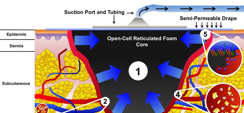
Dent. J. 2016, 4, 30                                                                                                          2 of 13
   Dent. J. 2016, 4, 30                                                                                                    2 of 12

viagrowth
     chemotaxis
            factor[17].    The second
                      (PDGF),             phase of wound
                                 which stimulates                healing,
                                                        recruitment         inflammation,
                                                                        of neutrophils,         starts with the
                                                                                            macrophages,            aggregation
                                                                                                                fibroblasts,  and of
neutrophils    at  the  site of  injury  within     the first 24   h of injury    [18].  The  neutrophils
   myofibroblasts via chemotaxis [17]. The second phase of wound healing, inflammation, starts with            work    to eliminate
foreign   material, of
   the aggregation      bacteria,   injured
                           neutrophils         cells,
                                          at the  site and   damaged
                                                       of injury    withinmatrix
                                                                            the firstcomponents
                                                                                       24 h of injurywithin
                                                                                                        [18]. The the  wound site
                                                                                                                     neutrophils
in work   to eliminate
    addition    to mast foreign     material,
                            cells and            bacteria,
                                        monocytes,          injured
                                                          while        cells, and damaged
                                                                    macrophages                  matrixto
                                                                                      are recruited       components
                                                                                                             the wound     within
                                                                                                                             site by
   the wound
T-cells  [19,20].site
                   Theinactivation
                           addition of to macrophages
                                           mast cells andleads monocytes,      while macrophages
                                                                     to the release     of PDGF and are        recruited togrowth
                                                                                                          transforming         the
   wound     site  by   T-cells  [19,20].  The   activation     of  macrophages       leads   to  the
factor-beta (TGF-β) [21]. The expression of TGF-β is critical in leading to the initiation of the thirdrelease    of  PDGF    and
   transforming
phase,               growth
         proliferation.     Infactor-beta    (TGF-β)phase,
                               the proliferation         [21]. The   expression
                                                                 collagen           of TGF-β
                                                                             is deposited     andis critical  in leading
                                                                                                    cross-linked           to the
                                                                                                                      by enzymes,
   initiation  of   the  third  phase,   proliferation.     In  the   proliferation     phase,   collagen
such as lysyl oxidase [22]. Additionally, angiogenesis initiates enabling the formation of granulation       is  deposited    and
   cross-linked    by  enzymes,    such   as lysyl   oxidase   [22]. Additionally,      angiogenesis
tissue [23,24]. The epidermis (or the intraoral mucosa) begins to re-epithelialize. The fibroblast      initiates   enabling   the is
primarily responsible for producing new extracellular matrix components such as collagens to
   formation     of   granulation     tissue    [23,24].   The     epidermis     (or   the  intraoral    mucosa)       begins    and
   re-epithelialize. The fibroblast is primarily responsible for producing new extracellular matrix
glycosaminoglycans (GAGs) to stabilize the structure and mechanical integrity of the tissue. In the
   components such as collagens and glycosaminoglycans (GAGs) to stabilize the structure and
final phase, remodeling, wound contracture and scar formation initiate. Excess cells undergo apoptosis,
   mechanical integrity of the tissue. In the final phase, remodeling, wound contracture and scar
while collagen fibrils, according to Clark et al. [25] orient with the initial alignment of fibrin fibers,
   formation initiate. Excess cells undergo apoptosis, while collagen fibrils, according to Clark et al. [25]
which may be influenced by mechanical tension acting directly on the wound site [26]. Myofibroblasts
   orient with the initial alignment of fibrin fibers, which may be influenced by mechanical tension
synthesize    alpha-smooth muscle actin (α-SMA), which enables collagen to contract and pull wound
   acting directly on the wound site [26]. Myofibroblasts synthesize alpha-smooth muscle actin
edges   together
   (α-SMA),     which[27].  Cellular
                         enables       debristoand
                                   collagen            compromised
                                                   contract                extracellular
                                                              and pull wound         edges matrix
                                                                                             togetherfibers
                                                                                                         [27]. are   destroyed
                                                                                                                 Cellular  debrisby
matrix   metalloproteinase        (MMPs)      produced      by   fibroblasts,   endothelial
   and compromised extracellular matrix fibers are destroyed by matrix metalloproteinase (MMPs)  cells, and    macrophages       [28].
Disruptions     in remodeling      where    excess    collagen    is deposited     can   lead  to hypertrophic
   produced by fibroblasts, endothelial cells, and macrophages [28]. Disruptions in remodeling where                 or keloid   scar
formation    [29–31].is deposited can lead to hypertrophic or keloid scar formation [29–31].
   excess collagen

          Figure 1. Phases of wound healing. The four phases of wound healing are illustrated. Immediately
      Figure    1. Phases of wound healing. The four phases of wound healing are illustrated. Immediately
          after a wound forms, hemostasis begins. Platelets form a clot, and are bound together by fibrin.
      after a wound forms, hemostasis begins. Platelets form a clot, and are bound together by fibrin.
          Several cytokines are released, which recruit neutrophils and other leukocytes to the site of injury to
      Several cytokines are released, which recruit neutrophils and other leukocytes to the site of injury
          start the inflammation phase. Leukocytes begin clearing the wound of bacteria, debris, and other
      to start the inflammation phase. Leukocytes begin clearing the wound of bacteria, debris, and other
          foreign contaminants. T-cells infiltrate the wound and recruit macrophages, which release PDGF and
      foreign contaminants. T-cells infiltrate the wound and recruit macrophages, which release PDGF and
          TGF-β to signal fibroblasts and myofibroblasts to start the proliferation phase. Granulation tissue
      TGF-β to signal fibroblasts and myofibroblasts to start the proliferation phase. Granulation tissue forms,
          forms, and fibroblasts begin developing new extracellular fibers by producing collagen and GAGs.
      and fibroblasts begin developing new extracellular fibers by producing collagen and GAGs. In addition
          In addition angiogenesis begins, and new blood is supplied to the site of injury. After proliferation,
      angiogenesis begins, and new blood is supplied to the site of injury. After proliferation, the final
          the final phase, remodeling, occurs in which extracellular fibers align, the wound contracts, and
      phase,    remodeling,
          fibroblasts  release occurs
                                enzymesin which  extracellular
                                          to remove   damaged fibers     align, extracellular
                                                                  extraneous    the wound contracts,     and fibroblasts
                                                                                              matrix. Epithelial  cells,
      release    enzymes   to remove    damaged    extraneous    extracellular   matrix.  Epithelial cells,
          despite their defining role in coverage, healing, and controlling “crosstalk” with fibroblasts,   despite  their
                                                                                                                  have
      defining    role in coverage,  healing, and  controlling   “crosstalk”   with fibroblasts, have
          been deliberately left out of this schematic representation for the sake of clarity. (PDGF =been   deliberately
      leftPlatelet-Derived
            out of this schematic
                             Growthrepresentation   for the sake
                                      Factor, GF = Growth         of clarity.
                                                             Factor, GAG = (PDGF     = Platelet-Derived Growth Factor,
                                                                              Glycosaminoglycan).
      GF = Growth Factor, GAG = Glycosaminoglycan).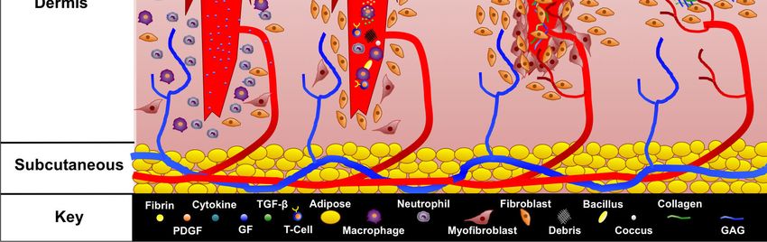
Dent. J. 2016, 4, 30 3 of 13
1.2. Wound Dressings
Since ancient times, wound dressings have been used, and have traditionally been made out of
cotton and more recently nylon gauze. Gauze dressings are hydrophilic, used as a wound contact
layer, and to remove exudate, and as a barrier to outside contamination. In 1962, Dr. George D. Winter
demonstrated in his seminal study that the use of a polyethylene film as a wound dressing, helped
keep the wound site moist, which reduced the time for re-epithelialization [32]. Dr. Winter’s study
emphasized the importance of keeping the wound site moist and spurred the development and
investigation of new materials, including custom polymers (e.g., polyvinyl alcohol (PVA), polyethylene
glycol (PEG), polyurethane (PUR), etc.), biological materials (e.g., collagens, carboxymethyl cellulose,
alginate, hyaluronic acid, etc.), and composite materials in the forms of woven-meshes and foams of
any imaginable pore size and shape, films, and hydrogels [33–36]. In addition, as the development and
construction of biomaterials has advanced for medical applications, dressings have been infused
with different antimicrobial agents, such as silver or iodine, and have been designed to work
in concert with topical therapies, negative pressure therapies, or instillation [37]. Li et al. [38]
reported that silver nanoparticles can be used to destroy the bacterial walls and prevent bacterial
replication, and do not exhibit any toxic effects on mammalian cells. Furthermore, technologies, such
as nanofiber electrospinning and precision particle fabrication, enable the ability to precisely load
different polymers with antimicrobial agents, growth factors, or vitamins to actively interact with the
wound environment [39–41]. Currently, construction and selection of dressings focus on maintaining a
moist wound environment, decreasing bacterial burden, and stimulating cell proliferation to aide the
wound healing progression [42–44].
1.3. Vacuum Assisted Closure
In 1997, building on advancements in wound dressings and drains, Drs. Argenta and Morykwas
introduced a groundbreaking device that applied a subatmospheric pressure through an open-cell
reticulated foam dressing to acute, sub-acute, and chronic wounds in both an animal and clinical
study [45,46]. The subatmospheric device used by Drs. Argenta and Morykwas dramatically
accelerated formation of granulation tissue. The dressing system kept wound environments moist
while reducing edema/exudate, promoting angiogenesis, and possibly reducing bacterial burden.
The dressing system was further developed, refined, and termed “Vacuum Assisted Closure (V.A.C.)”
by Kinetic Concepts Inc. (KCI, San Antonio, TX, USA), and distributed as the first NPWT by KCI.
The V.A.C. system has had a tremendous impact on the field of wound healing [47–49]. The V.A.C.
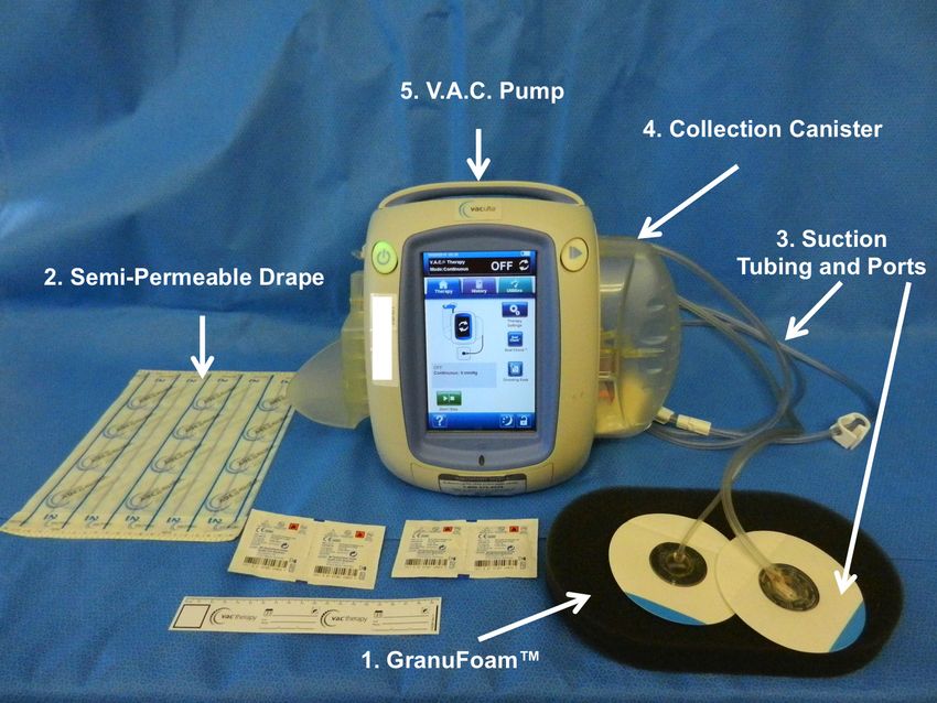
system is comprised of five basic components: (1) The open reticulated polyurethane ether foam
dressing (GranuFoam™); (2) semi-permeable adhesive film; (3) suction tubing; (4) collection canister;
and (5) vacuum pump (Figure 2). The GranuFoam™ is traditionally custom cut to fit the wound site
by the healthcare provider and placed within the wound so that GranuFoam™ completely interfaces
with the wound bed. The semi-permeable occlusive film drape is adhesively secured around and
over the wound site and the GranuFoam™ creating an airtight seal. A small opening is made
in the semi-permeable film drape, and the suction tubing and its port are placed at the opening.
The suction tubing is attached to the collection canister, which is fitted to the vacuum pump. When the
vacuum pump is activated, a suction differential of 75 to 125 mmHg is applied to the entire wound
surface. Under the vacuum, the GranuFoam™ compresses, which results in wound edges being pulled
closer together macroscopically while also causing microdeformations at the interface between the
GranuFoam™ and wound tissue, which stimulates the healing process [50]. The mechanical forces,
wound-dressing selection, and microenvironment interactions all contribute the success of V.A.C.,
and the primary mechanisms of action will be further expounded on to provide a context for increasing
the use of V.A.C. in maxillofacial applications.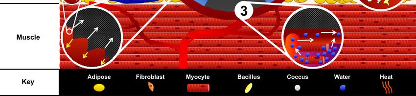
Dent. J. 2016, 4, 30                                                                                             4 of 13
   Dent. J. 2016, 4, 30                                                                                       4 of 12

        Figure 2. Vacuum Assisted Closure (V.A.C.) system. KCI Veraflow™ is the flagship model, and is
      Figure 2. Vacuum Assisted Closure (V.A.C.) system. KCI Veraflow™ is the flagship model, and is
        designed to provide instillation therapy in addition to standard V.A.C. therapy. The (1) black
      designed to provide instillation therapy in addition to standard V.A.C. therapy. The (1) black
        semi-occlusive dressing (GranuFoam™) along with the (2) semi-permeable drape used to isolate the
      semi-occlusive dressing (GranuFoam™) along with the (2) semi-permeable drape used to isolate
        wound and prevent escape of evaporative moisture. In addition, the (3) suction tubing and suction
      the wound and prevent escape of evaporative moisture. In addition, the (3) suction tubing and
        ports are connected to the (4) collection canister through which (5) the vacuum pump exerts suction
      suction ports are connected to the (4) collection canister through which (5) the vacuum pump exerts
        force.
      suction force.
   2. V.A.C. Mechanisms of Action
2. V.A.C. Mechanisms of Action
         The core events that occur during NPWT and affect the process of wound healing include: (1)
      The core events or
   macrodeformation       thatshrinkage
                                 occur during
                                           of theNPWT
                                                   wound;  and(2)affect  the process ofatwound
                                                                   microdeformation                  healing include:
                                                                                             the wound-dressing
(1)interface
    macrodeformation      or shrinkage
              exerting strain            of the wound;
                              on the extracellular        (2) microdeformation
                                                    components                     at the
                                                                    of the wound site;  (3)wound-dressing      interface
                                                                                            exudate fluid removal
exerting  strainreduction;
   and edema      on the extracellular  components
                            and (4) possible           of the
                                               reduction       wound site;
                                                           of infectious     (3) exudate
                                                                          material (Figurefluid  removal andthere
                                                                                            3). Additionally,    edema
reduction;  and (4)events
   are secondary     possible
                            thatreduction  of infectious
                                  occur that               materialthe
                                              aid in stabilizing     (Figure
                                                                        wound 3).environment
                                                                                  Additionally,andthere  are secondary
                                                                                                      accelerate  the
   wound
events      healing
         that  occur process,
                      that aid such  as mechanotransduction,
                                in stabilizing                     moisture control,
                                                the wound environment                  temperature
                                                                               and accelerate          stabilization,
                                                                                                 the wound      healing
   cell recruitment,
process,               and cell proliferation. Together,
          such as mechanotransduction,          moisture the    aforementioned
                                                           control,  temperature  events  affect the cell
                                                                                    stabilization,   inflammation,
                                                                                                          recruitment,
   proliferation,  and  remodeling   of the  wound   site as  healing  occurs.
and cell proliferation. Together, the aforementioned events affect the inflammation, proliferation,
and remodeling of the wound site as healing occurs.
   2.1. Macrostrain
2.1. Macrostrain
         Macrostrain or macrodeformation occurs when the TNPD shrinks in size, and the wound edges
   pull together. The
      Macrostrain   or GranuFoam™
                       macrodeformation is an interconnected
                                              occurs when the “mesh
                                                                 TNPD net”, containing
                                                                         shrinks       open
                                                                                 in size, andpores rangingedges
                                                                                              the wound     in
   size from  400 to  600 μm    in diameter  [51]. Scherer et al. [52] demonstrated   that GranuFoam™
pull together. The GranuFoam™ is an interconnected “mesh net”, containing open pores ranging               can
in shrink  by up
   size from  400toto80%
                      600 of
                          µm itsinoriginal
                                   diametervolume.  The deformation
                                              [51]. Scherer et al. [52]of the TNPD is directly
                                                                        demonstrated            related to thecan
                                                                                        that GranuFoam™
   volume of the material that is occupied by air. The pump evacuates the air, which enables shrinkage
shrink  by up to 80% of its original volume. The deformation of the TNPD is directly related to the
   of the TNPD. Thus, choosing an appropriate TNPD to interface with the wound bed is important for
volume of the material that is occupied by air. The pump evacuates the air, which enables shrinkage
   considerations of macrodeformation. However, not all tissues possess the same elasticity properties,
of the TNPD. Thus, choosing an appropriate TNPD to interface with the wound bed is important for
   which directly limits the degree to which wounds can contract. For example, a wound located on the
considerations of macrodeformation. However, not all tissues possess the same elasticity properties,
   head, such as the scalp, where the skin is already fixed to fascia, may not contract to the degree that a
which directly limits the degree to which wounds can contract. For example, a wound located on the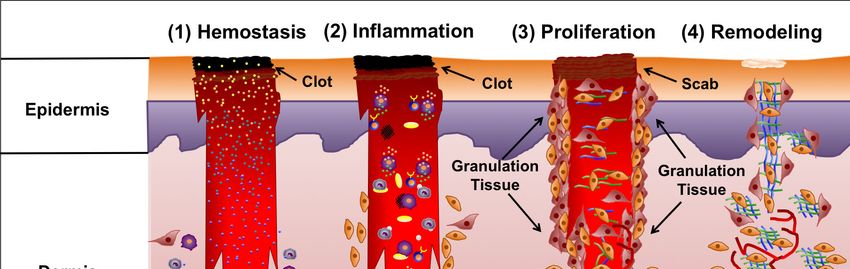
Dent. J. 2016, 4, 30 5 of 13
Dent. J. 2016, 4, 30 5 of 12
head, such as the scalp, where the skin is already fixed to fascia, may not contract to the degree that a
wound
wound on on the
theabdomen
abdomenwould wouldcontract
contractasasresult ofof
result thethe
different tensile
different andand
tensile elastic forces
elastic acting
forces on the
acting on
skin in both
the skin locations
in both [51,53].
locations [51,53].
In
In addition, while aa V.A.C.
addition, while V.A.C. system
system applies
applies subatmospheric
subatmospheric pressure
pressure toto aa wound
wound site,
site, the
the strain
strain
exerted
exerted at the wound surface itself may increases pressure in the underlying tissues proportional to
at the wound surface itself may increases pressure in the underlying tissues proportional to
the suction force. Kairinos et al. [54–56] reported the detection of increased atmospheric
the suction force. Kairinos et al. [54–56] reported the detection of increased atmospheric pressure in pressure
in surrounding
surrounding tissues
tissues in incircumferential
circumferentialnegative
negativepressure
pressurewound
wound therapy
therapy dressings;
dressings; however,
however,
Kairinos
Kairinos etet al.
al. [54–56]
[54–56] noted
noted thatthat the
the pressure
pressure gradually
gradually decreased
decreased over
over 48
48 hours
hours inin most
most wounds
wounds
analyzed.
analyzed. Hence, caution is advised in applying a V.A.C. system to a wound near or directly over
Hence, caution is advised in applying a V.A.C. system to a wound near or directly over
delicate
delicate vasculature
vasculature or or internal
internal organs,
organs, such
such as
as the
the dura/brain.
dura/brain.
Figure 3.
Figure 3. V.A.C.
V.A.C.mechanisms
mechanismsofofaction. action.An Anopen-cell
open-cell reticulated
reticulated foam foam is placed
is placed within
within the wound
the wound and
covered by a semi-permeable adhesive drape. The drape is adhesively secured around andand
and covered by a semi-permeable adhesive drape. The drape is adhesively secured around overover
the
the wound
wound to create
to create an airtight
an airtight seal. Aseal. A small
small hole ishole
madeis made
withinwithin
the center the ofcenter of the and
the drape, drape,the and the
suction
suction port and tubing connected to the collection canister are attached.
port and tubing connected to the collection canister are attached. Engaging the vacuum pump evacuates Engaging the vacuum
pump
the air evacuates
from the foam the airandfrom the foam
enables and enables (1) Macrodeformation
(1) Macrodeformation of the foam via shrinkage,of the foamwhichvia shrinkage,
pulls the
which pulls the wound edges together. At the interface between
wound edges together. At the interface between the foam and wound bed, (2) microstrain occurs the foam and wound bed, (2)
in
microstrain occurs in which cells are pulled into the pores of the foam while
which cells are pulled into the pores of the foam while an equal and opposing force acting on the struts an equal and opposing
force
of the acting
foam pusheson thecellsstruts
away.of The
the foam pushes
microstrain oncells away.
the cells The microstrain
initiates on the cells
mechanotransduction, initiates
which can
mechanotransduction,
stimulate cell proliferation which can stimulate
as illustrated cellthe
within proliferation
sub-inset ofasinset illustrated within the
2. Additionally, sub-inset of
engagement of
insetvacuum
the 2. Additionally,
facilitates theengagement
(3) movement of ofthefluid
vacuum
out of facilitates
interstitial thespaces,(3) thereby
movement of fluid
reducing edemaoutand
of
interstitial spaces, thereby reducing edema and increasing blood flow as
increasing blood flow as illustrated in inset 3. The V.A.C. system possibly (4) reduces bacterial burden;illustrated in inset 3. The
V.A.C. system
however, possibly (4)by
the mechanism reduces
which bacterial
bacteria areburden; however,
reduced is not the
fully mechanism
understood. by The
which bacteria are
destruction of
reduced is
bacteria is illustrated
not fully understood.
in inset 4. The TheV.A.C.
destruction
systemofcontributes
bacteria istoillustrated
(5) woundinstabilization
inset 4. Thethrough
V.A.C.
system contributes
secondary events. Insetto (5) wound stabilization
5 illustrates the movement through
of warm secondary
air downevents. through Inset
the 5semi-permeable
illustrates the
movement
drape into theof warm
woundair down
space, through
while the semi-permeable
isolating the wound from drape foreigninto the wound Furthermore,
contaminants. space, while
the semi-permeable drape prevents evaporative water loss, which aids in keeping the woundprevents
isolating the wound from foreign contaminants. Furthermore, the semi-permeable drape moist to
evaporative
enable water loss,
cell migration andwhich aidstransport.
nutrient in keeping the wound moist to enable cell migration and nutrient
transport.Dent. J. 2016, 4, 30 6 of 13
2.2. Microstrain
Microstrain is the force exerted directly on the cells via the extracellular matrix from the shrinkage
of the TNPD in the wound. The vacuum creates a microdeformation in which cells are pulled into the
pores of the foam while the struts provide an equal and opposing force that pushes cells away from the
foam, creating a quilting pattern at the wound interface [57]. Microdeformation is advantageous for the
proliferation phase of wound healing as the TNPD microdeformation acts on the extracellular matrix
initiating mechanosignaling within the fibroblasts, epithelial cells, and myofibroblasts to stimulate cell
proliferation, differentiation, angiogenesis, and neurogenesis [58]. Saxena and colleagues reported a
measured tissue strain on the surface of the wound of 5%–20% using finite element analysis [59].
2.3. Fluid Removal and Edema Reduction
Edema is an excess interstitial fluid trapped within tissue, which manifests as swelling. Edema can
be deleterious to the wound healing process, as the extracellular fluids can exert their own compressive
forces on microvasculature and cells. This can stall cell proliferation and reduce blood flow, as well as
increasing the diffusion distance for nutrients [60,61]. When the subatmospheric pressure is applied to
a porous TNPD, the excess fluid can be evacuated out. The reduction of edema and compressive burden
combined with the stimulation of mechanical signaling enables cells to proliferate [62]. Additionally,
the evacuation of fluid may reduce the toxins, cell debris, and disrupted bacteria from the wound site,
while increasing the efficiency of the volume of fluid drained by the lymphatic system by increasing
blood perfusion.
2.4. Reduction of Infectious Material
The reduction of infectious material within the wound site as a result of the application of V.A.C.
is controversial [63]. During the inflammation phase of wound healing, neutrophils, macrophages,
and lymphocytes work together to destroy foreign agents in the wound site to prevent excessive
invasion of pathogenic bacteria. However, if the bacterial burden is too high, and pathogenic bacteria
species invade the epithelium and disrupt the expression of MMPs and cytokines that promote
inflammation, effectively creating a continuous inflammation cycle [64]. The wound microenvironment
is dynamic and factors, such as moisture, pH, oxidation, and surface area, play a critical role in the
survival and invasion of pathogenic bacteria. Gram-positive bacteria, such as Staphylococcus aureus,
grow at an optimal pH of 6–7, while Pseudomonas aeruginosa and Enterococcus faecalis are capable of
growing at wider pH levels [65]. Wounds that contain foreign debris, eschar, or necrotic tissues are
at higher risk of bacterial infection as wound contaminates can provide attachment sites, nutrition,
or both for foreign bacteria [66]. Thus, cleaning the wound is important for limiting bacterial infection,
and aiding wound healing. Mertz et al. [67] reported a reduction in endogenous Gram-positive bacteria,
but not Gram-negative bacteria after 5 days when a semi-occlusive polyurethane film dressing was
used in partial thickness excisional wounds created on the back of pigs. In the original pig model
study published by Morykwas et al. [46] wound defects were inoculated with 108 organisms of a
human isolate of Staphylococcus aureus and a pig isolate of Staphylococcus epidermis. After 5 days of
NPWT, bacterial counts were reduced to fewer than 105 organisms per gram of tissue. However,
Mouës et al. [68] reported no significant differences in bacterial counts between patients that received
NPWT and patients that received standard therapy where a moist gauze dressing was changed twice
per day. Mouës et al. [68] have proposed that difference in bacterial counts may be a result of using a
biopsy sampling technique instead of superficial swabbing as was used in previous studies. The use
of GranuFoam™ impregnated with silver nanoparticles (GranuFoam Silver™) has been reported to
reduce bacterial load in chronic wounds [69]. Further examination of bacterial behavior and bacterial
quantification within wound sites is still required to fully understand if or how the NPWT is directly
reducing bacterial load.Dent. J. 2016, 4, 30 7 of 13
2.5. Wound Stabilization and Secondary Events
The removal of fluid and reduction of edema can act as a micro-debridement of the wound tissue,
which can be further enhanced by actual irrigation through a V.A.C. system. This allows cells to
re-establish homeostatic osmotic and oncotic gradients, in addition to establishing hypoxic conditions
that produce vascular endothelial growth factor (VEGF) gradients that direct angiogenesis [70].
Increased blood flow and perfusion allow for the critical supply of nutrients to stimulate cell
proliferation and extracellular matrix remodeling [71]. Furthermore, the semi-permeable occlusive
drape isolates the wound site, and prevents contamination as well as acting as a thermal insulator
to keep wound temperatures optimal for healing. The semi-permeable drape in combination with
a hydrophobic foam core additionally prevents evaporative water loss, which is critical for keeping
the wound moist for cell migration and transport of nutrients. The combination of macrostrain
and microstrain act on cell shape, and induce mechanotransduction that can affect cell behavior
and expression of critical signals that promote cell proliferation, extracellular matrix deposition,
and remodeling [59]. Nuutila et al. [72], were the first group to report increases in gene expression
of inflammation signals (Interleukin 8 (IL8), IL24) and tissue remodeling signals (MMP1, MMP3,
and MMP10) in a clinical study in which patients were treated with NPWT. In addition, to wound
stabilization some studies have reported a reduction in the wound closure duration. Arti et al. [73]
reported a 1.5 day reduction in the duration of hospital stay and 19% reduction in wound surface for
patients receiving NPWT for skin graft or flap coverage as opposed to patients receiving conventional
wound dressings. In a separate study, a 13.7 day reduction in the duration of wound closure for cats
with open wounds was reported [74]. Additional studies examining treatment of open-wounds with
NPWT against other conventional therapies could be beneficial in determining the effect of NPWT on
wound closure duration.
3. Maxillofacial Considerations
The head and neck provides many unique problems to maxillofacial surgeons in the setting
of wound healing and reconstruction. Function and aesthetics are often equally important to most
patients. Simultaneous management of both function and aesthetics has driven a large market for
products such as TNPDs used in NPWT.
Use of NPWT as a surgical adjuvant in maxillofacial surgery was first described in 2006 [75,76].
This paper retrospectively reviewed the use of NPWT in several complicated maxillofacial situations,
such as exposed calvarial bone, bolster dressing for large facial skin grafts, and wound management
following necrotizing fasciitis debridement. NPWT was successful in all such clinical situations and
this work led to other published reports that expanded its use.
Palm et al. [77] reviewed 1502 peer-reviewed journal articles on “vacuum therapy” for which
37 articles pertained to maxillofacial surgery. They noted that studies were generally limited by
containing case reports or case series and NPWT was used as an adjuvant maxillofacial reconstructive
procedures and management of soft tissue defects of the neck. However, published reports on NPWT
use are in all areas of maxillofacial reconstruction. The largest study by Satteson et al. [78] reviewed
69 patients with 73 head and neck wounds resulting from cancer (86%), trauma (8%), infection (3%),
or burns (3%) that used V.A.C. in conjunction with skin grafts, Integra, and open debrided wounds.
Minor complications were reported in 56% of patients that received skin grafts, 33% of patients that
received Integra, and 29% of patients with open debrided wounds. Most complications were resolved
with follow-up treatment.
3.1. Upper Third Maxillofacial Reconstruction
NPWT use for scalp and forehead reconstruction was first described by Andrews et al. [75]. Its use
for skin and soft tissue defects has been further described by Hsia et al. [79]. Both studies demonstrated
that NPWT could be used to temporize wounds in the setting of trauma and contamination or definitelyDent. J. 2016, 4, 30 8 of 13
treat wounds by promoting granulation tissue formation and wound contracture/epithelialization.
More aggressive protocols have been used to manage exposed dura and/or brain when the calvarium
is missing [80–82]. Ahmed et al. [80] discussed technical nuances to manage cerebrospinal fluid (CSF)
compartmentalization and eventual soft tissue coverage of dural repairs in their case report.
3.2. Middle Third Maxillofacial Reconstruction
Soft tissue defects of the cheeks and orbit are particularly suitable for NPWT. NPWT can be used
to promote granulation tissue formation on the facial skeleton so that skin grafts can be applied, for
wound management and contracture, or for skin graft bolstering [75]. Although these applications
seem straightforward concerns arise in the midface maintaining an occlusive seal of any negative
pressure device secondary to the midface contours and the presence of the eyes, nose, mouth and ears.
3.3. Lower Third Maxillofacial Reconstruction
NPWT use on the lower jaw and its soft tissue has been well described. Zhang et al. [83] described
its use in the management of submandibular fistulas after osteoradionecrosis reconstruction. In their
study NPWT was successful closing small submandibular fistulas that developed in nine patients in 7
to 12 days. The proximity of the oral cavity and often a tracheostomy make utilization of NPWT in this
region difficult.
3.4. Neck Reconstruction
Cutaneous-oral fistulas are a rare and difficult complication of maxillofacial surgery. TNPDs have
been shown to be an effective means of closing these intraoral communications [75]. Long track
fistulas with collapsible non-radiated tissues have been shown to be most amenable to this mode of
closure. Yang et al. [84] used TNPDs to successfully close eight salivary fistulas with an average time
of treatment being 10.8 days. Tian et al. [85] further advanced the utilization of TNPD for salivary
fistula closure by employing dental paste intraorally to maintain an occlusive seal for the negative
pressure system. In their series, they successfully close salivary fistulas in 9 of 10 patients.
3.5. Technical Considerations
Establishing an occlusive environment and maintaining a negative pressure seal is one significant
obstacle in NPWT head and neck utilization. Occlusive dressings that avoid important functional
structures such as the eyes, ears, nose, and mouth should be employed. If necessary these structures can
be temporarily covered; however, they should be protected and direct negative pressure application
minimized to these structures (especially the eyes). Although NPWT has been used on exposed
brain/dura caution should be used in this application. A watertight CSF closure should be ensured
prior to its use as to not immediately empty the CSF reserves and cause herniation. Frequent dressing
changes in NPWT are needed in the head and neck and are often done daily at our institution
to maintain the occlusive seal. Currently, TNPDs for NPWT are manufactured as flat elliptical
foam sponges, most conducive for treating wounds located on the abdomen or limbs. Interestingly,
healthcare providers have successfully used NPWT and TNPDs to treat diabetic foot ulcers, which
may provide insights for using NPWT on wounds occurring on the head and neck. The ability to make
a more malleable or shapeable foam to fit the contours of the head and neck, will allow significant
opportunities to more easily utilize NPWT in maxillofacial applications.
4. Conclusions
NPWT accelerates wound healing by enhancing the inflammation, proliferation, and remodeling
phases of wound healing. The pressure differential facilitates movement of fluid and reduces edema
while increasing blood flow, which aids in lymphatic drainage and clearing of the wound to reduce
inflammation. Simultaneously, the macrostrain and microstrain initiate mechanotransduction, whichDent. J. 2016, 4, 30 9 of 13
stimulates fibroblasts, myofibroblasts, and epithelia cells to proliferate while building new extracellular
matrix and releasing enzymes to remodel and process the developing support matrix. In conjunction,
VEGF gradients are established as a result of hypoxia, and direct angiogenesis, which further increases
blood perfusion, and contributes to repairing the original tissue. Healthcare providers have a
well-established history of treating acute and chronic wounds occurring on the torso and limbs
with NPWT. However, while NPWT has been shown to be safely and effectively used in complicated
maxillofacial wounds, NPWT is under utilized in this due to the limitations in maintaining a negative
pressure seal when TNPDs are applied to the head and neck. Further work remains to develop next
generation TNPDs to fit the contours of the head and neck. These technologies have the potential to
increase the use of NPWT in maxillofacial applications, thereby benefitting patients with a wider range
of injuries.
Acknowledgments: Funding provided by the Department of Plastic Surgery at the University of Kansas Medical
Center. We would like to thank Rachel Vukas, for her assistance in helping the authors search and access literature.
Author Contributions: The scope and theme of the review was conceived by B.T.A. The manuscript was written
by A.J.M. and B.T.A. D.S.Z. was consulted on the technical content of the manuscript. The manuscript was
reviewed by A.J.M., D.S.Z., and B.T.A.
Conflicts of Interest: D.S.Z. declares that he has sold patents to the V.A.C. to KCI/Acelity and continues to receive
royalties on Prevena (KCI/Acelity). A.J.M. and B.T.A. declare no conflicts of interest.
References
1. Banwell, P.; Withey, S.; Holten, I. The use of negative pressure to promote healing. Br. J. Plast. Surg. 1998,
51, 79. [CrossRef]
2. Orgill, D.P.; Bayer, L.R. Update on negative-pressure wound therapy. Plast. Reconstr. Surg. 2011, 127
(Suppl. 1), 105S–115S. [CrossRef] [PubMed]
3. Asher, S.A.; White, H.N.; Golden, J.B.; Magnuson, J.S.; Carroll, W.R.; Rosenthal, E.L. Negative pressure
wound therapy in head and neck surgery. JAMA Facial Plast. Surg. 2014, 16, 120–126. [CrossRef] [PubMed]
4. Armstrong, D.G.; Andros, G. Use of negative pressure wound therapy to help facilitate limb preservation.
Int. Wound J. 2012, 9 (Suppl. 1), 1–7. [CrossRef] [PubMed]
5. Dumville, J.C.; Land, L.; Evans, D.; Peinemann, F. Negative pressure wound therapy for treating leg ulcers.
Cochrane Database Syst. Rev. 2015, 7, CD011354. [PubMed]
6. Evans, D.; Land, L. Topical negative pressure for treating chronic wounds: A systematic review. Br. J.
Plast. Surg. 2001, 54, 238–242. [CrossRef] [PubMed]
7. Stannard, J.P.; Gabriel, A.; Lehner, B. Use of negative pressure wound therapy over clean, closed surgical
incisions. Int. Wound J. 2012, 9 (Suppl. 1), 32–39. [CrossRef] [PubMed]
8. Poglio, G.; Grivetto, F.; Nicolotti, M.; Arcuri, F.; Benech, A. Management of an exposed mandibular plate
after fibula free flap with vacuum-assisted closure system. J. Craniofac. Surg. 2011, 22, 905–908. [CrossRef]
[PubMed]
9. Strub, G.M.; Moe, K.S. The use of negative-pressure therapy in the closure of complex head and neck wounds.
Facial Plast. Surg. Clin. N. Am. 2013, 21, 137–145. [CrossRef] [PubMed]
10. Yoon, B.W.; Yi, K.I.; Kang, J.H.; Kim, S.G.; Cha, W. Negative pressure wound therapy for cervical esophageal
perforation with abscess. Auris Nasus Larynx 2015, 42, 254–257. [CrossRef] [PubMed]
11. Agha, R.; Ogawa, R.; Pietramaggiori, G.; Orgill, D.P. A review of the role of mechanical forces in cutaneous
wound healing. J. Surg. Res. 2011, 171, 700–708. [CrossRef] [PubMed]
12. Pereira, R.F.; Bartolo, P.J. Traditional therapies for skin wound healing. Adv. Wound Care (New Rochelle) 2016,
5, 208–229. [CrossRef] [PubMed]
13. Rittie, L. Cellular mechanisms of skin repair in humans and other mammals. J. Cell Commun. Signal. 2016, 10,
103–120. [CrossRef] [PubMed]
14. Silver, I.A. The mechanics of wound healing. Equine Vet. J. 1979, 11, 93–96. [CrossRef] [PubMed]
15. Valero, C.; Javierre, E.; Garcia-Aznar, J.M.; Menzel, A.; Gomez-Benito, M.J. Challenges in the modeling of
wound healing mechanisms in soft biological tissues. Ann. Biomed. Eng. 2015, 43, 1654–1665. [CrossRef]
[PubMed]Dent. J. 2016, 4, 30 10 of 13
16. Broughton, G., 2nd; Janis, J.E.; Attinger, C.E. The basic science of wound healing. Plast. Reconstr. Surg. 2006,
117, 12S–34S. [CrossRef] [PubMed]
17. Velnar, T.; Bailey, T.; Smrkolj, V. The wound healing process: An overview of the cellular and molecular
mechanisms. J. Int. Med. Res. 2009, 37, 1528–1542. [CrossRef] [PubMed]
18. Galli, S.J.; Borregaard, N.; Wynn, T.A. Phenotypic and functional plasticity of cells of innate immunity:
Macrophages, mast cells and neutrophils. Nat. Immunol. 2011, 12, 1035–1044. [CrossRef] [PubMed]
19. Jameson, J.; Havran, W.L. Skin gammadelta T-cell functions in homeostasis and wound healing. Immunol. Rev.
2007, 215, 114–122. [CrossRef] [PubMed]
20. Wynn, T.A.; Vannella, K.M. Macrophages in tissue repair, regeneration, and fibrosis. Immunity 2016, 44,
450–462. [CrossRef] [PubMed]
21. Lech, M.; Anders, H.J. Macrophages and fibrosis: How resident and infiltrating mononuclear phagocytes
orchestrate all phases of tissue injury and repair. Biochim. Biophys. Acta 2013, 1832, 989–997. [CrossRef]
[PubMed]
22. Diegelmann, R.F.; Evans, M.C. Wound healing: An overview of acute, fibrotic and delayed healing.
Front. Biosci. 2004, 9, 283–289. [CrossRef] [PubMed]
23. Pietramaggiori, G.; Liu, P.; Scherer, S.S.; Kaipainen, A.; Prsa, M.J.; Mayer, H.; Newalder, J.; Alperovich, M.;
Mentzer, S.J.; Konerding, M.A.; et al. Tensile forces stimulate vascular remodeling and epidermal cell
proliferation in living skin. Ann. Surg. 2007, 246, 896–902. [CrossRef] [PubMed]
24. Tomasek, J.J.; Gabbiani, G.; Hinz, B.; Chaponnier, C.; Brown, R.A. Myofibroblasts and mechano-regulation of
connective tissue remodelling. Nat. Rev. Mol. Cell Biol. 2002, 3, 349–363. [CrossRef] [PubMed]
25. Clark, R.A. Cutaneous tissue repair: Basic biologic considerations. I. J. Am. Acad. Dermatol. 1985, 13, 701–725.
[CrossRef]
26. Yamaguchi, Y.; Yoshikawa, K. Cutaneous wound healing: An update. J. Dermatol. 2001, 28, 521–534.
[PubMed]
27. Bochaton-Piallat, M.L.; Gabbiani, G.; Hinz, B. The myofibroblast in wound healing and fibrosis: Answered
and unanswered questions. F1000 Res. 2016, 5. [CrossRef] [PubMed]
28. Pennacchi, P.C.; Almeida, M.E.; Gomes, O.L.; Faiao-Flores, F.; Crepaldi, M.C.; Dos Santos, M.F.; Barros, S.B.;
Maria-Engler, S.S. Glycated reconstructed human skin as a platform to study the pathogenesis of skin aging.
Tissue Eng. Part A 2015, 21, 2417–2425. [CrossRef] [PubMed]
29. Ding, J.; Tredget, E.E. The role of chemokines in fibrotic wound healing. Adv. Wound Care (New Rochelle)
2015, 4, 673–686. [CrossRef] [PubMed]
30. Ogawa, R.; Akaishi, S.; Kuribayashi, S.; Miyashita, T. Keloids and hypertrophic scars can now be cured
completely: Recent progress in our understanding of the pathogenesis of keloids and hypertrophic scars and
the most promising current therapeutic strategy. J. Nippon Med. Sch. 2016, 83, 46–53. [CrossRef] [PubMed]
31. Trace, A.P.; Enos, C.W.; Mantel, A.; Harvey, V.M. Keloids and hypertrophic scars: A spectrum of clinical
challenges. Am. J. Clin. Dermatol. 2016, 17, 201–223. [CrossRef] [PubMed]
32. Winter, G.D. Formation of the scab and the rate of epithelization of superficial wounds in the skin of the
young domestic pig. Nature 1962, 193, 293–294. [CrossRef] [PubMed]
33. Dabiri, G.; Damstetter, E.; Phillips, T. Choosing a wound dressing based on common wound characteristics.
Adv. Wound Care (New Rochelle) 2016, 5, 32–41. [CrossRef] [PubMed]
34. Gould, L.J. Topical collagen-based biomaterials for chronic wounds: Rationale and clinical application.
Adv. Wound Care (New Rochelle) 2016, 5, 19–31. [CrossRef] [PubMed]
35. Maessen-Visch, M.B.; van Montfrans, C. Wound dressings, does it matter and why? Phlebology 2016, 31,
63–67. [CrossRef] [PubMed]
36. Mayet, N.; Choonara, Y.E.; Kumar, P.; Tomar, L.K.; Tyagi, C.; Du Toit, L.C.; Pillay, V. A comprehensive review
of advanced biopolymeric wound healing systems. J. Pharm. Sci. 2014, 103, 2211–2230. [CrossRef] [PubMed]
37. Ovington, L.G. Advances in wound dressings. Clin. Dermatol. 2007, 25, 33–38. [CrossRef] [PubMed]
38. Li, W.R.; Xie, X.B.; Shi, Q.S.; Duan, S.S.; Ouyang, Y.S.; Chen, Y.B. Antibacterial effect of silver nanoparticles
on staphylococcus aureus. Biometals 2011, 24, 135–141. [CrossRef] [PubMed]
39. Abrigo, M.; McArthur, S.L.; Kingshott, P. Electrospun nanofibers as dressings for chronic wound care:
Advances, challenges, and future prospects. Macromol. Biosci. 2014, 14, 772–792. [CrossRef] [PubMed]
40. Berkland, C.; Kim, K.; Pack, D.W. Fabrication of PLG microspheres with precisely controlled and
monodisperse size distributions. J. Control. Release 2001, 73, 59–74. [CrossRef]Dent. J. 2016, 4, 30 11 of 13
41. Berkland, C.; Kim, K.; Pack, D.W. PLG microsphere size controls drug release rate through several competing
factors. Pharm. Res. 2003, 20, 1055–1062. [CrossRef] [PubMed]
42. Percival, S.L.; McCarty, S.M. Silver and alginates: Role in wound healing and biofilm control. Adv. Wound
Care (New Rochelle) 2015, 4, 407–414. [CrossRef] [PubMed]
43. Salamone, J.C.; Salamone, A.B.; Swindle-Reilly, K.; Leung, K.X.; McMahon, R.E. Grand challenge in
biomaterials-wound healing. Regen. Biomater. 2016, 3, 127–128. [CrossRef] [PubMed]
44. Tartarini, D.; Mele, E. Adult stem cell therapies for wound healing: Biomaterials and computational models.
Front. Bioeng. Biotechnol. 2015, 3, 206. [CrossRef] [PubMed]
45. Argenta, L.C.; Morykwas, M.J. Vacuum-assisted closure: A new method for wound control and treatment:
Clinical experience. Ann. Plast. Surg. 1997, 38, 563–576. [CrossRef] [PubMed]
46. Morykwas, M.J.; Argenta, L.C.; Shelton-Brown, E.I.; McGuirt, W. Vacuum-assisted closure: A new method
for wound control and treatment: Animal studies and basic foundation. Ann. Plast. Surg. 1997, 38, 553–562.
[CrossRef] [PubMed]
47. Gupta, S. Optimal use of negative pressure wound therapy for skin grafts. Int. Wound J. 2012, 9 (Suppl. 1),
40–47. [CrossRef] [PubMed]
48. Mendez-Eastman, S. Negative pressure wound therapy. Plast. Surg. Nurs. 1998, 18, 27–37. [CrossRef]
[PubMed]
49. Orgill, D.P.; Bayer, L.R. Negative pressure wound therapy: Past, present and future. Int. Wound J. 2013, 10
(Suppl. 1), 15–19. [CrossRef] [PubMed]
50. Wood, B.C.; Molnar, J.A. Subatmospheric pressure therapy: Basic science review. J. Surg. Orthop. Adv. 2011,
20, 168–175. [PubMed]
51. Huang, C.; Leavitt, T.; Bayer, L.R.; Orgill, D.P. Effect of negative pressure wound therapy on wound healing.
Curr. Probl. Surg. 2014, 51, 301–331. [CrossRef] [PubMed]
52. Scherer, S.S.; Pietramaggiori, G.; Mathews, J.C.; Prsa, M.J.; Huang, S.; Orgill, D.P. The mechanism of action of
the vacuum-assisted closure device. Plast. Reconstr. Surg. 2008, 122, 786–797. [CrossRef] [PubMed]
53. Subotic, U.; Kluwe, W.; Oesch, V. Community-associated methicillin-resistant staphylococcus aureus-infected
chronic scalp wound with exposed dura in a 10-year-old boy: Vacuum-assisted closure is a feasible option:
Case report. Neurosurgery 2011, 68, E1481–E1483. [CrossRef] [PubMed]
54. Kairinos, N.; Solomons, M.; Hudson, D.A. Negative-pressure wound therapy I: The paradox of
negative-pressure wound therapy. Plast. Reconstr. Surg. 2009, 123, 589–598. [CrossRef] [PubMed]
55. Kairinos, N.; Solomons, M.; Hudson, D.A. The paradox of negative pressure wound therapy–in vitro studies.
J. Plast. Reconstr. Aesthet. Surg. 2010, 63, 174–179. [CrossRef] [PubMed]
56. Kairinos, N.; Voogd, A.M.; Botha, P.H.; Kotze, T.; Kahn, D.; Hudson, D.A.; Solomons, M. Negative-pressure
wound therapy II: Negative-pressure wound therapy and increased perfusion. Just an illusion? Plast. Reconstr.
Surg. 2009, 123, 601–612. [CrossRef] [PubMed]
57. Lancerotto, L.; Bayer, L.R.; Orgill, D.P. Mechanisms of action of microdeformational wound therapy.
Semin. Cell Dev. Biol. 2012, 23, 987–992. [CrossRef] [PubMed]
58. Huang, C.; Holfeld, J.; Schaden, W.; Orgill, D.; Ogawa, R. Mechanotherapy: Revisiting physical therapy
and recruiting mechanobiology for a new era in medicine. Trends Mol. Med. 2013, 19, 555–564. [CrossRef]
[PubMed]
59. Saxena, V.; Hwang, C.W.; Huang, S.; Eichbaum, Q.; Ingber, D.; Orgill, D.P. Vacuum-assisted closure:
Microdeformations of wounds and cell proliferation. Plast. Reconstr. Surg. 2004, 114, 1086–1096. [CrossRef]
[PubMed]
60. Hanson, D.; Langemo, D.; Thompson, P.; Anderson, J.; Hunter, S. Understanding wound fluid and the phases
of healing. Adv. Skin Wound Care 2005, 18, 360–362. [CrossRef] [PubMed]
61. Kadohama, T.; Nishimura, K.; Hoshino, Y.; Sasajima, T.; Sumpio, B.E. Effects of different types of fluid shear
stress on endothelial cell proliferation and survival. J. Cell. Physiol. 2007, 212, 244–251. [CrossRef] [PubMed]
62. DeFranzo, A.J.; Argenta, L.C.; Marks, M.W.; Molnar, J.A.; David, L.R.; Webb, L.X.; Ward, W.G.; Teasdall, R.G.
The use of vacuum-assisted closure therapy for the treatment of lower-extremity wounds with exposed bone.
Plast. Reconstr. Surg. 2001, 108, 1184–1191. [CrossRef] [PubMed]
63. Weed, T.; Ratliff, C.; Drake, D.B. Quantifying bacterial bioburden during negative pressure wound therapy:
Does the wound VAC enhance bacterial clearance? Ann. Plast. Surg. 2004, 52, 276–279. [CrossRef] [PubMed]Dent. J. 2016, 4, 30 12 of 13
64. Scalise, A.; Bianchi, A.; Tartaglione, C.; Bolletta, E.; Pierangeli, M.; Torresetti, M.; Marazzi, M.; di Benedetto, G.
Microenvironment and microbiology of skin wounds: The role of bacterial biofilms and related factors.
Semin. Vasc. Surg. 2015, 28, 151–159. [CrossRef] [PubMed]
65. Daeschlein, G. Antimicrobial and antiseptic strategies in wound management. Int. Wound J. 2013, 10
(Suppl. 1), 9–14. [CrossRef] [PubMed]
66. Reed, B.R.; Clark, R.A. Cutaneous tissue repair: Practical implications of current knowledge. II. J. Am.
Acad. Dermatol. 1985, 13, 919–941. [CrossRef]
67. Mertz, P.M.; Eaglstein, W.H. The effect of a semiocclusive dressing on the microbial population in superficial
wounds. Arch. Surg. 1984, 119, 287–289. [CrossRef] [PubMed]
68. Moues, C.M.; Vos, M.C.; van den Bemd, G.J.; Stijnen, T.; Hovius, S.E. Bacterial load in relation to
vacuum-assisted closure wound therapy: A prospective randomized trial. Wound Repair Regen. 2004,
12, 11–17. [CrossRef] [PubMed]
69. Sachsenmaier, S.; Peschel, A.; Ipach, I.; Kluba, T. Antibacterial potency of V.A.C. Granufoam silver(® )
dressing. Injury 2013, 44, 1363–1367. [CrossRef] [PubMed]
70. Potter, M.J.; Banwell, P.; Baldwin, C.; Clayton, E.; Irvine, L.; Linge, C.; Grobbelaar, A.O.; Sanders, R.;
Dye, J.F. In vitro optimisation of topical negative pressure regimens for angiogenesis into synthetic dermal
replacements. Burns 2008, 34, 164–174. [CrossRef] [PubMed]
71. Petzina, R.; Gustafsson, L.; Mokhtari, A.; Ingemansson, R.; Malmsjo, M. Effect of vacuum-assisted
closure on blood flow in the peristernal thoracic wall after internal mammary artery harvesting. Eur. J.
Cardiothoracic. Surg. 2006, 30, 85–89. [CrossRef] [PubMed]
72. Nuutila, K.; Siltanen, A.; Peura, M.; Harjula, A.; Nieminen, T.; Vuola, J.; Kankuri, E.; Aarnio, P. Gene
expression profiling of negative-pressure-treated skin graft donor site wounds. Burns 2013, 39, 687–693.
[CrossRef] [PubMed]
73. Arti, H.; Khorami, M.; Ebrahimi-Nejad, V. Comparison of negative pressure wound therapy (NPWT) &
conventional wound dressings in the open fracture wounds. Pak. J. Med. Sci. 2016, 32, 65–69. [PubMed]
74. Nolff, M.C.; Fehr, M.; Reese, S.; Meyer-Lindenberg, A.E. Retrospective comparison of negative-pressure
wound therapy and silver-coated foam dressings in open-wound treatment in cats. J. Feline Med. Surg. 2016.
[CrossRef] [PubMed]
75. Andrews, B.T.; Smith, R.B.; Goldstein, D.P.; Funk, G.F. Management of complicated head and neck wounds
with vacuum-assisted closure system. Head Neck 2006, 28, 974–981. [CrossRef] [PubMed]
76. Schuster, R.; Moradzadeh, A.; Waxman, K. The use of vacuum-assisted closure therapy for the treatment of a
large infected facial wound. Am. Surg. 2006, 72, 129–131. [PubMed]
77. Palm, H.G.; Hauer, T.; Simon, C.; Willy, C. Vacuum-assisted closure of head and neck wounds. HNO 2011,
59, 819–830. [CrossRef] [PubMed]
78. Satteson, E.S.; Crantford, J.C.; Wood, J.; David, L.R. Outcomes of vacuum-assisted therapy in the treatment
of head and neck wounds. J. Craniofac. Surg. 2015, 26, e599–e602. [CrossRef] [PubMed]
79. Hsia, J.C.; Moe, K.S. Vacuum-assisted closure therapy for reconstruction of soft-tissue forehead defects.
Arch. Facial Plast. Surg. 2011, 13, 278–282. [CrossRef] [PubMed]
80. Ahmed, F.; Swan, M.C.; Flynn, M.; Tiernan, E.P. Retained vac therapy sponge as a complication of
abdominoplasty. J. Plast. Reconstr. Aesthet. Surg. 2010, 63, e497–e499. [CrossRef] [PubMed]
81. Powers, A.K.; Neal, M.T.; Argenta, L.C.; Wilson, J.A.; DeFranzo, A.J.; Tatter, S.B. Vacuum-assisted closure
for complex cranial wounds involving the loss of dura mater. J. Neurosurg. 2013, 118, 302–308. [CrossRef]
[PubMed]
82. Prince, N.; Blackburn, S.; Murad, G.; Mast, B.; Sapountzis, S.; Shaw, C.; Werning, J.; Singhal, D.
Vacuum-assisted closure therapy to the brain: A safe method for wound temporization in composite
scalp and calvarial defects. Ann. Plast. Surg. 2015, 74 (Suppl. 4), S218–S221. [CrossRef] [PubMed]
83. Zhang, D.M.; Yang, Z.H.; Zhuang, P.L.; Wang, Y.Y.; Chen, W.L.; Zhang, B. Role of negative-pressure wound
therapy in the management of submandibular fistula after reconstruction for osteoradionecrosis. J. Oral
Maxillofac. Surg. 2016, 74, 401–405. [CrossRef] [PubMed]Dent. J. 2016, 4, 30 13 of 13
84. Yang, Y.H.; Jeng, S.F.; Hsieh, C.H.; Feng, G.M.; Chen, C.C. Vacuum-assisted closure for complicated wounds
in head and neck region after reconstruction. J. Plast. Reconstr. Aesthet. Surg. 2013, 66, e209–e216. [CrossRef]
[PubMed]
85. Tian, B.; Khoo, D.; Tay, A.C.; Soo, K.C.; Tan, N.C.; Tan, H.K.; Iyer, N.G. Management of orocutaneous fistulas
using a vacuum-assisted closure system. Head Neck 2014, 36, 873–881. [CrossRef] [PubMed]
© 2016 by the authors; licensee MDPI, Basel, Switzerland. This article is an open access
article distributed under the terms and conditions of the Creative Commons Attribution
(CC-BY) license (http://creativecommons.org/licenses/by/4.0/).You can also read