SINGLE-DOSE SARS-COV-2 VACCINATIONS WITH EITHER BNT162B2 OR AZD1222 INDUCE DISPARATE TH1 RESPONSES AND IGA PRODUCTION
←
→
Page content transcription
If your browser does not render page correctly, please read the page content below
Müller et al. BMC Medicine     (2022) 20:29
https://doi.org/10.1186/s12916-022-02240-4

 RESEARCH ARTICLE                                                                                                                                     Open Access

Single-dose SARS-CoV-2 vaccinations with
either BNT162b2 or AZD1222 induce
disparate Th1 responses and IgA
production
Michael Müller1†, Johann Volzke1*† , Behnam Subin2, Silke Müller3, Martina Sombetzki4, Emil C. Reisinger4 and
Brigitte Müller-Hilke1

  Abstract
  Background: While vaccination programs against the severe acute respiratory syndrome virus 2 (SARS-CoV-2) are
  globally ongoing, disparate strategies for the deployment of spike antigen show varying effectiveness.
  Methods: In order to explore this phenomenon, we sought to compare the early immune responses against
  AZD1222 and BNT162b2. SARS-CoV-2 seronegative participants received a single dose of either vaccine and were
  analyzed for immune cell, effector T cell, and antibody dynamics.
  Results: AZD1222 induced transient leukopenia and major changes among innate and adaptive subpopulations.
  Both vaccines induced spike protein-specific effector T cells which were dominated by type 1 helper T cell
  responses following AZD1222 vaccination. A significant reduction of anti-inflammatory T cells upon re-stimulation
  was also restricted to AZD1222 vaccinees. While IgM and IgG were the dominant isotypes elicited by AZD1222,
  BNT162b2 led to a significant production of IgG and IgA.
  Conclusions: Our results suggest that the strategy for spike protein delivery impacts on how and to what extent
  immune priming against the main SARS-CoV-2 antigen proceeds.
  Keywords: SARS-CoV-2, Vaccination, AZD1222, BNT162b2, COVID-19, Type 1 helper T cells, Cytotoxic T cells

Background                                                                               several vaccines. These collectively aim at the production
The highly transmissible severe acute respiratory                                        of antibodies that will neutralize the binding of the viral
syndrome coronavirus 2 (SARS-CoV-2), which initially                                     spike glycoprotein to its angiotensin-converting enzyme
emerged in December 2019, has led to an unprecedented                                    2 (ACE2) receptor and thereby prevent cellular entry
pandemic that caused over 4 million casualties [1, 2].                                   and subsequent infection [3–5].
The prevailing occurrence of coronavirus disease 2019                                      The urgent need to develop safe and efficient vaccines
(COVID-19) and its dramatic hazard for global health                                     led to the deployment of various strategies, some of
and economy has since spiked the rapid development of                                    which were well established and others, like adenoviral
                                                                                         vectors or mRNA, were novel. Among the early vaccines
                                                                                         authorized by the European Medicines Agency (EMA)
* Correspondence: johann.volzke@med.uni-rostock.de
†
 Michael Müller and Johann Volzke contributed equally to this work.                      were the first-generation adenoviral vector AZD1222
1
 Core Facility for Cell Sorting and Cell Analysis, Rostock University Medical            that utilizes the simian dsDNA adenovirus ChAdOx1 as
Center, Rostock, Germany                                                                 a vector for antigen delivery [6]. The first vaccine
Full list of author information is available at the end of the article

                                          © The Author(s). 2022 Open Access This article is licensed under a Creative Commons Attribution 4.0 International License,
                                          which permits use, sharing, adaptation, distribution and reproduction in any medium or format, as long as you give
                                          appropriate credit to the original author(s) and the source, provide a link to the Creative Commons licence, and indicate if
                                          changes were made. The images or other third party material in this article are included in the article's Creative Commons
                                          licence, unless indicated otherwise in a credit line to the material. If material is not included in the article's Creative Commons
                                          licence and your intended use is not permitted by statutory regulation or exceeds the permitted use, you will need to obtain
                                          permission directly from the copyright holder. To view a copy of this licence, visit http://creativecommons.org/licenses/by/4.0/.
                                          The Creative Commons Public Domain Dedication waiver (http://creativecommons.org/publicdomain/zero/1.0/) applies to the
                                          data made available in this article, unless otherwise stated in a credit line to the data.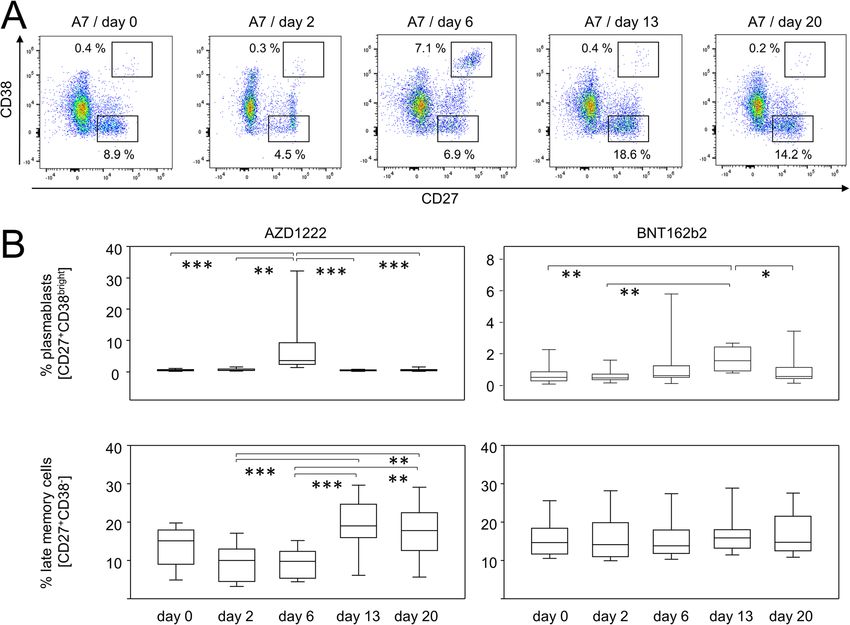
Müller et al. BMC Medicine           (2022) 20:29                                                                                Page 2 of 14

authorized by EMA was the nucleic acid-based                                     distribution of sex and comparable age ranges between
BNT162b2, a spike protein encoding N1-methyl-                                    both vaccination groups.
pseudouridine (m1Ψ) nucleoside-modified mRNA envel-                                 In order to delineate the early immune cell responses
oped by lipid nanoparticles [7, 8].                                              to both vaccines, we performed 24-dimensional flow cy-
   Complete vaccination with either of the vaccines, which                       tometry at each time point. Remarkably, by examining
includes two doses at varying intervals, was shown to                            major immune cell compositions, we found in samples
efficiently protect from symptomatic COVID-19 [9, 10].                           that were available for all consecutive time points a sig-
Although early data hint at similar efficiencies after a                         nificant reduction (2.3-fold) in peripheral leukocytes on
single dose of either vaccine, booster immunization with                         day 2 after vaccination with AZD1222 (Fig. 1A). This
BNT162b2 achieved somewhat higher rates of thwarting                             leukopenia resulted from significant reductions in granu-
viral breakthrough [9–13]. With the emergence of SARS-                           locytes and B-lymphocytes as well as CD4- and CD8-
CoV-2 variants that accumulate mutations in the spike                            positive T cells (see Additional file 1: Table S1). When
glycoprotein [14–16], the discrepancies between both                             compared to baseline, leukocyte counts were still slightly
vaccines grew even more pronounced with BNT162b2                                 reduced on day 6 yet back to normal on days 13 and 20.
leading to superior protection against the 1.351 (β) and                         In contrast, vaccination with BNT162b2 did not result
1.617.2 (δ) variants [15, 17–19].                                                in any significant fluctuations among immune cell quan-
   We were curious about the molecular and cellular                              tities (Additional file 1: Table S2).
immune modules capable of mediating superior                                        In order to survey qualitative alterations in the im-
neutralization of SARS-CoV-2 and therefore aimed at                              mune responses to either vaccine, we performed dimen-
exploring the immediate immune dynamics after a single                           sion reductions on our multiparametric data set by using
dose of either AZD1222 or BNT162b2. To that extent,                              the embedding algorithm “uniform manifold approxima-
we investigated the proportions of peripheral leukocytes                         tion and projection” (UMAP). Figure 1B summarizes all
among innate and adaptive compartments over the first                            data from all time points and shows the topological dis-
3 weeks after immunization. To investigate the adaptive                          tribution of immune cell subpopulations based on sur-
immune response in more detail, we surveyed the devel-                           face antigen expression patterns. Upon uncompressing
opment of spike-protein-specific plasma immunoglobu-                             the various time points, the overlay of both vaccine re-
lins as well as the re-activation and cytokine production                        sponses illustrates ample variation for the abundance of
of spike-specific effector T cells.                                              granulocyte, monocyte, and B cell subpopulations pri-
                                                                                 marily after administration of AZD1222, while differ-
Results                                                                          ences regarding T- and NK cell subpopulations were less
Immune responses to AZD1222 and BNT162b2 differ                                  prominent for both vaccination regimens (Fig. 1C).
quantitatively and qualitatively                                                 Taken together, our data show that vaccination of
A total of 40 participants were recruited from the local                         SARS-CoV-2 seronegative participants with AZD1222—
coordination center for clinical studies. Twenty of these                        unlike BNT162b2—resulted in a transient reduction of
participants were vaccinated with AZD1222 (Vaxzevria/                            peripheral leukocytes and led to alterations in immune
Astrazeneca) and BNT162b2 (Comirnaty/Biontech), re-                              cell compositions.
spectively. Two participants from each group had to be
excluded retrospectively. One individual from the                                AZD1222 vaccination led to significant alterations among
AZD1222 group was excluded because this subject was                              innate immune cell proportions
tested positive for antibodies at baseline and the others                        To further substantiate the time lines of early immune
withdrew their consent for unknown reasons. Blood                                events following AZD1222 and BNT162b2 vaccination,
samples were obtained by venipuncture on the day of                              we analyzed the major immune cell populations in more
vaccination (day 0) and 2, 6, 13, and 20 days later.                             detail. Live monocytes were identified by their sideward
Among all participants, 28 were available for all five con-                      scatter properties before alterations of CD14 and CD16
secutive venipunctures, five for four, two for three, and                        expression patterns were analyzed at various time points.
one for two venipunctures. Table 1 lists the demo-                               Figure 2A illustrates the time line for one participant re-
graphic data of all participants, showing an even                                ceiving AZD1222. Figure 2B documents a transient yet
                                                                                 statistically significant increase in CD14+CD16+ pro-
                                                                                 inflammatory monocytes on day 2 for the AZD1222
Table 1 Demographics of study participants
                                                                                 group (p < 0.0001). In contrast, there was no alteration
                       AZD1222 (n = 18)        BNT162b2 (n = 18)       p-value
                                                                                 among the proportions of pro-inflammatory monocytes
Sex [male/female]      9/9                     6/12                    0.5236*   following vaccination with BNT162b2 (Fig. 2B).
Age [mean ± SD]        36.7 ± 11.8             39.2 ± 11.5             0.4998#     Likewise, there were significant changes among gran-
*Resulting from Fisher’s exact test, #resulting from unpaired t-test             ulocyte subpopulations and these were restricted to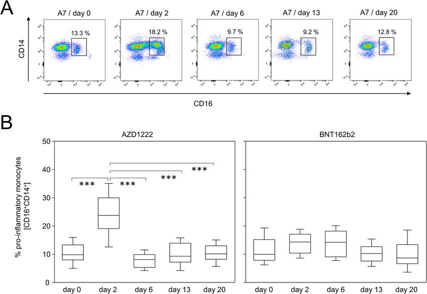
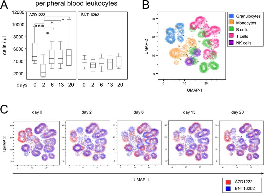
Müller et al. BMC Medicine     (2022) 20:29                                                                                        Page 3 of 14

 Fig. 1 Vaccination with AZD1222 induced a transient reduction of peripheral leukocytes and displacements of major immune cell populations. A
 Leukocyte counts in the peripheral blood after vaccination with AZD1222 (n = 13, left panel) and BNT162b2 (n = 15, right panel). p-values
 resulting from multiple group comparisons were 0.0005 (AZD1222/Kruskal-Wallis and Dunn’s multiple comparisons tests) and 0.5306 (BNT162b2/
 one-way ANOVA), respectively. **p < 0.01, ***p < 0.001. B UMAP of surface antigen expression and clustering of major immune cell populations
 for all time points after vaccination. C UMAPs on immune cell compositions for each time point

AZD1222-vaccinated subjects only (Additional file 1:                     Changes among adaptive immune cell populations were
Figs. S1 and S2). In detail, CD177–CD11b+ among                          most prominent after the AZD1222 vaccination
CD14+CD16– granulocytes were significantly elevated on                   In order to characterize the response of adaptive
days 2 and 13 following vaccination (Additional file 1:                  immune cells following vaccination, we next
Fig. S1). By day 20, this subpopulation was still increased              investigated the proportions of B- and T-lymphocyte
to some extent however, due to high variance, the differ-                subpopulations. Figure 3A shows representative data of
ence to baseline did not reach statistical significance.                 CD19+CD45RA+ B cells and illustrates for a partici-
Interestingly, dynamics of CD177–CD11b– among                            pant vaccinated with AZD1222 a shift of subpopula-
CD14+CD16+ granulocytes followed an opposing trend                       tions expressing CD27 and CD38, respectively. While
with proportions being decreased on days 2 and 13 be-                    CD27+CD38bright plasmablasts were significantly
fore returning to baseline by day 20 after vaccination                   enriched on day 6 following vaccination with AZD1222
(Additional file 1: Fig. S2). In summary, this expression                and reached a median of 3.08%, a plasmablast peak
data show that vaccination with BNT162b2 had almost                      after BNT162b2 vaccination was detectable on day 13,
no impact on the peripheral innate immune compart-                       yet reached a median of 1.57% only (Fig. 3B, upper
ment, whereas vaccination with AZD1222 led to marked                     panels). The increase in plasmablasts after the
alterations in the compositions of granulocyte and                       AZD1222 vaccination was flanked by an increase in
monocyte subpopulations.                                                 CD27+CD38– late memory B cells on days 13 and 20
Müller et al. BMC Medicine     (2022) 20:29                                                                                        Page 4 of 14

 Fig. 2 AZD1222 vaccination induced the enrichment of pro-inflammatory monocytes. A Pseudocolor plots for the expression of CD14 and CD16
 on monocytes are representative for the AZD1222 vaccination group. B Proportions of CD16+CD14+ pro-inflammatory monocytes after
 vaccination with AZD1222 (n = 18, left panel) or BNT162b2 (n = 18, right panel). All FACS analyses were on gated live monocytes. Asterisks
 indicate significant differences between time points. p-values resulting from one-way ANOVA and Tukey-Kramer multiple comparisons tests were
 < 0.0001 for AZD1222 and 0.0146 for BNT162b2 analyses. ***p < 0.001

(Fig. 3B, lower panels). No such alterations were ob-                    AZD1222 and BNT164b2 vaccinations led to significantly
served after the BNT162b2 vaccination.                                   different helper and cytotoxic T cell polarizations
   Likewise, alterations among T cell subpopulations                     So far we have shown that a single dose of AZD1222
were restricted to AZD1222 vaccinees only (Additional                    was capable of significantly modifying immune cell com-
file 1: Figs. S3 and S4). In detail, we detected a shift in              positions. However, we assumed that both vaccines
the expression patterns of CD27 and CD38 on CD8+                         would on a small scale induce a specific cellular immune
cells especially 2 days after AZD1222 vaccination (Add-                  response towards the SARS-CoV-2 spike protein, which
itional file 1: Fig. S3A). This translated into a significant            would become detectable upon re-stimulation with the
enrichment of CD27–CD38+ terminally differentiated                       antigen. We therefore used both, recombinant spike pro-
cytotoxic T cells for this group (Additional file 1: Fig.                tein and BNT162b2 vaccine, and aimed to investigate
S3B). Additional file 1: Fig. S4A exemplifies for CD4+                   cytokine profiles as well as the expression of inducible
cells a change in the expression patterns for CD27 and                   activation markers. In case of the recombinant spike
CD127 on day 20 after AZD1222 administration. We                         protein, we expected it to be taken up by antigen-
thus discovered that CD4+CD127–CD27+ effector mem-                       presenting cells (APCs). Upon processing the protein in
ory T cells re-expressing RA (TEMRA) were enriched                       lysosomes, respective peptides would predominantly be-
towards the end of the observation period (Additional                    come displayed on human leukocyte antigen (HLA) class
file 1: Fig. S4B). Taken together, we here demonstrated                  II molecules and thus would be ready to activate spike
that significant changes among subpopulations of B- and                  protein-specific T helper cells. By employing the mRNA
T-lymphocytes were observed after vaccination with                       vaccine, we anticipated its cellular uptake, translation
AZD1222 only.                                                            into protein and then both, secretion for uptake by
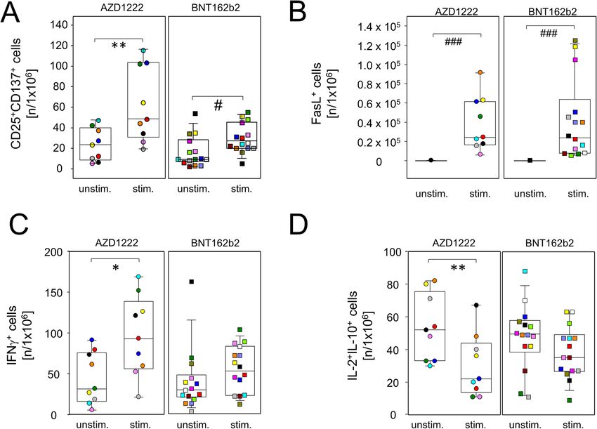
Müller et al. BMC Medicine     (2022) 20:29                                                                                          Page 5 of 14

 Fig. 3 Vaccination with AZD1222 and BNT162b2 induced significant yet transient alterations among peripheral plasmablasts. AZD1222 also
 induced a significant increase in late memory B cells on days 13 and 20. A Pseudocolor plots for the expression of CD27 and CD38 on B cells are
 representative for the AZD1222 vaccination group. B Proportions of CD27+CD38bright plasmablasts (top) and CD27+CD38– late memory B cells
 (bottom) after vaccination with AZD1222 (n = 18, left) or BNT162b2 (n = 18, right). All FACS analyses were on CD19+CD45RA+ B cells. p-values
 resulting from one-way ANOVAs were < 0.0001 for both AZD1222 analyses. p-values resulting from Kruskal-Wallis and Dunn’s multiple
 comparisons tests were 0.0008 for the comparison of plasmablast and 0.6998 for the comparison of late memory B cells for the BNT162b2
 analyses. *p < 0.05, **p < 0.01, ***p < 0.0001

APCs and class II presentation as well as processing the                   with recombinant spike protein to confirm for both vac-
protein for presentation via HLA class I molecules and                     cination regimes increasing amounts of interferon (IFN)γ-
thereby re-stimulating cytotoxic T cells [20].                             secreting cells on day 20 compared to day 0. While Fig.
  In order to establish a working protocol, we used per-                   4A presents individual examples of ELISPOTs, Fig. 4B
ipheral blood mononuclear cells (PBMCs) from fully                         summarizes all results and indeed shows a significant in-
vaccinated or COVID-19 convalescent blood donors and                       crease in IFNγ-positive cells after BNT162b2 vaccination
investigated the expression of the activation marker                       (p = 0.0059). There was also a trend towards increased
CD137 on unstimulated cells compared to cells chal-                        IFNγ-positive cells after AZD1222 vaccination; however,
lenged with either the recombinant spike protein or                        this did not reach statistical significance (p = 0.0968).
BNT162b2. Indeed, we found a significant activation of                        We next sought to differentiate the AZD1222- and
CD4+ but not CD8+ cells after providing the recombin-                      BNT162b2-induced adaptive cellular immune responses
ant spike protein (Additional file 1: Fig. S5). In contrast,               in more detail. We were interested in the activation pro-
stimulation with the BNT162b2 vaccine led to a signifi-                    files of both CD4- and CD8-positive T cells and there-
cant increase of CD137 expressing cells among both,                        fore chose BNT162b2 for re-stimulation over the spike
CD4+ and CD8+ lymphocytes (Additional file 1: Fig. S5).                    protein because the latter failed to induce a response by
  In a first approach, we used a classical enzyme-linked                   CD8+ T cells (Additional file 1: Fig. S5). Therefore, we
immune absorbent spot (ELISPOT) assay in combination                       cultured day 20 PBMCs from both vaccination groups in
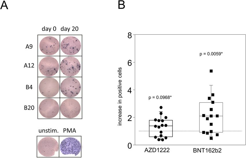
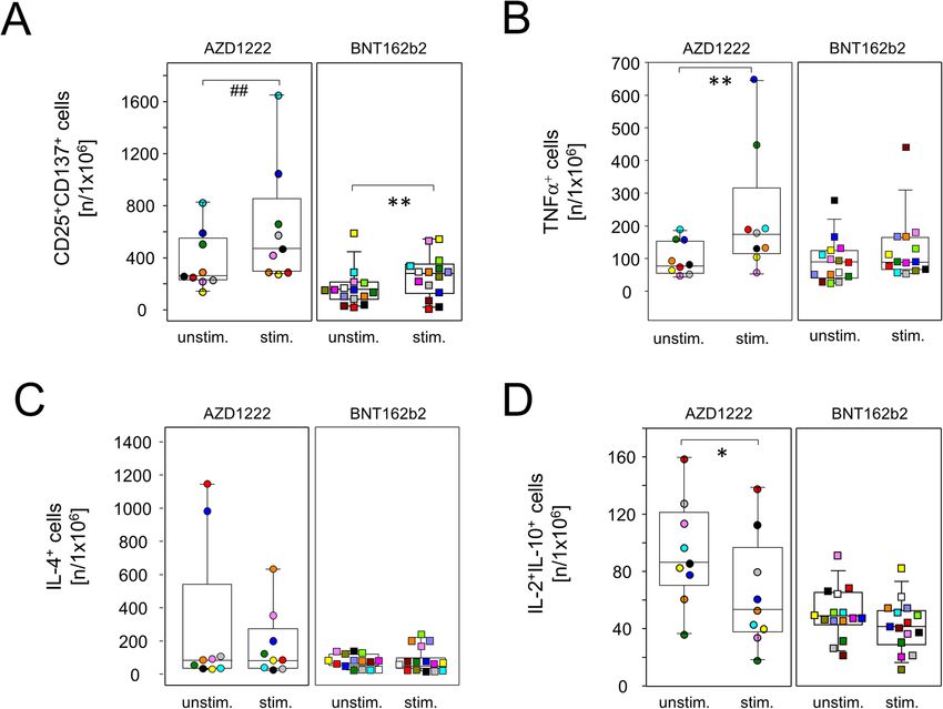
Müller et al. BMC Medicine      (2022) 20:29                                                                                              Page 6 of 14

 Fig. 4 Spike-specific IFNγ-producing lymphocytes were significantly increased after vaccination with BNT162b2. PBMCs were isolated at the day of
 vaccination (day 0) and 20 days later. Cells were then stimulated with recombinant SARS-CoV-2 spike protein for 24 h. Subsequently, an ELISPOT assay
 for the detection of IFNγ-producing cells was performed. A Representative shots of spot-forming cells from individuals vaccinated with either AZD1222
 (A9 and A12) or BNT162b2 (B4 and B20). PBMCs were stimulated with PMA as a positive control (bottom). B The counts of spot-forming cells from day
 20 were normalized to counts from day 0. Data show the relative increase in IFNγ+ cells after vaccination (n = 16 for AZD1222 and n = 15 for
 BNT162b2). p-values result from one-sample t-tests assessing the differences of group means to the hypothetical value of 1

the presence or absence of BNT162b2 and surveyed acti-                       spike-specific type 2 helper T cells as demonstrated
vation and cytokine profiles by flow cytometry after Bre-                    by a lack of inducible IL-4 production by CD4+ cells
feldin A-capture of secretory proteins. Figure 5A shows                      after re-stimulation (Fig. 6C). Similar to regulatory
that following both vaccination regimes, re-challenge                        CD8+ cells and again restricted to the AZD1222
with spike mRNA led to significantly increased amounts                       group, re-stimulation with spike mRNA statistically
of CD8+CD137+ T cell that also expressed CD25 (IL-                           significantly reduced anti-inflammatory IL-2 and IL-
2Rα), suggesting the differentiation to an effector pheno-                   10 co-production in a subset of CD4+ cells (Fig. 6D).
type. In addition, both vaccines facilitated the expansion                     In summary, these results demonstrate that single
of inducible spike-specific cytotoxic effector T cells as                    doses of either AZD1222 or BNT162b2 induced the
demonstrated by significantly increased percentages of                       polarization of spike-specific CD4+ and CD8+ effector T
FasL+CD8+ cells after in vitro re-stimulation (Fig. 5B).                     cells. More pronounced changes were again observed
We further detected a significant increase in IFNγ-                          after vaccination with AZD1222 that included reduced
producing CD8+ T cells from AZD1222- but not from                            proportions of IL-2 and IL-10 co-expressing CD4+ and
BNT162b2-vaccinated donors (p = 0.0325 vs 0.1514, Fig.                       CD8+ cells alike, combined with increased IFNγ-
5C). Finally, re-stimulation with spike mRNA decreased                       producing CD8+ and TNFα-producing CD4+ cells.
interleukin (IL-)2+IL-10+ co-expressing regulatory CD8+
cells for both AZD1222 and BNT162b2, the latter short                        BNT162b2 vaccination led to significantly more spike
of reaching statistical significance (p = 0.0058 vs 0.0768,                  protein-specific plasma IgG and IgA
Fig. 5D).                                                                    Even though both vaccines led to significant adaptive
  Significantly increased amounts of T helper cells                          immune activation, alterations to inducible effector func-
with an effector phenotype (CD4+CD25+CD137+) were                            tions followed distinct patterns for AZD1222 and
also detected for both vaccine regimens after in vitro                       BNT162b2, respectively. We therefore sought to investi-
re-stimulation (Fig. 6A). In contrast, an increase in                        gate whether these differences resulted in the production
pro-inflammatory helper T cells producing tumor ne-                          of diverse collections of immunoglobulin isotypes. To
crosis factor (TNF)α was restricted to re-stimulated                         that extent, spike protein-specific IgM, IgG, and IgA
cultures from day 20 AZD1222 donors (Fig. 6B). Of                            were assessed for all time points via an enzyme-linked
note, neither vaccination induced the expansion of                           immunosorbent assay (ELISA). As shown in Fig. 7A,
Müller et al. BMC Medicine      (2022) 20:29                                                                                           Page 7 of 14

 Fig. 5 Vaccination with single doses of AZD1222 or BNT162b2 induced the expansion of cytotoxic effector T cells. PBMCs were isolated on day
 20 after vaccination and stimulated with (stim.) or without (unstim.) spike protein-encoding mRNA (BNT162b2) for 24 h and analyzed by flow
 cytometry for the expression of inducible activation markers and intracellularly trapped cytokines among CD8+ cells. Sample sizes were n = 9 for
 AZD1222 and n = 15 for BNT162b2, respectively. Paired samples are illustrated by color coding. In vitro re-stimulation significantly increased
 CD25+CD137+ (A) and FasL expressing cells in both vaccination groups (B). INFγ–producers were increased (C) and IL-2 and IL-10 co-expressing
 cells were significantly reduced among AZD1222 vaccinees only (D). *p < 0.05, **p < 0.01 resulting from paired t-tests. #p < 0.05, ###p < 0.001
 resulting from Wilcoxon signed rank tests for matched pairs

both vaccines induced the production of detectable                          found that SARS-CoV-2 spike receptor binding domain
amounts of antibodies as early as day 13. However, signifi-                 neutralization was 1.6-fold higher for BNT162b2 (median
cant differences emerged between AZD1222 and                                784 IU/mL) when compared to AZD1222 (median 482 IU/
BNT162b2 vaccination concerning the distribution and                        mL, p = 0.0175). Interestingly, Spearman rank correlation
amounts of spike-specific antibody isotypes. In detail,                     analyses indicated strong relationships between neutralizing
AZD1222 predominantly induced IgM and IgG, while IgA                        antibodies and amounts of spike binding IgM (r = 0.6411, p
was virtually absent, even at day 20 after vaccination. In                  < 0.001), IgG (0.9328, p < 0.001), and IgA (0.6952, p <
contrast, although not significantly different from                         0.001), respectively. Collectively, disparate vaccine strategies
AZD1222, BNT162b2 elicited little IgM. There were                           for spike protein delivery impacted differently on the
though increased IgG and IgA titers in BNT162b2-                            humoral immune response and shaped distinctive antibody
vaccinated subjects and these differences reached statistical               isotype layouts as well as virus neutralization capacities
significance on day 20 for IgG and already on day 13 for                    after single doses of AZD1222 and BNT162b2.
IgA (Fig. 7A). When looking at response rates instead of Ig
titers, there were significantly more IgM and significantly                 Discussion
less IgA responders to AZD1222 compared to BNT162b2                         We here compared early immune reactions to the
as calculated via Fisher’s exact test (Additional file 1: Table             primary vaccination against SARS-CoV-2 with either
S3). When testing for surrogate virus neutralization, we                    AZD1222 from AstraZeneca or BNT162b2 from BioNtech
Müller et al. BMC Medicine      (2022) 20:29                                                                                            Page 8 of 14

 Fig. 6 AZD1222 vaccination supported the induction of type 1 helper T cells. PBMCs were isolated on day 20 after vaccination and stimulated
 with (stim.) or without (unstim.) spike protein encoding mRNA (BNT162b2) for 24 h and analyzed by flow cytometry for the expression of
 inducible activation markers and intracellularly trapped cytokines among CD4+ cells. Sample sizes were n = 9 for AZD1222 and n = 15 for
 BNT162b2, respectively. Paired samples are illustrated by color coding. In vitro re-stimulation significantly increased CD25+CD137+ cells in both
 vaccination groups (A) and TNFα-producing cells in the AZD1222 vaccinated group, only B. C Neither vaccination allowed for the re-stimulation
 of IL-4-producing cells. D IL-2 and IL-10 co-expressing cells were significantly reduced among AZD1222 vaccinees only. *p < 0.05, **p < 0.01,
 resulting from paired t-test. ##p < 0.01, resulting from Wilcoxon signed rank test for matched pairs

[6, 8]. While both vaccines elicited strong cellular and                    two separate studies in which a human adenoviral vector
humoral responses, the individual impacts on the periph-                    was applied in rhesus macaques and mice, respectively
eral immune compartment were strikingly different. In de-                   [22, 25].
tail, the adenoviral vector AZD1222 led to a transient yet                     Likewise, the AZD1222-induced changes among per-
profound leukopenia on day 2, involving significant reduc-                  ipheral immune cell proportions were reminiscent of
tions of B and T lymphocytes as well as granulocytes. As                    viral infections. For instance, short-term enrichments of
decreased leukocyte counts have previously been reported                    pro-inflammatory monocytes have been observed in pa-
for both, regular adenoviral infections [21] and                            tients infected with dengue or human immunodeficiency
adenovirus-mediated gene therapeutic approaches [22,                        virus, respectively [26, 27]. In detail, Kwissa and col-
23], we consider it likely that the leukopenia observed here                leagues have shown that CD14+CD16+ monocytes were
can be attributed to the viral vector rather than the spike                 increased in both humans and non-human primates
protein [24]. For example, Park and colleagues observed                     shortly after infection with the dengue virus and that
in an outbreak of adenoviral pneumonia among 191                            these cells were able to stimulate the differentiation of
otherwise healthy adults a high percentage of patients that                 plasmablasts [28]. Both the enrichment of CD4+
exhibited febrile leukopenia [21]. Furthermore, Varnavski                   TEMRA and terminally differentiated cytotoxic CD8+ T
et al. observed a transient decrease of leukocyte counts in                 cells have been associated with human cytomegalovirus
Müller et al. BMC Medicine       (2022) 20:29                                                                                              Page 9 of 14

 Fig. 7 A single dose of BNT162b2 induced significantly higher amounts of spike protein binding IgG, IgA, and receptor binding domain
 neutralizing antibodies than AZD1222. A SARS-CoV-2 spike binding antibodies were detected by ELISA and absorbance readouts were normalized
 to calibrator values to obtain arbitrary units. Gray lines depict the positive response threshold within a range of 0.8 to 1.1 arbitrary units. Sample
 sizes were n = 18 for AZD1222 and n = 18 for BNT162b2, respectively. p-values resulting from one-way ANOVA and Tukey-Kramer multiple
 comparisons tests were < 0.0001 for all three isotype analyses. B Neutralizing antibodies were determined by the surrogate virus competitive
 ELISA. Absorbance readouts from BNT162b2 (n = 17) and AZD1222 (n = 17) were calibrated with WHO standards to obtain international units
 (IU)/mL. *p < 0.05 resulting from Mann-Whitney U test and ***p < 0.001 resulting from Dunn’s test

[29–31]. In addition, the class-switched, late memory B                       towards the SARS-CoV-2 spike protein whereas the lar-
cells that we observed after AZD1222 vaccination have                         ger enrichment observed with AZD1222 likely reflects a
previously been associated with an efficient control of                       combined response towards the spike protein and the
viral infections [32, 33]. Together, our data suggest that                    adenoviral vector. In order to verify this hypothesis, fu-
it is the adenoviral vector rather than the encoded spike                     ture studies should examine the proportions of periph-
protein that elicits a significant immune cell response in                    eral plasmablasts and memory B cells that express a B
the periphery.                                                                cell receptor specific for either the spike protein or
   By comparison, peripheral immune cell proportions                          adenoviral antigens, respectively. In fact, a study by Cia-
appeared mostly unaffected by BNT162b2 administra-                            battini and colleagues demonstrated for subjects that
tion, which corroborates the observation that this vac-                       were vaccinated with BNT162b2 spike-specific memory
cine is globally well tolerated among first dose recipients                   B cells that persisted in the circulation for at least 6
[9]. Indeed, the BNT162b2 encoded mRNA bears an                               months after the second dose [36].
m1Ψ modification which attenuates innate immune                                 When comparing the specific antibody responses
sensing [34]. We therefore assume that the relatively                         against the spike protein elicited by both vaccines, we
small enrichment of peripheral plasmablasts after                             found that the isotypes produced were significantly dif-
BNT162b2 vaccination, which can also be observed dur-                         ferent with AZD1222 inducing primarily IgM and IgG
ing COVID-19 [35], resulted from an adaptive response                         compared to predominantly IgG and IgA by BNT162b2.
Müller et al. BMC Medicine   (2022) 20:29                                                                     Page 10 of 14

Furthermore, we found that the amounts of plasma neu-           unstimulated day 20 T cells, they show that both vac-
tralizing antibodies were significantly elevated for            cines induced Th1 responses. In addition, we found a re-
BNT162b2 at day 20 after vaccination. We also demon-            duction of IL-2 and IL-10 co-producing CD4+ and CD8+
strated that receptor binding domain neutralization             T cells upon in vitro re-stimulation which however only
strongly correlated with spike binding IgM, IgG, and IgA        reached significance in the case of AZD1222 vaccination.
which is in line with previous data [5, 37–39]. Even            IL-10 is a hallmark of regulatory T cells and exerts anti-
though we cannot yet predict whether this trend will be         inflammatory effects via suppressing not only effector T
continued beyond the first 3 weeks after vaccination, a         cells, but also antigen presentation and the secretion of
pronounced IgA response combined with a higher virus            inflammatory cytokines by APCs [45–47]. We like to
neutralization capacity following BNT162b2 vaccination          speculate that this significant reduction of IL-10 expres-
may explain its superior effectiveness in preventing            sion is not restricted to in vitro re-challenge but may
symptomatic COVID-19 after both infection with wild             also occur after booster immunization and perpetuate a
type SARS-CoV-2 and its variants [15, 17, 19, 40]. In-          Th1 response that impedes an IgA promoting cytokine
deed, Chan et al. demonstrated that, following vaccin-          milieu.
ation with BNT162b2, IgA is detectable in nasal swabs             This study has a few limitations, among them the
and that plasma IgA levels correlate with their capacity        small sample sizes. Furthermore, we did not account
for SARS-CoV-2 receptor binding inhibition which was            for any possible underlying conditions nor did we
also demonstrated for patients who recovered from               document the general health status of the study par-
COVID-19 [35, 41]. Optimizing existing vaccines might           ticipants that might influence the variance within
therefore also aim at alternative antigen delivery, e.g., to-   and between both vaccine groups. Nonetheless, our
wards mucosal sites, in order to support not only IgA           results depict for both vaccines significantly dispar-
production but also tissue-resident effector cells which        ate effects on the peripheral immune layout and on
will help contain viral loads at the nasopharyngeal entry       the regulation of T cell effector molecules and we
sites [42, 43].                                                 assume that these differences were generated by the
   Optimizing vaccines may also aim at modifying poten-         different strategies for spike antigen delivery. An-
tial bystander effects. When analyzing the spike protein-       other limitation is the lack of virus neutralization
specific T cell responses, we observed that both vaccines       data using SARS-CoV-2. However, recent data dem-
elicited functional immune responses. Both vaccination          onstrated that readouts from surrogate virus
regimes expanded effector cells to a comparable degree          neutralization robustly correlate with conventional
as documented by significant increases in activated             virus neutralization and are therefore a suitable
CD25 and CD137 co-expressing CD4+ and CD8+ T                    measure for humoral protection from infection [48].
lymphocytes as well as FasL expressing CD8+ cells upon          In summary, we consider the description of disparate
re-challenge. However, when looking at intracellular            vaccine effects on the immediate immune response
cytokine production, AZD1222 induced a prominent                the strength of our study and we believe that our
type 1 helper T cell (Th1) response as illustrated by           results will be of use for further optimization of
significant increases in IFNγ and TNFα, respectively.           vaccination strategies.
Because adenoviral vectors have previously been shown
to facilitate strong cellular immunity towards the deliv-       Conclusions
ered antigen and drive the expansion of Th1 cells [6], we       We here show that, after a single dose, the SARS-CoV-2
believe that this Th1 reaction towards the adenoviral           vaccines AZD1222 and BNT162b2 impact differently on
vector exerted some bystander effect on the response            the peripheral immune compartment. Although both
against the spike protein. However, an inordinate Th1           vaccines elicited the induction of spike-specific effector
response may foster a cytokine layout that is hardly sup-       cells and spike binding antibodies, the different compil-
portive of class-switch recombination towards IgA [44].         ation of these immunological features suggests that the
On the other hand, we assessed neither IL-5 nor trans-          strategy for spike delivery impacts on how and to what
forming growth factor (TGF)β and are therefore are not          extent immune priming against the main SARS-CoV-2
yet able to verify whether BNT162b2 indeed induced              antigen proceeds. We propose that the induction of
more IgA promoting cytokines.                                   higher quantities of IgA might be associated with
   Of note, our ELISPOT experiments addressing spike            superior protection against breakthrough infections
protein-specific Th1 cells before and after vaccination         after booster injections. Conclusively, additional in-
found an even stronger induction of IFNγ-producing T            vestigations are needed that further our understand-
helper cells among BNT162b2 compared to AZD1222                 ing about the immunization mechanisms that lead to
vaccinees. Even though these results seemingly contra-          a favorable humoral and cellular layout that is effect-
dict the intracellular cytokine readout of stimulated vs        ive against COVID-19.
Müller et al. BMC Medicine   (2022) 20:29                                                                   Page 11 of 14

Methods                                                       Antibodies were incubated for 15 min on ice in the
Study design                                                dark. Subsequently, ApotrackerTM Green (BioLegend)
This explorative study was designed to compare the          was added according to the manufacturer’s instruction
immediate immune response towards a single-dose             without washing, followed by incubation for 30 min on
vaccination with either AZD1222 or BNT162b2. For            ice. In order to lyse erythrocytes, 2.2 mL Fixative-Free
that, study participants were recruited from the            Lysing Solution (Thermo Fisher Scientific) was added
coordination center for clinical studies at the             and incubated for 20 min at room temperature. Subse-
Rostock University Medical Center. Individuals with         quently, 0.03 μg 4′,6-diamidino-2-phenylindole (DAPI,
a study-independent appointment at a vaccination            BioLegend) was added as a live/dead discriminator and
center were eligible to participate. No other specific      incubated for 5 min. Finally, data acquisition was per-
inclusion criteria were set. Apart from their age and       formed on the Cytek® Aurora flow cytometer running on
sex, no other personal data (i.e., underlying medical       the SpectroFlo Software version 2.2.0.3 (Cytek Biosci-
conditions) were documented. Blood samples were             ences). Analysis of flow cytometry data was done using
obtained by consecutive venipuncture immediately            FlowJo software version 10.7 (FlowJo). The gating
before vaccination (day 0) and on days 2, 6, 13, and        scheme is shown in Additional file 1: Fig. S6. Dimension
20 thereafter. Participants that were positive for          reduction of down-sampled (5 × 10⁴ live cells per sam-
SARS-CoV-2 spike protein binding IgG were ex-               ple) and concatenated data sets was performed using the
cluded from further analyses. PBMCs were isolated           FlowJo plugin for the algorithm UMAP [49].
from anti-coagulated blood by density gradient
centrifugation using Ficoll-PaqueTM PLUS to the             Interferon gamma ELISPOT
manufacturer’s instructions (Cytiva). PBMCs were            PBMCs from day 0 and day 20 were thawed, centrifuged,
subsequently suspended in fetal calf serum (Thermo          and suspended in a complete RPMI medium containing
Fisher Scientific) containing 10% dimethyl sulfoxide        10% FCS, 1% penicillin/streptomycin, 2 mM L-glutamine
(Sigma-Aldrich) and were frozen at −70 °C until fur-        (Thermo Fisher Scientific), 10 mM HEPES, and 1 mM
ther use. Plasma samples were obtained by centrifu-         sodium pyruvate (PAN-Biotech). Cell counts were
gation of anti-coagulated blood and were frozen             determined cytometrically on the Cytek® Aurora (Cytek
afterwards. This study was approved by the ethics           Biosciences) using DAPI (BioLegend) as a live/dead dis-
committee of the Rostock University Medical Center          criminator. Five hundred thousand PBMCs were pipet-
under file number A 2020-0086. Written informed             ted into a 96-well U-bottom plate and centrifuged for 5
consent was provided by all participants.                   min at 4 °C and 400×g. Subsequently, supernatants were
                                                            removed by carefully blotting the plate on a paper tissue.
Flow cytometric analyses on surface markers                 Cells were then suspended in a 36-μL complete RPMI
For the analysis of surface expression markers, 100 μL of   medium containing 0.2 μg of the SARS-CoV-2 trimeric
anti-coagulated whole blood was used. In order to re-       spike protein (R&D Systems). Afterwards, PBMCs were
duce unspecific antibody-conjugate binding, 10 μL FCS,      transferred into a 96-well ELISPOT assay plate coated
5 μL True-Stain Monocyte BlockerTM, and 5 μL anti-Fc        with capture antibodies specific for human IFNγ (R&D
receptor TruStain FcXTM (BioLegend) were added and          Systems). After incubating the cells for 30 min at 37 °C,
incubated for 15 min on ice. The following amounts of       164 μL of complete RPMI medium was added to all wells
antibody:fluorophore combinations were used: 0.25 μg        followed by 24-h incubation at 37 °C in a CO2 incubator
CD127:APC/R700 (clone HIL-7R-M21), 1 μg CD147:              (BINDER). The ELISPOT assay was then performed ac-
BV421 (TRA-1-85), 0.5 μg CD45RO:BV480 (UCHL1, BD            cording to the manufacturer’s guidelines. The numbers
Biosciences), 1 μg CD11b:PerCP/Cy5.5 (ICRF44), 0.8 μg       of IFNγ-producing cells were determined by automated
CD11c:BV785 (3.9), 0.56 μg CD14:BV510 (63D3), 0.13 μg       counting using the ImmunoSpot® analyzer running on
CD16:BV650 (3G8), 0.06 μg CD19:APC/Fire810 (HIB19),         the ImmunoSpot® Software version 5.0.9.15 (CTL
0.13 μg CD20:SparkNIR685 (2H7), 0.5 μg CD27:BV605           Europe). The counts of IFNγ-positive cells were normal-
(O323), 0.25 μg CD3:SparkBlue550 (SK7), 0.25 μg             ized to their respective paired sample from d0.
CD304:AlexFluor647 (12C2), 0.03 μg CD4:BV750 (SK3),
0.5 μg CD45RA:APC/Fire750 (HI100), 0.13 μg CD56:            T cell re-stimulation and intracellular cytokine staining
BV711 (5.1.H11), 0.13 μg CD8:BV570 (RPA-T8), 0.5 μg         assay
CD95:PE/Cy5 (DX2), 0.13 IgD:PE/Dazzle594 (IA6-2),           For the establishment of T cell re-stimulation and intra-
0.13 μg PD-1:APC (A17188B, BioLegend), 0.06 μg CD38:        cellular cytokine staining assays, blood was collected
PerCP/eFluor710 (HB7, Thermo Fisher Scientific),            from six vaccinated subjects 2 to 29 weeks after the last
0.06 μg CD177:PE/Vio770 (REA258), and 0.05 μg CD25:         dose and from one patient who had recovered from
PE (REA570, Miltenyi Biotec).                               COVID-19 presumably 24 weeks prior to venipuncture.
Müller et al. BMC Medicine (2022) 20:29 Page 12 of 14 In detail, one subject was vaccinated with a single dose Intracellular Staining Permeabilization Wash Buffer (BioLe- of Ad26.COV2.S (Johnson & Johnson), three individuals gend). After blocking unspecific binding sites as described received two doses of BNT162b2 (BioNTech), one re- above, 0.5 μg Granzym B:AlexaFluor647 (clone 6B11), ceived one dose of BNT162b2 and one received one 2.5 μg IFNγ:PerCP/Cy5.5 (4S.B3), 0.63 μg IL-2:BV650 dose of AZD1222 (AstraZeneca) followed by one dose of (MQ1-17H12), 0.3 μg IL-4:PE/Dazzle594 (MP4-25D2), 1 μg BNT162b2. PBMCs were isolated and frozen until fur- IL-10:BV421 (JES3-907, BioLegend), and 0.13 μg TNFα:PE/ ther use in the same fashion as described above. For Cy7 (Mab11, Thermo Fisher Scientific) were added and in- assaying the vaccinated study participants, day 20 cubated for 30 min at room temperature. Finally, cells were PBMCs were used. Upon thawing, PBMCs were counted washed twice in RB. Data acquisition and analysis of ex- as described above and aliquots of 0.8 million were pression data was performed as described above. The gating seeded into single wells of 96-well U-bottom plates. scheme for the intracellular cytokine staining assay is Every sample was stimulated at least in duplicates. After shown in Additional file 1: Fig. S7. centrifugation, cells were re-suspended and stimulated in a total volume of 36-μL complete RPMI medium with SARS-CoV-2 trimeric spike-specific antibodies either 1 μg of the BNT162b2 vaccine or 0.2 μg of the Plasma samples from all time points were thawed on ice SARS-CoV-2 trimeric spike protein or left without any and centrifuged at 10,000×g in order to remove precipi- stimulation. Pooled leftovers of the BNT162b2 vaccine, tates. For the detection of SARS-CoV-2 trimeric spike which are not allowed to be used for administration in protein-specific IgG and IgA levels, plasma was diluted Germany, were kindly provided by staff members of the 101-fold. The ELISA for these isotypes was conducted Rostock vaccination center. Re-stimulation with after the manufacturer’s specifications (EUROIMMUN). BNT162b2 was preferred over re-stimulation with In order to determine IgM levels, plasma samples were AZD1222 because we expected an activation of T cells diluted 1000-fold and the ELISA was performed to the that are reactive against adenoviral antigens from the manufacturer’s instructions (Thermo Fisher Scientific). vector which would obscure the isolated response to- The absorbance was detected at 450 nm (A450) on the wards the spike protein primarily in the group of partici- Infinite® 200 automated plate reader (Tecan Group). pants that were vaccinated with AZD1222. Phorbol 12- Absorbance readouts were normalized to calibrator myristate-13-acetate (PMA, 10 ng/ mL) and Ionomycin values and were reported as arbitrary units. According (1 μg/mL) stimulated samples were processed in parallel to the manufacturer’s guidelines, calibrated sample as positive controls. After adding 164 μL of complete values between 0.8 and 1.1 were considered borderline RPMI medium, cells were incubated for 20 h under 5% and above 1.1 were considered clearly positive. Samples CO2 atmosphere at 37 °C. One microgram of Brefeldin from individuals with arbitrary unit values of less than A (Sigma-Aldrich) was added thereafter followed by in- 0.8 were considered non-responders. cubation for another 4 h. Successive incubation steps were performed in the Surrogate virus neutralization dark. Duplicate samples were pooled, washed in PBS Plasma samples were processed like described above. For (Thermo Fisher Scientific), suspended in PBS containing standardization, we ran plasma samples from the World 2000-fold diluted ZombieNIR dye (BioLegend), and in- Health Organization (WHO) Reference Panel in parallel cubated for 20 min at room temperature. Thereafter, [50]. For the determination of antibody neutralization, we cells were washed and suspended in autoMACS® Run- utilized the SARS-CoV-2 Surrogate Neutralization Test ning Buffer (RB, Miltenyi Biotec). Subsequently, unspe- (sVNT) Kit (GenScript) to the manufacturer’s guidelines. cific antibody-conjugate binding sites were blocked by In brief, plasma samples and standards were diluted 10- adding FCS, True-Stain Monocyte BlockerTM, and anti- fold and were incubated with a horseradish peroxidase- Fc receptor TruStain FcXTM (BioLegend) for 10 min at conjugated SARS-CoV-2 receptor binding domain for 30 room temperature. Surface antigens were stained by in- min at 37 °C. Subsequently, sample mixtures were pipetted cubating the cells with the following antibody:fluoro- onto an ELISA plate coated with the ACE2 receptor. After phore combinations: 1.25 μg CD3:FITC (clone UCHT1), incubation at 37 °C for 15 min, wells were washed four 0.02 μg CD4:BV750 (SK3), 0.06 μg CD8:BV570 (RPA- times and color reaction was initiated by the addition of T8), 0.5 μg Fas-L:PE (NOK1), 1 μg CD25:APC (BC96, TMB substrate. Absorbances at 450 nm were detected on BioLegend), 1.25 μL CD127:APC/R700 (HIL-7R-M21), the automated plate reader after the reaction was and 0.25 μg CD137:BV480 (4B4-1, BD Biosciences) for quenched. A450 from a series of WHO standards with 15 min at room temperature. Cells were subsequently known neutralization capacity were fitted with an expo- washed in RB, suspended in 100 μL Fixation Buffer nential model by the formula: International Units per mL (BioLegend), and incubated at room temperature for 20 (IU/mL) = a × exp(b × A450). IU/mL for plasma samples min. Cells were washed twice and then suspended in were computed with this model.
Müller et al. BMC Medicine         (2022) 20:29                                                                                                   Page 13 of 14

Statistical analysis                                                            Funding
Data analyses were performed using R (version 3.5.1) and                        This study was financially supported by the Federal State of Mecklenburg-
                                                                                Western Pomerania via the “Sondervermögen des MV Schutzfonds Säule Ge-
InStat version 3.10 (GraphPad Software). All data were an-                      sundheit.” Open Access funding enabled and organized by Projekt DEAL.
alyzed by two-sided testing. Data sets were evaluated for
Gaussian distribution using the Kolmogorov-Smirnov test.                        Availability of data and materials
Under the assumption of normally distributed sample                             All data generated or analyzed during this study are included in this
                                                                                published article and its supplementary information file.
data, multiple independent groups were compared by
one-way analysis of variance (ANOVA) followed by post
                                                                                Declarations
hoc pairwise comparisons using the Tukey-Kramer test.
Data, which did not follow Gaussian distribution, was                           Ethics approval and consent to participate
compared by the Kruskal-Wallis one-way analysis of vari-                        This study was approved by the ethics committee of the Rostock University
                                                                                Medical Center under file number A 2020-0086. Written informed consent
ance combined with Dunn’s test for multiple comparisons.                        was provided by all participants.
The one-sample t-test was performed to compare single
group means to a hypothetical value. Differences between                        Consent for publication
dependent samples were assessed by the paired t-test                            Not applicable
given normal distribution and by the Wilcoxon signed
rank test in case of deviation from Gaussian distribution.                      Competing interests
                                                                                The authors declare that they have no competing interests.
Correlation analyses were performed using the Spearman
rank method. A p-value of less than 0.05 was considered                         Author details
                                                                                1
statistically significant. Data visualization was performed                      Core Facility for Cell Sorting and Cell Analysis, Rostock University Medical
                                                                                Center, Rostock, Germany. 2Department of Cardiology, Rostock University
with SigmaPlot version 13.0 (Systat Software).                                  Medical Center, Rostock, Germany. 3Institute of Pharmacology and
                                                                                Toxicology, Rostock University Medical Center, Rostock, Germany. 4Division of
Abbreviations                                                                   Tropical Medicine and Infectious Diseases, Center of Internal Medicine II,
ACE2: Angiotensin-converting enzyme 2; APC: Antigen-presenting cell;            Rostock University Medical Center, Rostock, Germany.
COVID-19: Coronavirus disease 2019; DAPI: 4’,6-Diamidino-2-phenylindole;
ELISA: Enzyme-linked immunosorbent assay; ELISPOT: Enzyme-linked immune         Received: 21 October 2021 Accepted: 6 January 2022
absorbent spot [assay]; EMA: European Medicines Agency; HLA: Human
leukocyte antigen; IFNγ: Interferon γ; IL: Interleukin; m1ψ: N1-methyl-
pseudouridine; PBMC: Peripheral blood mononuclear cell; PMA: Phorbol 12-
myristate-13-acetate; RB: Running Buffer; SARS-CoV-2: Severe acute              References
respiratory syndrome coronavirus 2; TEMRA: Effector memory T cells re-          1. Dong E, Du H, Gardner L. An interactive web-based dashboard to track
expressing RA; TGFβ: Transforming growth factor β; Th1: Type 1 helper T cell;       COVID-19 in real time. Lancet Infect Dis. 2020;20(5):533–4. https://doi.org/1
TNFα: Tumor necrosis factor α; UMAP: Uniform manifold approximation and             0.1016/S1473-3099(20)30120-1.
projection; WHO: World Health Organization                                      2. Muralidar S, Ambi SV, Sekaran S, Krishnan UM. The emergence of COVID-19
                                                                                    as a global pandemic: understanding the epidemiology, immune response
                                                                                    and potential therapeutic targets of SARS-CoV-2. Biochimie. 2020;179:85–
Supplementary Information                                                           100. https://doi.org/10.1016/j.biochi.2020.09.018.
The online version contains supplementary material available at https://doi.    3. Hanrath AT, Payne BAI, Duncan CJA. Prior SARS-CoV-2 infection is associated
org/10.1186/s12916-022-02240-4.                                                     with protection against symptomatic reinfection. J Infect. 2021;82(4):e29–30.
                                                                                    https://doi.org/10.1016/j.jinf.2020.12.023.
 Additional file 1. Supplementary figures and tables                            4. Okba NMA, Müller MA, Li W, Wang C, GeurtsvanKessel CH, Corman VM,
                                                                                    et al. Severe acute respiratory syndrome coronavirus 2−specific antibody
                                                                                    responses in coronavirus disease patients. Emerg Infect Dis. 2020;26(7):
Acknowledgements                                                                    1478–88. https://doi.org/10.3201/eid2607.200841.
We thank all our participants for taking part in this study and providing       5. Wajnberg A, Amanat F, Firpo A, Altman DR, Bailey MJ, Mansour M, et al.
blood samples. The authors want to particularly thank Wendy Bergmann                Robust neutralizing antibodies to SARS-CoV-2 infection persist for months.
(Core Facility for Cell Sorting and Cell Analysis, Rostock University Medical       Science. 2020;370(6521):1227–30. https://doi.org/10.1126/science.abd7728.
Center) for her help during flow cytometry panel design and setup.              6. van Doremalen N, Lambe T, Spencer A, Belij-Rammerstorfer S, Purushotham
Organizational and documentary support was provided by the coordination             JN, Port JR, et al. ChAdOx1 nCoV-19 vaccine prevents SARS-CoV-2
center for clinical studies and Manja Ehmke (Division of Tropical Medicine          pneumonia in rhesus macaques. Nature. 2020;586(7830):578–82. https://doi.
and Infectious Diseases, Center of Internal Medicine II) from Rostock               org/10.1038/s41586-020-2608-y.
University Medical Center. Blood samples were also collected by Anxhela         7. Schoenmaker L, Witzigmann D, Kulkarni JA, Verbeke R, Kersten G, Jiskoot W,
Muca and Stefanie Brigitte Amann-Stegbauer (both Core Facility for Cell Sort-       et al. mRNA-lipid nanoparticle COVID-19 vaccines: structure and stability. Int
ing and Cell Analysis, Rostock University Medical Center).                          J Pharm. 2021;601:120586. https://doi.org/10.1016/j.ijpharm.2021.120586.
                                                                                8. Vogel AB, Kanevsky I, Che Y, Swanson KA, Muik A, Vormehr M, et al.
Authors’ contributions                                                              BNT162b vaccines protect rhesus macaques from SARS-CoV-2. Nature. 2021;
MM, BM-H, and ECR designed the experiments. BM-H, BS, SM, and MS re-                592(7853):283–9. https://doi.org/10.1038/s41586-021-03275-y.
cruited vaccinees and documented their data. BS and SM collected periph-        9. Polack FP, Thomas SJ, Kitchin N, Absalon J, Gurtman A, Lockhart S, et al.
eral blood samples. MM and JV performed flow cytometry and                          Safety and efficacy of the BNT162b2 mRNA Covid-19 vaccine. N Engl J Med.
immunosorbent and immunospot assays, executed intracellular cytokine                2020;383(27):2603–15. https://doi.org/10.1056/NEJMoa2034577.
staining assays, processed peripheral mononuclear cells, and analyzed the       10. Voysey M, Clemens SAC, Madhi SA, Weckx LY, Folegatti PM, Aley PK, et al.
data. BM-H and JV performed statistical analyses. BM-H and JV wrote the first       Safety and efficacy of the ChAdOx1 nCoV-19 vaccine (AZD1222) against
draft of the manuscript. MM wrote sections of the manuscript. All authors           SARS-CoV-2: an interim analysis of four randomised controlled trials in Brazil,
contributed to manuscript revision and read and approved the submitted              South Africa, and the UK. Lancet. 2021;397(10269):99–111. https://doi.org/1
version.                                                                            0.1016/S0140-6736(20)32661-1.
Müller et al. BMC Medicine (2022) 20:29 Page 14 of 14
11. Amit S, Regev-Yochay G, Afek A, Kreiss Y, Leshem E. Early rate reductions of 32. Hammarlund E, Thomas A, Amanna IJ, Holden LA, Slayden OD, Park B, et al.
SARS-CoV-2 infection and COVID-19 in BNT162b2 vaccine recipients. Lancet. Plasma cell survival in the absence of B cell memory. Nat Commun. 2017;
2021;397(10277):875–7. https://doi.org/10.1016/S0140-6736(21)00448-7. 8(1):1781. https://doi.org/10.1038/s41467-017-01901-w.
12. Dagan N, Barda N, Kepten E, Miron O, Perchik S, Katz MA, et al. BNT162b2 33. Sanz I, Wei C, Lee FE-H, Anolik J. Phenotypic and functional heterogeneity
mRNA Covid-19 vaccine in a nationwide mass vaccination setting. N Engl J of human memory B cells. Semin Immunol. 2008;20(1):67–82. https://doi.
Med. 2021;384(15):1412–23. https://doi.org/10.1056/NEJMoa2101765. org/10.1016/j.smim.2007.12.006.
13. Voysey M, Clemens SAC, Madhi SA, Weckx LY, Folegatti PM, Aley PK, et al. 34. Karikó K, Muramatsu H, Welsh FA, Ludwig J, Kato H, Akira S, et al.
Single-dose administration and the influence of the timing of the booster Incorporation of pseudouridine into mRNA yields superior
dose on immunogenicity and efficacy of ChAdOx1 nCoV-19 (AZD1222) nonimmunogenic vector with increased translational capacity and
vaccine: a pooled analysis of four randomised trials. Lancet. 2021;397(10277): biological stability. Mol Ther. 2008;16(11):1833–40. https://doi.org/10.1038/
881–91. https://doi.org/10.1016/S0140-6736(21)00432-3. mt.2008.200.
14. Cele S, Gazy I, Jackson L, Hwa S-H, Tegally H, Lustig G, et al. Escape of SARS- 35. Sterlin D, Mathian A, Miyara M, Mohr A, Anna F, Claër L, et al. IgA dominates
CoV-2 501Y.V2 from neutralization by convalescent plasma. Nature. 2021; the early neutralizing antibody response to SARS-CoV-2. Sci Transl Med.
593(7857):142–6. https://doi.org/10.1038/s41586-021-03471-w. 2021;13:eabd2223. https://doi.org/10.1126/scitranslmed.abd2223.
15. Madhi SA, Baillie V, Cutland CL, Voysey M, Koen AL, Fairlie L, et al. Efficacy of 36. Ciabattini A, Pastore G, Fiorino F, Polvere J, Lucchesi S, Pettini E, et al.
the ChAdOx1 nCoV-19 Covid-19 vaccine against the B.1.351 variant. N Engl Evidence of SARS-CoV-2-specific memory B cells six months after
J Med. 2021;384(20):1885–98. https://doi.org/10.1056/NEJMoa2102214. vaccination with the BNT162b2 mRNA vaccine. Front Immunol. 2021;12.
16. Tegally H, Wilkinson E, Giovanetti M, Iranzadeh A, Fonseca V, Giandhari J, https://doi.org/10.3389/fimmu.2021.740708.
et al. Detection of a SARS-CoV-2 variant of concern in South Africa. Nature. 37. Folegatti PM, Ewer KJ, Aley PK, Angus B, Becker S, Belij-Rammerstorfer S,
2021;592(7854):438–43. https://doi.org/10.1038/s41586-021-03402-9. et al. Safety and immunogenicity of the ChAdOx1 nCoV-19 vaccine against
17. Abu-Raddad LJ, Chemaitelly H, Butt AA. Effectiveness of the BNT162b2 SARS-CoV-2: a preliminary report of a phase 1/2, single-blind, randomised
Covid-19 vaccine against the B.1.1.7 and B.1.351 variants. N Engl J Med. controlled trial. Lancet. 2020;396(10249):467–78. https://doi.org/10.1016/S014
2021;385(2):187–9. https://doi.org/10.1056/NEJMc2104974. 0-6736(20)31604-4.
18. Lopez Bernal J, Andrews N, Gower C, Gallagher E, Simmons R, Thelwall S, et al. 38. Groß R, Zanoni M, Seidel A, Conzelmann C, Gilg A, Krnavek D, et al.
Effectiveness of Covid-19 vaccines against the B.1.617.2 (Delta) variant. N Engl J Heterologous ChAdOx1 nCoV-19 and BNT162b2 prime-boost vaccination
Med. 2021;385(7):585–94. https://doi.org/10.1056/NEJMoa2108891. elicits potent neutralizing antibody responses and T cell reactivity. medRxiv.
19. Wall EC, Wu M, Harvey R, Kelly G, Warchal S, Sawyer C, et al. Neutralising 2021:21257971. https://doi.org/10.1101/2021.05.30.21257971.
antibody activity against SARS-CoV-2 VOCs B.1.617.2 and B.1.351 by 39. Jalkanen P, Kolehmainen P, Häkkinen HK, Huttunen M, Tähtinen PA,
BNT162b2 vaccination. Lancet. 2021;397(10292):2331–3. https://doi.org/10.1 Lundberg R, et al. COVID-19 mRNA vaccine induced antibody responses
016/S0140-6736(21)01290-3. against three SARS-CoV-2 variants. Nat Commun. 2021;12(1):3991. https://
20. Sahin U, Karikó K, Türeci Ö. mRNA-based therapeutics — developing a new doi.org/10.1038/s41467-021-24285-4.
class of drugs. Nat Rev Drug Discov. 2014;13(10):759–80. https://doi.org/10.1 40. Mlcochova P, Kemp SA, Dhar MS, Papa G, Meng B, Ferreira IATM, et al.
038/nrd4278. SARS-CoV-2 B.1.617.2 Delta variant replication and immune evasion. Nature.
21. Park JY, Kim B-J, Lee EJ, Park KS, Park HS, Jung SS, et al. Clinical features and 2021;599(7883):114–9. https://doi.org/10.1038/s41586-021-03944-y.
courses of adenovirus pneumonia in healthy young adults during an 41. Chan RWY, Liu S, Cheung JY, Tsun JGS, Chan KC, Chan KYY, et al. The
outbreak among Korean military personnel. PLoS ONE. 2017;12(1):e0170592. mucosal and serological immune responses to the novel coronavirus (SARS-
https://doi.org/10.1371/journal.pone.0170592. CoV-2) vaccines. Front Immunol. 2021;12. https://doi.org/10.3389/fimmu.2
22. Varnavski AN, Calcedo R, Bove M, Gao G, Wilson JM. Evaluation of toxicity 021.744887.
from high-dose systemic administration of recombinant adenovirus vector 42. Afkhami S, D’Agostino MR, Zhang A, Stacey HD, Marzok A, Kang A, et al.
in vector-naïve and pre-immunized mice. Gene Ther. 2005;12(5):427–36. Single-dose respiratory mucosal delivery of next-generation viral-vectored
https://doi.org/10.1038/sj.gt.3302347. COVID-19 vaccine provides robust protection against both ancestral and
23. Wirth T, Hedman M, Makinen K, Manninen H, Immonen A, Vapalahti M, variant strains of SARS-CoV-2. bioRxiv. 2021:452721. https://doi.org/10.1101/2
et al. Safety profile of plasmid/liposomes and virus vectors in clinical gene 021.07.16.452721.
therapy. Curr Drug Saf. 2006;1(3):253–7. https://doi.org/10.2174/1574 43. Boyaka PN. Inducing Mucosal IgA: A challenge for vaccine adjuvants and
88606777934440. delivery systems. J Immunol. 2017;199(1):9–16. https://doi.org/10.4049/
jimmunol.1601775.
24. Tatsis N, Ertl HCJ. Adenoviruses as vaccine vectors. Mol Ther. 2004;10(4):616–
44. Eckmann L, Morzycka-Wroblewska E, Smith JR, Kagnoff MF. Cytokine-
29. https://doi.org/10.1016/j.ymthe.2004.07.013.
induced differentiation of IgA B cells: studies using an IgA expressing B-cell
25. Varnavski AN, Zhang Y, Schnell M, Tazelaar J, Louboutin J-P, Yu Q-C, et al.
lymphoma. Immunology. 1992;76(2):235–41. http://www.ncbi.nlm.nih.gov/
Preexisting immunity to adenovirus in rhesus monkeys fails to prevent
pubmed/1634247.
vector-induced toxicity. J Virol. 2002;76(11):5711–9. https://doi.org/10.1128/
45. Moore KW, de Waal MR, Coffman RL, O’Garra A. Interleukin-10 and the
JVI.76.11.5711-5719.2002.
interleukin-10 receptor. Annu Rev Immunol. 2001;19(1):683–765. https://doi.
26. Green AM, Harris E. Monocyte-plasmablast crosstalk during dengue. Cell
org/10.1146/annurev.immunol.19.1.683.
Host Microbe. 2014;16(1):7–9. https://doi.org/10.1016/j.chom.2014.06.011.
46. Trinchieri G. Interleukin-10 production by effector T cells: Th1 cells show self
27. Kazer SW, Walker BD, Shalek AK. Evolution and diversity of immune
control. J Exp Med. 2007;204(2):239–43. https://doi.org/10.1084/jem.20070104.
responses during acute HIV infection. Immunity. 2020;53(5):908–24. https://
47. Yu Y, Ma X, Gong R, Zhu J, Wei L, Yao J. Recent advances in CD8+
doi.org/10.1016/j.immuni.2020.10.015.
regulatory T cell research (Review). Oncol Lett. 2018;15(6):8187–94. https://
28. Kwissa M, Nakaya HI, Onlamoon N, Wrammert J, Villinger F, Perng GC, et al.
doi.org/10.3892/ol.2018.8378.
Dengue virus infection induces expansion of a CD14+CD16+ monocyte
48. Tan CW, Chia WN, Qin X, Liu P, Chen MI-C, Tiu C, et al. A SARS-CoV-2
population that stimulates plasmablast differentiation. Cell Host Microbe.
surrogate virus neutralization test based on antibody-mediated blockage of
2014;16(1):115–27. https://doi.org/10.1016/j.chom.2014.06.001.
ACE2–spike protein–protein interaction. Nat Biotechnol. 2020;38(9):1073–8.
29. Gordon CL, Miron M, Thome JJC, Matsuoka N, Weiner J, Rak MA, et al. Tissue https://doi.org/10.1038/s41587-020-0631-z.
reservoirs of antiviral T cell immunity in persistent human CMV infection. J 49. McInnes L, Healy J, Melville J. UMAP: uniform manifold approximation and
Exp Med. 2017;214(3):651–67. https://doi.org/10.1084/jem.20160758. projection for dimension reduction. 2018. http://arxiv.org/abs/1802.03426.
30. Tian Y, Babor M, Lane J, Schulten V, Patil VS, Seumois G, et al. Unique 50. Kristiansen PA, Page M, Bernasconi V, Mattiuzzo G, Dull P, Makar K, et al.
phenotypes and clonal expansions of human CD4 effector memory T cells WHO International Standard for anti-SARS-CoV-2 immunoglobulin. Lancet.
re-expressing CD45RA. Nat Commun. 2017;8(1):1473. https://doi.org/10.103 2021;397(10282):1347–8. https://doi.org/10.1016/S0140-6736(21)00527-4.
8/s41467-017-01728-5.
31. Weiskopf D, Bangs DJ, Sidney J, Kolla RV, De Silva AD, de Silva AM, et al.
Dengue virus infection elicits highly polarized CX3CR1 + cytotoxic CD4 + T Publisher’s Note
cells associated with protective immunity. Proc Natl Acad Sci. 2015;112(31): Springer Nature remains neutral with regard to jurisdictional claims in
E4256–63. https://doi.org/10.1073/pnas.1505956112. published maps and institutional affiliations.You can also read