Similar HAGOS profile in patients with long-standing hip and groin pain and alpha angle under or over 60 degrees
←
→
Page content transcription
If your browser does not render page correctly, please read the page content below
Similar HAGOS profile in patients with
long-standing hip and groin pain and
alpha angle under or over 60 degrees

     Maria Biesert, ST-läkare, Ortopedkliniken Ljungby,
               maria.biesert@kronoberg.se

                     Supervisor: Ioannis Kostogiannis, MD, PhD
                                        Specialistläkare ortopedi
Abstract Background: Alpha angle is the most commonly used radiological measurement to evaluate and define cam-morphology. The cut-off value for a pathological alpha angle differ in the literature between 50 to 80 degrees. The purpose of this explorative study is to investigate if patients with long standing hip and groin pain and a high alpha angle (≥60 degrees) report worse patient-reported outcome scores compared to patients with a low alpha angle. Methods: Fifty-seven patients (females= 28, males =29) with a mean age of 35.7 years (range 19- 53) referred to the Department of Orthopedics at Skåne University Hospital for longstanding hip and groin pain (LHGP) were radiological examined and evaluated according to different radiological measurements, Alpha angle, LCE-angle, head neck offset ratio, coxa profunda and crossover sign. Plan X-ray films were used with Lauenstein and anteroposterior pelvic projections. All patients completed the PRO:s The Copenhagen Hip and Groin Outcome Score (HAGOS) as well as The Hip Sports Activity Scale (HSAS). Patients with LHGP and alpha angle
Table of contents

Introduction .................................................................................................................................................. 3
    Aim of study ............................................................................................................................................. 3

    Methods..................................................................................................................................................... 4

        Study design ......................................................................................................................................... 4

        Radiographic evaluation ..................................................................................................................... 5

        Patient-reported outcome measures (PRO:s) ................................................................................. 6

        Statistics ................................................................................................................................................ 7

    Etics ........................................................................................................................................................... 7

    Time plane ................................................................................................................................................ 7

    Fundings .................................................................................................................................................... 7

Results ............................................................................................................................................................ 8
    Patient characteristics and radiographic evaluation ............................................................................ 8

    Patient-reported outcomes (PRO:s) ...................................................................................................... 9

    Patients with bilateral symptomatic LHGP ....................................................................................... 10

Discussion ................................................................................................................................................... 12
    Limitations .............................................................................................................................................. 15

    Conclusion .............................................................................................................................................. 15

References ................................................................................................................................................... 16

                                                                                  2
Introduction
Femoroacetabular impingement (FAI) syndrome is a common cause of hip and groin pain in the
young active population and is a diagnosis and patient group that is growing fast. Ganz et. al. (1)
presented in 2003 a new surgical approach for FAI syndrome. Since then hip arthroscopic surgery
has emerged and new options with joint preservation treatment can be offered more frequently.
(2). The definition and management of FAI syndrome differ around the world and a lack of clarity
of diagnosis criteria creates ambiguity. To guide the medical field the Warwick Agreement was
established at an international congress In 2016. Consensus was made and femoroacetabular
impingement (FAI) syndrome was described as a triad of symptoms, clinical signs as well as imaging
findings (2).
The prevalence of radiographic signs that can predispose for FAI syndrome are reported in the
literature between 1% up to 95% with an increased prevalence in the athletic population. FAI
syndrome can relate to three different types of morphology: pincer, cam and mixed and can be
identified and graded with different types of measuring techniques and x-ray views (3). In a review
by Dickenson et al. (4) current studies show estimates of cam-morphology ranging from 5% to
75%, but are not truly population based and therefore the true prevalence and its relationship to
hip pain cannot be determined. Alpha angle is the most commonly used radiological measurement
to evaluate and define cam-morphology (5). In a study of collegiate athletes Larsen et. al (3) showed
increasing alpha angle was predictor for hip and groin pain (3). However a threshold value for a
pathological alpha angle has a range from 50 degrees to 80 degrees (5). Today no studies have
evaluated the threshold for a pathological alpha angle in a patient population with hip and groin
pain in respect to patient-reported outcomes.
Aim of study
The aim of this study was to investigate if patients with long-standing hip and groin pain and high
alpha value (≥60 degrees) report worse PRO:s compared to patients with LHGP and low alpha
angles.
3Methods
Study design
An explorative cross-sectional study was conducted between October 2014 to January 2017. All
patients with longstanding hip/groin pain (LHGP) that were referred to the Department of
Orthopedics, Skåne University Hospital in Malmö, Sweden were recruited and screened for
eligibility according to the following inclusion criteria: Hip/groin pain for more than three months,
age 18-55 years, no previous hip surgery. Exclusion criteria were patients with hip pathology ( i.e
Perthes’s disease), verified moderate or severe osteoarthritis (Tönnis >1), patients that had received
intra-articular or peri-articular injection with corticosteroids during the last 2 months, palpable
hernia or low-back pain with positive Lasegue test, MR – verified pathology in the lower
back/spine ( i.e spinal stenosis and disc herniation), other musculoskeletal co-morbidities and
patients with co-morbidities excluding physical activity and training, psychosocial disorders, drug
abuse or not understanding the language of interest.
The patient selection is demonstrated in Figure 1. A total of 68 patients were initially included in
this study. Lauenstein projections were missing for 5 participants, leaving 63 patients included for
the radiographic evaluation. After excluding 6 participants for missing PRO:s 57 patients were
included and presented in this study.
4Patients experiencing bilateral pain were analyzed separately since the results were not possible to interpret correctly compare to the unilateral hip/groin pain. The alpha angle of the hip that presented most symptoms was analyzed. 13 patients experiences bilateral pain. Lauenstein projection was missing from 1 participant and PRO:s were missing from 3 leaving 9 patients with bilateral symptoms included in the study. Radiographic evaluation Patients underwent imaging tests (plain X-rays films) prior to examination. All radiographs were analyzed by the same radiologist who was not involved in the care of the patients. The alpha angle, head-neck offset ratio, lateral centre-edge angle, coxa profunda and crossover sign were identified and analyzed in accordance with a report by Clohisy et al. (6). The Lauenstein (frog-leg lateral) projection was used to obtain the alpha angle and the head-neck offset ratio and the anteroposterior pelvic view (AP) was used to obtain the remaining measurements. The preliminary analysis of the radiographic evaluations reported by Pålsson et al. (7) showed excellent reliability. Alpha angle The alpha angle was calculated by drawing the best fitting circle around the femoral head, thus identifying the centre of the head. Then, a line was drawn from the centre of the femoral head to the center of the femoral neck. A second line was drawn form the center of the femoral head to the point where the head loses its spherical appearance anterolaterally. The angle was then calculated between these lines and values ≥ 60 degrees were used as a cut- off defining a cam deformity (8). Head-neck offset ratio Firstly, a line “A” was drawn from the centre of the femoral head through the centre of the femoral neck. Secondly, a parallel line “B” was drawn touching the most anterior part of the femoral neck. Thirdly, a line “C” parallel to the other lines was drawn through the most anterior aspect of the femoral head. The head-neck offset ratio was obtained by measuring the distance between lines “B” and “C” and the dividing it by the diameter of the femoral head. Cam morphology was defined by an offset ratio
Lateral centre-edge angle ( Wiberg’s angle, LCE-angle)
A first line “A” was drawn connecting the inferior part of the acetabular teardrops. Then a line
“B”, perpendicular to “A” was drawn through the centre of the femoral head. Finally a line ”C”
was drawn from the centre of femoral head through the sclerotic part of the superolateral sourcil
of the acetabulum. The angle between line “B” and “C” was calculated and an LCE >40 indicated
a presence of a pincer deformity (6).

Crossover sign
The crossover sign was recognized if the anterior aspect of the acetabular rim was crossing the
posterior line of the acetabular rim (6).

Coxa profunda
The ilioischial line and the floor of the acetabular fossa was identified. If the border of the
acetabular fossa was medial to the ilioischial line it was classified as coxa profunda (6).

Patient-reported outcome measures (PRO:s)
All patients were asked to fill in a short history form about the duration and onset of symptoms,
the Copenhagen Hip and Groin Outcome Score (HAGOS) (9) and Hip Sports Activity Scale
(HSAS) (10) for patients reported outcome measures.

The Copenhagen Hip and Groin Outcome Score (HAGOS)
The HAGOS was developed for young active individuals with hip/groin pain in 2011 (11). The
validated and adapted Swedish version was used in this study (9). It consists of 6 separately scored
subscales: pain, symptoms, activity of daily living, function in sport/recreation, participation in
physical activities, and hip-related quality of life. All questions contains standardized answers from
a scale from 0-4 in each subscale. A maximum score of 100 indicates no symptoms or disability
(9). For comparison the results from the Wörner et al. (12) study with 33 healthy controls are
demonstrated.

The Hip Sports Activity Scale (HSAS)
A Swedish version of the validated sports activity scale designed for patients with hip/groin pain
and femoroacetabular impingement was used to assess patients’ sports activity level during early

                                                   6
adolescence (10-15 years), pre-injury as well as current activity level. The scale include nine levels range from 0 (no recreational or competitive sports) to 8 (competitive sports at elite level) (10). Statistics The statistical analysis was performed using the software program IBM SPSS 22:0 for MAC. Descriptive data were used for patient demographics. All variables were tested for skewness. The mean or median and appropriate distribution measurements will be presented depending on the nature of the data. Group-comparisons consisting of normally distributed data was analyzed using the Independent sample t-test and non-normally distributed data was analyzed with the Mann- Whitney’s test. The HAGOS and HSAS levels were calculated with the Mann-Whitney’s test. Friedman’s test was used to determine change in HSAS levels over time. P
Results

Patient characteristics and radiographic evaluation
Fifty-seven patients (females= 28, males =29) with a mean age of 35.7 years (range 19-53) were
included in the study. Patient characteristics are presented in table 1. The prevalence of abnormal
radiological sign in the symptomatic hip is presented in table 2. All patients included had some kind
of radiological abnormality. Twenty-eight patients (49.1%) had an increased alpha angle (≥60
degrees), among them 7 females, p = 0.676. The mean alpha angle in the symptomatic hip was 51.8
(95% CI: 47.3-56.2) among females and 65.5 (95% CI 60.6-70.4) among males p=0.000.

Table 1. Patients characteristics for all patients with unilateral pain, patients with an alpha angle
< 60 degrees and patients with an alpha angle ≥60 degrees. Data is expressed as mean and
standard deviation (SD) unless otherwise stated

                                                            Patients with an alpha   Patients with an alpha
 Descriptive data                 All patients (n=57)       angle
Table 2. The prevalence of abnormal radiological signs in all patients with unilateral symptoms Radiographic data Patients n Present n (%) Absent n (%) Alfa angle >60 (CAM morphology) 57 28 (49.1) 29 (50.9) LCE angle >40 (Pincer morphology) 56a 13 (22.8) 43 (75.4) Head-neck offset ratio 60 degrees are presented in table 3. The worst scores on the HAGOS were reported for subscale Physical activity and Quality of Life and the best score could be seen for the subscale Activity of daily living, Table 3. Both groups scored similar in all HAGOS subscales and thus no statistically significant differences could be seen between the groups. Both groups had significant worse score in all subscales compared to the normal controls (12) (p=0.000). Table 3. HAGOS score for all patients with unilateral pain with an alpha angle
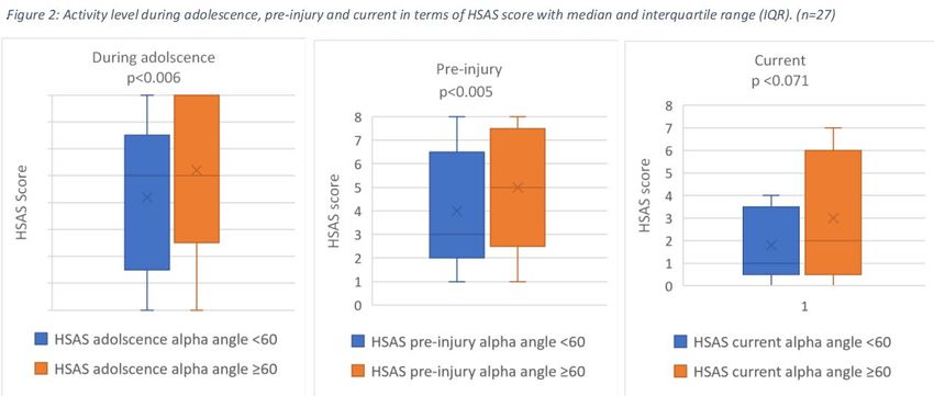
The HSAS level, presented as median (interquartile range) can be seen in Figure 2.

Patients with bilateral symptomatic LHGP
Patients with bilateral symptomatic hip/groin pain were calculated separately. Descriptive data is
presented in Table 4. Three (33.3 %) patients had an increased alpha angle (> 60 degrees), all male.
The mean alpha angle in the symptomatic hip was 43.8 (95% CI 37.5-50.1) among females and 67
(95% CI 47.4-86.6) among males p=0.058.

Table 4. Patients characteristics for all patients with bilateral pain, patients with an alpha angle
< 60 degrees and patients with an alpha angle ≥60 degrees. Data is expressed as mean and
standard deviation (SD) unless otherwise stated

                                    All patients    Patients with an alpha   Patients with an alpha
 Descriptive data                   (n=9)           angle
67.9), ADL 75 (35-87.5), sports 43.7 (23.4-57.8), Physical activities 12.5 (0-56.2), QoL 25 (20-35).
Only three patients presented with an increased alpha angle. The HSAS level among patients with
bilateral symptomatic LHGP was 6 (5-7) during adolescence, 5 (4.5-7) pre-injury, and present level
was 3 (1-5.5). No significant change in HSAS can been seen since the patient group is too small.
11Discussion
This exploratory study is to our knowledge, the first to investigate the alpha angle’s association, in
a symptomatic patient group, to validated PRO:s. The results suggest that an alpha angle >60
degrees is not associated with worse patient-reported outcomes in patients with long-standing hip
and groin pain. Patients with LHGP showed overall lower results in all HAGOS subscales
compared to a control group (12) but very similar HAGOS results were seen between patients with
LHGP with an alpha angle 60 degrees. Kopec et al. (13)
examined the association between radiographic measurements of hip morphology (cam and pincer)
among a general population with and without hip pain. The alpha angle was defined in the 45
degree bilateral Dunn view and the LCE angle was defined in the AP view. Data from 500 subjects
were obtained. Even if a higher alpha angle indicated worse HAGOS scores no significant
difference could be seen in the HAGOS profile between those with an alpha angle 60 degrees. Measuring the alpha angle in plain radiographs is simpler, cheaper and less-
time consuming than CT or MRI. Nepple et al. (14) describe that plain radiographs effectively
identify femoral head-neck malformation. The Dunn view showed highest sensitivity of detecting
abnormal alpha angles (71-80%) but the lauenstein (frog-leg lateral) showed the best specificity (91-
100%). Pålsson et al. (7) have demonstrated that measuring the alpha angle in the Lauenstein
position is simple and reproducible, even in unexperienced examiners. Even if we have the same
alpha angle criteria as Kopec et al.(13) the inclusion criteria differed and our study only contained
symptomatic subjects. By using the Dunn view Kopec et al.(13) might have categorized a larger
patient group with a pathological alpha angle compared to our study that used the Lauenstein
projection. Our study presented overall a lower score in all HAGOS subscales compared to Kopec
et al.(13). However our patient population might have had worse radiographic findings and
therefore more symptoms which is shown in the HAGOS profile. This can be an obvious reason
why our patient group had overall a worse HAGOS profile. Sansone et al. (15) compared HAGOS
scores pre-operative with PRO:s two(15) and five(16) years after hip arthroscopic surgery. All
patients met inclusion criterias for surgery but no alpha angle was described. The pre-operative
HAGOS results were very similar to ours.
The alpha angle is commonly used as a radiologic sign that defines a CAM-morphology and
although the cut-off for pathological alpha angle differs in different studies, a common used
12threshold is 60 degrees. It is therefore used as an indication for surgery (5). However, several studies
have failed to demonstrate a correlation between alpha angle correction after surgery and patient-
reported outcomes, as well as patient satisfaction. Stähelin et al. (17) included 22 patients under-
going hip arthroscopic offset correction. A normal alpha angle was defined as 50 degrees or less.
Restoration was consider accurate if a normal angle or a reduction of 20 degrees or more was
achieved. No more than 20% of the femoral neck diameter was removed to avoid risk of fracture.
The mean alpha angle preoperative was 75.1 + 12.7 (range 58-100) degrees and postoperative 53.8
+ 9.2 (40-72) degrees. Postoperative no significant difference could be seen clinically and in PRO:s
between patients with an alpha angle reduced to 50 degrees. Neither a difference depending on the precent of correction.
However all patients experienced improved mobility and less pain postoperative (17). Briggs et al
(Briggs) presented similar result with 230 patients included. The median preoperative alpha angle
was 72 (50-105) degrees and the median postoperative alpha angle was 45 (30-100) degrees. Two
groups were compared, patients that postoperative had an alpha angle 55
degrees. PRO:s 5 years after surgery did not show that patients with a larger postoperative alpha
angle had lower patient-reported outcomes (18). A recent systematic review compared relationship
between alpha angle correction and outcomes. A reduction of alpha angle to 55 degree or less were
recommended and should improve outcome scores. However the review also point out that a no
reduction more than 20 degrees should be considered. Mixed evidence on the ability of the alpha
angle to predict patient-reported outcomes were also highlighted (19).
Beck et al. (20) described that the main difference between a normal hip and a hip with FAI
syndrome is abnormal joint morphology. Threshold values defining normal from pathological is
therefore difficult. However since many patients without symptoms have an increased alpha angles
it is important that the threshold value is considered as one of several classification criteria that
supports the diagnosis of FAI syndrome and not a diagnostic criteria for surgery. (5, 21, 22).
Threshold values differs between literature partly due to different measuring techniques. Most
studies use CT or MRI but some use plain radiographs. Different radiographic views are also
debatable in order to obtained optimal scans. The frog-lateral, Lauenstein or Dunn-view is the
most recommended (14, 23). Pollard et. al (8) examined the reference interval for cam deformity
in the cross-table lateral view and found a mean value of 46-49 degrees (sex dependent) and a 95%
confidence interval of 32-62 degrees. Nötzli et al. (24) reported an average alpha angle using MRI
scans of 74 degrees among patients and 42 degrees among controls but recommended a threshold
value of 55 degrees. Sutter et al. (22) showed a great overlap in alpha angle measurement between
13symptomatic and asymptomatic patients who underwent MRI. While 55 degrees alpha angles had
a good sensitivity the specificity was low. By increasing the cutoff value to 60 degrees the false
positive results were reduced but a good sensitivity still remained. Barrientos et. al. (25) reported
using CT an average angle of 67 degrees among patients compared to 58 degrees among controls.
They used a cutoff value of 57 degrees. Allen et al. (26) chose in 2009 a cutoff value of 55 degrees
for defining a cam-deformity using CT-imaging. However this study also showed that painful hips
were more likely to have an alpha angle over 60 degrees with an odds ratio of 2.59 compared to
hips with alpha angles of 60 degrees or less.
One theory is that the range of alpha angles depends on gender and that males overall have higher
alpha angle value compared to females (8, 22, 27). Discussions if different cut-off values depending
on gender have been proposed (28) but overall a non-sex specific threshold value are described in
the literature (5).
There is a risk with overanalyzing radiographic findings, since common radiographic signs of FAI
syndrome are prevalent even in asymptomatic individuals (29). In our study 100% of the patients
had one or more signs of impingement such as high alpha angle, low head-neck offset, pincer or
coxa profunda. The risk of using only radiological findings as diagnostic criterions could risk over-
diagnosing. Palsson et al. (7) present, from the same data as this report, that only about half of the
patients referred to tertiary care were potential candidates for surgery.
Both patient groups had a lower current activity level compared to pre-injury level as well as
adolescence. The patients with an alpha angle >60 degrees showed a significant higher activity level
during both adolescence, pre-injury and current compared to the patients with and alpha angle <
60 degrees. Indications of high impact training at a young age has been showed in previous studies
to be a predispose factor to develop CAM morphology during skeletal maturation and thereby
results in increased alpha angles (30-32).
The same patient group has a higher pre-injury and higher current HSAS score which could indicate
that the group has greater physical demands since youth. However the scale has categorized
different sports into different levels depending on the assumed load of the hip joint and range of
motion and do not describe duration of activity and frequency of exercise making it more difficult
to make larger clinical assumptions (10).
14Limitations
This is an exploratory study. The number of patients included in the study is small and therefore
there is a risk for type II statistical error.
All patients had been referred to the Orthopedic Department of a University hospital, serving as a
regional hospital, to an orthopedic surgeon for assessment for surgery. Therefore the patient group
had server hip related symptoms where prior treatments had failed. One can therefore argue that
these patients had worse symptoms compared to the average FAI syndrome patient.
By including patients with long-standing hip and groin pain prior to diagnosis, one can argue that
our patient population is heterogenic.
Since we only used plain radiographs, we may have underestimated the size of the CAM-lesion in
some patients. Furthermore, our threshold values can be used in order to detect patients with more
symptoms, although a number of patients with alpha angles lower than 60 degrees may also suffer
from FAI syndrome.
HSAS was used to investigate patient-reported activity level. However the patient reported
outcome measure do not report the load of the hip as duration of activity, frequency and intensity.
Therefore comparison between patients can be difficult. The pre-injury activity level as well as
adolescence activity level is reported retrospectively.
Conclusion
In this exploratory study patients with longstanding hip and groin pain and alpha angles over 60
degrees report similar outcomes to patients with alpha angles under 60 degrees.
15References
1. Ganz R, Parvizi J, Beck M, Leunig M, Nötzli H, Siebenrock KA. Femoroacetabular
impingement: a cause for osteoarthritis of the hip. Clin Orthop Relat Res. 2003(417):112-20.
2. Griffin DR, Dickenson EJ, O'Donnell J, Agricola R, Awan T, Beck M, et al. The Warwick
Agreement on femoroacetabular impingement syndrome (FAI syndrome): an international
consensus statement. Br J Sports Med. 2016;50(19):1169-76.
3. Larson CM, Sikka RS, Sardelli MC, Byrd JW, Kelly BT, Jain RK, et al. Increasing alpha angle
is predictive of athletic-related "hip" and "groin" pain in collegiate National Football League
prospects. Arthroscopy. 2013;29(3):405-10.
4. Dickenson E, Wall PD, Robinson B, Fernandez M, Parsons H, Buchbinder R, et al.
Prevalence of cam hip shape morphology: a systematic review. Osteoarthritis Cartilage.
2016;24(6):949-61.
5. van Klij P, Reiman MP, Waarsing JH, Reijman M, Bramer WM, Verhaar JAN, et al.
Classifying Cam Morphology by the Alpha Angle: A Systematic Review on Threshold Values.
Orthop J Sports Med. 2020;8(8):2325967120938312.
6. Clohisy JC, Carlisle JC, Beaulé PE, Kim YJ, Trousdale RT, Sierra RJ, et al. A systematic
approach to the plain radiographic evaluation of the young adult hip. J Bone Joint Surg
Am. 2008;90 Suppl 4(Suppl 4):47-66.
7. Pålsson A, Kostogiannis I, Lindvall H, Ageberg E. Hip-related groin pain, patient
characteristics and patient-reported outcomes in patients referred to tertiary care due to
longstanding hip and groin pain: a cross-sectional study. BMC Musculoskelet Disord.
2019;20(1):432.
8. Pollard TC, Villar RN, Norton MR, Fern ED, Williams MR, Simpson DJ, et al.
Femoroacetabular impingement and classification of the cam deformity: the reference
interval in normal hips. Acta Orthop. 2010;81(1):134-41.
9. Thomeé R, Jónasson P, Thorborg K, Sansone M, Ahldén M, Thomeé C, et al. Cross-cultural
adaptation to Swedish and validation of the Copenhagen Hip and Groin Outcome Score
(HAGOS) for pain, symptoms and physical function in patients with hip and groin disability
due to femoro-acetabular impingement. Knee Surg Sports Traumatol Arthrosc.
2014;22(4):835-42.
1610. Naal FD, Miozzari HH, Kelly BT, Magennis EM, Leunig M, Noetzli HP. The Hip Sports
Activity Scale (HSAS) for patients with femoroacetabular impingement. Hip Int.
2013;23(2):204-11.
11. Thorborg K, Hölmich P, Christensen R, Petersen J, Roos EM. The Copenhagen Hip and
Groin Outcome Score (HAGOS): development and validation according to the COSMIN
checklist. Br J Sports Med. 2011;45(6):478-91.
12. Wörner T, Nilsson J, Thorborg K, Granlund V, Stålman A, Eek F. Hip Function 6 to 10
Months After Arthroscopic Surgery: A Cross-sectional Comparison of Subjective and
Objective Hip Function, Including Performance-Based Measures, in Patients Versus
Controls. Orthop J Sports Med. 2019;7(6):2325967119844821.
13. Kopec JA, Qian H, Cibere J, Wong H, Li LC, Barber M, et al. Relationship Between Hip
Morphology and Hip-Related Patient-Reported Outcomes in Young and Middle-Aged
Individuals: A Population-Based Study. Arthritis Care Res (Hoboken). 2019;71(9):1202-8.
14. Nepple JJ, Martel JM, Kim YJ, Zaltz I, Clohisy JC. Do plain radiographs correlate with CT
for imaging of cam-type femoroacetabular impingement? Clin Orthop Relat Res.
2012;470(12):3313-20.
15. Sansone M, Ahldén M, Jónasson P, Thomeé C, Swärd L, Öhlin A, et al. Outcome after hip
arthroscopy for femoroacetabular impingement in 289 patients with minimum 2-year follow-
up. Scand J Med Sci Sports. 2017;27(2):230-5.
16. Öhlin A, Ahldén M, Lindman I, Jónasson P, Desai N, Baranto A, et al. Good 5-year
outcomes after arthroscopic treatment for femoroacetabular impingement syndrome. Knee
Surg Sports Traumatol Arthrosc. 2020;28(4):1311-6.
17. Stähelin L, Stähelin T, Jolles BM, Herzog RF. Arthroscopic offset restoration in
femoroacetabular cam impingement: accuracy and early clinical outcome. Arthroscopy.
2008;24(1):51-7.e1.
18. Briggs KK, Soares E, Bhatia S, Philippon MJ. Postoperative alpha angle not associated with
patient-centered midterm outcomes following hip arthroscopy for FAI. Knee Surg Sports
Traumatol Arthrosc. 2019;27(10):3105-9.
19. de Sa D, Urquhart N, Philippon M, Ye JE, Simunovic N, Ayeni OR. Alpha angle correction
in femoroacetabular impingement. Knee Surg Sports Traumatol Arthrosc.
2014;22(4):812-21.
20. Beck M, Kalhor M, Leunig M, Ganz R. Hip morphology influences the pattern of damage
to the acetabular cartilage: femoroacetabular impingement as a cause of early osteoarthritis
of the hip. J Bone Joint Surg Br. 2005;87(7):1012-8.
1721. Mascarenhas VV, Ayeni OR, Egund N, Jurik AG, Caetano A, Castro M, et al. Imaging
Methodology for Hip Preservation: Techniques, Parameters, and Thresholds. Semin
Musculoskelet Radiol. 2019;23(3):197-226.
22. Sutter R, Dietrich TJ, Zingg PO, Pfirrmann CW. How useful is the alpha angle for
discriminating between symptomatic patients with cam-type femoroacetabular impingement
and asymptomatic volunteers? Radiology. 2012;264(2):514-21.
23. Saito M, Tsukada S, Yoshida K, Okada Y, Tasaki A. Correlation of alpha angle between
various radiographic projections and radial magnetic resonance imaging for cam deformity
in femoral head-neck junction. Knee Surg Sports Traumatol Arthrosc. 2017;25(1):77-83.
24. Nötzli HP, Wyss TF, Stoecklin CH, Schmid MR, Treiber K, Hodler J. The contour of the
femoral head-neck junction as a predictor for the risk of anterior impingement. J Bone Joint
Surg Br. 2002;84(4):556-60.
25. Barrientos C, Barahona M, Diaz J, Brañes J, Chaparro F, Hinzpeter J. Is there a pathological
alpha angle for hip impingement? A diagnostic test study. J Hip Preserv Surg.
2016;3(3):223-8.
26. Allen D, Beaulé PE, Ramadan O, Doucette S. Prevalence of associated deformities and hip
pain in patients with cam-type femoroacetabular impingement. J Bone Joint Surg Br.
2009;91(5):589-94.
27. Kapron AL, Anderson AE, Aoki SK, Phillips LG, Petron DJ, Toth R, et al. Radiographic
prevalence of femoroacetabular impingement in collegiate football players: AAOS Exhibit
Selection. J Bone Joint Surg Am. 2011;93(19):e111(1-10).
28. Gosvig KK, Jacobsen S, Palm H, Sonne-Holm S, Magnusson E. A new radiological index
for assessing asphericity of the femoral head in cam impingement. J Bone Joint Surg Br.
2007;89(10):1309-16.
29. Frank JM, Harris JD, Erickson BJ, Slikker W, 3rd, Bush-Joseph CA, Salata MJ, et al.
Prevalence of Femoroacetabular Impingement Imaging Findings in Asymptomatic
Volunteers: A Systematic Review. Arthroscopy. 2015;31(6):1199-204.
30. Agricola R, Bessems JH, Ginai AZ, Heijboer MP, van der Heijden RA, Verhaar JA, et al.
The development of Cam-type deformity in adolescent and young male soccer players. Am
J Sports Med. 2012;40(5):1099-106.
31. Siebenrock KA, Ferner F, Noble PC, Santore RF, Werlen S, Mamisch TC. The cam-type
deformity of the proximal femur arises in childhood in response to vigorous sporting activity.
Clin Orthop Relat Res. 2011;469(11):3229-40.
1832. Philippon MJ, Ho CP, Briggs KK, Stull J, LaPrade RF. Prevalence of increased alpha angles
as a measure of cam-type femoroacetabular impingement in youth ice hockey players. Am J
Sports Med. 2013;41(6):1357-62.
19You can also read