Selenium in Human Health and Gut Microflora: Bioavailability of Selenocompounds and Relationship With Diseases
←
→
Page content transcription
If your browser does not render page correctly, please read the page content below
REVIEW
                                                                                                                                                     published: 04 June 2021
                                                                                                                                              doi: 10.3389/fnut.2021.685317

                                               Selenium in Human Health and Gut
                                               Microflora: Bioavailability of
                                               Selenocompounds and Relationship
                                               With Diseases
                                               Rannapaula Lawrynhuk Urbano Ferreira 1 , Karine Cavalcanti Maurício Sena-Evangelista 1,2 ,
                                               Eduardo Pereira de Azevedo 3 , Francisco Irochima Pinheiro 3,4 , Ricardo Ney Cobucci 3,4
                                               and Lucia Fatima Campos Pedrosa 1,2*
                                               1
                                                Postgraduate Program in Nutrition, Federal University of Rio Grande do Norte, Natal, Brazil, 2 Department of Nutrition,
                                               Federal University of Rio Grande do Norte, Natal, Brazil, 3 Graduate Program of Biotechnology, Laureate International
                                               Universities - Universidade Potiguar, Natal, Brazil, 4 Medical School, Laureate International Universities - Universidade
                                               Potiguar, Natal, Brazil

                                               This review covers current knowledge of selenium in the dietary intake, its bioavailability,
                                               metabolism, functions, biomarkers, supplementation and toxicity, as well as its
                                               relationship with diseases and gut microbiota specifically on the symbiotic relationship
                                               between gut microflora and selenium status. Selenium is essential for the maintenance
                                               of the immune system, conversion of thyroid hormones, protection against the harmful
                            Edited by:         action of heavy metals and xenobiotics as well as for the reduction of the risk of
                        Lucia A. Seale,        chronic diseases. Selenium is able to balance the microbial flora avoiding health damage
    University of Hawaii, United States
                                               associated with dysbiosis. Experimental studies have shown that inorganic and organic
                        Reviewed by:
                       Yasumitsu Ogra,
                                               selenocompounds are metabolized to selenomethionine and incorporated by bacteria
              Chiba University, Japan          from the gut microflora, therefore highlighting their role in improving the bioavailability of
                        Dalia El Khoury,       selenocompounds. Dietary selenium can affect the gut microbial colonization, which in
         University of Guelph, Canada
                                               turn influences the host’s selenium status and expression of selenoproteoma. Selenium
                   *Correspondence:
        Lucia Fatima Campos Pedrosa            deficiency may result in a phenotype of gut microbiota that is more susceptible to
                lfcpedrosa@gmail.com           cancer, thyroid dysfunctions, inflammatory bowel disease, and cardiovascular disorders.
                                               Although the host and gut microbiota benefit each other from their symbiotic relationship,
                    Specialty section:
          This article was submitted to        they may become competitors if the supply of micronutrients is limited. Intestinal bacteria
             Nutrition and Metabolism,         can remove selenium from the host resulting in two to three times lower levels of host’s
                a section of the journal
                                               selenoproteins under selenium-limiting conditions. There are still gaps in whether these
                    Frontiers in Nutrition
                                               consequences are unfavorable to humans and animals or whether the daily intake of
            Received: 24 March 2021
             Accepted: 11 May 2021             selenium is also adapted to meet the needs of the bacteria.
            Published: 04 June 2021
                                               Keywords: selenium, gut microbiota, selenocompounds, selenoproteins, selenium metabolism
                              Citation:
Ferreira RLU, Sena-Evangelista KCM,
            de Azevedo EP, Pinheiro FI,
Cobucci RN and Pedrosa LFC (2021)
                                               SELENIUM FORMS, FOOD SOURCES, AND BIOAVAILABILITY
  Selenium in Human Health and Gut
            Microflora: Bioavailability of
                                               The organic forms of Se are found as a sulfur amino acid analog, selenomethionine (SeMet),
 Selenocompounds and Relationship              selenocysteine (SeCys), and as methylated derivatives. The inorganic forms correspond to
With Diseases. Front. Nutr. 8:685317.          Se salts such as selenate (SeO−2                    −2
                                                                              4 ) and selenite (SeO3 ) (1). SeMet is found in plant- and
       doi: 10.3389/fnut.2021.685317           animal-origin products as well as in some food supplements (2). On the other hand, SeCys is

Frontiers in Nutrition | www.frontiersin.org                                           1                                               June 2021 | Volume 8 | Article 685317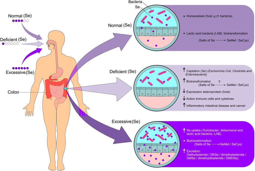
Ferreira et al. Selenium and Gut Microflora
found primarily in animal-derived food (3), whereas SELENIUM ABSORPTION, METABOLISM,
selenium-methylselenocysteine (SeMeCys) is a natural EXCRETION, AND BIOMEDICAL
monomethylated organic Se found in some vegetables
APPLICATIONS
such as garlic, onion, broccoli, and leeks (2–4). Among
the inorganic forms, selenite is present mainly in food Dietary Se intake from either organic or inorganic origin
supplements, while selenate is found in plant and fish is absorbed in the gastrointestinal tract and subsequently
sources (3). These forms of Se have been used to biofortify transported to the liver, where it is metabolized and used for
some vegetables (5, 6). producing selenoproteins, followed by its distribution to other
Brazil nuts, cereals, meat, fish, seafood, milk, and nuts are tissues of the body. Selenoamino acids are actively transported
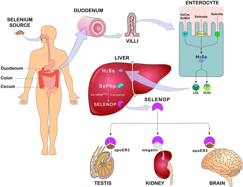
the best sources of Se (7) (Figure 1). The interaction of fish in the duodenum, cecum, and colon through various membrane
and seafood with mercury results in insoluble Se derivatives transport mechanisms, whereas selenate is transported by anion
that can reduce its bioavailability (8). In fact, the bioavailability exchangers from the family of the SLC26 gene. On the other
of Se depends primarily on its chemical form. In general, hand, there is insufficient evidence on the transport of the
the organic forms are more quickly absorbed and are usually other forms of Se (14). Se absorption, metabolism and body
used for the biosynthesis of selenoproteins (9). In addition, the distribution are represented in Figure 3.
amount of protein, fat, and heavy metal in the diet influence Although the metabolic route differs depending of the Se
the bioavailability of Se (7–10). High levels of Se are present in source, all the absorbed Se is converted to hydrogen selenide
some herbal plants such as Astragalus bisulcatus and Brassicaceae (H2 Se) in the enterocytes before the specific incorporation of
(broccoli) (11). Bertholletia excelsa, known as Brazil nut, is one selenocysteine takes place in the active site of the selenoproteins
of the highest sources of Se with concentrations that range (15). SeMet undergoes transulfurization reactions, in which
from 1.80 to 320.80 µg Se/g (12). In addition, the content cystathionine beta-synthetase catalyzes the formation of
of Se in the soil has a major influence on the amount of selenocystathionin, being further converted to SeCys by
this metal in food, being related to its deficiency and toxicity cystathionine gamma-lyase followed by conversion to H2 Se
in some regions. The Se content in the soil usually ranges by selenocysteine lyase. SeCys, from both food and the SeMet
from 1 to 1.5 µg Se/g, reaching 5.0 µg Se/g in seleniferous pathway, will also be reduced to H2 Se (16, 17). Alternatively,
soils (13) (Figure 2). SeMet can also be incorporated non-specifically into proteins,
FIGURE 1 | Foods rich in Se with their relative proportions of SeMet, SeCys, SeMeCys, selenate, selenite, and γ-GluMeSeCys. The figure shows the predominance of
SeMet in Brazil nuts, wheat grain, fish cod, and chicken (breast). Lamb meat (kidney) is rich in Se mainly in the SeCys form, whereas onion has Se almost exclusively
in the form of selenate. Garlic, potato, and broccoli have a balanced proportion of the various forms of Se. Se, selenium; SeMet, selenomethionine; SeCys,
selenocysteine; SeMeCys, selenium-methylselenocysteine; γ-GluMeSeCys, γ-glutamyl-Se-methyl-selenocysteine (Figure illustration by Francisco Irochima Pinheiro).
Frontiers in Nutrition | www.frontiersin.org 2 June 2021 | Volume 8 | Article 685317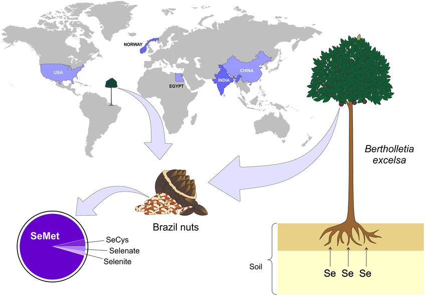
Ferreira et al.                                                                                                                              Selenium and Gut Microflora

  FIGURE 2 | Main countries with documented Se-rich soils. The presence of Se-rich soil is not uniform in the world, but some countries such as Egypt, China, India,
  Norway, and USA stand out among the others. On the other hand, some plants are more capable of retaining Se, for example - Bertholletia excelsa (Brazil nuts), which
  is a typical tree of the northern region of Brazil. Se, selenium; SeMet, selenomethionine; SeCys, selenocysteine (Figure illustration by Francisco Irochima Pinheiro).

such as albumin and hemoglobin, replacing methionine (3).                               provides information in a targeted manner to the ribosomes that
As for the inorganic forms, selenate is converted to selenite                           translate mRNAs (messenger ribonucleic acid) to selenoproteins
followed by reduction to H2 Se by thioredoxin reductase                                 (3, 18–20). Se is transported to tissues such as brain, kidneys,
(TXNRD) and thioredoxin, as well as by glutathione to form                              and testicles, mainly in the form of selenoprotein P (SELENOP)
selenodiglutathione (GS-Se-SG). Glutathione reductase converts                          through endocytosis mediated by apolipoprotein E receptor 2
the latter to glutathioselenol (GS-SeH) which reacts with                               (apoE2) and megaline (14).
glutathione to form H2 Se (3, 18–20). On the other hand,                                   H2 Se can also be methylated by thiol-S-methyltransferase
SeMeCys and the synthetic Se derivatives selenobetaine,                                 before being excreted. The main form of Se excretion is through
methylseleninic acid, and methylselenocyanate are converted                             urine, however, in cases of excessive consumption, respiratory
into methylselenol (CH3 )SeH through the enzyme cystathione                             excretion might occur. Excretion by the lungs occurs when
gamma-lyase, followed by demethylation to become H2 Se                                  the elimination of Se in the form of trimethyl selenonium
(21, 22).                                                                               (CH3 )3 Se in the urine becomes saturated, whose elimination
    In view of this cascade of reactions, all H2 Se regardless of                       occurs mainly in the form of volatile dimethyl selenide (CH3 )2 Se
its origin will be transported in the blood linked to VLDL and                          (23). In situations of moderate consumption of Se, the main
LDL fractions as well as to other proteins (albumin and alpha-                          monomethylated compound eliminated through kidneys is a
globulin). In the liver, H2 Se is converted to selenophosphate                          seleno sugar namely 1β-methylseleneN-acetyl-D-galactosamine.
(SePhp) via selenophosphate synthetase (SEPHS), which will                              The non-absorbed Se from food is incorporated into the bile,
be incorporated into selenoproteins in the form of SeCys. For                           pancreatic, and intestinal secretions, being eliminated in the
selenocysteine synthesis, the UGA codon (TGA) is used as an                             feces (23).
initiation codon, requiring a specialized tRNA (ribonucleic acid                           Se functionality occurs in the form of selenoproteins that are
carrier), which, after several reactions from the seryl-tRNA,                           encoded by the insertion of SeCys by the UGA codon in mRNA

Frontiers in Nutrition | www.frontiersin.org                                        3                                              June 2021 | Volume 8 | Article 685317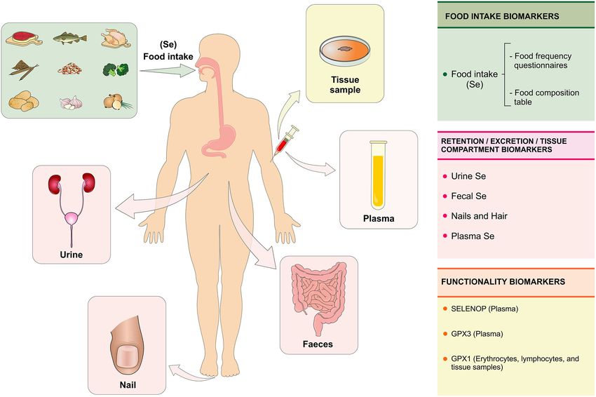
Ferreira et al.                                                                                                                              Selenium and Gut Microflora

  FIGURE 3 | Se absorption, metabolism, and distribution. After eating Se-rich foods in its organic and/or inorganic form, Se absorption occurs in the duodenum,
  cecum, and colon. In enterocytes, SeMet and SeCys are absorbed by active transport (systems B0 and b0 + rBAT) while selenate is absorbed by passive transport
  (anion changers of the SLC26 gene family). After absorption, all forms of Se are converted to H2 Se through reactions that occur in the enterocyte and transported in
  the blood bound LDL, VLDL (mainly). In the liver, H2 Se is converted to SePhp and incorporated into selenoproteins in the form of SeCys. Transport to other tissues
  such as testis, kidneys, and brain occurs mainly in the form of SELENOP through receptor-mediated endocytosis - apoE2 and megaline. Se, selenium; SeMet,
  selenomethionine; SeCys, selenocysteine; H2 Se, hydrogen selenide; LDL, low-density lipoprotein; VLDL, Very low-density lipoprotein; SePhp, selenophosphate;
  SELENOP, selenoprotein P; apoE2, apolipoprotein E receptor 2 (Figure illustration by Francisco Irochima Pinheiro).

under specific conditions. Most of these selenoproteins are                             levels using biomarkers of intake, retention/excretion, and
involved in the regulation of redox signaling and are grouped into                      concentration on tissues as well as biomarkers of functionality
families such as glutathione peroxidases (GPXs), iodothyronine                          (22) (Figure 4).
deiodinases (DIOs), TXNRDs, and SELENOP. Thus, the main
biomedical applications attributed to Se are related to its                             Biomarkers of Intake
antioxidant activity, regulation of thyroid hormone metabolism,                         The assessment of Se intake can be performed using methods
anticarcinogenic property, and prevention of cardiovascular                             of assessing food consumption, such as the food frequency
diseases. SELENOP acts as the main Se transporter for peripheral                        questionnaire. The Se content on foods is estimated using food
tissues in addition to performing extracellular antioxidant                             composition tables (22). Algeria ’s population consumes a wide
function (14, 22).                                                                      variety of Se-rich foods, such as seafood, meat, eggs, milk,
                                                                                        and legumes, however, no significant associations were found
SELENIUM BIOMARKERS                                                                     between dietary patterns and Se biomarkers, such as Se in
                                                                                        plasma, SELENOP, and GPX. Predicting the Se status from food
The evaluation of Se status determines the amount of this                               consumption remains a challenge due to the lack of precision
biologically active nutrient as a function of intake, retention,                        of nutrient content in food composition tables, considering the
and metabolism. Thus, the Se status can be assessed at three                            variation in the Se concentration of foods as a function of Se

Frontiers in Nutrition | www.frontiersin.org                                        4                                              June 2021 | Volume 8 | Article 685317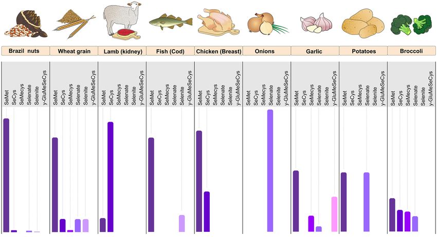
Ferreira et al.                                                                                                                                Selenium and Gut Microflora

  FIGURE 4 | Se biomarkers. The Se status is evaluated with the purpose of quantifying the biologically active or potentially active nutrient as a function of Se intake,
  retention, and metabolism in the body. The Se status can be assessed at three levels using specific biomarkers: (1) biomarkers of intake (assessment of food
  consumption through the food frequency questionnaire); (2) biomarkers of retention/excretion and concentration in tissues (urine, feces, nails, hair, and plasma) and (3)
  biomarkers of selenium functionality (SELENOP and GPX3 in plasma and GPX1 in erythrocytes, lymphocytes, and tissue samples). Se, selenium; SELENOP,
  selenoprotein P; GPX1, glutationa peroxidase; GPX3, glutationa peroxidase 3 (Figure illustration by Francisco Irochima Pinheiro).

content in the soil (22, 24). In addition, Se from the diet affects                      (28). The evaluation of Se in urine can be a sensitive parameter for
colonization of the microbial intestine, which in turn influences                        occupational exposures of Se in the short term, but the knowledge
the host’s Se status and selenoproteoma expression (25).                                 about specificity and kinetics of this elimination pathway is still
                                                                                         little explored (27).
Biomarkers of Retention/Excretion and Se                                                     The measurement of the Se concentration in urine is
Concentrations in Tissues                                                                considered as a potentially viable biomarker of Se status in
The retention of Se in the body can be assessed by the difference                        population studies. Additionally, the concentration of Se in the
between the amount of Se ingested and the sum of Se in the                               urine can be used to identify regional variations in the status
urine and feces, which requires the collection of total urine and                        of Se and might reflect differences in the amount of Se in food
excreted feces for a few days. Alternatively, it is recommended                          according to the type of soil. This evidence supports the need
to evaluate the concentration of creatinine in the urine to reduce                       for reviewing the policies of national systems for monitoring
the error associated with the variation in urinary excretion. Renal                      micronutrient deficiencies including Se (26).
excretion is the main route of elimination of absorbed Se (22).                              The concentration of Se in the nail is considered a superior
Genetic and environmental factors, as well as body size, age, and                        biomarker of Se status, as it provides an integrated measurement
sex can influence the retention and excretion of Se in the urine                         of long term exposure (up to 1 year), while blood biomarkers
(26). However, studies on Se in urine for biological monitoring                          indicate a short term exposure (29). Toenails are considered non-
are scarce, especially with regard to occupational exposure, in                          invasive matrices and are used in large epidemiological studies
which inhalation is the main route of exposure. After inhalation                         because they present slow growth, easy collection and have
of high concentrations of Se by workers, inflammatory effects                            less influence from external contamination. The standardization
were observed in the respiratory tract (27). An increased intake of                      of sample collection, quality control, and analytical techniques
Se is reflected rapidly in the increased excretion of Se in the urine                    are important to consolidate the usefulness of this matrix in

Frontiers in Nutrition | www.frontiersin.org                                         5                                               June 2021 | Volume 8 | Article 685317
Ferreira et al. Selenium and Gut Microflora
epidemiological studies (30). The Se content in nails has a direct SELENIUM AND DISEASES
relationship with SELENOP and most organic forms of Se,
especially SeCys, whereas it has an inverse relationship with the Se plays a crucial role in normal physiology and contributes to the
amount of the inorganic forms, such as selenite and selenate. This pathophysiology of various diseases. Due to its antioxidant and
opposite behavior may be related to the composition of human anti-inflammatory properties, several studies have evaluated the
nails, which are mainly made of proteins rich in cysteine, the impact of Se status in conditions characterized by inflammation
latter being able to form complex with Se (31). and oxidative stress, which includes diabetes, metabolic
There are controversies about the use of Se content in nails and syndrome, cancer, cardiovascular, and neurodegenerative
hair as a way to assess the effectiveness of Se supplementation. A diseases (38).
systematic review performed with 18 Se supplementation studies Inadequate serum Se levels may increase the risk for
found no evidence to support the use of the Se content in the development of several diseases, especially cardiovascular
the nail and hair as a reliable measurement of effectiveness of disorders, but it also may lead to cancer, liver diseases, and
Se supplementation (32). Se content in hair has been used to arthropathies. On the other hand, excessive consumption of Se
assess long-term Se status in epidemiological studies, offering can cause selenosis, which leads to symptoms such as fatigue,
the advantage of being a low-cost method and easy to store the tachycardia, nausea, and diarrhea. Chronic selenosis can cause
samples. The Se concentration in hair and nails are excretory liver and kidney necrosis, neurological disorders and might
forms of Se. Therefore, both reflect the previous status, being compromise the reproductive and immune systems (39).
more useful as biomarkers in studies of populations with stable In three large cohorts, the high serum Se concentration was
dietary patterns (22). associated with reduced mortality (40). Another larger study,
Plasma Se concentration is a more useful biomarker to in which more than 13,000 adults were followed for 12 years,
assess Se status in humans, considering the stability of Se in revealed that serum Se greater than or equal to 135 µg/L
this compartment (22). A systematic review has recommended were associated with reduced cancer mortality (41, 42). Meta-
the use of plasma Se concentration as a reliable biomarker analysis involving 16 prospective studies demonstrated an inverse
in supplementation studies with adults of both sexes. The relationship between Se status and cardiovascular risk (43).
measurement of Se in plasma has shown to be effective in Likewise, a systematic review with meta-analysis involving 13
reflecting changes in the amount intake (supplementation) in studies revealed that high physiological levels of Se are associated
individuals with intermediate or high Se concentrations at with lower incidence and lower mortality from cardiovascular
baseline. In addition, this review highlights the usefulness of Se disease (CVD) (44). In another meta-analysis in which more
in erythrocytes and whole blood as markers of Se status, both of than 40 thousand participants in randomized clinical trials were
which are reported as markers of long-term status (32). included, the authors found that Se supplementation decreases
the serum levels of C-reactive protein and increases the levels of
GPX, suggesting a positive effect on reduction of inflammation
Biomarkers of Selenium Functionality and oxidative stress in cardiovascular diseases (45).
Biomarkers of Se functionality include SELENOP, which Selenium-binding protein 1 (SELENBP1), an intracellular
comprises 20–70% of Se in plasma; GPX3, which comprises 10 protein involved in Se metabolism and redox control, has been
to 25% of Se in plasma and GPX1, which can be tested on identified as a circulating biomarker for cardiac events in patients
erythrocytes, lymphocytes, oral cells, and tissue biopsy specimens with suspected acute coronary syndrome. At the molecular level,
(22). Plasma SELENOP has been considered a useful biomarker it seems that hypoxia acts as a modulator of SELENBP1, therefore
of Se status in populations with relatively low Se intake, but not reducing the oxidative stress and controlling the lower oxygen
in populations with high intake that already had high levels of supply (46).
Se before supplementation began (32). SELENOP has shown to Previous studies have shown that circulating Se plays
be a reliable and sensitive Se status biomarker, providing dose an important role in the pathogenesis of abnormal glucose
response that can be used to estimate the Se intake required to metabolism, especially at high concentrations (47, 48). High
reach its plateau in the plasma (33). It seems that SELENOP exposure to Se can affect the expression of the main regulators
reaches a plateau after supplementation with selenite at doses of glycolysis and gluconeogenesis, through actions mediated
around 400 µg/day (34). by the GPX1 (49), as shown in studies that evidenced
GPX is one of the main selenoproteins that belongs to the that the overexpression of this selenoprotein causes insulin
cellular antioxidant defense system. The recommended Se intake resistance (50).
was calculated based on optimal plasma GPX3 activity due to A review study has elucidated the relationship between Se
the hierarchy of selenoproteins. It also considers the necessary status and cerebral Se homeostasis via SELENOP. In fact,
amounts of Se for normal concentrations of other biologically Se SELENOP may be involved in some brain disorders, in particular
compounds (35). A cohort study conducted with 51 participants in Alzheimer’s disease, providing Se for brain tissue to produce
with adequate Se intake investigated the association between selenoproteins. In addition, it competes with amyloid-β for metal
plasma Se, GPX activity, and SELENOP. The results were ions and redox-active metals, such as copper and iron. This
discrepant between plasma Se concentrations and GPX activity, study points out the involvement of SELENOP in signaling
suggesting other factors may impact the activity of this enzyme pathways in neuronal and glial tissues, including neuronal
such as genetic polymorphisms (36, 37). calcium homeostasis and excitotoxicity (51).
Frontiers in Nutrition | www.frontiersin.org 6 June 2021 | Volume 8 | Article 685317Ferreira et al. Selenium and Gut Microflora
SELENIUM SUPPLEMENTATION
61.3 (57.7–65.1)d
57.2 (45.8–67.5)c
53.6 ± 20.4a
22.8 ± 5.0a
GPX (Ug/Hb) erythrocyte
Brazil nuts (Bertholletia excelsa, family Lecythidaceae) are
After
known to be the richest source of Se with high SeMet
content and therefore, it has been widely used in studies
of Se supplementation. Regular consumption of Brazil
d
c
nuts results in optimum plasma Se and erythrocytes
48.7 (37.5–57.6)
61.8 (58.8–65.1)
20.6 ± 4.4ª,b
a
36.6 ± 17.0
concentrations, as well as in better activity of selenoenzymes
Before
(52–55) antioxidant state (56), muscular retention (57), and
inflammation status (54, 58). It is important to consider genetic
variants in selenoprotein genes (55) and pre-stratification
of the population prior to starting the trials as a way to
c
55.5(37.1–150.6)
d
avoid possible differentiated responses depending on the
3.9 (3.7–4.1)
After
Se status in each individual (59). Studies on the effects of
SELENOP (ng/mL)
supplementation with Brazil-nuts on selenium biomarkers are
shown in Table 1.
The effect of Brazil nuts on the human intestinal microbiota is
3.4 (3.2–3.5) mg/L
still unknown. It is well-known, however, that Brazil nuts contain
c
37.7 (16.1–51.9)
fiber, unsaturated fatty acids, and polyphenols that may impact
d
Before
the composition of the gut microbiota and overall gut health.
A systematic review with meta-analysis including randomized
controlled trials on nut consumption investigated the intake of
almonds (n = 5 studies), walnuts (n = 3 studies), and pistachios
d
267.0 (252.8–282.0)
(n = 1 study) and demonstrated a significant increase in the
150.81 ± 17.4a
c
a
244 (226–278)
132.5 ± 34.9
gut content of genera Clostridium, Dialister, Lachnospira, and
After
Roseburia, as well as a significant decrease in Parabacteroides.
Se in plasma (µg/L)
The nuts did not show any significant influence on bacterial
phyla, bacterial diversity or stool output (60). Other studies
have only reported an increase in the abundance of butyrate-
d
producing bacteria after nuts (61, 62) and pistachios (63) intake,
90.7 (86.4–95.2)
c
90.8 ± 12.63a
87.1 (82–97.7)
a
55.7 ± 13.3
without demonstrating any effect on the overall composition of
Before
the microbiome.
The Nutritional Prevention of Cancer (NPC) trial showed the
TABLE 1 | Studies on the effects of supplementation with Brazil-nuts on selenium biomarkers.
effectiveness of supplementing 200 µg/day of Se, as selenized
yeast, in reducing the risk of prostate, lung, and colorectal cancers
period (w)
Ingestion
(64). Se supplementation was also reported to decrease CVD and
12
related mortalities (65, 66). In a group of healthy New Zealand
8
8
8
men, Se supplementation as selenized yeast (Selplex, 200 µg/day)
in the form of SeMet, significantly increased Se levels, improved
Supplemented
blood GPx was assayed as a measure of erythrocyte GPx activity.
the TXNDR activity and enhanced DNA stability (59). Due to the
dose (µg)
1261.4
positive results from the NPC trial, the Selenium and Vitamin
100
290
300
E Cancer Prevention Trial (SELECT) was undertaken in 35,000
healthy US men randomly assigned to 4 groups (selenium-−200
mcg/d from L-selenomethionine, vitamin E-−400 IU/d of all rac-
New Zealand 59 adult men
Brazil 130 healthy adults
alpha-tocopheryl acetate, selenium + vitamin E, and placebo).
Brazil 37 obese women
Brazil 55 obese women
Country population
Neither selenium nor vitamin E, alone or in combination, was
able to prevent prostate cancer in this population (67).
Observational studies and randomized clinical trials
conducted with high-dose Se supplementation have shown
controversy over the effects on diabetes mellitus, indicating that
c Median (interquartile interval).
d Geometric means (CI 95%).
both excess and deficiency of Se may be associated with higher
risks of diabetes mellitus type 2 (DM2) (68–71). In a NPC trial,
Cominetti et al. (53)
Thomson et al. (52)
participants were randomly assigned to receive 200 µg/day of Se
Donadio et al. (55)
Duarte et al. (54)
(as high-selenium yeast). This group were more likely to develop
± SD.
DM2 than those assigned to placebo. In a randomized controlled
Studies
study involving patients with DM2 and cardiovascular disease,
b Whole
a Mean
supplementation with 200 µg Se/day resulted in a significant
Frontiers in Nutrition | www.frontiersin.org 7 June 2021 | Volume 8 | Article 685317Ferreira et al. Selenium and Gut Microflora
decrease in insulin, HOMA-IR, C-reactive protein and an The microbiome is capable of encoding more than three
increase in total antioxidant capacity (70). million genes. It carries out a variety of metabolic functions not
attainable by the human host, which includes the production of
some types of vitamins and bioactive compounds, the synthesis
SELENIUM TOXICITY of essential and non-essential amino acids, the metabolism
of non-digestible carbohydrates and the activity in neural,
Se toxicity can affect individuals as a result of occasional overdose
hormonal, and immunological signaling through the gut-brain
that usually occurs with intake of incorrectly formulated
axis. Furthermore, it acts on the absorption of nutrients and as
supplements (72) or due to the excess of Se intake in randomized
an epithelial barrier for pathogens (78). In this sense, imbalances
clinical trials, in which doses of 200 µg/day or more are
in the intestinal ecosystem or in two-way communication with
administered for a substantial period of time (73). Acute toxicity
the brain are associated with gastrointestinal disorders, metabolic
from excessive Se exposure causes stomach pain, headache,
diseases, and neurobehavioral disorders. Therefore, strategies
respiratory symptoms, changes in blood pressure, vomiting, and
have been developed to manipulate the microbiome, with the
nausea. Chronic oral intake of high amounts of Se results in
aim of preventing and/or reversing conditions that are harmful
selenosis, a condition characterized by hair loss, deformation and
to health (81).
loss of nails, tooth discoloration, garlic breath, gastrointestinal
Comparative genomics provides a powerful tool for
disturbances, skin rash, numbness, paralysis, and occasional
investigating genes, pathways and evolutionary changes across
hemiplegia (74). Other outcomes have been reported such as
multiple lineages (82). In the past decade, studies have been
dermatitis, increased mortality (73), DM2 (68) and increased
conducted evaluating the use of Sec Trait in ∼600 bacterial and
incidence of prostate cancer (67), which are also observed in
archaeal genomes, in which the organisms rich in selenoproteins
Se deficiency.
were the anaerobic Deltaproteobacteria and Clostridia classes,
Increased mortality has been reported at the highest dose of Se
especially Syntrophobacter fumarroxidans, with the highest
in the Danish PRECISE, a randomized, double-blinded, placebo-
prokaryotic selenoproteoma reported (83).
controlled, clinical trial performed with four groups treated with
Traces of Se and related key genes have been evaluated
100, 200, or 300 µg Se/day as Se-enriched yeast or placebo yeast.
in over 2,300 bacterial and archaea genomes, identifying a
The results of this study warn that a 300-µg/day dose of Se (as
phylogenetic and genomic mosaic pattern among organisms
Se yeast) taken for 5 years in a country with moderately low Se
using Se in different forms. This profile suggests new genes whose
status can increase all-cause mortality by 10 years later (73).
encoded proteins participate in Se metabolism and homeostasis
The levels of dietary exposure that is able to induce selenosis
in prokaryotes, such as YedE involved in Se transport, YedF
and Se toxicity is difficult to establish due to the fact that toxicity
which transcribes redox protein and LysR Se known as specific
is affected by the chemical form of Se and its bioavailability.
transcriptional Se-regulator (84).
Furthermore, interactions of Se with other dietary components,
Evolutionary trends in the use of Se and selenoproteins
the individual’s genotype and intestinal microbiota are also
indicate more than 5,200 bacterial genomes, with the majority
factors that influence the Se toxicity. Even in the face of
being related to the host, resulting in the largest Se utilization
Danish PRECISE, some populations exposed to excess Se
map in this realm. However, of this total, 2/3 of the bacteria
did not develop adverse effects. Such conditions suggest that
do not use Se, suggesting that this ability has been lost over
there are mechanisms for genetic adaptation that might be
time. Environmental factors and use of Se were also investigated,
involved in oscillations in the Se intake, which are mediated by
revealing that Se-cofactor trait (68%) and Sec Trait (37%) appear
polymorphisms, complexation of SELENOP with toxic elements
to favor the conditions of host-associated bacteria, while SeU trait
such as cadmium, arsenic, and mercury forming products of Se
prefers aquatic species that have been isolated mainly from the
excretion (75, 76). The metabolism of Se by intestinal bacteria
sea or freshwater (85). These macro-evolutionary trends extend
also favors the excretion of excess of Se (41, 77).
to cell respiration and temperature characteristics, in which
anaerobic conditions can significantly promote the use of the
SELENIUM AND GUT MICROBIOTA Se-cofactor trait and lead to the evolution of new selenoprotein
genes. Temperature seems to affect the use of Se, in which
The human digestive tract is inhabited by several microorganisms thermophilic (Ferreira et al. Selenium and Gut Microflora
of medicines (88). Specific foods and dietary patterns can SeMet, selenohomolanthionine (SeHLan), selenocenoine
influence the abundance of different types of bacteria in the (SeCys2), 1β-methyl-acetyl-D-galactosamine (SeSug1), except
intestine. For instance, the low intake of FODMAPs (Fermentable for trimethylselenonium ion (TMSe) administered orally. The
Oligosaccharides, Disaccharides, Monosaccharides, and Polyols) authors discussed these findings based on mechanisms related to
has been identified as a nutritional therapy indicated for the relief gastrointestinal enzymes that can degrade bioselenocompounds
of gastrointestinal symptoms reported by patients with irritable into selenocompounds in the intestine (100).
bowel syndrome (IBS) and non-celiac sensitivity to gluten (89). Germ-free mice that were fed with diets with adequate
Foods rich in fructans (wheat, rye, garlic, and onion) lactose and high Se levels modified their selenoproteoma expression
(milk and dairy products), fructose (fruits and processed foods in a similar way to that of the control group but showed
containing syrups), sorbitol, xylitol red fruits, and mushrooms higher levels and activity of GPX1 and methionine-R-sulfoxide
are fermented by intestinal bacteria (Actinobacteria) and yeasts reductase 1 (MSRB1) in the liver, suggesting partial sequestration
producing hydrogen and methane gases, resulting in bloating of Se by intestinal microorganisms, therefore resulting in
symptoms, abdominal pain, and diarrhea (90). In a meta-analysis limited availability to the host. In these experiments, the
study with randomized clinical trials, the low FODMAP diet genus Parabacteroides of the phylum Bacteriodetes, showed an
was beneficial for remission of gastrointestinal symptoms in opposite correlation with Se dietary supplementation. The study
patients with IBS (91). However, the restriction of several foods concluded that dietary Se affects both the composition of the gut
may lead to a potential inadequacy of micronutrients in patients microflora and the colonization of the gastrointestinal tract (99).
who follow this dietary recommendation, resulting in significant Zhai et al. (101) compared the effects of different levels of Se
changes in the microbiota and metabolome, whose duration and dietary supplements (deficient, adequate, and supranutritional)
clinical relevance are still unknown (92, 93). on the intestinal microbiota of mice. The animals’ fecal
microbiota transplantation was performed in one of the
experiments. Supplementation conducted with different amounts
Selenium as a Modulating Agent of of Se did not significantly alter the mice’s intestinal microbiota.
Intestinal Flora It rather induced significant changes in the composition of
Dietary Se influences both the host’s selenium status and the gut microbiota. In comparison to the Se-deficient diet,
selenoproteoma expression. The intestinal microbiota can use supranutritional Se supplementation significantly decreased the
the ingested Se for the expression of its own selenoproteins. Se abundance of Dorea sp. and increased the levels of microbes with
affects the composition and colonization of the gut microbiota, potential protective effects against colitis and intestinal barrier
which may interfere with the diversity of the microbiota and dysfunction, such as Turicibacter and Akkermansi. Dorea sp. is
cause unique effects on microbial composition. About 1/4 of all one of the most common species of the intestinal microbiota
bacteria have genes that encode selenoproteins. Some of them, that supplies hydrogen and carbon dioxide in the intestine. The
such as Escherichia coli, Clostridia, and Enterobacteria classes, are authors concluded that Se supplementation can optimize the
able to colonize the gastrointestinal tract of humans and animals intestinal flora to protect against intestinal dysfunction.
(94). Selenocysteine synthase (SelA) is a pyridoxal phosphate-
dependent enzyme (PLP) (95) which catalyzes the formation of
selenocysteinyl-tRNA in bacteria from a UGA decoding tRNASec Microbiota as an Environment That Affects
(SelC) loaded with serine and selenophosphate, the product of Selenium Status
the enzyme selenophosphate synthetase (SelD). Along with SelB, Although the host and the intestinal microbiota mutually
a specific translation factor of selenocysteinyl-tRNA, SelA, SelC, benefit from a symbiotic relationship, these environments can
and SelD are components of bacterial Sec decoding, allowing the become competitors when the supply of micronutrients becomes
incorporation of Sec into specific UGA codons followed by a limited. On the other hand, the intestinal microbiota favors the
sequence of insertion of Sec elements (SECIS) (96). biotransformation of Se compounds, characterizing a dubious
The composition of the microbiota can also be modulated by situation (Figure 5). The Se uptake by intestinal bacteria can
metals that participate in microbial growth through respiratory negatively influence the expression of selenoproteins in the
mechanisms, as a source of energy for autotrophic growth, as host, which results in a two to three times lower levels of
well as to transfer and storage of electrons between cells (86). selenoproteins under Se limiting conditions. The unfavorable
Manganese, zinc, selenium, and iron act as critical cofactors consequences of this effect for humans and animals have not
for bacterial enzymes responsible for DNA replication and yet been evidenced. In view of the high propagated intake of
transcription, antioxidant action, and cellular respiration (97). probiotics, the metabolism of Se in these organisms should be
Iron and zinc are the metals used by almost all living organisms investigated in order to assess whether a higher Se intake is
in metabolic and oxidation-reduction processes (98). Some recommended (94).
species require Se for normal metabolic functions, for instance, A study conducted with animal models indicated that the
Escherichia coli has three selenoproteins in its structure (99). gut microbiota may affect the status of Se and the expression
Selenocompounds are found in animal and plant sources of selenoproteins. The colonization of germ-free (GF) mice has
with distinct bioavailability. In experimental models using shown to induce the expression of the gastrointestinal form of
rats, no differences in nutritional availability were observed several selenoproteins, even under conditions of Se-deficient diet.
between selenite, selenate, selenocyanate (SeCN), SeMeCys, GF mice showed higher GPX and TXNRD1 activities in the
Frontiers in Nutrition | www.frontiersin.org 9 June 2021 | Volume 8 | Article 685317Ferreira et al. Selenium and Gut Microflora
FIGURE 5 | Modulation of the gut microbiota dependent on Se status and biotransformation of Se derivatives. Given the adequate intake of Se, homeostasis occurs
due to the beneficial relationship between intestinal and host bacteria resulting in the biotransformation of Se compounds (Se salts metabolized into SeMet and
SeCys). Se deficiency results in increased Se uptake by bacteria (Escherichia coli, Clostridia, and Enterobacteria), biotransformation of Se compounds (Se salts
metabolized into SeMet and SeCys), decreased expression of selenoproteins by the host, decreased activation of Se immune cells, increased pro-inflammatory
cytokines, and increased risk for IBD and cancer. On the other hand, excessive intake of Se causes increased uptake by bacteria such as Turicibacter, Akkermansi,
and Lactic acid bacteria (LAB), biotransformation of Se compounds such as selenite (SeO2− 2−
3 ) and selenate (SeO4 ) which are metabolized into SeMet and SeCys, and
increased excretion of volatile compounds from Se. Se, selenium; SeMet, selenomethionine; SeCys, selenocysteine; IBD, inflammatory bowel diseases (Figure
illustration by Francisco Irochima Pinheiro).
intestine and liver, greater expression of GPX1 in the liver and Some bacterial species are able to benefit from Se by triggering
GPX2 in the proximal and distal jejunum and colon, as well some effects on bacterial pathogenesis. Faced with an infection
as greater activity of GPX1 and GPX2 in the colon. The study by this type of bacteria, a complex interaction takes place
indicated that GF animals have less need for Se for selenoprotein between the host’s immune response, the microbial pathogen,
biosynthesis than conventionally colonized animals. In addition, the microbiota, and the host’s Se status. Bacteria that have Se-
it has been observed that colonized animals have a higher risk dependent enzymes can survive under anaerobic conditions in
for developing selenoprotein deficiency when the supply of Se the mammalian gut. As a result, these bacteria benefit from the
becomes limited (94). host by using Se to increase its virulence and pathogenicity (103).
Another study has demonstrated that several inorganic Se deficiency can leave the individual immunocompromised,
and organic selenocompounds were metabolized to SeMet by allowing the survival of bacteria that do not need Se to establish
the gut microflora of rats and that SeMet was incorporated an infection and cause disease. The host’s microbiota may also
into bacterial proteins. Proteins containing SeMet, available differ in the presence of Se, which can prevent infection by
as a Se pool for the host animal, were accumulated in Se-dependent bacteria, either by competition for Se or by the
the gut microflora. The main urinary selenometabolite, production of toxic metabolites that can be harmful to pathogenic
SeSug1, was transformed into a nutritionally available bacteria (103).
selenocompound by the intestinal microflora. Finally, positive
effects on the bioavailability of some bioselenocompounds, Selenium, Microbiota, and Toxicity
such as SeCN, MeSeCys, and SeSug1, were observed in the The role of the intestinal microbiota in the excretion of SeMet
gut microflora (102). and selenite has been investigated in rats. It has been reported
Frontiers in Nutrition | www.frontiersin.org 10 June 2021 | Volume 8 | Article 685317Ferreira et al. Selenium and Gut Microflora
that the excretion of excess of SeMet and selenite occurs through and secretion, visceral hypersensitivity (hyperalgesia), and failure
the production of methylated derivatives of Se and elemental Se in the intestinal-brain communication (111). Se deficiency is
from the biotransformation of L-selenomethionine and selenite common among patients with IBD, reaching 30.9% of cases (112).
(104). Another study corroborates this hypothesis by showing The importance of Se in improving IBD is attributed to the ability
that the gut microflora of rats can metabolize L-SeMet to some of the selenoproteins in reducing the inflammatory response
metabolites (77). (113, 114).
Bacterial count and protein analysis have shown that the The nuclear factor erythroid factor 2-related factor 2 (Nrf2)
number of cells and protein concentrations in the cecum also appears to contribute to redox homeostasis in epithelial
and colon suspensions of rats are similar, but the cecum cells (115). In a study conducted using an animal model of IBD,
microbiota of these animals may contain more metabolically the lack of Nrf2 led to increased expression of inflammatory
active microorganisms for SeMet and selenite compared to those cytokines, such as TNFα and IL6 and increased expression
in the colon microbiota. Given the much larger relative size of the of COX2 (116). Nrf2 can also stimulate the expression of
colon in humans, the metabolism of Se compounds in the human TXNRD and GPX under adequate concentrations of Se (117).
intestine is likely to occur mainly in the colon. The formation of This relationship was explored in another study that found a
these volatile compounds of methylated and elemental Se in the positive association between plasma concentration of Se and
intestinal tract points to the role of the microbiota in protecting the expression of Nrf2-related genes (118). In addition, a study
the host from toxicity due to high doses of Se supplements (104). showed that the lack of Nrf2 increases NF-?B activity, further
Significant increase in the absorption and distribution of intensifying oxidative stress (119).
cadmium and lead in the blood, gastrointestinal tract, kidneys, Zhu et al. (120) investigated the protective effect of Se
liver, and spleen were seen in germ-free mice exposed to nanoparticles with Ulva lactuca polysaccharide (ULP-SeNPs)
cadmium or lead (5, 20, and 100 ppm) for 6 weeks in comparison on DSS-induced acute colitis in mice. The main benefits were
to non-exposed animals. Thus, it seems that the microbiota act as the reduction of CD68 in the colon, modulation of IL-6 and
a protective factor against heavy metals (105). TNF-α, inactivation of macrophages and suppression of nuclear
The role of Se has also been investigated against translocation of NF-κB.
methylmercury (MeHg) poisoning though the modulation Bacteria with pro-inflammatory activity, such as Escherichia
of gut flora and decomposition of this compound. Treatment and Fusobacterium, are increased in patients with IBD, whereas
with selenite for 90 days of rats poisoned with MeHg showed anti-inflammatory species such as Faecalibacterium, Roseburia
a modulation of flora abundance, specially Bacteroidetes and Clostridium coccoides, Clostridium leptum, prausnitzii, and
Firmicutes phyla. An increase in total mercury (THg) was found Bifidobacterium are reduced in this disease (121, 122). Other
in fecal samples after treatment with Se on the 30th day. The phyla of bacteria have been associated with the ingestion of Se
percentage of MeHg in the poisoned group was between 81 in individuals with IBD. Dietary Se was positively correlated
and 105%, while 65–84% was found in the Se treated group, with the presence of Firmicutes and negatively correlated with
suggesting an increase in MeHg decomposition after treatment Verrucomicrobia in patients with Crohn’s disease and ulcerative
with Se (106). colitis, respectively (123).
Animals treated with SeCys and selenocystine showed a
Selenium, Microbiota, and Diseases reduction in the concentration of ROS and malondialdehyde
Se and selenoproteins may play an important role in signaling (MDA), as well as an increase in intestinal activity of SOD and
pathways that are involved in the pathogenesis of some diseases, GPX, which seems to indicate a protective effect against damage
especially IBD (107), cancer (108), thyroid dysfunction (109), to the gut mucosa. In addition, the levels of IL-1, MCP, IL-6,
and neurogenerative disorders (110). The Se status may impact and TNF-α were significantly reduced in the group treated with
the expression of nuclear factor-κB (NF-κB) transcription factors SeCys (124).
and peroxisome proliferator activated receptor (PPAR) γ, which Xu et al. (125) reported that the administration of Se
are involved in immune cell activation that ultimately results in nanoparticles with Lactobacillus casei ATCC 393 (L. casei 393-
various stages of inflammation (107). Thus, Se deficiency and SeNPs) protected mice from intestinal barrier dysfunction and
inadequate selenoprotein expression impair innate and adaptive oxidative stress associated with enterotoxigenic Escherichia coli
immune responses, especially at the colonic level where an infection K88 (ETEC K88), when compared to the animals
increase in inflammatory cytokines is observed (25). In addition, supplemented with L. casei alone. These findings suggest
low intake of Se might result in a phenotype of the gut microbiota the ability of L. casei 393-SeNPs in maintaining intestinal
that is more susceptible to colitis and infection by Salmonella epithelial integrity.
typhimurium. On the other hand, a diet with sufficient or high
levels of Se can optimize the gut microflora for protection against Selenium, Microbiota, and Cancer
intestinal dysfunctions and chronic diseases (101). The specific link between gut microbiota, selenium status, and
cancer is difficult to establish, and multiple mechanisms may be
Selenium, Microbiota, and Inflammatory Bowel involved in the complex interplay between microbiome, diet, and
Diseases human host. It has been demonstrated that dietary Se affects
Crohn’s disease and ulcerative colitis are IBD characterized by both composition of the intestinal microbiota and colonization
microbial dysbiosis that result in changes in intestinal motility of the gastrointestinal tract, which, in turn, influence the host
Frontiers in Nutrition | www.frontiersin.org 11 June 2021 | Volume 8 | Article 685317Ferreira et al. Selenium and Gut Microflora
Se status and selenoproteome expression (99). The effect of the carcinogenic and inflammatory bacterial strains were observed.
gut microbiota on selenoproteins and other molecules linked In addition, the composition of the gut microbiota has a major
to redox homeostasis and those linked to the WNT/β-catenin influence on the availability of essential micronutrients for
signaling pathway may have an impact on the regulation of the thyroid gland such as Se and zinc, which are co-factors
oxidative stress, apoptosis, inflammation, and immune response, for deiodination reactions that convert thyroxine (T4) into
suggesting a direct influence on increased risk of cancer (108). triiodothyronine (T3). Deficiency of these minerals might result
Considering that Se uptake by intestinal microbiota occurs in from restrictive or unbalanced diets at any stage of life, which
conditions of imbalance, it might negatively impact the supply leads to a decreased production of thyroid hormones (135, 136).
of Se to the host, therefore predisposing to cancer and gut The microbiota influences the uptake of Se and may alter the
dysfunctions. Deficiency of selenoproteins and molecules linked availability of L-thyroxine and toxicity of propylthiouracil (PTU)
to redox homeostasis can lead to a gut microbiota phenotype (134). In case of normal levels of Se, the thyoredoxin reductase
that is more vulnerable to colitis, pathogen infections, and system and SH-Px protect the thyrocytes from the activity of
cancer (101). Lower expression of different selenoproteins have peroxides, whereas the apoptotic response to H2 O2 is increased
been described in colorectal adenomas and cancer tissues, while with Se deficiency (136). For instance, the decrease in the levels of
higher SELENOP concentrations were inversely associated with Lactobacillus can interfere with the formation of iodothyronine
colorectal cancer risk (126). deiodinases (DIOs) and, consequently, might result in thyroid
Bacteria of the Dorea sp. genus, one of the most common dysfunctions (109, 137).
species of the gut microbiota, are increased in conditions of Several species of Lactobacillus are able to keep sodium
deficiency of Se (101) and are associated with IBS, cancer, selenite intracellularly as SeCys and SeMet, thus providing a
multiple sclerosis, and non-alcoholic liver disease (101, 127–130). more bioavailable form of Se, whose absorption by human
Se deficiency and inadequate selenoprotein expression impairs cells is usually poor in its inorganic form (138). Therefore, the
innate and adaptive immune responses with higher levels of decrease in the amount of Lactobacillus in patients with thyroid
inflammatory cytokines, especially at colonic level. The effect of disease might impart the bioavailability of Se and its role in the
the gut microbiota on selenoproteins and other molecules linked transformation of activated thyroid hormone. In addition, Se
to the redox homeostasis may have an impact in the regulation of protects against oxidative damage during the synthesis of other
oxidative stress, apoptosis, inflammation, and immune response, hormones (109).
which appears to have a direct influence on cancer risk and In a cohort study, the relationship between the gut
development (108, 131). microbiome, thyroid cancer, and thyroid nodules was confirmed.
On the other hand, the administration of probiotics enriched Among the findings, a relative abundance of Butyricimonas
with organic Se seems to by a promising alternative for (p < 0.001) and a significant lower amount of Lactobacillus (p
elimination of pathogenic bacteria in the case of IBD and < 0.001) was observed in the group with thyroid cancer and
colon cancer (132). Likewise, Porto et al. (133) showed that in the group of thyroid nodules, respectively (86). The authors
oral administration of Saccharomyces cerevisiae enriched with point out to the fact that Lactobacillus is an important genus in
Se reduced eosinophil peroxidase activity, histopathological the human intestine that is able to improve the concentration of
tissue damage and oxidative stress (lipid peroxidation various metals in human cells including Se.
and nitrite production) in the small intestine of mice. In human and rats, it has been proven that large amounts
Therefore, clinical studies involving the biological function of conjugated iodothyronines can be hydrolyzed in fecal
and bioaccessibility/bioavailability/bioactivity of selenoproteins suspension. The diversity and structure of gut microbiota may
and selenometabolites in different functional foods enriched play several roles in regulating the drug-controlled thyroidal
with Se and nutraceuticals are highly recommended in order to metabolism (139). Some studies have corroborated that thyroid
confirm the findings of preclinical studies. disorders are the causal factor in the relationship with gut
microbes. Other studies have demonstrated that bacteria might
Selenium, Microbiota, and Thyroid Dysfunctions act as the motivating factor, as thyroid function may be impaired
The thyroid gland contains the highest amount of Se per mg in patients with small intestinal bacterial overgrowth (140, 141).
of tissue in the body. Several proteins involved in thyroid However, the causative role of Se deficiency, thyroid and gut
metabolism contain Se, namely GPX (type I and II), DIOs, microbiota has not been thoroughly ascertained yet and further
and TXNRD. Resident microbes of the colon metabolize Se, clinical studies are highly recommended.
which is not absorbed by the host in the upper gastrointestinal
tract. Microbes influence thyroid levels by regulating iodine Selenium, Microbiota, and Cardiovascular Diseases
uptake, degradation, and enterohepatic cycling. In addition, The metabolic potential of gut microbiota has been identified
some minerals play an important role on interactions between as a contributing factor for the development of CVD (142).
host and microbiota, particularly selenium, iron, and zinc (134). The intestinal microbiota produces signaling molecules such
Besides having beneficial effects on the activity of the immune as lipopolysaccharide (LPS) and peptidoglycans that interact
system, a healthy gut microbiota positively influences the thyroid with host mucosal surface cells, often through the pattern
function. Although dysbiosis has been found in autoimmune recognition receptors (PRR) (143). In addition, the gut
thyroid diseases (AITDs), it has also been reported in patients microbiota interacts with the host through the trimethylamine
with thyroid carcinoma, in which an increased number of (TMA)/trimethylamine-N-oxide (TMAO) and short-chain fatty
Frontiers in Nutrition | www.frontiersin.org 12 June 2021 | Volume 8 | Article 685317Ferreira et al. Selenium and Gut Microflora
acids routes as well as through other routes related to biliary Selenium, Microbiota, and Glycemic Disorders
acids. Some of these molecules have shown to functionally A study has shown that mice fed with a high-fat diet presented
interact with ghrelin, leptin, glucagon-like peptide 1 (GLP-1), and high plasma concentrations of LPS, which is a gram-negative
peptide YY (PYY), and to stimulate the parasympathetic nervous bacterial translocation marker that is strongly related to insulin
system. Such activities impact the metabolic processes related to resistance, obesity, and diabetes. However, such LPS-induced
the development of risk factors for CVD (142). metabolic responses were not observed in CD14 mutant mice,
TMAO has gained considerable attention as a potential suggesting that the LPS/CD14 system may define the intensity
promoter of atherosclerosis, cardiometabolic diseases, arterial of insulin sensitivity and related diseases (154). In this context,
hypertension, ischemic stroke, atrial fibrillation, heart failure, the presence of Bifidobacterium was associated with lower
and acute myocardial infarction (142, 144). Mice supplemented concentrations of LPS in the intestine, which resulted in a lower
with choline or TMAO showed increased risk of thrombosis, incidence of metabolic diseases (155). In addition to reducing
in contrast to germ-free mice under the same diet, suggesting the systemic inflammatory response, Bifidobacterium reduces
that gut microbiota and specific dietary nutrients that enhance intestinal permeability in patients with DM2 (156).
TMAO generation seems to modulate platelet function and The antidiabetic effects of Bifidobacterium were more
thrombosis potential in vivo (145). responsive when administered together with Se. Bifidobacterium
Phosphatidylcholine and L-carnitine are metabolized by the enriched with sodium selenite (B. longum DD98, Se-B) mitigated
intestinal microbiota producing trimethylamine gas (TMA), oral glucose tolerance in diabetic mice, suggesting increased
being further metabolized to TMAO by the liver enzymes of the insulin sensitivity and protection of pancreatic β cells. These
host (144). A variety of enzymes are involved in the production of effects were dose dependent indicating the importance of
TMA from dietary components (146). Glycine betaine reductase administering adequate doses for better effectiveness of B.
(GrdH) is an enzyme that requires Se and is responsible for the longum DD98, Se-B (157). Wei et al. (69) also evaluated
production of TMA from glycine betaine (147). However, the role the combined supplementation of Se with microorganisms in
of Se in the TMA-generating pathways remains to be elucidated. diabetic C57BL/6 mice, reporting that treatment with aqueous
The role of gut microbiota in the oxidative stress process extracts of selenium-enriched Auricularia auricular (AESA)
occurs through the uric acid metabolism. Higher levels of relieved liver damage triggered by oxidative stress in mice
Escherichia coli result in greater uric acid decomposition, whereas with DM.
elevated serum uric acid levels in patients with coronary heart Other mechanism involved in the prevention and treatment
disease are related to gut microbiota dysfunction. High levels of insulin resistance relates to the production of short-chain fatty
of serum uric acid increase the production of oxygen free acids (SCFAs), especially butyrate (158). Increased concentration
radical and induce endothelial dysfunction (148). Circulating Se of butyrate in DM2 mice supplemented with live multi-
is inversely associated with acid uric levels, suggesting the role of strain probiotics was able to reduce HbA1C levels, improving
selenium in regulating the intracellular redox status (149). glucose tolerance and insulin resistance (159). In addition, the
Patients with type 2 diabetes and coronary heart disease have administration of Se nanoparticles (0.9 mg/kg) demonstrated
shown reduced hs-CRP, fasting blood glucose, insulin levels, an increase in butyrate and in the amounts of beneficial
HOMA-IR and increased nitric oxide (NO), total antioxidant bacteria such as Lactobacillus and Faecalibacterium (160). High
capacity (TAC), and glutation (GSH) after use of 200 µg/day of concentrations of butyric acid, acetic acid, and isobutyric acid
Se and 8 x 10⁹ CFU/day of Lactobacillus acidophilus, Lactobacillus were identified in the feces of mice after oral administration of
reuteri, Lactobacillus fermentum, and Bifidobacterium bifidum (2 B. longum DD98, Se-B (157).
x 109 CFU/g each) for 12 days (150). The positive effects of butyrate on insulin seems to be
The bioavailability of Se on Enterococcus faecium CCDM 922A associated with an increase in the levels of GLP-1, which in turn
(EF) and Streptococcus thermophilus CCDM 144 (ST) and their lowers blood glucose in patients with DM2 (161) demonstrated
respective forms enriched with Se, SeEF, and SeST, improved their that the administration of probiotic, VSL # 3, prevented
antioxidant status in animal models (151). Selenium works by and treated obesity and diabetes in mice. The mechanism
blocking the activation of nuclear factor-kB through modulation discussed involves the probiotic-gut flora-butyrate-GLP-1 axis
of expression of selenoprotein genes and by inhibiting the which is capable of promoting enhanced metabolic efficiency.
production of reactive oxygen species (ROS) (152). Moreover, Considering that supplementation with B. longum DD98, Se-B
probiotic may reduce inflammatory factors and oxidative damage also resulted in increased secretion of GLP-1 and protected β
by producing short chain fatty acids in the gut and by decreasing cells, it has been speculated whether Se acts on this axis as a
the production of free radicals (153). Probiotic and Se co- modulator of the deleterious effects caused by DM (157).
supplementation in diabetic patients with coronary heart disease
showed beneficial effects on indicators of metabolic profiles Selenium, Microbiota, and Neurological Diseases
related to cardiovascular disease. With the discovery that some bacteria species produce chemicals
Despite multiple human clinical studies revealing associations similar to hormones and monoaminal neurotransmitters in the
between gut microbiota composition with the development of intestine, the microbiota-intestine-brain axis became evident.
cardiovascular diseases, few studies have provided mechanistic This bidirectional interaction allows the brain to influence the
or causal evidence of a direct role of Se in gut microbiota in gastrointestinal functions as well as the immune functions (162).
this context. Oral administration of heat-killed Candida kefyr decreased the
Frontiers in Nutrition | www.frontiersin.org 13 June 2021 | Volume 8 | Article 685317You can also read


