Scutellarin-induced A549 cell apoptosis depends on activation of the transforming growth factor- β1/smad2/ROS/caspase-3 pathway - De Gruyter
←
→
Page content transcription
If your browser does not render page correctly, please read the page content below
Open Life Sciences 2021; 16: 961–968
Research Article
Guang-Yan Zhang*, Wei-Yong Chen, Xiao-Bo Li, Hua Ke, Xue-Lin Zhou
Scutellarin-induced A549 cell apoptosis depends
on activation of the transforming growth factor-
β1/smad2/ROS/caspase-3 pathway
https://doi.org/10.1515/biol-2021-0085
received April 26, 2021; accepted July 14, 2021
1 Introduction
Abstract: Scutellarin plays an anti-tumor role in A549 lung Lung cancer is diagnosed in 1.04 million cases and
cancer cells, but the underlying mechanism is unclear. causes 921,000 deaths every year in China [1]. Non-small
In this study, scutellarin was used to treat A549 cells for cell lung cancer accounts for 85% of all lung cancer cases
12, 24, and 48 h, followed by the addition of Tempo, a and is the most common type [2]. At present, although
selective scavenger of mitochondrial reactive oxygen spe- methods such as chemotherapy and radiotherapy have
cies (ROS) and SB431542, a transforming growth factor been partially successful, lung cancer has still not been
(TGF)-β1 receptor inhibitor. A dihydroethidium fluores- cured.
cence probe was used to measure the intracellular ROS Scutellarin, extracted from the perennial herb, is a
level, Cell Counting Kit-8 (CCK-8) was used to detect cell
flavonoid with a free hydroxyl in the 7 position [3]. One
viability, and flow cytometry was performed to examine
study reported that in malignant glioma, breast carcinoma,
apoptosis. Western blots were used to detect the total pro-
and prostate cancer, scutellarin inhibited cell viability and
tein level of TGF-β1, p-smad2, and cleaved caspase-3 in
increased the apoptosis rate [4]. Guo et al. reported that
A549 cells. The results showed that scutellarin signifi-
scutellarin generated mitochondrial reactive oxygen species
cantly inhibited cell viability and increased apoptosis.
(ROS) which resulted in apoptosis of human colon cancer
Scutellarin also promoted intracellular ROS production,
HCT116 cells [5]. Moreover, an inhibitory effect on cell pro-
TGF-β1/smad2 signaling pathway activation, and cleaved
liferation was observed following scutellarin treatment of
caspase-3 expression, which was partly reversed by Tempo.
lung cancer cells [6]. However, the underlying mechanism
Moreover, scutellarin-induced intracellular ROS produc-
tion and cleaved caspase-3 expression were inhibited by remains unclear.
blocking the TGF-β1/smad2 pathway with SB431542. In con- It was reported that scutellarin promoted the produc-
clusion, scutellarin promoted apoptosis and intracellular tion of ROS which selectively reduced the survival rate of
ROS accumulation, which could be abrogated by Tempo multiple myeloma cells and induced apoptosis without
and SB431542 treatment in A549 cells. Our study indicated affecting non-malignant cells [7]. Das et al. demonstrated
that scutellarin induced A549 cell apoptosis via the TGF-β1/ that transforming growth factor (TGF)-β1 induced oxida-
smad2/ROS/caspase-3 pathway. tive stress to generate ROS production through a mito-
chondrial-dependent pathway and induced cell apoptosis
Keywords: scutellarin, A549 cells, TGF-β1/smad2, ROS, via cleavage of caspase-3 [8]. TGF-β is a secreted growth
cleaved caspase-3, apoptosis differentiation factor that binds to TGF-β receptor II and
recruits and phosphorylates TGF-β receptor I [9]. Acti-
vated TGF-β receptor I phosphorylates two different
smad proteins in the case of TGF-β, smad2 and smad3
[10]. These smad proteins then interact with smad4 to
* Corresponding author: Guang-Yan Zhang, Respiratory form the oligomeric complexes smad2/smad4 [11] and
Department, The Chengdu Seventh People’s Hospital, Wuhou Smad3/Smad4 [12], which translocate to the nucleus,
District, Chengdu, Sichuan 610000, People’s Republic of China,
bind DNA, and regulate transcription. Because TGF-β
e-mail: ZGYDXC123@163.com
Wei-Yong Chen, Xiao-Bo Li, Hua Ke, Xue-Lin Zhou: Respiratory
has potent growth inhibitory activity in a variety of cells,
Department, The Chengdu Seventh People’s Hospital, Wuhou it is considered a tumor suppressor [13]. The TGF-β/smad
District, Chengdu, Sichuan 610000, People’s Republic of China signaling pathway plays important roles in cancer cell
Open Access. © 2021 Guang-Yan Zhang et al., published by De Gruyter. This work is licensed under the Creative Commons Attribution 4.0
International License.
962 Guang-Yan Zhang et al.
differentiation, proliferation, and apoptosis [14]. The acti- cultured in medium with and without scutellarin for 12,
vation of the TGF-β/smad signaling pathway mediates 24, and 48 h, followed by addition of CCK-8 reagent and
apoptosis, contributing to its anti-oncogenic effect in incubation at 37°C for 1 h. A miniature flat panel reader
lung cancer, which is supported by studies showing (BioTEK, Winooski, VT, USA) was used to detect absor-
that TGF-β1 plays an important role in mediating apop- bance at 490 nm.
tosis of small cell lung cancer cells [15] and that the
TGF-β1/smad2 signaling pathway is responsible for dex-
amethasone-induced apoptosis of human lung A549
adenocarcinoma cells [16]. Therefore, we hypothesized 2.3 Annexin/PI staining
that the TGF-β/smad2 signaling pathway may be respon-
sible for A549 cells’ apoptosis, which is also induced by Flow cytometry (Annexin/PI staining) was used to detect
scutellarin. the A549 apoptosis rate. In short, about 1 × 10⁵ cells were
In our study, Cell Counting Kit-8 (CCK-8) prolifera- harvested and washed twice with PBS. Binding buffer
tion assays were performed to detect the cell viability (100 μL) containing Annexin V-FITC and PI (Beyotime,
of scutellarin-exposed A549 cells. Annexin/propidium Shanghai, China) was added to stain the cells. A FACScan
iodide (PI) staining was used to detect apoptosis of flow cytometer (BD Biosciences, Franklin Lakes, NJ, USA)
A549 cells. A dihydroethidium (DHE) probe was added was used to quantify Annexin V-FITC and PI staining on
to scutellarin-exposed A549 cells to quantify the intracel- channels FL-1 and FL-3, respectively, and CellQuest Pro
lular ROS levels. Western blots were then performed to software (BD Biosciences) was used for analysis.
detect the protein levels of cleaved caspase-3, TGF-β1,
and phosphorylated (p)-smad2. The results demonstrate
that scutellarin induces A549 cell apoptosis through the
TGF-β1/smad2/ROS/caspase-3 pathway. 2.4 Detection of ROS levels by fluorescence
microscopy
A DHE fluorescence probe (DCFH-DA; Beyotime, Shanghai,
2 Materials and methods China) was used to determine the level of intracellular ROS.
In short, the cells were treated with DCFH-DA (10−5 M final
2.1 Cell culture and reagents concentration) and incubated in a light-protected humidifi-
cation chamber at 37°C for 30 min and then washed with
Dulbecco’s modified Eagle’s medium with 10% of fetal PBS. In this experiment, the excitation wavelength (488 nm)
bovine serum (Thermo Fisher Scientific, Waltham, MA, and emission wavelength (525 nm) were set by the fluor-
USA) was used to culture A549 cells (American Type escence microscope (Axio Observer Z1Magol, Carl Zeiss,
Culture Collection, Manassas, VA, USA). When the con- Oberkochen, Germany). The fluorescence intensity was
fluency of A549 cells reached 80–90%, the cells were pas- quantified by Image-Pro Plus version 6.0 software (Media
saged. CCK-8 reagent was purchased from the Tongren Cybernetics, Rockville, MD, USA) to compare the results
Institute of Chemistry (Dojindo, Japan). Primary antibodies between groups.
against GAPDH, TGF-β1, p-smad2, smad2, and cleaved cas-
pase-3 (all diluted 1:1,000) were purchased from Abcam
(Cambridge, MA, USA). Scutellarin, Tempo, and SB431542
2.5 Western blotting analysis
were obtained from MedChemExpress (Monmouth Junction,
NJ, USA).
The cells were treated with cleavage buffer containing pro-
tease and phosphate inhibitors (Beyotime) for 30 min.
The lysate was collected and centrifuged at a speed of
2.2 CCK-8 proliferation assay 12,000 rpm at 4°C for 15 min. The supernatant was col-
lected and boiled with loading buffer (Beyotime). Cell pro-
The cell viability of A549 cells was tested in CCK-8 pro- teins (30 μg) were subjected to SDS-PAGE in 10% of gels
liferation assays. In short, A549 cells were inoculated in and transferred to polyvinylidene fluoride membrane.
96-well microplates at a concentration of 1 × 10⁴ cells/ The blots were blocked in 5% of skimmed milk, incu-
well, incubated at 37°C with 5% of humidified CO2, then bated with the primary antibody overnight at 4°C and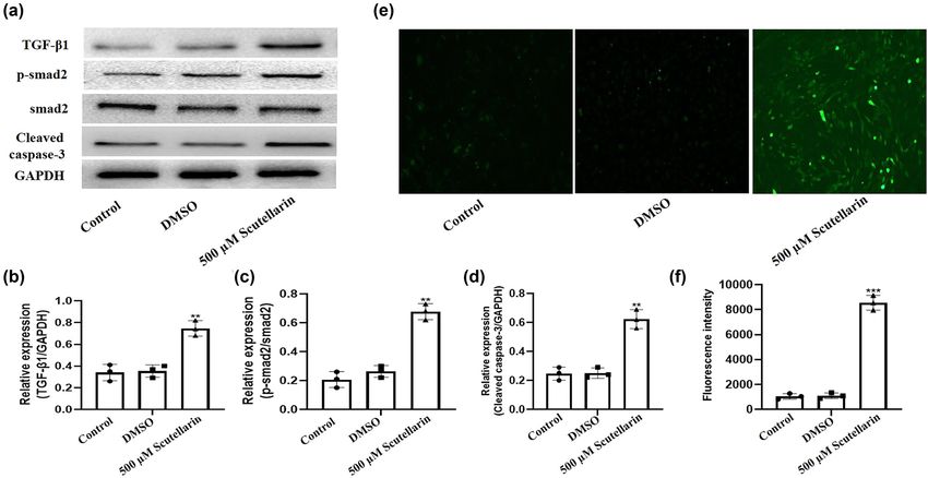
Scutellarin induces lung cancer apoptosis via TGF-β1/smad2 pathway 963
then incubated with the secondary antibody for 2 h at assays at 48 h. With the increase in scutellarin concen-
room temperature. The immunoreactive proteins were tration, the proliferative activity of A549 cells gradually
visualized by chemiluminescence. decreased, which indicated that scutellarin decreased
A549 cell proliferation in a concentration-dependent manner
(Figure 1a). We also observed that scutellarin reduced A549
2.6 Statistical analysis cell proliferation in a time-dependent manner (Figure 1b).
The Annexin/PI staining results showed that the apoptosis
Data were expressed as mean values ± SD from three rate of the scutellarin-treated group was higher than the
independent experiments. Statistical significance between untreated group (Figure 1c and d).
groups was assessed by one-way analysis of variance fol-
lowed by Tukey’s test and Student’s t-test using Prism 5.01
(GraphPad Software, San Diego, CA, USA). A P-value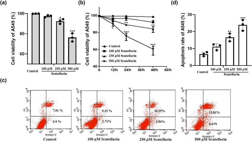
964 Guang-Yan Zhang et al.
Figure 2: Intracellular ROS level and expression of TGF-β1, smad2, and cleaved caspase 3 in A549 cells were increased after scutellarin
exposure. A549 cells were treated with 500 µM scutellarin for 48 h. (a–d) Western blots were prepared to detect the expression of TGF-β1,
p-smad2, smad2, and cleaved caspase-3, and the results were analyzed by Prism version 5.01 software. (e and f) The intracellular ROS level
was detected using a DHE fluorescence probe and then analyzed. **P < 0.01 and ***P < 0.001 vs control.
are cleaved by the corresponding cytoplasmic and nuclear 3.4 Intracellular ROS levels and apoptosis in
substrates and eventually lead to apoptosis, which was scutellarin-exposed A549 cells were
measured after scutellarin treatment. Scutellarin signi- inhibited by blocking the TGF-β1/smad2
ficantly increased the expression of cleaved caspase-3
pathway with SB431542
(Figure 2d). The intracellular ROS level was detected by
DHE fluorescence probe and was significantly increased by
We next examined whether ROS and TGF-β1/smad2 induced
scutellarin (Figure 2e and f).
apoptosis through independent or similar mechanisms.
SB431542, a TGF-β1 receptor inhibitor, was added to A549
cells before scutellarin treatment to block the TGF-β1
3.3 Scutellarin-induced intracellular ROS receptor. SB431542 inhibited scutellarin-induced TGF-β1/
smad2 signaling pathway activation and cleaved caspase-3
level and cleaved caspase-3 expression
expression (Figure 4a–d). SB431542 treatment also inhib-
were inhibited by Tempo ited apoptosis in scutellarin-treated A549 cells (Figure 4e
and f) and ROS accumulation (Figure 4g and h).
In order to explore the mechanism of apoptosis in scu-
tellarin-treated A549 cells, we treated A549 cells with
500 μM of scutellarin and then added 100 μM of TEMPO,
a selective scavenger of mitochondrial ROS. Western blots 4 Discussion
were then performed to detect the expression of TGF-β1,
p-smad2, smad2, and cleaved caspase-3. TEMPO treatment In the present study, we discovered that scutellarin decreased
significantly inhibited scutellarin-induced caspase-3 clea- the viability and enhanced the apoptosis of A549 cells in a
vage without affecting TGF-β1, p-smad2, or smad2 expres- dose- and time-dependent manner. Our data analysis
sion (Figure 3a–d). The intracellular ROS level was then showed that scutellarin also significantly increased the
detected using the DHE fluorescence probe and analyzed. intracellular ROS level and cleaved caspase-3 production,
ROS production induced by scutellarin exposure was inhib- which could be abrogated by Tempo treatment. Moreover,
ited by Tempo treatment (Figure 3e and f). blocking the TGF-β1/smad2 pathway with SB431542Scutellarin induces lung cancer apoptosis via TGF-β1/smad2 pathway 965
Figure 3: Intracellular ROS level and expression of cleaved caspase-3 induced by scutellarin were reversed by Tempo. (a–d) A549 cells were
exposed to 500 µM of scutellarin with and without 100 µM of Tempo for 48 h, and western blots were prepared to detect the protein level of
TGF-β1, p-smad2, and cleaved caspase-3. (e and f) The intracellular ROS level was detected using a DHE fluorescence probe and then
analyzed. ***P < 0.001 comparing the scutellarin + Tempo group vs the scutellarin group.
reversed cleaved caspase-3 expression, intracellular ROS cardiac dysfunction of infarct rats. However, the effect of
accumulation, and apoptosis of A549 cells. scutellarin on expression of TGF-β1 in cancer cells has not
Scutellarin is a flavonoid isolated from the traditional yet been examined. Our study observed that scutellarin
Chinese medicine Erigeron breviscapus, which displays activates the TGF-β1/smad2 signaling pathway and pro-
activity against cancer cells [17,18]. Cao et al. [19] reported motes the expression of cleaved caspase-3 in A549 cells.
that scutellarin plays a role in inhibiting proliferation and Intracellular ROS induces DNA damage and cell apop-
promoting apoptosis in A549 cells. Sun et al. [20] reported tosis [29]. Scutellarin has different effects on the accumu-
that scutellarin induced apoptosis of lung cancer cells lation of ROS in normal cells and tumor cells. Scutellarin
in vitro and in vivo. It has also been reported that scutel- reduced apoptosis and ROS production to protect cardio-
larin ameliorated the drug resistance of A549/DDP cells myocyte ischemia-reperfusion injury [30]. However, scu-
to cisplatin by promoting apoptosis [21]. In our study, tellarin treatment promoted apoptosis in multiple myeloma
we found that scutellarin significantly suppressed the pro- cells by inducing ROS accumulation [7]. Our study observed
liferation of A549 cells in a dose- and time-dependent that intracellular ROS levels and the apoptosis rate were
manner and promoted cell apoptosis. significantly increased in A549 cells after scutellarin expo-
TGF-β is dysregulated in cancer, and as an important sure. We also observed that scutellarin induced upregula-
growth factor plays a crucial role in regulating tissue tion of intracellular ROS levels and expression of cleaved
development and dynamic balance [22]. TGF-β exerts caspase-3 were significantly inhibited by Tempo, a ROS
tumor-suppressive functions primarily resulting in apop- scavenger.
tosis in the early phase of tumorigenesis [23]. TGF-β acti- TGF-β serves as a mediator of intracellular ROS pro-
vates or inhibits the transcription of target genes through duction. Veith et al. [31] reported that TGF-β1 induced
binding to its ligand and activating smads in the nucleus mitochondrial ROS production in human bronchial epithe-
[24,25]. Studies have reported that TGF-β1 regulates apop- lial cells, which subsequently contributed to the epithelial
tosis by activating smad2 [16,26,27]. Pan et al. [28] revealed injury and fibrotic lung scarring. TGF-β1 promoted the
that scutellarin suppressed TGF-β1 production to alleviate invasion and migration of cervical cancer cells by966 Guang-Yan Zhang et al.
Figure 4: Intracellular ROS level and apoptosis of A549 cells induced by scutellarin were significantly decreased by blocking the TGF-β1/
smad2 pathway with SB431542. (a–d) A549 cells were treated with 500 µM of scutellarin with and without 10 mM of SB431542 for 48 h and
then the expressions of TGF-β1, p-smad2, and cleaved caspase-3 were examined in western blots. (e and f) Apoptosis of A549 cells was
tested by flow cytometry and analyzed. (g and h) The intracellular ROS level was detected using a DHE fluorescence probe and then
analyzed. ***P < 0.001 (scutellarin group vs control group) and P < 0.05 (scutellarin + SB431542 group vs scutellarin group).
activating NOX4 to generate ROS production [32]. Our scutellarin may be a promising anti-tumor agent in the
study provides evidence that blocking the TGF-β1/smad2 future.
pathway significantly inhibited the scutellarin-induced
increase in intracellular ROS levels and apoptosis in A549 Funding information: This research was supported by the
cells. Taken together, these data suggest that the TGF-β1/ Chengdu Medical Scientific Research Project (2019099).
smad2 pathway was activated by scutellarin, which subse-
quently induced the production of ROS and eventually Conflict of interest: The authors state no conflict of
resulted in A549 cell apoptosis. interest.
To our knowledge, this study is the first to demon-
strate that the TGF-β1/smad2/ROS/capsase-3 signaling Data availability statement: The datasets generated
pathway is involved in scutellarin-induced apoptosis of during and/or analyzed during the current study are
A549 cells. Considering its anti-tumor effect on tumor available from the corresponding author on reasonable
cells and its protective effect in normal tissue cells, request.Scutellarin induces lung cancer apoptosis via TGF-β1/smad2 pathway 967
References mesenchymal transition in A549 cell lines. Int J Oncol.
2016;49(2):700–8. doi: 10.3892/ijo.2016.3547.
[1] Feng RM, Zong YN, Cao SM, Xu RH. Current cancer situation in [15] Zheng M, Niu Y, Bu J, Liang S, Zhang Z, Liu J, et al. ESRP1
China: good or bad news from the 2018 global cancer statis- regulates alternative splicing of CARM1 to sensitize small cell
tics? Cancer Commun. 2019;39:1–2. doi: 10.1186/s40880-019- lung cancer cells to chemotherapy by inhibiting TGF-beta/
Smad signaling. Aging (Albany NY). 2021;12:3554.
0368-6.
doi: 10.18632/aging.202295.
[2] Wu B, Chang N, Xi H, Xiong J, Zhou Y, Wu Y, et al. viaPHB2
[16] Feng XL, Fei HZ, Hu L. Dexamethasone induced apoptosis of
promotes tumorigenesis via RACK1 in non-small cell lung
A549 cells via the TGF-beta1/Smad2 pathway. Oncol Lett.
cancer. Theranostics. 2021;11(7):3150–66. doi: 10.7150/
2018;15(3):2801–6. doi: 10.3892/ol.2017.7696.
thno.52848.
[17] Wang S, Fu JL, Hao HF, Jiao YN, Li PP, Han SY. Metabolic
[3] Shi X, Chen G, Liu X, Qiu Y, Yang S, Zhang Y, et al. Scutellarein
reprogramming by traditional Chinese medicine and its role in
inhibits cancer cell metastasis in vitro and attenuates the
effective cancer therapy. Pharmacol Res. 2021;170:105728.
development of fibrosarcoma in vivo. Int J Mol Med.
doi: 10.1016/j.phrs.2021.105728.
2015;35(1):31–8. doi: 10.3892/ijmm.2014.1997.
[18] Ke Y, Bao T, Wu X, Tang H, Wang Y, Ge J, et al. Scutellarin
[4] Parajuli P, Joshee N, Rimando AM, Mittal S, Yadav AK. In vitro
suppresses migration and invasion of human hepatocellular
antitumor mechanisms of various Scutellaria extracts and
carcinoma by inhibiting the STAT3/Girdin/Akt activity.
constituent flavonoids. Planta Med. 2009;75(1):41–8.
Biochem Biophys Res Commun. 2017;483(1):509–15.
doi: 10.1055/s-0028-1088364.
doi: 10.1016/j.bbrc.2016.12.114.
[5] Guo F, Yang F, Zhu YH. Scutellarein from Scutellaria barbata
[19] Cao P, Liu B, Du F, Li D, Wang Y, Yan X, et al. Scutellarin
induces apoptosis of human colon cancer HCT116 cells
suppresses proliferation and promotes apoptosis in A549 lung
through the ROS-mediated mitochondria-dependent pathway.
adenocarcinoma cells via AKT/mTOR/4EBP1 and STAT3 path-
Nat Prod Res. 2019;33(16):2372–5. doi: 10.1080/
ways. Thorac Cancer. 2019;10(3):492–500. doi: 10.1111/1759-
14786419.2018.1440230. 7714.12962.
[6] Cheng CY, Hu CC, Yang HJ, Lee MC, Kao ES. Inhibitory effects of
[20] Sun C, Li C, Li X, Zhu Y, Su Z, Wang X, et al. Scutellarin induces
scutellarein on proliferation of human lung cancer A549 cells apoptosis and autophagy in NSCLC cells through ERK1/2 and
through ERK and NFkappaB mediated by the EGFR pathway. AKT signaling pathways in vitro and in vivo. J Cancer.
Chin J Physiol. 2014;57(4):182–7. doi: 10.4077/ 2018;9(18):3247–56. doi: 10.7150/jca.25921.
CJP.2014.BAC200. [21] Sun CY, Zhu Y, Li XF, Wang XQ, Tang LP, Su ZQ, et al.
[7] Shi L, Wu Y, Lv DL, Feng L. Scutellarein selectively targets Scutellarin increases cisplatin-induced apoptosis and auto-
multiple myeloma cells by increasing mitochondrial super- phagy to overcome cisplatin resistance in non-small cell lung
oxide production and activating intrinsic apoptosis pathway. cancer via ERK/p53 and c-met/AKT signaling pathways. Front
Biomed Pharmacother. 2019;109:2109–18. doi: 10.1016/ Pharmacol. 2018;9:92. doi: 10.3389/fphar.2018.00092.
j.biopha.2018.09.024. [22] Zi Z. Molecular engineering of the TGF-beta signaling pathway.
[8] Das R, Xu S, Quan X, Nguyen TT, Kong ID, Chung CH, et al. J Mol Biol. 2019;431(15):2644–54. doi: 10.1016/
Upregulation of mitochondrial Nox4 mediates TGF-beta- j.jmb.2019.05.022.
induced apoptosis in cultured mouse podocytes. Am J Physiol [23] Gu S, Feng XH. TGF-beta signaling in cancer metastasis.
Renal Physiol. 2014;306(2):F155–67. doi: 10.1152/ Acta Biochim Biophys Sin (Shanghai). 2018;50(10):941–9.
ajprenal.00438.2013. doi: 10.1093/abbs/gmx123.
[9] Hill CS. Transcriptional control by the SMADs. Cold Spring [24] Xue L, Deng D, Zheng S, Tang M, Yang Z, Pei H, et al. Design,
Harb Perspect Biol. 2016;8(10):a022079. doi: 10.1101/ synthesis and discovery of 2(1H)-quinolone derivatives for the
cshperspect.a022079. treatment of pulmonary fibrosis through inhibition of TGF-
[10] Derynck R, Budi EH. Specificity, versatility, and control of beta/smad dependent and independent pathway. Eur J Med
TGF-beta family signaling. Sci Signal. 2019;12(570):aav5183. Chem. 2020;197:112259. doi: 10.1016/j.ejmech.2020.112259.
doi: 10.1126/scisignal.aav5183. [25] Sakai S, Ohhata T, Kitagawa K, Uchida C, Aoshima T, Niida H,
[11] David CJ, Massague J. Contextual determinants of TGFbeta et al. Long noncoding RNA ELIT-1 Acts as a Smad3 cofactor to
action in development, immunity and cancer. Nat Rev facilitate TGFbeta/Smad signaling and promote epithelial-
Mol Cell Biol. 2018;19(7):419–35. doi: 10.1038/s41580-018- mesenchymal transition. Cancer Res. 2019;79(11):2821–38.
0007-0. doi: 10.1158/0008-5472.CAN-18-3210.
[12] Yao Y, Yuan Y, Lu Z, Ma Y, Xie Y, Wang M, et al. Effects of [26] Chen H, Liao K, Cui-Zhao L, Qiang-Wen F, Feng-Zeng X, Ping-
nervilia fordii extract on pulmonary fibrosis through TGF-beta/ Wu F, et al. Cigarette smoke extract induces apoptosis of rat
Smad signaling pathway. Front Pharmacol. 2021;12:659627. alveolar Type II cells via the PLTP/TGF-beta1/Smad2 pathway.
doi: 10.3389/fphar.2021.659627. Int Immunopharmacol. 2015;28(1):707–14. doi: 10.1016/
[13] Koo BH, Kim Y, Je Cho Y, Kim DS. Distinct roles of transforming j.intimp.2015.07.029.
growth factor-beta signaling and transforming growth factor- [27] Wu J, Xing C, Zhang L, Mao H, Chen X, Liang M, et al. Autophagy
beta receptor inhibitor SB431542 in the regulation of p21 promotes fibrosis and apoptosis in the peritoneum during
expression. Eur J Pharmacol. 2015;764:413–23. doi: 10.1016/ long-term peritoneal dialysis. J Cell Mol Med.
j.ejphar.2015.07.032. 2018;22(2):1190–201. doi: 10.1111/jcmm.13393.
[14] Zeng Y, Zhu J, Shen D, Qin H, Lei Z, Li W, et al. Repression of [28] Pan Z, Zhao W, Zhang X, Wang B, Wang J, Sun X, et al.
Smad4 by miR205 moderates TGF-beta-induced epithelial- Scutellarin alleviates interstitial fibrosis and cardiac968 Guang-Yan Zhang et al.
dysfunction of infarct rats by inhibiting TGF-beta1 cing apoptosis and oxidative stress. Life Sci. 2016;157:200–7.
expression and activation of p38-MAPK and ERK1/2. doi: 10.1016/j.lfs.2016.01.018.
Br J Pharmacol. 2011;162(3):688–700. doi: 10.1111/j.1476- [31] Veith C, Hristova M, Danyal K, Habibovic A, Dustin CM,
5381.2010.01070.x. McDonough JE, et al. Profibrotic epithelial TGF-beta1 signaling
[29] Hu LL, Liao BY, Wei JX, Ling YL, Wei YX, Liu ZL, et al. involves NOX4-mitochondria cross-talk and redox-mediated
Podophyllotoxin exposure causes spindle defects and DNA activation of the tyrosine kinase FYN. Am J Physiol Lung Cell Mol
damage-induced apoptosis in mouse fertilized oocytes and Physiol. 2020;320:L356–67. doi: 10.1152/ajplung.00444.2019.
early embryos. Front Cell Dev Biol. 2020;8:600521. [32] Ryu D, Lee JH, Kwak MK. NRF2 level is negatively correlated
doi: 10.3389/fcell.2020.600521. with TGF-beta1-induced lung cancer motility and migration via
[30] Wang Z, Yu J, Wu J, Qi F, Wang H, Wang Z, et al. Scutellarin NOX4-ROS signaling. Arch Pharm Res. 2020;43(12):1297–310.
protects cardiomyocyte ischemia-reperfusion injury by redu- doi: 10.1007/s12272-020-01298-z.You can also read