Salmonella Salamae and S. Waycross isolated from Nile perch in Lake Victoria show limited human pathogenic potential
←
→
Page content transcription
If your browser does not render page correctly, please read the page content below
www.nature.com/scientificreports
OPEN Salmonella Salamae and S.
Waycross isolated from Nile perch
in Lake Victoria show limited
human pathogenic potential
Yaovi Mahuton Gildas Hounmanou1,2*, Zebedayo Baniga3, Vanesa García1,4 &
Anders Dalsgaard1*
Non-enterica subspecies of Salmonella enterica are rarely associated with human infections.
Paradoxically, food safety legislations consider the entire genus Salmonella as pathogenic to humans.
Globally, large amounts of seafoods are rejected and wasted due to findings of Salmonella. To inform
better food safety decisions, we investigated the pathogenicity of Salmonella Salamae 42:r- and
Salmonella Waycross isolated from Nile perch from Lake Victoria. Genome-wide analysis revealed
absence of significant virulence determinants including on key Salmonella pathogenicity islands in
both serovars. In epithelial cells, S. Salamae showed a weak invasion ability that was lower than the
invH mutant of S. Typhimiurium used as negative control. Similarly, S. Salamae could not replicate
inside macrophages. Moreover, intracellular replication in S. Waycross strains was significantly lower
compared to the wild type S. Typhimurium. Our findings suggest a low pathogenicity of S. Salamae
reinforcing the existing literature that non-enterica subspecies are avirulent. We propose that food
legislations and actions taken on findings of Salmonella are revisited to avoid wasting valuable sea-
and other foods.
The genus Salmonella is classified into two species, Salmonella enterica and Salmonella bongori. S. enterica,
contains over 2600 serovars and is subdivided into six subspecies which are enterica I, salamae II, arizonae IIIa,
diarizonae IIIb, houtenae IV, and indica VI1. Serovars belonging to subspecies S. enterica, which are around 1600,
are mainly non-typhoid Salmonella associated with gastroenteritis and salmonellosis in humans and a nimals1,2.
The hosts of S. enterica subsp. enterica are warm-blooded animals and their occurrence in aquatic environments
is often associated with fecal pollution due to influx of fecal wastes from humans and a nimals3–5. In contrast,
the hosts of the Salmonella non-enterica subspecies are commonly cold-blooded animals such as reptiles and
amphibians, and they rarely cause infection in animals and h umans6–8.
Although Salmonella enterica and non-enterica subspecies can be isolated from many sources, it is the serovars
of subspecies enterica hosted and transmitted by terrestrial animals that constitute the lead causes of salmonellosis
in humans9. The relative contribution of seafood including fish to the global epidemiology of human salmonel-
losis is very low and mostly limited to S. Weltevreden in Asia; a serovar which is also an enterica subspecies9,10.
In a recent study, we identified S. Waycross of subspecies enterica and the serovar 42:r- of subspecies Salamae
from Nile perch (Lates niloticus) and water sampled far off the shores of Lake Victoria containing low levels of
fecal contamination11.
In East Africa, the Nile perch industry has suffered economic losses due to failure to comply with the microbi-
ological standards of the European Union (EU), the main export market for the Lake Victoria’s fisheries. Through
the Rapid Alert System for Food and Feed (RASFF) of the EU (https://e c.e uropa.e u/f ood/s afety/r asff_e n), imports
of fresh and frozen Nile perch products from Lake Victoria have often been suspended due to findings of Salmo-
nella spp. which has led to rejections or notification of consignments and severe economic l osses11,12. Since the
existing food safety legislation in the EU and elsewhere is based on identification of Salmonella at the genus level
1
Department of Veterinary and Animal Sciences, University of Copenhagen, Frederiksberg, Denmark. 2Research
Unit of Applied Microbiology and Pharmacology of Natural Substances, University of Abomey-Calavi, Godomey,
Benin. 3Department of Veterinary Medicine and Public Health, Sokoine University of Agriculture, Morogoro,
Tanzania. 4Laboratorio de Referencia de Escherichia coli (LREC), Departamento de Microbioloxía e Parasitoloxía,
Facultade de Veterinaria, Instituto de Investigación Sanitaria de Santiago de Compostela (IDIS), Universidade de
Santiago de Compostela (USC), 27002 Lugo, Spain. *email: gil@sund.ku.dk; adal@sund.ku.dk
Scientific Reports | (2022) 12:4229 | https://doi.org/10.1038/s41598-022-08200-5 1
Vol.:(0123456789)
www.nature.com/scientificreports/
Sample number A7 A8 A9 B6 D5 D6 I1 A1 C2 D3 E5 E9 G5 H6
Sample type Fish intestines Fish intestines Fish intestines Fish intestines Lake water Lake water Fish intestines Fish surface Fish intestines Fish intestines Fish intestines Fish intestines Fish intestines Lake water
# Contigs 150 118 172 148 94 123 79 116 67 82 70 83 78 93
Genome size (bp) 4,775,198 4,792,107 4,797,164 4,763,962 4,800,001 4,788,828 4,799,696 4,737,395 4,742,170 4,742,542 4,741,533 4,741,231 4,711,795 4,700,978
Coverage 52 64 46 62 91 56 86 42 61 79 86 79 70 71
Accession No SAMEA5927480 SAMEA5927481 SAMEA5927482 SAMEA5927483 SAMEA5927486 SAMEA5927487 SAMEA5927492 SAMEA5927479 SAMEA5927484 SAMEA5927485 SAMEA5927488 SAMEA5927489 SAMEA5927490 SAMEA5927491
Subspecies II(Salamae) II(Salamae) II(Salamae) II(Salamae) II(Salamae) II(Salamae) II(Salamae) enterica enterica enterica enterica enterica enterica enterica
Serovars 42:r:- 42:r:- 42:r:- 42:r:- 42:r:- 42:r:- 42:r:- Waycross Waycross Waycross Waycross Waycross Waycross Waycross
H1-phase-1
r r r r r r r z4,z23 z4,z23 z4,z23 z4,z23 z4,z23 z4,z23 z4,z23
flagella
MLST 1208 1208 1208 1208 1208 1208 1208 2460 2460 2460 2460 2460 3691 3691
Resistance parC:p.T57S; parC:p.T57S; parC:p.T57S; parC:p.T57S; parC:p.T57S; parC:p.T57S; parC:p.T57S; parC:p.T57S; parC:p.T57S; parC:p.T57S; parC:p.T57S; parC:p.T57S; parC:p.T57S; parC:p.T57S;
genotypes aac(6’)-Iaa aac(6’)-Iaa aac(6’)-Iaa aac(6’)-Iaa aac(6’)-Iaa aac(6’)-Iaa aac(6’)-Iaa aac(6’)-Iaa aac(6’)-Iaa aac(6’)-Iaa aac(6’)-Iaa aac(6’)-Iaa aac(6’)-Iaa aac(6’)-Iaa
Phenotypic
None SMX None None None None None NAL; SMX SMX None None None NAL; SMX NAL;SMX
resistancea
Table 1. Genomic characteristics of S. Salamae 42:r:- and S. Waycross. a NAL, nalidixic acid; SMX,
sulfamethoxazole.
and actions are taken irrespective of which sub-species or serovar is found, fish products containing S. Waycross
and S. Salamae would be rejected by food safety authorities in the importing countries. Such actions are taken
despite that the public health importance of these and other non-enterica Salmonella serovars is still uncertain.
Due to their rare involvement in human and animal infections, it has been proposed to consider the non-
enterica Salmonella subspecies like S. Salamae, as opportunistic p athogens13. Moreover, S. Waycross, although
a member of the enterica subspecies, is not commonly reported associated with human infection14,15. From a
food safety and public health point of view, it is therefore important to elucidate the full pathogenic potential of
non-enterica subspecies but also rare serovars of the enterica subspecies to inform better food safety decisions
without unnecessary losses of safe foods and sale. Knowledge about non-enterica subspecies is still relatively
limited but studies carried out reveal that their virulence and capacity to colonize humans is very l imited13. Few
genomic studies have been conducted on non-enterica subspecies and little is known about the differences in
for instance the Salmonella pathogenicity islands between enterica and non-enterica subspecies16. Using whole
genome sequence analysis coupled with epithelial and macrophage cell lines infection, we reveal in this report
a potentially poor pathogenicity of Salmonella Salamae and S. Waycross isolated from Nile perch and water
obtained in the Tanzanian basin of Lake Victoria.
Results
Genetic profiles of S. Waycross and S. Salamae. The genome data confirmed the initial serotyping
result that isolates were S. enterica subsp. Salamae serovar 42:r- and S. enterica subsp. enterica serovar Waycross
(Table 1). All S. Salamae 42:r- were assigned to sequence type (ST) 1208, while S. Waycross were ST2460 (five
strains) and ST3691 (two strains) (Table 1).
The antimicrobial resistance typing data revealed that the strains only harbored the aac(6’)laa gene encoding
aminoglycoside resistance and a mutation in parC (p.T57S) encoding fluoroquinolone resistance. However, the
previous phenotypic resistance data established by MIC indicated that five S. Salamae 42:r- and three S. Waycross
strains were susceptible to all tested antimicrobials (Table 1). The remaining strains showed phenotypic resistance
to sulfamethoxazole and nalidixic acid (Table 1). The genomes contained no plasmid replicons.
The in-silico analysis of the S. Salamae 42:r- genomes resulted in the prediction of six prophage regions in each
strain (but seven for strain A9) (Table S1). The genome sizes of these prophage regions varied from 4.8 to 66.1 kb,
with a GC content ranging from 48.0 to 53.7%. Of the total 43 predicted prophages, 21 were intact, whereas 17
were incomplete and the remaining five were questionable. The number of prophages differed between S. Sala-
mae and S. Waycross as the latter showed more phage regions ranging from eight to 11 in each of the analyzed
strains. The phages identified in S. Waycross varied in size from 6.1 to 41.8 kb and their GC content from 41.9 to
54.3%. Of the 69 predicted prophages in S. Waycross, 28 were intact, whereas 29 were incomplete and 12 were
questionable. In S. Salamae 42:r-, the prophage regions identified showed similarity to known Enterobacteriaceae
prophages where the predominant ones were sal3 (NC_031940, 32.6%) followed by HP2 (NC_003315, 17.8%),
phiE255 (NC_009237), Gifsy1 (NC_010392) and SEN34 (NC_028699) with the three latter occurring at 16.4%
proportions. Of the many prophage regions identified in the S. Waycross genomes, the most prevailing ones
showed similarity to vB_SosS_Oslo (NC_018279, 15.9%), followed by Gifsy2 (NC_010393, 11.6%), and HP2
(NC_003315), sal3 (NC_031940) and BTP1 (NC_042346) with the three latter occurring in 10.1% proportions.
Overall, S. Waycross genomes in contrast with S. Salamae harbored higher number and more diverse prophage
regions as they seem to have experienced more phage insertions than S. Salamae.
Pathogenicity determinants. Virulence in Salmonella is mainly regulated by the SPIs (Salmonella Patho-
genicity Islands) with SPI1 encoded genes involved in host cell invasion and SPI2 genes in survival of Salmo-
nella in the host cell. Our S. Waycross and S. Salamae 42:r- strains, like most Salmonella enterica harbored SPI1
and SPI2 genes as well as their translocated effectors (Fig. 1). However, a number of significant deletions were
observed in the two pathogenicity islands of the serovars. On SPI1, genes missing in most strains included invB,
invF, invH, invI, orgA, orgB, orgC, prgI, prgJ prgK, sicA, sicP, spaP, spaQ, and sprB (Fig. 1). On SPI2 important
genes involved in survival in host cells including ssaE, ssaG, ssaH, ssaI, ssaJ, ssaM, and ssaV were absent in
Scientific Reports | (2022) 12:4229 | https://doi.org/10.1038/s41598-022-08200-5 2
Vol:.(1234567890)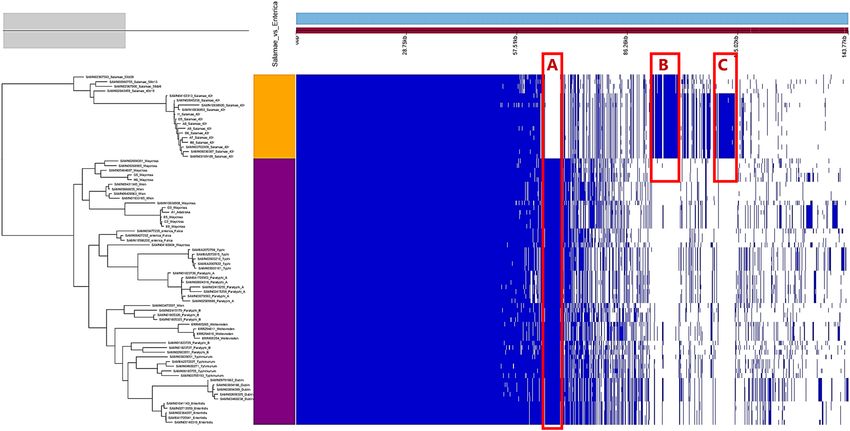
www.nature.com/scientificreports/
Figure 1. Presence/absence map of genes on SPI1 and SPI2 as well as their translocated effectors. The full red
boxes represent presence of the indicated gene.
most of the S. Salamae 42:r- and S. Waycross strains. Clinically important type-three secretion system (T3SS)
translocated effectors in Salmonella virulence such as avrA, sopE, sopD, gogB, and sseK were also absent in the
two serovars (Fig. 1) despite lowering the detection threshold to 50%. The deletions on the pathogenicity islands
occurred more in S. Salamae 42:r- than in S. Waycross as some S. Waycross strains showed full presence of the
targeted genes, but both serovars completely lacked the spv locus with its five related genes (spvA, B, C, D, and
R) indicating a low virulence potential of these serovars. Besides SPI1 and SPI2, both serovars contained SPI3,
4, 5 and 9 encoded genes.
Genome‑wide content comparison. The pan-genome analysis of 75 enterica and non-enterica subspe-
cies of Salmonella enterica identified 14,438 total coding sequences (CDS), divided into 2,931 core CDS (shared
by > 95% of strains), 38 soft core CDS, 2,984 shell CDS and 8,485 cloud CDS. The overall gene presence and
absence data from the pangenome is provided in Table S2. In the accessory genome, the output gene pres-
ence/absence map (Fig. 2) revealed subspecies and/or serovar-based clustering with three characteristic unique
regions in the genomes corresponding to the enterica subspecies including S. Waycross (Part A, Fig. 2), the S.
Salamae subspecies (Part B) and the S. Salamae 42:r- serovar (Part C). These unique regions represent CDS spe-
cific to the subspecies and/or the serovars and were further analyzed for their potential role in virulence.
The unique CDS were retracted as described in Materials and Methods and mapped back to reference
genomes of each of the three groups to identify their location and analyze their involvement in potential genetic
evolution. This mapping revealed that the unique CDS did not correspond to one specific region in the genomes
of S. Waycross and S. Salamae, but could rather be seen throughout the genomes with query covers between 81
and 100% across the entire genomes with variable GC contents indicating their potential external acquisition
as a result of horizontal gene transfer (Fig. S1). This indicates that evolution/divergences at serovar level occurs
not in a specific region of the genomes but across the genomes and should be further studied for their functional
role in each targeted subspecies and / or serovar.
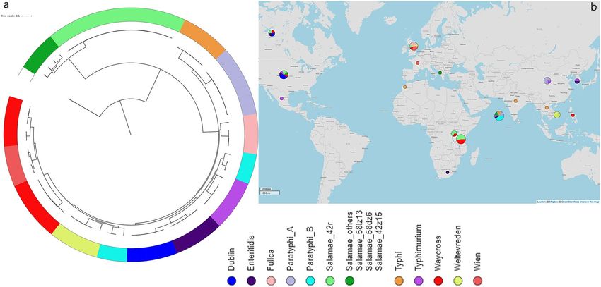
To provide understanding in the role of these regions specific to S. Waycross and S. Salamae 42:r-, the targeted
CDS unique to these serovars i.e. Part A and Part C from the pangenome comparison were further analyzed. The
results supports that S. Waycross clustering with other common pathogenic enterica-subspecies, contain genes
encoding for virulence factors as well as type three secretion system genes and T3SS translocated effectors that
are absent in S. Salamae 42:r- (Fig. 3). This reinforces that S. Waycross as an enterica-subspecies contains more
pathogenicity markers compared to S. Salamae 42:r- despite the many deletions in its pathogenicity islands.
On the other hand, in the unique CDS of S. Salamae 42:r-, the genetic content shows gene clustering corre-
sponding to two main prophage elements and the rest are hypothetical proteins (Fig. S2). We could not recon-
struct any intact phage from these gene clusters. However, region #1 contains a number of phage particles such
as repressors of immunity control, phage integrase, phage tail assembly proteins, endolysins and various phage
hypothetical proteins (Fig. S2). Likewise, the second region of gene clusters specific to S. Salamae 42:r- contained
phage components such as phage tail fiber assembly proteins, phage related transmembrane proteins, holin and
Ner-like proteins. Overall, none of the components of these clusters point to virulence determinants.
Scientific Reports | (2022) 12:4229 | https://doi.org/10.1038/s41598-022-08200-5 3
Vol.:(0123456789)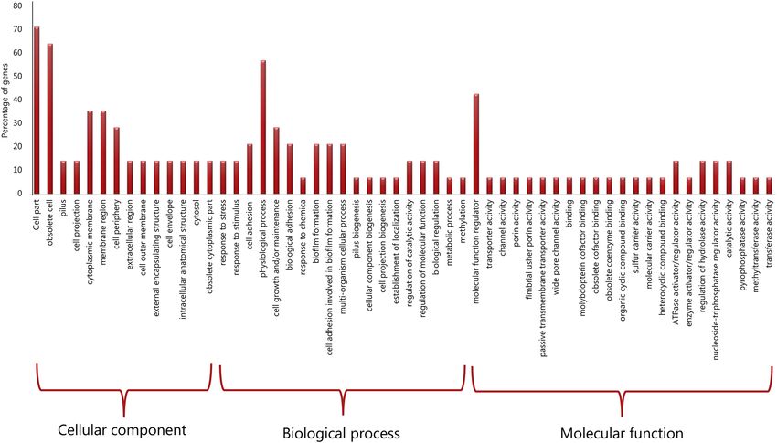
www.nature.com/scientificreports/

                                           Figure 2.  Pangenome view of 75 non-enterica and enterica subspecies of Salmonella enterica. All S. Salamae
                                           strains are in the clade colored orange while all subspecies enterica are colored purple. Unique coding sequences
                                           observed in the accessory genome are represented by the zones A, B and C.

                                           Figure 3.  Annotated genes encoded in the unique CDS of the enterica species group (including S. Waycross) in
                                           Part A of Fig. 2. The solid marks in the upper image is displayed in the lower table with the actual genes names.

                                              A much closer look into each of the observed unique parts in the accessory genomes unique to the serovars
                                           was done with a GO annotation analysis and revealed that the functional GO-terms rather point to a variety of
                                           gene enrichment in the biological processes and molecular functions. A specific look into Part C reveals a low
                                           level of gene enrichment in the pathways of biofilm formation, response to stress and stimulus, which indicates
                                           limited environmental adaptation and pathogenicity (Fig. 4, portion biological process).

          Scientific Reports |   (2022) 12:4229 |              https://doi.org/10.1038/s41598-022-08200-5                                                  4

Vol:.(1234567890)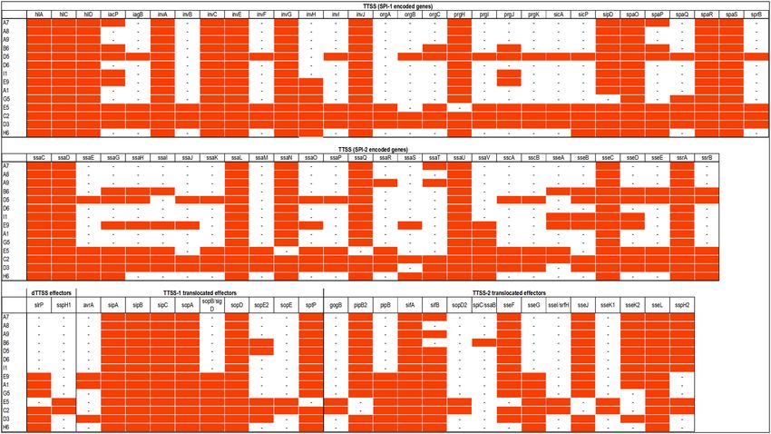
www.nature.com/scientificreports/
Figure 4. Gene oncology enrichment depicted from Part C representing unique CDS for S. enterica subsp.
salamae serovar 42:r-.
Figure 5. Phylogeny of S. Waycross and S. Salamae from Lake Victoria in a global S. enterica context; (a) SNP-
tree showing clustering of the strains colored by serovars; (b) geographical distributions of the serovars included
in the analysis. Bigger circles illustrate geographical areas with higher number of strains included in the analysis.
The Map in panel (b) is generated from Microreact (https://microreact.org/) using geographical coordinates of
the place of isolation of analyzed strains.
Genetic variations in our strains within Salmonella enterica genealogy. The phylogenetic analy-
sis of gene sequences from 75 enterica and non-enterica subspecies of Salmonella enterica isolated from 1985 to
2018 revealed two large clusters based on subspecies irrespective of the source of isolation i.e. environmental or
clinical origin (Fig. 5). This suggests that it is unlikely that S. Salamae 42:r- and S. Waycross are natural occurring
aquatic microorganisms as they showed wide-genetic variations as compared with for instance S. Weltevreden,
Scientific Reports | (2022) 12:4229 | https://doi.org/10.1038/s41598-022-08200-5 5
Vol.:(0123456789)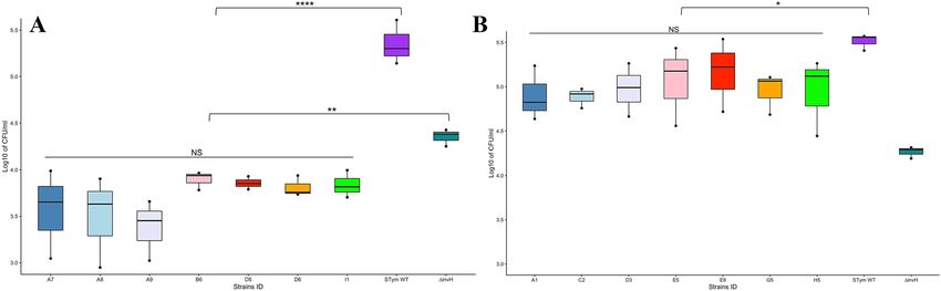
www.nature.com/scientificreports/
Figure 6. Invasion of S. Salamae (A) and S. Waycross (B) in human intestinal epithelial cell line INT-407. Each
box plot represents one strain with values from three biological replicates and two technical replicates at each
round. NS: not significant; p > 0.05; *p < 0.05; **p < 0.01,****p < 0.0001.
Figure 7. Intracellular replication of S. Salamae (A) and S. Waycross (B) in mouse macrophage J774 cell lines.
NS: not significant; p > 0.05; *p < 0.05; ***p < 0.0001; ****p < 0.00001.
which frequently are isolated from the aquatic environment. The analysis also showed a further convergence of
Salmonella strains by serovars as shown in the sub-clades on the tree (Fig. 5).
Invasion of intestinal epithelial cells. The log distribution of bacteria counts for the three experimental
replicates from inoculum, adhesion and invasion studies are shown in Fig. S3 and Fig. S4, respectively for S. Sala-
mae and S. Waycross. The adhesion to the INT-407 epithelial cells ranged from 0.01% to 0.06% for S. Salamae
strains, while 0.23 to 0.43% was recorded as adhesion rates for the wild type S. Typhimurium (Table S3). The
cell invasion analysis shows that within S. Salamae tested strains, the number of bacteria that invaded epithelial
cells varied non-significantly from 1.6 × 10³ to 9.8 × 10³ CFU/mL (p = 0.3). However, when compared to the wild
type S. Typhimurium (1.39 × 10⁵ to 4.06 × 10⁵ CFU/mL), the Salamae serovar strains show significantly negligi-
ble invasion rates (p = 0.000000011) and even the invH mutant of S. Typhimurium (used as a negative control)
(1.78 × 10⁴ to 2.67 × 10⁴) was more invasive in epithelial cells than S. Salamae (p = 0.004) (Fig. 6A).
On the other hand, the epithelial cell adhesion rates in S. Waycross varied from 0.07% to 1.3%; however, there
was no significant intra-serovar variation for S. Waycross in their cell invasion ability (2.78 × 10⁴ to 2.72 × 10⁵ CFU/
mL, p = 0. 956). Nevertheless, compared to S. Typhimurium wild type (2.56 × 10⁵ to 3.72 × 10⁵ CFU/mL), the S.
Waycross strains showed a slightly lower cell invasion levels (p = 0.014). However, S. Waycross remain signifi-
cantly more invasive than the invH mutant of S. Typhimurium (p = 0.0009) as opposed to S. Salamae (Fig. 6B).
Intracellular survival and replication. After the J774 macrophages phagocytized the experimental bac-
terial strains, we recorded 0.17–0.9% as intra-cellular survival rate among S. Salamae 42-r strains. However,
these could not replicate to higher number inside macrophages after 18 h post infection as their fold replica-
tion ranged between 0.22% and 3.2% (Table S3). This explains the significant difference when compared to the
intracellular fold replication rate of wild type S Typhimurium that varied from 77.5% to 80% (p = 3.02 × 10⁻³⁴,
Fig. 7A). Nevertheless, of 0.26–1.6% of S. Waycross strains that were obtained during intracellular survival, up
Scientific Reports | (2022) 12:4229 | https://doi.org/10.1038/s41598-022-08200-5 6
Vol:.(1234567890)www.nature.com/scientificreports/
to 18.6–45.9% were able to replicate within the macrophages. The S. Waycross strains showed a significantly
higher intracellular replication compared to ssaV mutant of S. Typhimurium, whose fold replication rates varied
between 2.4% to 3.8% (p = 3.57 × 10⁻⁴). The fold replication rates observed in S. Waycross were however not as
high as the wild type positive control (86.2–97.5%, Table S3, Fig. 7B).
Discussion
We isolated two rare serovars S. Salamae 42:r- ST 1208 and S. Waycross ST 2460 and ST 3691 in Nile perch and
water samples collected from deep offshore waters in Lake Victoria in Tanzania11. The presence of Salmonella in
Nile Perch is often the cause of restrictions on export of the fish products into the international markets. Recent
reports of Nile perch products from Tanzania exported to EU member states such products were rejected or
detained due to contamination by Salmonella spp.11. Salmonella spp. have been found in fresh and processed
Nile perch from Lake Victoria in K enya3,12. Existing evidence from the global epidemiology of Salmonella how-
ever suggests that only serovars within the subspecies enterica of Salmonella enterica are implicated in human
salmonellosis9. It is therefore important to determine the subspecies of Salmonella strains isolated in seafood
otherwise rejections of fish/fish products containing Salmonella spp. could result in unnecessary food waste
and economy loss.
Existing literature have shown that Salmonella Salamae serovar Sofia were able of colonizing chicken organs
but could not cause symptomatic d isease17. Other studies have also reported that non-enterica subspecies such
as subspecies arizonae and diarizonae show significant reduction of intestinal colonization, persistence and
systemic spread in murine models with subsequent decrease in faecal s hedding18. These characteristics docu-
ment avirulent features of the non-enterica subspecies. The characterization of non-enterica Salmonella strains
at subspecies level is therefore important to determine their pathogenic potential. The genomic analysis of our
strains revealed only the presence of the aac(6’)laa gene encoding aminoglycoside resistance and a mutation in
parC (p.T57S) known to be associated with nalidixic acid resistance observed in some strains11. A few isolates
showing phenotypic resistance to sulfamethoxazole (Table 1) were not found to contain any encoding resist-
ance gene, a discrepancy that is increasingly reported in many b acteria10. Overall, our strains were susceptible
to most antimicrobials tested. This suggests that the strains may not have been exposed to antimicrobials, which
is further supported by the fact that they were isolated in off-shore lake waters containing low levels of fecal
contamination11. Knowing that bacteria can pick-up genetic material from each other, it is possible that, when
in the same niche, epidemic strains could transfer virulence or antimicrobial resistant genes to non-pathogenic
strains through their mobile genetic elements.
The pathogenicity of Salmonella is mainly dependent upon the two Salmonella encoded T3SSs (Type III
Secretion systems), i.e. SPI-1 and SPI-2, which are required for different stages of salmonellosis, namely cell
invasion and intracellular r eplication19,20. SPI-1 and SPI-2 were present in our strains of both S. Salamae and in
S. Waycross. However, the significant deletions recorded in the two pathogenicity islands (Fig. 1) especially in
S. Salamae corroborates existing literature that this subspecies lacks key virulence genes in these pathogenicity
islands required causing human s almonellosis21. With cell invasion assays, we further confirmed the low patho-
genicity of the S. Salamae strains as they could not invade the INT-407 epithelial cells, not even close to the levels
observed in the invH mutant of S. Typhimurium 4/74. In fact, all the S. Salamae strains lacked the invH gene that
has been knocked-out in the negative control, confirming the role of these set of inv genes in the invasion ability
of Salmonella13. While the tested S. Waycross strains, as an enterica subspecies seem to invade the epithelial cells
at increased levels compared to S. Salamae, the wild type S. Typhimurium 4/74 were significantly more invasive.
The deletions observed mainly in the SPI-2 encoding genes of S. Salamae explain why they are not able to
survive and replicate within the murine J774 macrophages. Studies have shown that non-enterica subspecies
serovars including S. Salamae do not internalize well and do not replicate in macrophages and also demonstrate
a severely reduced intestinal colonization and intestinal persistence13,21. Such inabilities are attributable to the
deletions of important genes that are found in the subspecies enterica, explaining why S. Waycross strains showed
higher intracellular replication rates although not as high as the wild type S. Typhimurium.
Apart from the T3SSs, the ability to form biofilm has been documented as important factor for environmental
persistence and virulence in Salmonella as it favours the survival of the bacteria under harsh conditions including
low-nutrient conditions, acidic pH, and varying t emperatures22; all factors increasing ability to infect a host. In
this context, both S. Waycross and S. Salamae contained few genes associated with biofilm formation suggesting
a potentially reduced ability to form biofilm, a feature that could impede their overall long term environmental
adaptation as opposed to S. Weltevreden10.
While our characterizations of the S. Waycross isolated from fish and water documented some pathogenic
potential, the findings show that S. Salamae should not be regarded as an important human pathogen. It is pro-
posed that Salmonella spp. isolated from seafood should be further characterized to determine their pathogenic
potential. Such characterizations are currently not done by most food safety laboratories because the existing
food safety legislations still consider all sub-species and serovars of the genus Salmonella pathogenic to humans.
Our data together with existing literature supports that not all Salmonella subspecies and serovars are patho-
genic, and that efforts should be made towards revisiting legislations and equipping food safety laboratories
with knowledge and analytical methods to ensure less waste of food while still ensuring food safety. Currently
most laboratories responsible for microbiological food safety analysis and issuing export certifications do not
perform whole genome sequencing. Therefore, based on the outcomes of the accessory genomes studied a more
practical suggestion would be to design subspecies-specific primers targeting for instance unique CDS on SPI-1
and SPI-2 genes for PCR assays that can be performed at any laboratory to distinguish the non-enterica subspe-
cies. Overall, the outcomes of this study provide useful background information, especially for public health
Scientific Reports | (2022) 12:4229 | https://doi.org/10.1038/s41598-022-08200-5 7
Vol.:(0123456789)www.nature.com/scientificreports/
authorities and researchers that can found the basis for designing and implementing further investigations that
address the limitations and add further perspectives to the present study.
Materials and methods
Genomic analysis. Salmonella isolates, DNA extraction and whole genome sequencing. Salmonella enterica
subsp. enterica serovar Waycross and S. enterica subsp. Salamae serovar 42:r:- were previously isolated from Nile
perch (Lates niloticus) and water samples collected far offshore in Lake Victoria in waters with low levels of fecal
contamination; details on sampling strategy are in the previous study11. The isolates were originally serotyped
based on the White-Kauffmann-Le Minor (WKL) scheme23, and tested for susceptibility to 14 antimicrobial
agents by the MIC method11. All seven S. Salamae and seven of the S. Waycross isolated in that study were se-
lected for whole genome sequencing. Selection of the S. Waycross strains was based on differences in sampling
dates, sample types and MIC results. DNA was extracted from exponential bacterial cultures using the Maxwell
RSC culture cell’s DNA kit following the manufacturer’s protocol and the automated Maxwell RSC machine
(Promega, Wisconsin, USA). The complete genomes were sequenced with the MiSeq instrument (Illumina, Inc,
San Diego, CA, USA). The sequence reads were submitted to the European Nucleotide Archive under the project
accession number PRJEB34642.
Read processing and assembly. Raw sequence reads were trimmed with b bduk224, using the score cut-
off of 20 and the reads quality was evaluated with FastQC v0.11.5 before and after quality check. Trimmed reads
were assembled with Spades v3.13.025 using error correction, coverage cut-off = 2 and the kmer sizes 21, 33, 55,
77, 99 and 127. Contigs shorter than 200 bases were discarded and the quality of the de novo assembled contigs
was analysed using Quast (v4.5)26.
Characterization of the genomes. A serovar prediction analysis for confirmation of the two serovars
was performed applying Salmonella in-silico Typing Resource (SISTR)27. MLST was determined based on the
Achtman seven housekeeping genes MLST scheme from Enterobase28. General characteristics of the assembled
genomes were determined using tools available at Center for Genomic Epidemiology (https://cge.cbs.dtu.dk/
services/). These included SPIFinder v1.0 for the detection of Salmonella Pathogenicity Islands (SPIs), Plasmid-
Finder v2.029, and ResFinder v4 (using Abricate) for the detection of antimicrobial resistance genes30 in com-
parison with the phenotypic MIC microdilution test initially reported11. The in-silico analyses of the genomes
also included identification of prophages using PHASTHER31 to detect and compare prophage insertions within
the genomes of each strains.
Comparative genomics of virulence determinants. Pathogenic markers encoded in the main Sal-
monella Pathogenicity Islands (SPIs) were first determined using the Pathogenicity Islands Database PAIDB
v.2.032, and then the VFanalyser of the Virulence finder database (http://www.mgc.ac.cn/cgi-bin/VFs/v5/main.
cgi?func=VFanalyzer) was used to further characterize virulence determinants and compare them with the S.
Waycross and S. Salamae genomes. Genes located on the SPIs were further investigated one-by-one by local
BLAST search against our genomes with low thresholds set at 50% query cover and 50% percent ID to avoid
eventual “false negative” gene absence outcomes. Moreover, the detected genes on the SPIs were confirmed in
Artemis33 by the absence of premature stop codons in the annotation.
A pangenome analysis was performed to describe the specificities of the genomes of 75 Salmonella strains
isolated from humans, animals, food and the environment obtained from different countries from 1985 to 2018
(up to the isolation date of our strains) (Table S4). These include S. Typhi, S. Typhimurium, S. Paratyphi A and
B, S. Wien, S. Waycross, S. Salamae, S. Dublin, S. Enteritidis and S. Weltevreden. All genomes were annotated
using Prokka34 with the annotated GFF3 files used as an input to the Roary (v.3.7.0)35 pangenome analysis tool
in a Linux interface. We then used the binary presence/absence data of the accessory genome produced in Roary
to calculate the associations between all genes in the accessory genome and the source types, as well as serovars
of the identified isolates by employing Scoary v.1.6.1136.
The genes presence/absence in the pangenome along with the accessory genome was visualized in
Phandango37. The unique coding sequence (CDS) blocks observed per subspecies and/or serovar in Phan-
dango were extracted. We then applied the ‘query_pan_genome’ function of Roary to retract them as multi-
fasta files. Using a blast atlas analysis from the GView server (https://server.g view.ca), the block of CDS that
was unique to the enterica subspecies where S. Waycross belonged, was mapped back to the reference S. Way-
cross genome SAMN04160804. The same analysis was done for the unique CDS of all S. Salamae using the
reference SAMEA2665118 and more specifically for the serovar S. Salamae 42:r- using the reference genome
SAMN10638893.
To identify functional roles of the CDS unique to each subspecies and/or serovar we performed a functional
gene ontology annotation of the targeted CDS with eggNOG-mapper38. This tool performs annotation with
similar precision as the widely used homology-based approaches: BLAST and InterProScan, but runs about
15 × faster than BLAST and at least 2.5 × faster than I nterProScan38. The obtained functional profiles based on
GO-terms were classified in biological and molecular functions and reported for each GO-annotated gene of
the unique CDS to determine pathways related to pathogenicity.
To understand the role of the targeted unique CDS in each subspecies and/or serovar, the extracted multi-
fasta files were used as input and analysed with the VRprofile39 server that generates rapid information on
virulence and antimicrobial resistance determinants within pathogenic bacterial genomes. The resulting data
was visualized as graphics using Microsoft excel and the Gview server (https://server.g view.ca/). The unique
Scientific Reports | (2022) 12:4229 | https://doi.org/10.1038/s41598-022-08200-5 8
Vol:.(1234567890)www.nature.com/scientificreports/
CDS files were further re-assessed ResFinder, PHASTER and RAST to identify potential virulence, resistance
and environmental persistence factors.
Phylogenetic analysis across enterica and non‑enterica subspecies. All 14 sequenced strains
along with the public genomes used in the pangenome analysis were included to construct a phylogenetic tree.
This aimed to investigate genetic diversities within and between the Salmonella serovars mainly to understand
how our serovars diverge from the enterica-subspecies of Salmonella enterica. We used FastTree for SNP call-
ing through CSIphylogeny40 where Salmonella bongori (accession SAMN02603391) was used as outgroup. The
newick file of the tree was visualized in Microreact (https://microreact.org/showcase) to display the spatio-tem-
poral distribution of the strains collections along with the SNP-tree. The pairwise SNPs data are shown in the in
supplementary Table S5.
Comparative genomics for identification of environmental adaptation markers. Since the
strains were isolated from fish and water collected at deep waters with low levels of fecal contamination11, a
function-based comparison was made between S. Waycross and S. Salamae and the aquatic bacterium Vibrio
cholerae O1 strain N16961 based on the comparison of the metabolic reconstruction from the RAST server41
that allows to compare the functioning parts of two organisms.
Cell infection studies. Bacterial strains and cell lines. All S. Salamae 42:r- and S. Waycross strains ana-
lyzed by WGS were included in experimental cell infection studies. The reference strain S. Typhimurium 4/74
was used as positive control in all experiments. The isogenic mutant for invH of S. Typhimurium 4/74 served as
negative control for the epithelial cells infection. This strain has a mutation in the invH gene, reducing its rate of
invasion compared to the wild type. S. Typhimurium 4/74 ΔssaV was used as negative control for the intracel-
lular macrophage infection study. This strain is deficient in ssaV, a structural component of the SPI2-encoded
T3SS reducing its rate of intracellular replication with regard to the wild type. The wild type and mutants were
described in a previous study42.
The human embryonic intestinal epithelial cell line INT-407 (HeLa-derived epithelial cells) served to assess
the invasion ability of S. Salamae and S. Waycross in comparison to the control strains. On the other hand,
intracellular survival and replication within macrophages was investigated for our strains using the mouse
monocyte-derived macrophage cells J774.
Infection of epithelial cell lines. The epithelial cells were cultivated in DMEM (Gibco) supplemented
with 10% (v/v) heat-inactivated fetal bovine serum (FBS, Fertile Bovine Serum, Invitrogen) and 25 µg/mL gen-
tamicin to prevent bacterial contamination. The cells were grown in a humidified 37 °C and 5% CO2 incubator.
Twenty-four hours prior to infection, the INT-407 cells (Cell Lines Service (CLS), Heidelberg, Germany) were
seeded in two 24-well plates (one plate for determination of adhesion and the other for invasion) in a concen-
tration of 2.5 × 10⁵ cells per mL. The bacteria (experimental strains as well as controls) were grown for 16 h at 5
rcf and 37 °C in Luria Broth (Difco, Maryland, USA). Bacterial cultures were centrifuged at 8,228 rcf for 8 min.
The suspensions were adjusted to O D600 = 0.25 (2.5 × 10⁸ bacteria/ml) in 0.9% NaCl and added to monolayers
of the eukaryotic cells in both 24-well plates (labeled T1 and T2) at a multiplicity of infection of 100:1 (bacte-
ria to eukaryotic cell) without antibiotic. Inoculum counts were verified by plating aliquots of the inoculated
suspension on LB agar plates (counts before infection). After 30 min of infection at 37 °C, 5% CO2, the media
from both plates was removed and monolayers were washed twice with 0.9% NaCl. At this point, T1 plates are
processed straight away for plating while fresh media containing 100 µg/mL gentamicin was added to T2 plates
to kill extracellular bacteria then they were incubated for further 2 h at 37 °C, 5% C O2. To enumerate adhered
(T1 = 30 min) and invaded bacteria (T2 = 2 h), cells were washed twice with 0.9% NaCl and lysed in 1 mL 0.1%
Triton X-100 (v/v). The viable bacteria were enumerated by colony counts of lysate dilutions plated on LB agar.
The experiments were performed in triplicates for biological replication with two technical replicates during
each round.
Infection of macrophages. Macrophage cell lines J774.1 (Cell Lines Service (CLS), Heidelberg, Germany)
were cultured in RPMI (Gibco) supplemented with 10% (v/v) heat-inactivated FBS and 25 µg/mL gentamicin.
Cells were incubated in a humidified 37 °C, 5% C O2 incubator. The bacteria were grown in LB to a stationary
phase, and harvested at 8,228 rcf for 5 min and resuspended in 0.9% (w/v) NaCl. Following similar procedures as
in the epithelial cell infection, bacteria were added to eukaryotic cells at a multiplicity of infection of 10:1 (bacte-
ria/macrophage). The monolayer cells with bacteria were centrifuged at 12 rcf for 3 min immediately after addi-
tion of the bacteria followed by incubation for 25 min at 37 °C, 5% CO2 without antibiotic. Enumerations of the
bacteria in the inoculum were verified by plating onto LB agar plates (counted before infection). After 30 min,
the media was removed and monolayers were washed twice with 0.9% NaCl. At this time point (corresponding
to time 0 for phagocytosis) fresh RPMI containing 10% heat-inactivated FBS and 100 µg/mL gentamycin was
added to kill the extracellular bacteria and the plates were incubated for 1 h (T1) at 37 °C in 5% CO2 to assess
intracellular survival. Thereafter, cells in replication plates were washed twice with 0.9% NaCl and incubated
with RPMI containing 10% heat-inactivated FBS and 25 µg/mL gentamycin for 16 h (T2). For enumeration of
bacteria, eukaryotic cells were washed twice with 0.9% NaCl, subsequently lysed in 0.1% Triton X-100 (v/v).
The viable intracellular bacteria were enumerated by colony counts of lysate dilutions plated on LB agar plates.
For intracellular survival rates, bacteria were enumerated at t = 1 h (1 h post-uptake or post-phagocytosis), and
for intracellular replication, bacteria were counted at t = 16 h (~ 18 h post-infection). The experiments were per-
formed in triplicates for biological replication with two technical replicates during each round.
Scientific Reports | (2022) 12:4229 | https://doi.org/10.1038/s41598-022-08200-5 9
Vol.:(0123456789)www.nature.com/scientificreports/
Statistical analysis. In the epithelial cell experiments, rates of adhesion and invasion are expressed as the
CFU/mL after 30 min and 2 h post infection with respect to the initial inoculum (Table S3). In the macrophage
infections, the intracellular survival was determined as the CFU/mL at T1 with respect to the initial inoculum,
while the fold replication was calculated as the ratio of bacteria recovered from host cells at T2 and T1 (Replica-
tion/Survival). Statistical significance of the differences between the strains was determined using R and RStudio
1.1.1717 package, where comparison within our experimental strains was performed using one-way ANOVA
and comparisons between experimental strains and controls was performed using the pairwise t test compari-
son. The Bonferroni adjusted p value was used for significance.
Ethics statement. The present study required no ethical approval because as indicated in the Materials and
Methods, this study only analyzed archived bacterial strains initially isolated by a previous study11. All methods
were carried out in accordance with relevant guidelines and regulations.
Received: 15 December 2021; Accepted: 3 March 2022
References
1. Ashton, P. M. et al. Identification of Salmonella for public health surveillance using whole genome sequencing. PeerJ 4, e1752
(2016).
2. Tessari, E. N. C. et al. Important aspects of Salmonella in the poultry industry and in public health. In Salmonella—A Dangerous
Foodborne Pathogen (ed. Barakat, S. M.) (InTech, 2012). https://doi.org/10.5772/30812.
3. David, O. M., Wandili, S., Kakai, R. & Waindi, E. N. Isolation of Salmonella and Shigella from fish harvested from the Winam Gulf
of Lake Victoria, Kenya. J. Inf. Dev. Ctries. 3, 1. https://doi.org/10.3855/jidc.56 (2009).
4. Mdegela, R. H., Mhongole, O. J., Kamundia, P. W., Byarugaba, D. & Mbuthia, P. G. Identification of Salmonella and Vibrio in water
and Oreochromis niloticus in Mwanza Gulf, Lake Victoria, Tanzania. Int. J. Curr. Res. 7, 18087–18092 (2015).
5. Prasad, V. R., Srinivas, T. N. R. & Sarma, V. V. S. S. Influence of river discharge on abundance and dissemination of heterotrophic,
indicator and pathogenic bacteria along the east coast of India. Mar. Pollut. Bull. 95, 115–125 (2015).
6. Brenner, F. W., Villar, R. G., Angulo, F. J., Tauxe, R. & Swaminathan, B. Salmonella nomenclature. J. Clin. Microbiol. 38, 2465–2467
(2000).
7. Nair, S., Wain, J., Connell, S., de Pinna, E. & Peters, T. Salmonella enterica subspecies II infections in England and Wales—The use
of multilocus sequence typing to assist serovar identification. J. Med. Microbiol. 63, 831–834 (2014).
8. Tomastikova, Z., Barazorda Romero, S., Knotek, Z. & Karpiskova, R. Prevalence and characteristics of Salmonella species isolated
from captive reptiles in the Czech Republic. Vet. Med. 62, 456–469 (2017).
9. Ferrari, R. G. et al. Worldwide epidemiology of Salmonella Serovars in animal-based foods: A meta-analysis. Appl. Environ.
Microbiol. https://doi.org/10.1128/AEM.00591-19 (2019).
10. Hounmanou, Y. M. G. et al. Molecular characteristics and zoonotic potential of Salmonella Weltevreden from cultured Shrimp
and Tilapia in Vietnam and China. Front. Microbiol. https://doi.org/10.3389/fmicb.2020.01985 (2020).
11. Baniga, Z., Mdegela, R. H., Lisa, B., Kusiluka, L. J. M. & Dalsgaard, A. Prevalence and characterisation of Salmonella Waycross and
Salmonella enterica subsp. salamae in Nile perch (Lates niloticus) of Lake Victoria, Tanzania. Food Control 100, 28–34 (2019).
12. Mungai, D., Mwatha, W. E. & Okemo, P. Salmonella and Vibrio cholerae in Nile perch (Lates niloticus) processing establishments
in Kenya. J. Trop. Microbiol. Biotechnol. 1, 79–88 (2002).
13. Lamas, A. et al. A comprehensive review of non-enterica subspecies of Salmonella enterica. Microbiol. Res. 206, 60–73 (2018).
14. Kagambèga, A. et al. Prevalence and characterization of Salmonella enterica from the feces of cattle, poultry, swine and hedgehogs
in Burkina Faso and their comparison to human Salmonella isolates. BMC Microbiol. 13, 253 (2013).
15. Stevens, A. et al. Epidemiological analysis of Salmonella enterica from beef sampled in the slaughterhouse and retailers in Dakar
(Senegal) using pulsed-field gel electrophoresis and antibiotic susceptibility testing. Int. J. Food Microbiol. 123, 191–197 (2008).
16. Desai, P. T. et al. Evolutionary genomics of Salmonella enterica subspecies. MBio https://doi.org/10.1128/mBio.00198-13 (2013).
17. Gan, E., Baird, F. J., Coloe, P. J. & Smooker, P. M. Phenotypic and molecular characterization of Salmonella enterica serovar Sofia,
an avirulent species in Australian poultry. Microbiology 157, 1056–1065 (2011).
18. Katribe, E., Bogomolnaya, L. M., Wingert, H. & Andrews-Polymenis, H. Subspecies IIIa and IIIb Salmonellae are defective for
colonization of Murine models of Salmonellosis compared to Salmonella enterica subsp. I Serovar Typhimurium. J. Bacteriol. 191,
2843–2850 (2009).
19. Lou, L., Zhang, P., Piao, R. & Wang, Y. Salmonella Pathogenicity Island 1 (SPI-1) and its complex regulatory network. Front. Cell.
Infect. Microbiol. https://doi.org/10.3389/fcimb.2019.00270 (2019).
20. Bleasdale, B. et al. The Salmonella pathogenicity Island 2-encoded type III secretion system is essential for the survival of Salmonella
enterica Serovar Typhimurium in free-living Amoebae. Appl. Environ. Microbiol. 75, 1793–1795 (2009).
21. Pollard, D. J. et al. The type III secretion system effector SeoC of Salmonella enterica subsp. salamae and S. enterica subsp. arizonae
ADP-ribosylates Src and inhibits opsonophagocytosis. Infect. Immun. 84, 3618–3628 (2016).
22. Balcázar, J. L., Subirats, J. & Borrego, C. M. The role of biofilms as environmental reservoirs of antibiotic resistance. Front. Microbiol.
6, 1216 (2015).
23. Grimont, P. A. D. & Weill, F.-X. Antigenic Formulae of the Salmonella serovar (Institut Pasteur, 2007).
24. Bushnell, B., Rood, J. & Singer, E. BBMerge—Accurate paired shotgun read merging via overlap. PLoS ONE 12, e0185056 (2017).
25. Bankevich, A. et al. SPAdes: A new genome assembly algorithm and its applications to single-cell sequencing. J. Comput. Biol. 19,
455–477 (2012).
26. Gurevich, A., Saveliev, V., Vyahhi, N. & Tesler, G. QUAST: Quality assessment tool for genome assemblies. Bioinformatics 29,
1072–1075 (2013).
27. Zhang, S. et al. Salmonella serotype determination utilizing high-throughput genome sequencing data. J. Clin. Microbiol. 53,
1685–1692 (2015).
28. Achtman, M. et al. Multilocus sequence typing as a replacement for serotyping in Salmonella enterica. PLoS Pathog. 8, e1002776
(2012).
29. Carattoli, A. et al. In silico detection and typing of plasmids using PlasmidFinder and plasmid multilocus sequence typing. Anti-
microb. Agents Chemother. 58, 3895–3903 (2014).
30. Zankari, E. et al. Identification of acquired antimicrobial resistance genes. J. Antimicrob. Chemother. 67, 2640–2644 (2012).
31. Arndt, D. et al. PHASTER: A better, faster version of the PHAST phage search tool. Nucleic Acids Res. 44, W16-21 (2016).
Scientific Reports | (2022) 12:4229 | https://doi.org/10.1038/s41598-022-08200-5 10
Vol:.(1234567890)www.nature.com/scientificreports/
32. Yoon, S. H., Park, Y.-K. & Kim, J. F. PAIDB v2.0: Exploration and analysis of pathogenicity and resistance islands. Nucleic Acids
Res. 43, D624–D630 (2015).
33. Carver, T. J. et al. ACT: The Artemis Comparison Tool. Bioinformatics 21, 3422–3423 (2005).
34. Seemann, T. Prokka: Rapid prokaryotic genome annotation. Bioinformatics 30, 2068–2069 (2014).
35. Page, A. J. et al. Roary: Rapid large-scale prokaryote pan genome analysis. Bioinformatics 31, 3691–3693 (2015).
36. Brynildsrud, O., Bohlin, J., Scheffer, L. & Eldholm, V. Rapid scoring of genes in microbial pan-genome-wide association studies
with Scoary. Genome Biol. 17, 238 (2016).
37. Hadfield, J. et al. Phandango: An interactive viewer for bacterial population genomics. Bioinformatics 34, 292–293 (2018).
38. Huerta-Cepas, J. et al. eggNOG 4.5: A hierarchical orthology framework with improved functional annotations for eukaryotic,
prokaryotic and viral sequences. Nucleic Acids Res 44, D286–D293 (2016).
39. Li, J. et al. VRprofile: Gene-cluster-detection-based profiling of virulence and antibiotic resistance traits encoded within genome
sequences of pathogenic bacteria. Brief Bioinform. 19, 566–574 (2018).
40. Kaas, R. S., Leekitcharoenphon, P., Aarestrup, F. M. & Lund, O. Solving the problem of comparing whole bacterial genomes across
different sequencing platforms. PLoS ONE 9, e104984 (2014).
41. Overbeek, R. et al. The SEED and the Rapid Annotation of microbial genomes using Subsystems Technology (RAST). Nucleic
Acids Res. 42, D206-214 (2014).
42. Herrero-Fresno, A. et al. The homolog of the gene bstA of the BTP1 phage from Salmonella enterica Serovar Typhimurium ST313
is an Antivirulence Gene in Salmonella enterica Serovar Dublin. Infect. Immun. 86, e00784-17 (2018).
Acknowledgements
Gratitudes are expressed to John E. Olsen and Robinson H. Mdegela for their comments and contributions. The
authors acknowledge the financial support from the Danish International Development Agency (DANIDA)
through the Innovations and Markets for Lake Victoria Fisheries (IMLAF) project (DFC file no. 14-P01-TAN),
for supporting this research work. The authors also acknowledge the technical assistance offered by staff at
the Department of Veterinary and Animal Sciences, University of Copenhagen during DNA preparation and
sequencing of the Salmonella isolates. V.G. acknowledges the Consellería de Cultura, Educación e Ordenación
Universitaria, Xunta de Galicia, for her post-doctoral grant (Grant Number ED481B-2018/018).
Author contributions
Z.B. Collected samples and conducted preliminary isolation, wrote and revised manuscript. Y.M.G.H. Performed
Genomic DNA extraction, WGS, Genomic analysis, Cell infection, infection data analysis, wrote and revised
manuscript. V.G. contributed in cell infection and wrote and revised manuscript. A.D. supervised the work,
acquired funding, wrote and revised manuscript. All authors revised the manuscript.
Competing interests
The authors declare no competing interests.
Additional information
Supplementary Information The online version contains supplementary material available at https://doi.org/
10.1038/s41598-022-08200-5.
Correspondence and requests for materials should be addressed to Y.M.G.H. or A.D.
Reprints and permissions information is available at www.nature.com/reprints.
Publisher’s note Springer Nature remains neutral with regard to jurisdictional claims in published maps and
institutional affiliations.
Open Access This article is licensed under a Creative Commons Attribution 4.0 International
License, which permits use, sharing, adaptation, distribution and reproduction in any medium or
format, as long as you give appropriate credit to the original author(s) and the source, provide a link to the
Creative Commons licence, and indicate if changes were made. The images or other third party material in this
article are included in the article’s Creative Commons licence, unless indicated otherwise in a credit line to the
material. If material is not included in the article’s Creative Commons licence and your intended use is not
permitted by statutory regulation or exceeds the permitted use, you will need to obtain permission directly from
the copyright holder. To view a copy of this licence, visit http://creativecommons.org/licenses/by/4.0/.
© The Author(s) 2022
Scientific Reports | (2022) 12:4229 | https://doi.org/10.1038/s41598-022-08200-5 11
Vol.:(0123456789)You can also read