Renal Apoptosis in the Mycotoxicology of Penicillium polonicum and Ochratoxin A in Rats - MDPI
←
→
Page content transcription
If your browser does not render page correctly, please read the page content below
life

Article
Renal Apoptosis in the Mycotoxicology of Penicillium polonicum
and Ochratoxin A in Rats
Ana Miljkovic and Peter Mantle *

                                          Biochemistry Department, Imperial College London, London SW7 2AZ, UK; anabrake@gmail.com
                                          * Correspondence: p.mantle@imperial.ac.uk

                                          Abstract: Penicillium polonicum K. M. Zaleski, which is common on foodstuffs in Balkan regions
                                          that are notable for their history of endemic nephropathy, has been shown experimentally to cause
                                          a striking histopathological renal change in rats that are given feed contaminated by this fungus.
                                          The nephrotoxic agent(s) are only partially characterized. The principal change seen in the cortico-
                                          medullary region is karyocytomegaly, but apoptosis, identified with the ApopTag® methodology,
                                          is the first response to a dietary extract of P. polonicum-molded wheat after a few days of exposure.
                                          Chromatin debris migrates along the nephrons into the medulla, but whether the damaged epithelial
                                          fate is via autophagy is unclear. In intermittent exposure experiments, renal apoptosis was resolved
                                          with the cessation of exposure and was restored with renewed exposure. Apoptosis became less
                                          evident after 3 months of chronic exposure. In contrast, a relatively high dose of dietary ochratoxin
                                          A, a potent nephrocarcinogen in male rats after many months of dietary exposure, gave no evidence
                                          of apoptosis in asymptomatic weanlings over a few days of dietary exposure. This was attributed to
                                          a masking effect by concomitant marked histological disruption in renal tissue. However, in young
                                          adults, renal apoptosis was a primary outcome of dietary exposure to either the P. polonicum extract
                                          or to ochratoxin A, but the histopathological response to the former was less distorted. The apparent
                                          conflicted use in the literature of P. polonicum as a descriptor is highlighted.

                                   Keywords: Penicillium polonicum; karyocytomegaly; apoptosis; confocal microscopy; propidium
Citation: Miljkovic, A.; Mantle, P.       iodide; nephropathy; Penicillium aurantiogriseum; ochratoxin A; DNA adduct
Renal Apoptosis in the
Mycotoxicology of Penicillium
polonicum and Ochratoxin A in Rats.
Life 2022, 12, 352. https://doi.org/      1. Introduction
10.3390/life12030352
                                                The topic of renal apoptosis in response to fungus is contemporary with the history
Academic Editor: Pietro Di Fazio          of mycotoxicology, founded partly by the discovery of ochratoxin A (OTA) [1] and its
Received: 18 January 2022
                                          relevance to porcine renal disease [2]. It also encompasses the Balkan endemic nephropa-
Accepted: 22 February 2022
                                          thy (BEN), which was first recognized in Bulgaria in the 1950s as a silent bilateral renal
Published: 28 February 2022
                                          atrophy and manifested as the principal cause of human mortality in particular agricultural
                                          communities. These communities were usually in low-altitude (flood plain) geographical
Publisher’s Note: MDPI stays neutral
                                          clusters, as well as in Romania and the former Yugoslavia. An early observation of the
with regard to jurisdictional claims in
                                          increase in deaths during those years experiencing high rainfall [3] prompted a forensic
published maps and institutional affil-
                                          focus on molds. Despite the prevailing geopolitical limitations, a study in the hyperen-
iations.
                                          demic Romanian village of Erghevita [4] enabled the sampling of mycobiota associated
                                          with human foodstuffs, from which a common green-sporing fungus, identified as Peni-
                                          cillium verrucosum var. cyclopium Westling (IMI 180992), formed the basis for subsequent
Copyright: © 2022 by the authors.
                                          experimental toxicology [5]. The principal findings were renal toxicity in rats when given
Licensee MDPI, Basel, Switzerland.        as a dietary additive, and the specific growth inhibition of renal cells in tissue culture. The
This article is an open access article    former was expressed as karyocytomegaly in nephron epithelia in the cortico-medullary
distributed under the terms and           region. Concurrently, since there was increasing recognition of an association of urothelial
conditions of the Creative Commons        tumors with some cases of Balkan nephropathy, the karyomegaly in Penicillium-treated
Attribution (CC BY) license (https://     rats was seen as possibly a pre-tumor change. Meanwhile, taxonomic revision within the
creativecommons.org/licenses/by/          Penicillium subgenus Penicillium [6] revised P. verrucosum var. cyclopium to within P. auran-
4.0/).                                    tiogriseum Dierckx, in response to which further study focused on its nephrotoxicity and

Life 2022, 12, 352. https://doi.org/10.3390/life12030352                                                    https://www.mdpi.com/journal/life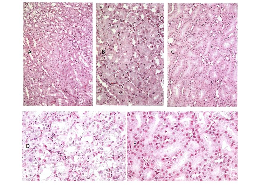
Life 2022, 12, 352                                                                                           2 of 19

                     abundance in households in the Croatian village of Kaniza [7], notable as hyperendemic
                     for BEN. The same fungus was common in an analogous cluster of villages in the Vratza
                     region in NW Bulgaria [8]. Attempts to characterize the toxin(s) causing rat renal kary-
                     omegaly and its histopathology continued [9], while its failure to affect the hamster was
                     also demonstrated [10]. Renewed experimental attention to compare rat nephropathies of
                     P. aurantiogriseum with that of the mycotoxin OTA resumed in the 1990s. Apoptosis located
                     amid karyomegalic changes then became possible with the ApopTag staining methodology
                     and raised questions about any etiological application to the renal atrophy of BEN [11].
                     Unfortunately, merely a brief explanation and color illustration of this in situ apoptosis,
                     revealed by the fluorescent TUNEL staining of 30 -OH caspase, now appears online only
                     and without its color.
                           Further taxonomic revision of P. aurantiogriseum [12] defined four new species descrip-
                     tions while retaining that name for a more restricted group of Penicillia. One of the new
                     redefined species is P. polonicum; rat nephrotoxicity has seemed exclusive to this species
                     and that descriptor has accordingly been applied ever since.
                           An exceptional opportunity for a comparison of rat renal histopathology, in response
                     to an extract of P. polonicum fermentation, with that of a vervet monkey in South Africa
                     found no change in the primate in contrast to the striking renal histopathological changes
                     in the rat [13]. This at least implied no apparent human genotoxic risk from susceptibility to
                     a P. polonicum karyocytomegaly, even when a primate model had been given an excessively
                     large dose of extract relative to the rat model. However, the expression of karyomegaly in
                     non-human primates has only been recorded infrequently [14] and, in general, a chemically
                     induced karyomegalic response in the rat does not necessarily predict a similar change in
                     human kidneys. Hard [14] also recommended that the threshold for diagnosing renal tubule
                     karyomegaly in animal studies should be accepted as requiring at least four times that of
                     normal nuclear size. This was not quite satisfied for one rat renal example attributed to
                     P. polonicum [15], being just one of a small group of rat renal tumors caused experimentally
                     by OTA and subjected to special DNA ploidy distribution measurements. Several aneuploid
                     nuclei above tetraploid occurred. Nevertheless, with hindsight, an exploration of apoptosis
                     in the primate experiment cited for P. polonicum [13] should have been attempted.
                           In contrast, OTA has a huge literature on toxicity in pigs and poultry, the potent
                     experimental causation of renal cancer in male rats and mice, in vitro experimental toxi-
                     cology, sophisticated analytical detection in food and feed components, and regulations
                     and legislations for human protection, but there is no proven case of human disease. An
                     experimental diagnosis of renal apoptosis in rats has been noted for OTA according to
                     Haematoxylin and Eosin (H&E) histology [16] and forms a timely basis for comparison
                     here, revealed by specific TUNEL staining, with P. polonicum/rat renal histopathology. To
                     avoid any misunderstanding, it should be stated that the P. polonicum isolate does not
                     produce OTA.
                           The present aim has been to illustrate a modern renal histopathological diagnosis of
                     apoptosis in the mycotoxicology of P. polonicum in rats after the semi-acute and chronic
                     ingestion of a selective culture extract. Tests for analogous histopathology in response to
                     OTA were also planned. For P. polonicum, this study serves to support a prospective report
                     further defining its mycotoxin(s).

                     2. Results
                     2.1. Preliminary Experiments Using P. polonicum-Molded Shredded Wheat
                           Preliminary studies had explored the importance of rat variety (Fischer, Sprague-
                     Dawley, Lewis, Wistar), gender and age, young adults (~200 g) or weanlings, for expressing
                     the familiar histopathological response to dietary P. polonicum. No clear differences were
                     evident except that Sprague-Dawley weanlings in the weight range of 25–40 g expressed
                     the most marked response and were used in some experiments. For continuity with the
                     literature [11] and as a preliminary study, adult male Sprague-Dawley rats were given feed
                     containing either a 20% P. polonicum-molded component or a 5% component for 5 days. For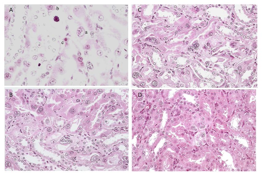
Life 2022, 12, 352                                                                                             3 of 19

                     the 20% component, toluidine blue-stained sections showed nuclei with condensed and/or
                     fragmented chromatin, i.e., “mitotic figures”, as well as enlarged nuclei, i.e., changes
                     typical of an acute response, in the renal cortico-medullary region where the S3 segment of
                     nephrons is a dominant component. The changes contrasted with the regular histology of a
                     control rat.
                           A qualitatively similar pathology was observed in rats given the 5% P. polonicum
                     feed, but the frequency of lesions was much lower. The outer cortex and medulla in
                     both treatment cases showed no changes. Confirming the well-established features of
                     karyomegaly, necrotic cells, and cells arrested in cell division in the kidneys of rats exposed
                     to a feed with a P. polonicum-molded component raised the question of apoptosis as a
                     mechanism of cell death in this context.
                           The histopathology of rat kidney following 5 days of consuming feed containing a 20%
                     component of shredded wheat molded by P. polonicum presents, in the cortico-medullary
                     region, as a complex picture. This involves karyocytomegalic epithelial cells in nephrons,
                     large irregular condensed chromatin bodies that are suggestive of epithelial mitosis and,
                     apparently, necrotic luminal cellular debris in nephrons. The present report is intended to
                     focus on the latter, but this must be set in the context of the other histopathological changes.
                     To establish confidence in the efficient recognition of apoptosis by ApopTag staining,
                     negative controls that were stained only by propidium iodide showed no spontaneous green
                     fluorescence when viewed via the appropriate blue filter. Control rats, given only normal
                     feed, showed only very rare fluorescence in the kidney sections; the observed fluorescence
                     in P. polonicum-treated rats was, thus, confidently regarded as indicating apoptosis.
                           Kidney sections of the rat receiving the 20% component were tested for apoptosis
                     using the ApopTag direct labeling kit (Merck Life Sciences, Gillingham, UK), which clearly
                     showed apoptotic nuclei stained the characteristic green-yellow by fluorescein (Figure 1A),
                     in contrast to other nuclei only stained red with propidium iodide. Actually, all the nuclei
                     were stained red by propidium iodide, but the intense green-yellow fluorescence of fluores-
                     cein masks the red staining of apoptotic bodies. Apoptotic nuclei, often located toward the
                     lumen of tubules, were confined to the cortico-medullary junction among the many cells
                     with condensed and/or fragmented chromatin. These were often located adjacent to the
                     tubular basement membranes, which were not fluorescein-labeled (Figure 1B).
                           Further exploratory studies using P. polonicum or OTA employed groups of weanling
                     rats (25–50 g).
                           For OTA, the mycotoxin was given in the feed (0.2 mg or 0.8 mg daily) for 5 days.
                     Histopathological changes in response to the higher OTA dose were also confined to the
                     cortico-medullary junction and involved the extensive loss of nephron epithelia; many cells
                     were necrotic, with eosinophilic cytoplasm, but the changes (stained with toluidine blue,
                     Figure 2C) did not conform to those recorded in response to P. polonicum (Figure 2B), both
                     being markedly changed when compared with the control (Figure 2A).
                           The lower OTA dose elicited only slight changes. The other weanlings given the higher
                     (0.8 mg) OTA in feed were tested for apoptosis using the ApopTag protocol, but none was
                     detected (Figure 2D).

                     2.2. Experiments Using the Cell-Free Extract of P. polonicum/Wheat Fermentation: Recognizing
                     Apoptosis in Both P. Polonicum and OTA Nephrotoxicity after 5 Daily Doses to Groups of 2 or
                     3 Animals
                          Seven protocols were applied for administering the 200 g male rat oral intake:
                     •    P. polonicum extract from 45 g or 15 g shredded wheat substrate in 20 g feed;
                     •    P.polonicum extract from 15 g shredded wheat substrate in 0.75 mL water for gavage;
                     •    Ochratoxin A: 1 mg or 0.2 mg in bicarbonate (0.3 mL) in 20 g feed or for oral gavage,
                          (5 or 1 mg/kg body weight (b. wt.)).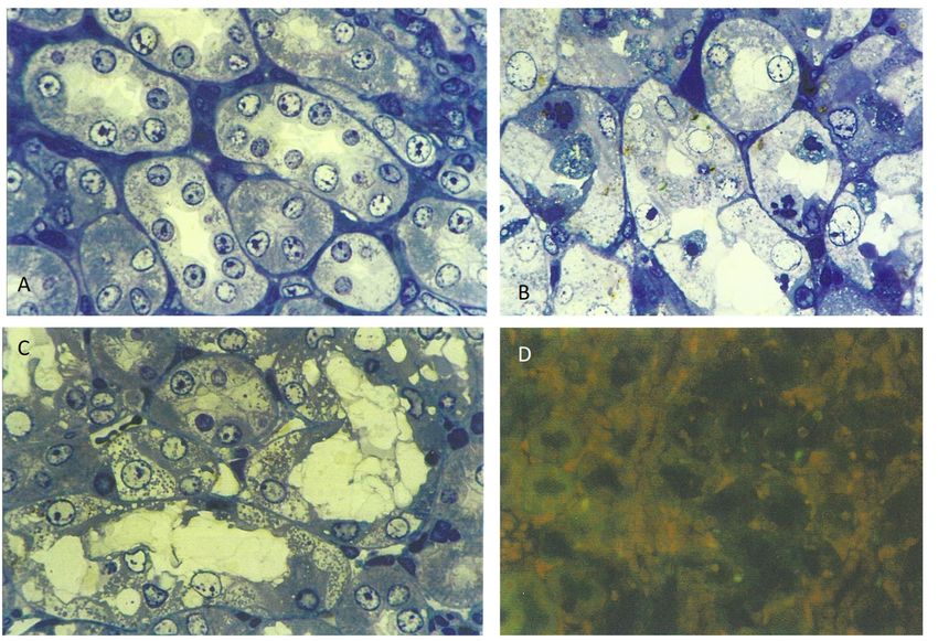
Life 2022,
Life 2022, 12,
12, 352
x FOR PEER REVIEW 44 of 21
of 19
Figure 1. Fluorescein-stained apoptotic nuclei in the renal cortico-medulla of an adult male rat given
a 20% P. polonicum-molded wheat diet for 5 days ((A), ×200). Apoptotic nuclei are depicted in a
tubule lumen. A propidium iodide-stained mitotic figure is shown ((B), ×450).
Further exploratory studies using P. polonicum or OTA employed groups of weanling
rats (25–50 g).
For OTA, the mycotoxin was given in the feed (0.2 mg or 0.8 mg daily) for 5 days.
Histopathological changes in response to the higher OTA dose were also confined to the
cortico-medullary junction and involved the extensive loss of nephron epithelia; many
cells were
Figure necrotic, with eosinophilic
1. Fluorescein-stained apoptotic cytoplasm,
apoptotic nuclei but
in the renal the changes of
cortico-medulla
cortico-medulla (stained
an adultwith
maletoluidine
rat given
blue, Figure
a 20% P. 2C) did not
P. polonicum-molded conform
polonicum-molded wheat to
wheat diet those
diet for recorded
for 55days
days((A), in response
((A),××200). to
200). Apoptotic P. polonicum
Apoptotic nuclei (Figure in
nuclei are depicted 2B),
a
both
tubule being
tubule lumen.
lumen. Amarkedly changed when
propidium iodide-stained
A propidium compared
iodide-stained mitotic with
mitotic figure
figure is the control
is shown
shown ((B), (Figure
((B), ×
×450).
450). 2A).
Further exploratory studies using P. polonicum or OTA employed groups of weanling
rats (25–50 g).
For OTA, the mycotoxin was given in the feed (0.2 mg or 0.8 mg daily) for 5 days.
Histopathological changes in response to the higher OTA dose were also confined to the
cortico-medullary junction and involved the extensive loss of nephron epithelia; many
cells were necrotic, with eosinophilic cytoplasm, but the changes (stained with toluidine
blue, Figure 2C) did not conform to those recorded in response to P. polonicum (Figure 2B),
both being markedly changed when compared with the control (Figure 2A).
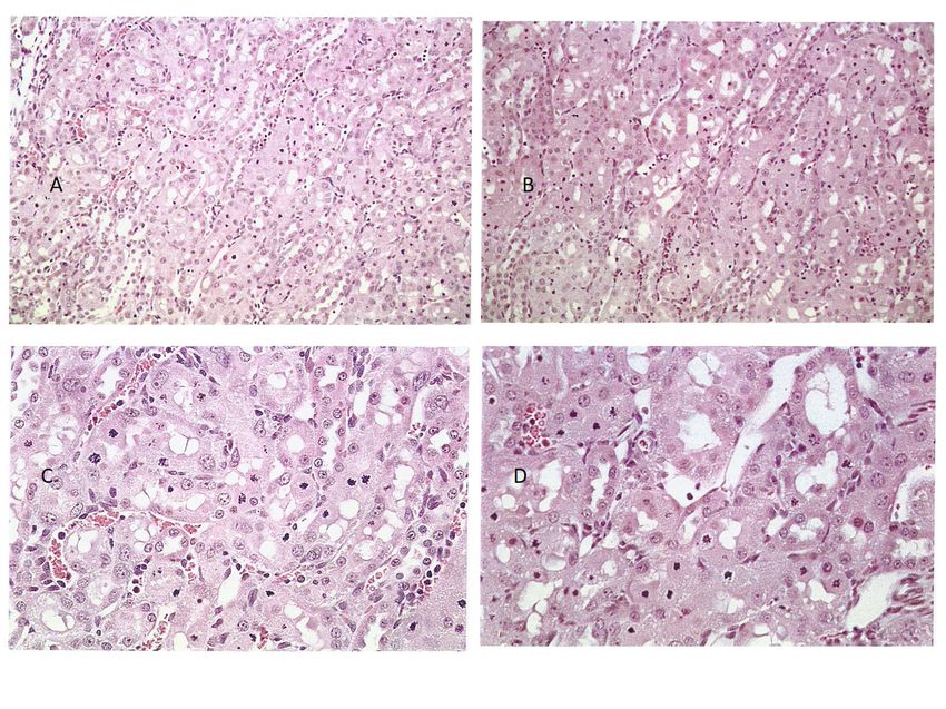
Figure 2. Comparative deviation in weanling rats from normal toluidine blue-stained cortico-
medullary histology: (A) by 5 days exposure to 20% P. polonicum-contaminated feed (B) or to ~25 mg
OTA/kg b. wt. (C), also in the feed. Note the absence of apoptosis, according to ApopTag staining in
a weanling rat given OTA (D); this may be a consequence of the marked necrotic damage illustrated
in (C). Settings: (A–C), 450×; (D), 200×.
General renal histopathology, illustrated in hematoxylin and eosin (H&E)-stained
sections, showed very frequent necrotic chromatin bodies in the cortico-medullary region
of a young adult male (200 g) given an extract from 45 g of shredded wheat molded by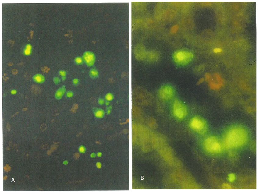
•    P.polonicum extract from 15 g shredded wheat substrate in 0.75 mL water for gavage;
                                         •    Ochratoxin A: 1 mg or 0.2 mg in bicarbonate (0.3 mL) in 20 g feed or for oral gavage,
                                              (5 or 1 mg/kg body weight (b. wt.)).
                                               General renal histopathology, illustrated in hematoxylin and eosin (H&E)-stained
                                         sections, showed very frequent necrotic chromatin bodies in the cortico-medullary region
      Life 2022, 12, 352                                                                                                           5 of 19
                                         of a young adult male (200 g) given an extract from 45 g of shredded wheat molded by P.
                                         polonicum daily for 5 days, within 20 g of the normal diet (Figure 3A,C). For comparison,
                                         the response in another rat given an extract of only 15 g of the molded feed was only
                                         P. polonicum
                                         slightly less daily for 5 days,
                                                        prominent        within
                                                                    (Figure     20 gWhere
                                                                            3B,D).   of the the
                                                                                            normal
                                                                                                 samediet (Figure
                                                                                                       extracts   3A,C).
                                                                                                                were     For comparison,
                                                                                                                     administered   once
                                         daily by oral gavage, there was still a similar pathology; however, this feed
                                         the  response   in another  rat given an  extract of  only 15  g  of the molded       was only
                                                                                                                          was clearer   at
                                         slightlymagnification
                                         higher    less prominent   (Figure4A,B),
                                                                  (Figure   3B,D).and
                                                                                    Where
                                                                                       was the
                                                                                             set same  extracts
                                                                                                 against        were administered
                                                                                                          the normality  of a controlonce
                                                                                                                                       rat
                                         daily by oral gavage, there was still a similar pathology; however, this was clearer at higher
                                         (Figure 4C). Both weight and condition were maintained among these experimental
                                         magnification (Figure 4A,B), and was set against the normality of a control rat (Figure 4C).
                                         animals.
                                         Both weight and condition were maintained among these experimental animals.

                                         Figure 3. Cortico-medullary histopathology of rats given a feed for 5 days containing extract from
Life 2022, 12, x FOR PEER REVIEW                                                                                             6 of 21
                                         45 g P. polonicum-molded wheat Haematoxylin and Eosin (H&E); (A) ×100, (C) ×200) or 15 g
                                         Figure 3. Cortico-medullary histopathology of rats given a feed for 5 days containing extract from
                                         P. polonicum-molded wheat (H&E; (B) ×100, (D) ×200).
                                         45 g P. polonicum-molded wheat Haematoxylin and Eosin (H&E); (A) ×100, (C) ×200) or 15 g P. po-
                                         lonicum-molded wheat (H&E; (B) ×100, (D) ×200).

                                         Figure 4. Comparative H&E histopathology in rats given a fermentation extract of 15 g shredded
                                   Figure 4. Comparative H&E histopathology in rats given a fermentation extract of 15 g shredded
                                         wheat by oral gavage daily for 5 days ((A), ×100, (B), ×200) with control ((C), ×100). Further
                                   wheat by oral gavage daily for 5 days ((A), ×100, (B), ×200) with control ((C), ×100). Further
                                         comparisons show the effect of 1 mg OTA by oral gavage ((D), ×200) and in feed ((E), ×200).
                                   comparisons show the effect of 1 mg OTA by oral gavage ((D), ×200) and in feed ((E), ×200).

                                        In rats given OTA, the greatest histopathological change was when 1 mg was given
                                   daily by oral gavage; there were frequent necrotic cells and eosinophilic debris within the
                                   lumen of tubules in the cortico-medullary region (Figure 4D). In contrast, administration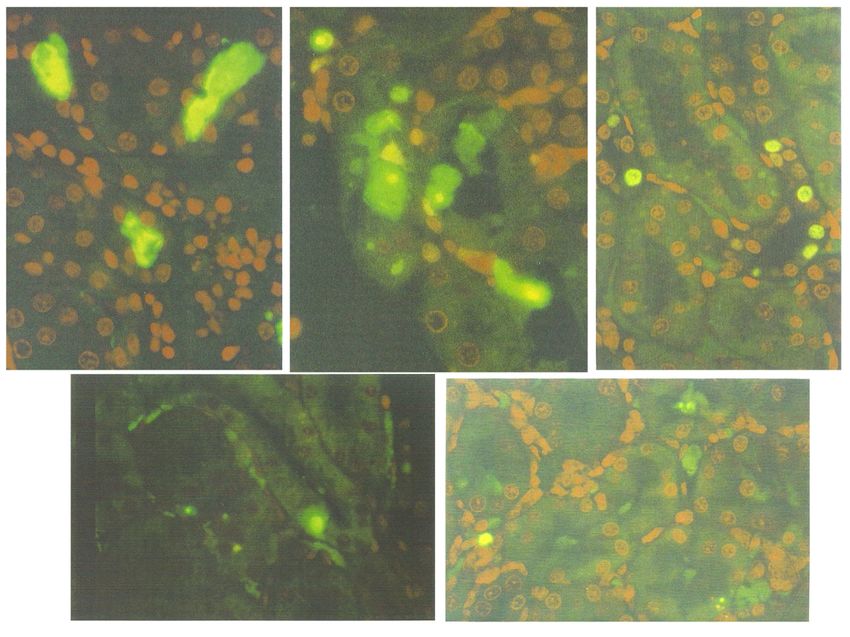
Life 2022, 12, 352 6 of 19
In rats given OTA, the greatest histopathological change was when 1 mg was given
daily by oral gavage; there were frequent necrotic cells and eosinophilic debris within the
lumen of tubules in the cortico-medullary region (Figure 4D). In contrast, administration
in the feed caused no obvious damage (Figure 4E) and 0.2 mg of the toxin by either route
similarly had no effect.
A more impressive response was revealed in the ApopTag-stained preparations, rep-
resented by the green-fluorescent bodies in the cortico-medullary region (Figure 5A,C),
seen in the young adult rat model that was given an extract from 45 g of shredded wheat
molded by P. polonicum, given daily for 5 days, within 20 g of the normal diet. This not
only verified apoptosis as an aspect of the complex renal histopathological change that can
occur in rats consuming feed contaminated by this fungus but also demonstrated that an
Life 2022, 12, x FOR PEER REVIEW
apoptotic factor could be extracted in alcoholic water. Other rats that were given one-third
7 of 21
of the above dose or an extract of the full dose by gavage also showed similar apoptosis
(Figure 5B,D) within the cortico-medullary region.
Figure5.5. Cortico-medullary
Figure Cortico-medullary region
region of
of aa rat
rat model,
model, given
given the
the higher
higher dose
dose (45
(45 g)
g) of
of P.P.polonicum
polonicum
fermentationextract
fermentation extract in the
in the diet,diet, showing
showing fluorescent
fluorescent apoptotic
apoptotic bodies, bodies, most of
most of which which
either either
protrude
protrude into the tubular lumen or are already free in it (A). As seen, also through a phase-contrast
into the tubular lumen or are already free in it (A). As seen, also through a phase-contrast blue filter
blueApopTag
((C), filter ((C), ApopTag
×200). ×200). The corresponding
The corresponding illustrationsillustrations
for the lowerfor the(15
dose lower dose in
g) given (15the
g) feed
given
arein
the feed are shown in (B), compared with administering by gavage (D). Setting: ×200.
shown in (B), compared with administering by gavage (D). Setting: ×200.
Sprague-Dawleyrats
Sprague-Dawley rats(200
(200g)g) given
given thethe higher
higher OTA OTAdosedose by oral
by oral gavage
gavage showedshowed
a fewa
few fluorescent (apoptotic) bodies oriented within the tubular epithelium,
fluorescent (apoptotic) bodies oriented within the tubular epithelium, as well as diffuse as well as
diffuse fluorescence
fluorescence within lumens
within tubular tubular (Figure
lumens 6A,B),
(Figurepossibly
6A,B), possibly representing
representing apoptoticapoptotic
debris.
debris.
Such Such histological
histological changes changes
extendedextended
not only not only
across theacross the cortico-medullary
cortico-medullary region butregion
also
but also into both the cortex and medulla. This could logically
into both the cortex and medulla. This could logically correlate with the 30 g weightcorrelate with the 30 g
weight loss sustained during the 5-day dosing period, reflecting
loss sustained during the 5-day dosing period, reflecting a general adverse response to a general adverse
response
the rather to the OTA
high ratherdose
high(5OTAmg/kgdoseb.(5 wt.).
mg/kg Inb.contrast,
wt.). In contrast,
no weight no loss
weight loss occurred
occurred in the
in theOTA
other otherregimens.
OTA regimens.
The lowerThe dose
lowerby dose by gavage
gavage and theand the higher
higher dose indose
feedinelicited
feed elicited
only
only
2–3 2–3 fluorescent
fluorescent bodies bodies
across anacross
entireanlongitudinal
entire longitudinal kidney (Figure
kidney section section 6C,D),
(Figureand 6C,D),
no
and no
such such fluorescent
fluorescent staining staining
occurredoccurred
after theafter
lowerthe lower
dose dose (1 b.
(1 mg/kg mg/kg b. wt., Figure
wt., Figure 6E). 6E).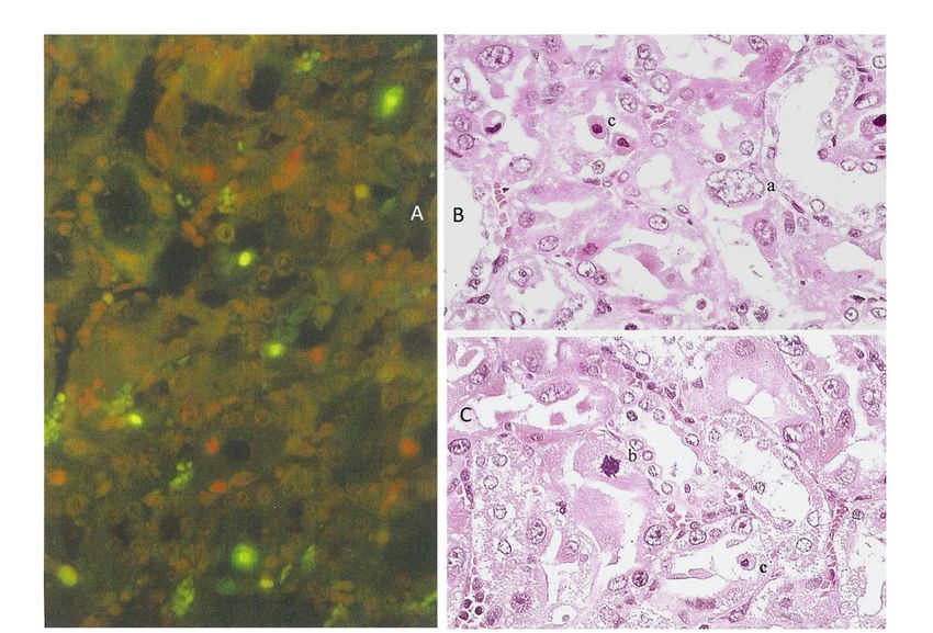
Life2022,
Life 2022,12,
12,352
x FOR PEER REVIEW 87 of 19
21
A B C
D E
Figure
Figure6.6. ApopTag
ApopTagstaining
stainingofofthe
therenal
renalcortico-medullary
cortico-medullary region ofof
region rats given
rats OTA
given OTAdaily forfor
daily 5 days, to
5 days,
show the fluorescent
to show apoptotic
the fluorescent apoptotic (×200).(×200).
bodiesbodies (A,B) After
(A,B)1After
mg by1oral
mg gavage,
by oralwith marked
gavage, withapoptosis
marked
in tubular in
apoptosis lumen andlumen
tubular and(×
epithelia 200). (C)(×200).
epithelia Staining
(C) after 0.2 mg
Staining by0.2
after oral
mggavage,
by oralshowing discrete
gavage, showing
apoptosis, particularly in the nephron epithelium (×200). (D,E) Staining after the administrationthe
discrete apoptosis, particularly in the nephron epithelium (×200). (D,E) Staining after of
1administration
mg and 0.2 mgofextract,
1 mg and 0.2 mg extract,
respectively, both respectively,
in feed, with both
very in feed, with
occasional very occasional
apoptosis (×200). apoptosis
(×200).
2.3. Application of Laser Scanning (Confocal) Microscopy
2.3. Application of Laser Scanning
Due to its technical superiority (Confocal) Microscopy
over light fluorescent microscopy, confocal microscopy
allowedDuefortotheitsrecording
technicalofsuperiority
fluorescenceoverin the ApopTag-labeled
light kidney sections
fluorescent microscopy, at a
confocal
much lower allowed
microscopy magnification,
for thesuch as ×10of(Figure
recording 7B–D), in
fluorescence withtheaApopTag-labeled
wider view of thekidneyrenal
tissue.
sectionsThis
at aenabled visualmagnification,
much lower proof of the extent
suchofasthe
×10small fluorescent
(Figure bodies
7B–D), with in the view
a wider cortico-
of
medullary region, illustrating dose-dependent response relationships
the renal tissue. This enabled visual proof of the extent of the small fluorescent bodies among rats givenin
high- or low-dose dietary
the cortico-medullary P. polonicum
region, extract;
illustrating an approximate
dose-dependent 140:30
response ratio of apoptotic
relationships among
cells is evident.
rats given high-Also illustrated
or low-dose is an analogous
dietary response
P. polonicum extract;evoked by a rather140:30
an approximate high regimen
ratio of
for OTA (1cells
apoptotic mg is byevident.
oral gavage); the daily is
Also illustrated dose approximates
an analogous to 5 mg/kg
response evokedb.bywt. and
a rather
approximately 65 apoptotic bodies were revealed in Figure 7D. This
high regimen for OTA (1 mg by oral gavage); the daily dose approximates to 5 mg/kg b. illustrates a cortico-
medullary background 65
wt. and approximately in apoptotic
which thebodies
regular arrangement
were revealed in ofFigure
propidium iodide-stained
7D. This illustrates a
nuclei showed the normal renal tubule conformation, as in
cortico-medullary background in which the regular arrangement of propidium Figure 7B,C. This experiment
iodide-
also verified
stained that
nuclei the apoptotic
showed P. polonicum
the normal metabolite
renal tubule is extractable
conformation, from
as in a fermentation
Figure 7B,C. This
matrix, simply
experiment with
also alcohol
verified and
that thewater.
apoptotic P. polonicum metabolite is extractable from a
Furthermore, in kidney sections
fermentation matrix, simply with alcohol stained
and with
water.an ApopTag Direct kit, all nuclei are
labeled red with propidium iodide, while the apoptotic cells are additionally labeled
yellow-green by fluorescein. However, because of the masking effect of fluorescein over
propidium iodide, the apoptotic cells are always seen as bright yellow-green-fluorescent
bodies under light fluorescence microscopy (Figure 8A). Nevertheless, the powerful laser of
the confocal microscope makes it possible to distinguish between the emissions from both
fluorescein and propidium iodide within the apoptotic cells (Figures 7A and 8C), allowing
differentiation between the genuine cells undergoing apoptosis and those solely carrying
green non-specific labeling. Nuclei with condensed chromatin (“mitotic figures”) that are
stained only with propidium iodide are also clearly visible in Figure 8.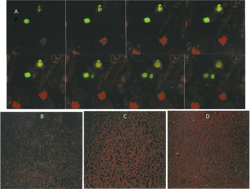
Life 2022, 12, 352                                                                                                                             8 of 19
   Life 2022, 12, x FOR PEER REVIEW                                                                                                      9 of 21

                                       Figure 7. Laser-scanned micrographs of ApopTag preparations for apoptosis caused by P.
                                      Figure 7. Laser-scanned micrographs of ApopTag preparations for apoptosis caused by P. polonicum.
                                       polonicum. A gallery of 8 sequential planes at 0.85 µm intervals through a 6 µm kidney section
                                      A gallery of 8 sequential planes at 0.85 µm intervals through a 6 µm kidney section showing the
                                       showing the change of color of certain bodies (labeled with propidium iodide (red) and fluorescein
                                      change
                                       (green)),ofdifferentiating
                                                   color of certain    bodies
                                                                  between      (labeled
                                                                           apoptotic    with and
                                                                                      (above) propidium     iodide figures
                                                                                                   mitotic (below)  (red) and  fluorescein
                                                                                                                           of constituent    (green)),
                                                                                                                                          bodies
                                       ((A) ×400). Low-magnification (×10) sections in the cortico-medullary region of rats given ×
                                      differentiating   between   apoptotic  (above) and  mitotic (below)  figures of constituent bodies  ((A) P.400).
Life 2022, 12, x FOR PEER REVIEW                                                                                                      10 of 21
                                       polonicum   extract in feed at higher (B) and one-third lower  (C) content or OTA  at 1 mg/day  ((D)
                                      Low-magnification (×10) sections in the cortico-medullary region of rats given P. polonicum extract in×10).
                                      feed at higher (B) and one-third lower (C) content or OTA at 1 mg/day ((D) ×10).
                                           Furthermore, in kidney sections stained with an ApopTag Direct kit, all nuclei are
                                      labeled red with propidium iodide, while the apoptotic cells are additionally labeled
                                      yellow-green by fluorescein. However, because of the masking effect of fluorescein over
                                      propidium iodide, the apoptotic cells are always seen as bright yellow-green-fluorescent
                                      bodies under light fluorescence microscopy (Figure 8A). Nevertheless, the powerful laser
                                      of the confocal microscope makes it possible to distinguish between the emissions from
                                      both fluorescein and propidium iodide within the apoptotic cells (Figures 7A and 8C),
                                      allowing differentiation between the genuine cells undergoing apoptosis and those solely
                                      carrying green non-specific labeling. Nuclei with condensed chromatin (“mitotic figures”)
                                      that are stained only with propidium iodide are also clearly visible in Figure 8.

                                                Light-fluorescentmicrograph
                                     Figure8.8.Light-fluorescent
                                   Figure                           micrographshowing
                                                                                 showing bright
                                                                                            bright yellow-green
                                                                                                   yellow-green apoptotic
                                                                                                                  apoptotic bodies
                                                                                                                              bodies and
                                                                                                                                      anda apropid-
                                   propidium    iodide-stained
                                     ium iodide-stained         condensed
                                                           condensed        chromatin
                                                                       chromatin        resembling
                                                                                   resembling       a mitotic
                                                                                                a mitotic     figure
                                                                                                          figure      at kidney
                                                                                                                 at the  the kidney cortico-
                                                                                                                                cortico-medullary
                                   medullary   junction
                                     junction of          of a rat
                                                  a rat treated    treated
                                                                with        with P. ((A)
                                                                      P. polonicum.  polonicum.
                                                                                          ×450).((A)  ×450). Laser-scanning
                                                                                                  Laser-scanning    micrographsmicrographs
                                                                                                                                   showing apop-
                                   showing apoptotic bodies labeled by propidium iodide only (red, (B)) and by fluorescein (green,
                                     totic bodies labeled by propidium iodide only (red, (B)) and by fluorescein (green, (C)) in ApopTag
                                   (C)) in ApopTag preparations (×400).
                                     preparations (×400).
                                   2.4. Intermittent Exposure to P. Polonicum for Three Months; Alternating Cycles with Normal
                                   Feed
                                        An experiment with four male Sprague-Dawley rats commenced concerning
                                   alternating cycles of the standard 5-day 20% P. polonicum-molded diet, followed by 3.5
                                   weeks toxin-free, offering opportunities for accumulated histopathological change or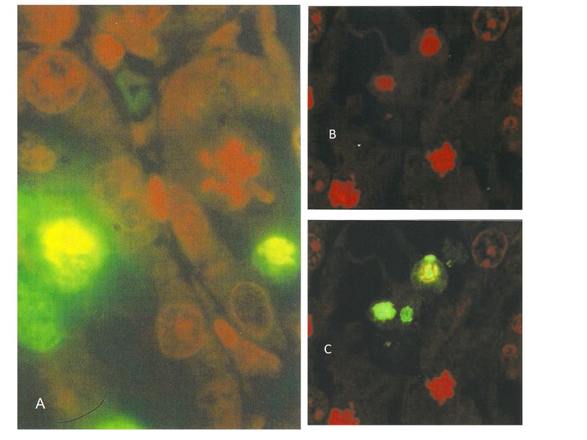
Life 2022, 12, 352 9 of 19
2.4. Intermittent Exposure to P. polonicum for Three Months; Alternating Cycles with
Normal Feed
An experiment with four male Sprague-Dawley rats commenced concerning alter-
nating cycles of the standard 5-day 20% P. polonicum-molded diet, followed by 3.5 weeks
toxin-free, offering opportunities for accumulated histopathological change or partial re-
gression. After the first toxin phase, renal H&E histology in one rat appeared typical of
the P. polonicum influence already described. The other three rats continued their normal
diet for a further 3.5 weeks, at which point two were euthanized for histology. No pyknotic
cells or cells with condensed and/or fragmented chromatin that was typical of the first rat’s
response to P. polonicum were seen. Nevertheless, cells with slightly enlarged nuclei that
Life 2022, 12, x FOR PEER REVIEW
were mildly karyomegalic occurred in the cortico-medullary region, implying 11 that some
of 21
nuclear division had occurred during and shortly after the initial P. polonicum insult and
had persisted. Notably, in kidney sections also subjected to ApopTag staining, a substantial
frequency of apoptotic bodies was apparent (Figure 9A).
Figure
Figure9. 9.ApopTag-revealed apoptosis after
ApopTag-revealed apoptosis aftera second
a second
5-day5-day exposure
exposure to P. polonicum
to P. polonicum (A).
(A). Accumulated
Accumulated
cortico-medullary histopathological changes, following the fourth cycle of 5-day P. polonicumP.sub-
cortico-medullary histopathological changes, following the fourth cycle of 5-day
polonicum sub-chronic exposure, as various populations of abnormal cells, such as karyocytomegalic
chronic exposure, as various populations of abnormal cells, such as karyocytomegalic ((B),a), large
((B),a), large mitotic figure ((C),b), or pyknotic apoptotic cells ((B),c), H&E (×200).
mitotic figure ((C),b), or pyknotic apoptotic cells ((B),c), H&E (×200).
Experiments
Experiments with thethe
with fourth rat rat
fourth continued
continued for for
twotwomore
more cycles, culminating
cycles, culminating in ain a
fourth
fourth 5-day P. polonicum treatment, with normal conditions continuing. Kidneyhis-
5-day P. polonicum treatment, with normal conditions continuing. Kidney H&E H&E
topathology showed
histopathology a striking
showed response
a striking during
response the three-month
during the three-month experiment,
experiment, wherein
whereina a
large proportion of abnormally enlarged cells had much-enlarged nuclei,
large proportion of abnormally enlarged cells had much-enlarged nuclei, thus constituting thus constitut-
ingkaryocytomegaly.
karyocytomegaly.Nephron Nephronepithelia
epithelia with
with condensed
condensed and/or
and/or fragmented
fragmented chromatin
chromatin were
were also prominent in the now-distorted cortico-medullary region.
also prominent in the now-distorted cortico-medullary region. Some of these were Some of these were
much
much larger
larger thanthan those
those in response
in response to atosingle
a single 5-day
5-day exposure
exposure (illustrated
(illustrated in Figure
in Figure 3). 3).
This
This suggestedthe
suggested thecumulative
cumulativebuilding
building of of specific
specific nuclei
nucleiduring
duringthe thecycles
cyclesof of
P. polonicum
P. polonicum
exposure.
exposure. Nevertheless,
Nevertheless, the eosinophilic appearance
the eosinophilic of the corresponding
appearance of the correspondingcytoplasm sug-
cytoplasm
gested a concomitant degree of necrosis. Another population of abnormal
suggested a concomitant degree of necrosis. Another population of abnormal cells had cells had round
or elliptical
round ornuclei in the
elliptical eosinophilic
nuclei cytoplasm cytoplasm
in the eosinophilic that protrudedthat into the nephron
protruded into thelumens,
nephron
extending
lumens,also into thealso
extending medulla (Figure
into the medulla9B,C). These
(Figure might
9B,C). be apoptotic
These might besince they resem-
apoptotic since they
bleresemble
fluorescein-stained bodies in bodies
fluorescein-stained a previous
in a ApopTag-stained example (Figure
previous ApopTag-stained example 9A). It was9A).
(Figure
concluded that intermittent
It was concluded exposure toexposure
that intermittent P. polonicum
to P.resulted in the
polonicum induction
resulted in theof promi-
induction
nently karyocytomegalic
of prominently cells throughcells
karyocytomegalic the cycles
throughof toxic insults,ofpresumably
the cycles toxic insults, exacerbated
presumably
by the last one. The persistent evidence takes the form of a combination of “mitotic fig-
ures” composed of condensed or fragmented chromatin.
2.5. Comparative Continuous-Exposure Responses to Dietary P. polonicum or Ochratoxin A
Male Sprague-Dawley rats (180 g) were given the 20% P. polonicum-contaminated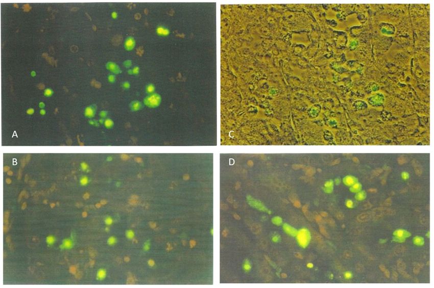
Life 2022, 12, 352 10 of 19
exacerbated by the last one. The persistent evidence takes the form of a combination of
“mitotic figures” composed of condensed or fragmented chromatin.
2.5. Comparative Continuous-Exposure Responses to Dietary P. polonicum or Ochratoxin A
Male Sprague-Dawley rats (180 g) were given the 20% P. polonicum-contaminated diet,
two for 3 weeks, one for 2 months, and one for 3 months. An additional rat was given a
diet contaminated with OTA (0.4 mg/rat daily: ~2 mg/kg b. wt.), also for 3 months, for
comparison. All treatments were well tolerated.
All rats given P. polonicum developed prominent karyocytomegaly, expressed both
in the frequency and magnitude of the histopathological change. Additionally, cells with
large condensed or fragmented chromatin were seen. A replicated example at 3 weeks is
Life 2022, 12, x FOR PEER REVIEW shown in Figure 10A. Nevertheless, these histological abnormalities were most marked in
12 of 21
the rat exposed to P. polonicum for the longest period, in which the proximal convoluted
tubules had lost their arrangement at the cortico-medullary junction and cells with multiple
nuclei were evident (Figure 10B,C). The rat that was exposed for two months presented
presented intermediate karyocytomegaly. Although ApopTag staining for the detection
intermediate karyocytomegaly. Although ApopTag staining for the detection of apoptosis
of apoptosis had not yet been performed, apoptotic cells were recognized morphologically
had not yet been performed, apoptotic cells were recognized morphologically according to
according to their appearance and location in H&E sections, but they were rare.
their appearance and location in H&E sections, but they were rare.
Figure
Figure 10. Cortico-medullary
Cortico-medullaryhistopathology
histopathologyafter
after 3 weeks
3 weeks of dietary
of dietary exposure
exposure to P. polonicum-
to P. polonicum-molded
molded feed, showing
feed, showing (A) abnormal
(A) various various abnormal cells: karyomegaly
cells: karyomegaly (a), large
(a), large mitotic mitotic
figure withfigure with
condensed
condensed chromatin (b), and necrotic cells (c). Histopathology after 3 months,
chromatin (b), and necrotic cells (c). Histopathology after 3 months, showing karyocytomegalic cells showing
karyocytomegalic cells and
and cells with multiple cells
nuclei inwith multiple
tubules, with nuclei
distortedin tubules, with distorted
conformation conformation
(B,C). Comparison with(B,C).
mild
Comparison with mild karyomegaly within the regular tubular conformation, after 3 months of
karyomegaly within the regular tubular conformation, after 3 months of dietary OTA (0.4 mg daily in
dietary OTA (0.4 mg daily in feed, (D) H&E (×200)).
feed, (D) H&E (×200)).
In
In the ratgiven
the rat giventhree
threemonths
months of dietary
of dietary OTA,
OTA, only only
mild mild karyomegaly
karyomegaly occurred
occurred within
within the regular cortico-medullary conformation (Figure 10D), contrasting with the
the regular cortico-medullary conformation (Figure 10D), contrasting with the distortions
distortions
from exposurefromtoexposure to P. polonicum.
P. polonicum.
3.
3.Discussion
Discussion
In additiontotosummarizing
In addition summarizing the the immediate
immediate experimental
experimental findingsfindings on rat
on rat renal renal
histopatho-
histopathological
logical responses responses to theofingestion
to the ingestion of P.extrolites,
P. polonicum polonicumalong
extrolites, alongcomparison
with some with some
comparison with the
with the responses to responses to OTA,evolution
OTA, the modern the modern evolution taxonomy
of Penicillium of Penicillium
will taxonomy
need to be
will
addressed to highlight some apparent uncertainties in using this taxonomy, particularlythis
need to be addressed to highlight some apparent uncertainties in using for
taxonomy,
P. polonicumparticularly for P. polonicum
and P. aurantiogriseum in theand P. aurantiogriseum
recent literature. in the recent literature.
Confirmation and refinement of the former general histopathology regarding the
dietary exposure of rats to a wheat substrate molded by P. polonicum, which is of Balkan
origin, has given us an opportunity to focus on the renal pyknotic nuclei and apoptosis
and to make some preliminary comparison with that caused by OTA. Finding thatLife 2022, 12, 352 11 of 19
Confirmation and refinement of the former general histopathology regarding the
dietary exposure of rats to a wheat substrate molded by P. polonicum, which is of Balkan
origin, has given us an opportunity to focus on the renal pyknotic nuclei and apoptosis and
to make some preliminary comparison with that caused by OTA. Finding that progressive
renal karyomegaly leading to karyocytomegaly can be evident in both weanling and adult
rats, simply via an extract in the feed, enables economy in the bioassays necessary to
recognize the elusive toxin(s). Notably, nephrotoxins will remain in an alcohol extract of
the P. polonicum-molded fermentation of wheat from which, after evaporation, the excess
fungal sterol can be precipitated with water. Incorporating the product into powdered feed
to mimic the natural human or animal intake also allows the opportunity for evaporation
of the traces of residual alcohol before consumption.
Since raising the question concerning any P. polonicum contribution by apoptosis to the
chronic renal atrophy of BEN [11], this study offers a more comprehensive demonstration
that at least most of the pyknotic nuclei in the cortico-medullary nephrons of the H&E-
stained kidneys of rats given OTA are apoptotic, according to TUNEL-based histology
that confirms apoptosis as contributing at least part of P. polonicum’s nephrotoxicity. One
subsequent report on apoptosis as a part of OTA nephropathy in the rat model [16] was
even more assertive concerning OTA’s putative involvement in the pathogenesis of Balkan
endemic nephropathy, offering findings after daily administration over several weeks at
an overall rate a little higher than the high dose of the NTP study [17]. Apoptosis was
diagnosed after intraperitoneal OTA toxicity was assigned to the small, condensed chro-
matin bodies deeply stained for such purposes in H&E histology preparations. The extent
to which this equates to the specificity of the TUNEL technique is an open question. How-
ever, in a further description of the same experiment, H&E staining specifically excluded
simple necrosis [18]. The enteral administration of OTA for the present experiments makes
direct comparison difficult, but the findings confirm apoptosis for P. polonicum as a not-
unexpected mycotoxicological attribute. Historically for OTA, the first major rat lifetime
exposure study [17] used oral gavage because of its accuracy of dosing in a toxicological
context, but predictably slowed bioavailability by delivering the dose in a corn oil vehicle.
The present OTA delivery used contrasting oral gavage in an aqueous vehicle or incorpora-
tion in the feed for that whole day. The Croatian study’s [16] use of an intraperitoneal route
would have given quite quick direct insults to the renal parenchyma. Such administration
near the kidneys, although needing only a short circulatory vascular pulse to the renal
artery, would of course enable maximum toxicological efficiency, while being non-natural.
A single 1 mg/kg dose to female Wistar rats caused a few cortical apoptoses across a kidney
section the next day, declining numerically during the following 9 days. After the same
dose daily three times per week for 4 weeks, ~100 apoptotic nuclei were recorded across a
kidney section, assuming that H&E staining always diagnoses TUNEL histopathology. Pre-
sumably, female rats were used because the incidence of endemic nephropathy in Croatia
is higher in women [18] and OTA might somehow generate the chronic fibrotic pathology
of the Balkan disease.
The recognition of apoptotic nuclei in Wistar male rat kidneys by TUNEL staining
after chronic exposure to OTA [19] was also achieved after 2 months of daily gavage in oil
at a dose slightly less than that of the mean daily high dose in the NTP study [17], which
had caused significant renal cancer much later in life. The exposure-related occasional
incidence of karyomegaly and pyknotic nuclei in cortical nephrons in H&E preparations
was illustrated, as were fluorescent apoptotic bodies identified specifically by the TUNEL
protocol. Another study [20], using an even higher gavage OTA dosage in oil to Sprague-
Dawley males for 2 weeks (0.5 mg/kg b. wt.), also caused renal apoptosis, as illustrated by
TUNEL staining. However, the serum OTA concentration achieved in the first experimental
study was nearly 10 µg/mL which is 10,000 times greater than that measured for a European
human exposed to a normal diet; it is important to have a realistic perspective when
extrapolating from an experimental rodent to a human model.Life 2022, 12, 352 12 of 19
In the present study, alternating short periods of dietary P. polonicum exposure of
young rats with subsequent longer periods of uncontaminated diet over 3 months led to
the progressive expansion of karyomegalic nuclei over the 3-month period. However, the
findings raise the question of whether the histological picture of progressively increasing
karyomegalic ploidy during repetitive exposure to P. polonicum is driven within replacement
nephron epithelial cells, consequent on replacing the apoptoses that had occurred during
the juvenile exposure phase. Continuous P. polonicum exposure for 3 months also supported
expansive karyocytomegaly; in contrast, continuous dietary OTA exposure at a relatively
high dose (2 mg/kg b. wt.) only caused mild karyomegaly within an otherwise undisturbed
nephron architecture.
The question arises, therefore, whether local nephron epithelial repair after apoptosis
in response to P. polonicum nephrotoxicity differs from that in response to OTA, which can
easily cause more extensive local cytotoxicity in the cortico-medullary region (Figure 2C).
Notably, the DNA ploidy distribution in rat kidneys after 4 weeks of P. polonicum dietary
exposure [15] caused several aneuploid nuclei in the tetraploid range, but also a few
toward octoploid. Such nuclei could be unstable and a potential matter of concern [21].
OTA in male rats is capable of forming renal tumors exhibiting a wide range of unstable
aneuploidy [15].
A two-week pathology study [22] with male Sprague-Dawley rats compared dietary
P. polonicum with oral gavage of the P. citrinum mycotoxin, citrinin, which shares its pen-
taketide structure with a similar moiety of OTA [23]. A 10% P. polonicum-molded shredded
wheat diet triggered the histopathological changes as presently described, combined with
only mild cytotoxicity, all in the same S3 kidney region. Citrinin (2.5 mg/kg) elicited
cytotoxicity but caused no nuclear changes when administered alone, but, when combined
with the P. polonicum regimen, pathological changes were only as those for P. polonicum. No
significant pathologies in the stomach, small intestine, spleen, thymus gland or lung were
associated with the P. polonicum regimen. For all regimens, urinary osmolarity decreased,
associated with slight glucosuria and an impaired concentration capacity of the kidneys.
Urinalysis showed the increased activity of y-glutamyl transpeptidase where rats received
P. polonicum, also demonstrating the elevated urinary composition of low-molecular-weight
proteins. The latter finding predates the proposed role of small serum proteins binding
OTA [24], together, salvaged into rat cortical nephrons’ proximal tubule epithelia from the
glomerular filtrate. A question now arises concerning whether any analogous mechanism
might be operating for a P. polonicum nephrotoxic mycotoxin.
The severity of nephrotoxic responses to environmental Penicillia, whether to OTA
from P. verrucosum or P. nordicum, or to the mycotoxins of P. polonicum, has long been
conditional not only on the dose magnitude but also on the delivery mode [25]. When there
is a marked response, oral gavage gives a greater response than natural delivery in feed; for
OTA, this has also been reflected in the mycotoxin’s plasma concentration after repetitive
dosing. The principle was extended to apply to the accumulation of DNA adducts in a
general exploration of rat kidney DNA in the specialist laboratory of Professor A. Leszkow-
icz, Toulouse, after a range of exposures to P. polonicum [26]. Although most experimental
permutations yielded unremarkable findings, the inclusion of P. polonicum fermentation
extract in the diet caused not only the characteristic histopathological changes already illus-
trated above but also created one prominent DNA adduct that was dose-related. This was
proportionately represented across a threefold difference in dose by a fivefold numerical
differential (Figure 11). Therefore, a further aspect of P. polonicum nephrotoxicology in
the rat model is added, although the amount of the genotoxin in the P. polonicum extract
consumed over 5 days by adult rats is unknown, as is whether there is any relationship
with karyomegaly. For other mammals, the P. polonicum histopathology has been seen in
guinea pigs and pigs but not in Balb-C mice [27]. Studies in hamsters, contemporary with
the rat experiments at Imperial College in the 1980s, showed no histopathological changes
after dietary exposure [10].difference in dose by a fivefold numerical differential (Figure 11). Therefore, a further
aspect of P. polonicum nephrotoxicology in the rat model is added, although the amount
of the genotoxin in the P. polonicum extract consumed over 5 days by adult rats is
unknown, as is whether there is any relationship with karyomegaly. For other mammals,
the P. polonicum histopathology has been seen in guinea pigs and pigs but not in Balb-C
Life 2022, 12, 352 13 of 19
mice [27]. Studies in hamsters, contemporary with the rat experiments at Imperial College
in the 1980s, showed no histopathological changes after dietary exposure [10].
Figure 11. Autoradiographs of polyethyleneimine-cellulose chromatography of 32 P-post-labeled
DNA adducts from rat kidney. Left, control. Center, 5 days’ feeding with a diet containing an extract
from 3 g shredded wheat substrate molded for 20 days by P. polonicum. The right image is as in the
center, but refers to 9 g shredded wheat extract. Principal samples—a specific adduct attributed to
P. polonicum has measured a 32 P disintegration ratio of 1:5. A minor spot to the right at a higher dose
has a similar ratio with the lower-dose image.
As is concurrent with the study of its nepropathic potential [5], the former Bulgar-
ian P. verrucosum var. cyclopium had been found to produce the alkaloid auranthine [28].
Another alkaloid, a benzodiazepine named anacine [29], was later described as a metabo-
lite of a Yugoslavian isolate (IMI 357488), collected in the hyperendemic nephropathy
village of Kaniza [7]) and authenticated as P. aurantiogriseum Dierckz [6,30]. Subsequently,
it is stated [12] that “the original isolate (IMI 180922A) investigated by Barnes et al. [5]
as P. aurantiogriseum was correctly identified”. It is not clear whether this amplification
of the original literature means that it was as recognized before or after its revised tax-
onomic status [12,31]; the suffix A, added to the simple IMI number as cited [12,30], is
also not explained. Ultimately, the culture of IMI 180922 had been supplied directly to
P.M. by P. Austwick [5] and revealed nephropathy in rats, enabling the first description of
auranthine as a co-metabolite with penicillic acid and verrucosidin [28].
Consequently, the previous discussion is relevant to the recent revision of the auran-
thine structure [32] since the Bulgarian P. verrucosum var. cyclopium = P. aurantiogriseum =
P. polonicum nomenclature, spanning over 40 years, implies that further study of auranthine
would need to be conducted with a modern P. polonicum. Thus, structural revision using
a modern, defined P. aurantiogriseum isolate (CBS 112021 [32]) could not necessarily be
expected to biosynthesize auranthine without access to a reference sample. Unfortunately,
none of that sample remains. However, the revised structure, aided by X-ray crystallog-
raphy, was based on biosynthetic conditions, including a substantial glutamine additive
(~15 g/L) to the medium. That additive might reasonably be regarded as not only enriching
the nitrogen source but also potentially directing the biosynthesis of a glutamine-derived
extrolite. Thus, in perhaps not using the correct fungus, and using a fermentation nu-
tritionally enriched to achieve an increased metabolite yield, the revised structure may
indeed widen its occurrence as a P. aurantiogriseum metabolite with a weak cytotoxicity
profile [32]. Notably, however, a co-metabolite, aurantiamine, markedly decreased the
viability of HepG2 cells at 30 µM and above [32]. In our experience, practical differentiation
in agar cultures between modern P. polonicum and P. aurantiogriseum is not easy; it is partly
conditioned by the individual perception of color, as was also problematic between the
former P. aurantiogriseum and P. commune in a Croatian study c. 30 years ago [7]—the two
were subsequently acknowledged as being synonymous [31]. Notably, P. commune isolates,
both from Yugoslavia and Bulgaria, and a P. aurantiogriseum from Yugoslavia had all had
been shown to produce auranthine [7]. While a structural revision after 40 years in the lightLife 2022, 12, 352 14 of 19
of new analytical findings is always welcome, it is vital to be sure that the recent revision
for auranthine actually relates to the same substance as formerly described [28]. The re-
appraisal of auranthine as a structurally characterized metabolite within the Penicillium
section, Viridicata series, Viridicata under simple cultural conditions by an authenticated
fungus and augmented by biosynthetic evidence would be helpful. Nevertheless, none of
the recognized P. polonicum extrolites (penicillic acid, verrucosidin, verrucofortines, aspter-
ric acid, anacine, puberulines, cyclopenins [33]) has apparently not yet been tested in terms
of the present rat nephropathy.
Notably for the original description of the rat nephropathy described here [5], foodstuff
crop samples were collected during the early 1970s in those Balkan areas hyperendemic
for the Balkan (endemic) nephropathy. Of three collected from Yugoslavia and Bulgaria
and identified as P. verrucosum var. cyclopium, only one, from maize in Bulgaria and
originally assigned within the P. cyclopium series [34] but later cited as P. verrucosum var.
cyclopium [35], was used for the nephropathy studies in rats [5], although the other two
isolates were similarly toxic. Twenty years later, in similar localities [7,8], similar fungi
were isolated and identified as P. aurantiogriseum and P. commune, according to the currently
revised Penicillium taxonomy [6] (still not yet embellished further by color illustration,
although their appearance was subsequently well illustrated [36]). Further taxonomic
revision followed [12,31], resolved partly according to the distinctive patterns of secondary
metabolites. This revision retained P. aurantiogriseum for a more limited application (notably
excluding Penicillia producing ochratoxin A) and revived P. polonicum Zaleski [37] as a
distinct entity. The P. aurantiogriseum forms coincided with the Balkan isolates that were
designated as such [7,8] in the early 1990s, many of which were shown to be nephrotoxic in
rats; however, others designated as P. commune on account of colony morphology on agar
media were also nephrotoxic. At least one P. aurantiogriseum or P. commune representative
from each foodstuff commodity, as studied in Yugoslavia and Bulgaria, demonstrated the
karyomegaly pathology in rats as described here, to which a high consistency in expressing
nephropathy might actually have occurred. The assignment of this nephropathy as a
taxonomic characteristic of both P. aurantiogriseum and P. polonicum, but not of P. commune,
was the situation in 2004 [33] but this remains to be re-evaluated.
Diverse examples of recent biochemical publications attributed for P. polonicum isolated
from different parts of the world and from both terrestrial and marine environments
may also be stretching the taxonomic criteria (for example, see [38–40]). Caution and
mycological rigor are also important in assigning natural isolates to P. polonicum [41]. A
specific illustration of the terverticillate sporophores of the nephropathic mold highlighted
by Barnes et al. [5] is given a decade later [42]. There is clearly a need for well-disseminated
genome characterization in assigning fungi to P. polonicum, bearing in mind that its original
description nearly a century ago was from continental Europe (Poland). For reference,
cultured material from the present studies, archived privately and probably suitable for
genome analysis, is available on request to P.M. An earlier (c. 1989) deposit, then designated
P. commune from Bulgaria and producing auranthine, is IMI.180922.
The recent notable publication of a Croatian study of fungal contaminants of traditional
dry-cured meat products characterized P. polonicum and P. commune as being among the
most abundant and widespread contaminants for which genome analyses were made [43].
However, the authors seemed understandably unaware of such Penicillia having a likely
similarity to or identity with those also taken from the Croatian village of Kaniza [7] (and
the manifest generosity of a dry-cured delicacy there), studied many years ago in London in
terms of nephrotoxic molds for rats. Clearly, there remains a basis for mutual interest here.
As a programmed cell death mechanism, apoptosis has been extended to include
pyroptosis, which is associated with the body’s response to infection and can be expressed
as fragmented DNA; it has recently been applied histologically to the in vivo response to
OTA [44]. OTA was administered to male mice intermittently by the intraperitoneal route
and has some parallels ([16], although that was not cited in [44]) except for the matter of
gender. Some small urinary proteins that have a vital role in sensory behavior in both ratsYou can also read