RAPID CHARACTERIZATION OF A DELTA-OMICRON SARS- COV-2 RECOMBINANT DETECTED IN EUROPE
←
→
Page content transcription
If your browser does not render page correctly, please read the page content below
Rapid characterization of a Delta-Omicron SARS-
CoV-2 recombinant detected in Europe
Etienne SIMON-LORIERE ( etienne.simon-loriere@pasteur.fr )
Institut Pasteur https://orcid.org/0000-0001-8420-7743
Xavier Montagutelli
Institut Pasteur https://orcid.org/0000-0002-9372-5398
Frederic Lemoine
Institut Pasteur https://orcid.org/0000-0001-9576-4449
Flora Donati
Institut Pasteur https://orcid.org/0000-0003-2862-9751
Franck Touret
UVE Aix Marseille Univ, IRD 190, INSERM 1207, IHU Méditerranée Infection https://orcid.org/0000-
0002-4734-2249
Jerome Bourret
Institut Pasteur
Matthieu Prot
Institut Pasteur
Sandie Munier
Institut Pasteur
Mikael Attia
Institut Pasteur
Laurine Conquet
Institut Pasteur
Scott Nguyen
District of Columbia https://orcid.org/0000-0001-6498-2232
faustine amara
institut pasteur
Anna Maisa
Santé publique France
Lucie Fournier
Santé publique France
Angela Brisbarre
Institut Pasteur
oceane dehan
institut pasteur
Page 1/26
Laurine Levillayer
 Institut Pasteur
Vithiagaran Gunalan
 Statens Serum Institut
The Danish COVID-19 Genome Consortium (DCGC)
 Statens Serum Institut
Jannik Fonager
 Department of Virus & Microbiological Special Diagnostics, Statens Serum Institut
 https://orcid.org/0000-0003-1688-1482
Morten Rasmussen
 Department of Virus & Microbiological Special Diagnostics, Statens Serum Institut
Stephan Kemeny
 Inovie GEN-BIO
Abdelali Zrhidri
 Inovie GEN-BIO
Thomas Duret
 Inovie GEN-BIO
Sylvie Behillil
 Institut Pasteur
Vincent Enouf
 Pasteur Institute
Christophe Rodriguez
 Hôpital Henri Mondor
Slim Fourati
 Hôpital Henri Mondor
Jean-Michel Pawlotsky
 Hôpital Henri Mondor
Nicolas Capron
 BioPath
Hugues Leroy
 BioPath
Elodie Alessandri-Gradt
 CHU Rouen https://orcid.org/0000-0002-8906-3416
Florian Juszczak
 National Platform bis UMONS
Laetitia Gheysen
 National Platform bis UMONS
Véronique Brodard
 Hôpital Robert Debré

                                               Page 2/26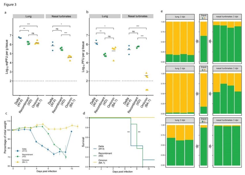
Hélène Moret
 Hôpital Robert Debré
Martine Bos
 inBiome B.V.
Matthijs Welkers
 Public Health Service Amsterdam
Claus Scholz
 Laboratory Dr. Wisplinghoff https://orcid.org/0000-0002-6406-7859
Sofia Paraskevopoulou
 Robert Koch Institute
Laurence Josset
 CHU Lyon
Claire Cervi
 Laboratoires Corcy et associés
Brigitte Couzon
 Centre Hospitalier de Versailles
Stéphanie Marque-Juillet
 Centre Hospitalier de Versailles
Deborah Delaune
 Institut Pasteur https://orcid.org/0000-0003-4970-9566
Slim El Khiari
 Institut Pasteur
Julien Fumey
 Institut Pasteur
Marie-Charlotte Hallouin-Bernard
 Institut Pasteur
Félix Rey
 Institut Pasteur https://orcid.org/0000-0002-9953-7988
Yazdan Yazdanpanah
 ANRS-MIE
Bruno Coignard
 Santé publique France
Xavier de Lamballerie
 UMR Émergence des Pathologies Virales (EPV: Aix-Marseille Université – IRD 190-INSERM 1207 –
EHESP – IHU Méditerranée Infection), Marseille, France https://orcid.org/0000-0001-7895-2720
Sylvie van der Werf
 Institut Pasteur https://orcid.org/0000-0002-1148-4456
Axelle Paquin
 Centre Hospitalier de Laval

                                              Page 3/26
Biological Sciences - Article
Keywords:
Posted Date: April 4th, 2022
DOI: https://doi.org/10.21203/rs.3.rs-1502293/v1
License: This work is licensed under a Creative Commons Attribution 4.0 International License.
Read Full License
Page 4/26Abstract
Recombination is a crucial process in the evolution of many organisms. Although the evolutionary
reasons behind its occurrence in RNA viruses are debated, this phenomenon has been associated with
major epidemiological events such as virus host range expansion, antigenic shift or variation in virulence
1,2, and this process occurs frequently in positive strand RNA viruses such as coronaviruses. The SARS-
CoV-2 pandemic has been associated with the repeated emergence of variants of concern presenting
increased transmissibility, severity or immune escape 3. The recent extensive circulation of Delta
worldwide and its subsequent replacement by viruses of the Omicron lineage 4 (BA.1 then BA.2), have
created conditions for genetic exchanges between viruses with both genetic diversity and phenotypic
specificities 5-7. Here we report the identification and in vitro and in vivo characterization of a Delta-
Omicron recombinant in Europe. This recombinant exhibits immune escape properties similar to Omicron,
while its behavior in mice expressing the human ACE2 receptor is more similar to Delta. This recombinant
provides a unique and natural opportunity to better understand the genotype to phenotype links in SARS-
CoV-2.
Main
The recombination process can occur when two genetically distinct viruses co-infect a cell, and
corresponds to the generation of a chimeric molecule classically as a result of template switching during
replication, although other mechanisms exist. The divergence between the parental sequences and the
length of the genomic regions involved strongly affect the possibility to detect such events. In addition,
recombination may bring together genetic variations that will not necessarily result in a functional and fit
virus, as short- or long-distance epistatic interactions in the genome and proteins might be disrupted. The
mechanistic process is thus likely much more frequent than the – epidemiologically relevant – fraction
that can be captured by genomic surveillance.
There is potent evidence that recombination is pervasive in the evolutionary history of many viral
families, including coronaviruses and the sarbecovirus subgenus to which SARS-CoV-2 belongs 8,9. There
are similarly increasing reports on the frequent occurrence of recombination throughout the COVID-19
pandemic 10-13.
Identification of a recombinant and putative parent lineages in Europe
In early 2022, multiple laboratories contributing to routine SARS-CoV-2 genomic surveillance in France
(the EMERGEN consortium) and in Europe, publicly shared sequences initially classified as Delta AY.4 and
21J by Pangolin 14 and Nextclade 15 respectively. Further examination of these genomes revealed a large
number of signature mutations characteristic of the BA.1 lineage in only a portion of the genome, highly
suggestive of a recombinant 16. Analysis of the raw sequencing data of primary samples and of an
isolate revealed the absence of co-infection or contamination and the correct assembly of the genome
Page 5/26(see methods). The lineage was designated as clade GKA by GISAID, then received the Pango lineage
designation XD and was classified as a variant under monitoring by ECDC on March 17, 2022 17.
The mosaic structure of the genome is described in Fig. 1 and Extended Data Fig. 1. We identified two
breakpoints, one at the beginning of the spike (in a window spanning nt 22035 to 22194) and one within
ORF 3a (nt 25469 to 25583). At the time of writing, the earliest sequence of this recombinant was
sampled on January 3rd, 2022 in Northern France during routine surveillance (random selection for
sequencing of a fraction of weekly cases, "Flash investigation"). As of March 18th, 2022, the XD lineage
has been detected in multiple regions of France, as well as in Denmark, the Netherlands, Belgium and
Germany, through random sampling and cluster investigations. The majority of cases investigated
reported no travel history, except for one case travelling from Italy, suggesting widespread but infrequent
circulation. In support of this, we observed signal of diversification within the lineage, with geographic
clustering within both France and Europe (Fig. 1b,c).
Data from ongoing epidemiological investigations were available from 32 confirmed XD cases and six
suspected cases who tested positive for SARS-CoV-2 between January 3rd and March 9th, 2022 in France
(Extended Data Fig. 2a). As comparison, we used epidemiological data recently obtained from
investigations of Omicron (BA.1) cases with the same methodology in France 18. The median age was
32.5 years (IQR 19.75-44.75), which was similar to Omicron cases (35 years), however, significantly more
XD cases were below 20 years of age (25.7%) compared to Omicron cases (11.3%, p = 0.026, Extended
Data Fig. 2b). Only two XD cases reported a previous SARS-CoV-2 infection (5.6%, Table 1), fewer than for
Omicron (14%). No XD case was over 70 years old, and no risk factor was reported. Only 6% of
recombinant cases were unvaccinated (vs 27% for Omicron cases), and 30% had received three doses (vs
8%), but the overall vaccination coverage of the population was different between the XD and Omicron
study periods. All XD cases reported here from France were symptomatic and most reported symptoms
were headache (61.1%), asthenia/fatigue (58.3%), cough (44.4%), fever (38.9%) and myalgia (33.3%,
Extended Data Fig. 2b). The main differences compared to Omicron cases were the higher rates of
ageusia (odds ratio OR 2.71 [1.064-6.514], p=0.024) and anosmia (OR 1.98 [0.66-5.251], p = 0.18). Two
XD cases were hospitalized, one for unrelated causes, but for less than 24h and without intensive care
admission.
Search for the putative parental sequences
Splitting conservatively the genome based on the largest breakpoint windows, we performed similarity
searches on the GISAID 19 EpiCoV database to identify (closely related) genomes that may represent the
lineages that gave rise to this recombinant virus. While the shorter region inherited from BA.1 returned a
large set of genomes from all over the world, likely due to the shorter genomic portion involved and the
relatively low diversity of the recently emerged BA.1, the Delta-like part of the genome led to a small
subset of sequences (n=69) within the large AY.4 sublineage. The closest relatives were predominantly
Page 6/26sampled in Europe (France, Germany and Sweden, Extended Data Fig. 3). Together with the closest
detected AY.4 genomes, the recombinant XD lineage (with the Omicron part of the genome masked)
forms a monophyletic cluster (branch support: 0.98).
Growing reports of potential SARS-CoV-2 recombinant sequences describe mosaic genomes with
breakpoints recurrently noted in specific regions of the genome such as the beginning of the spike.
Similar observations can be made in the evolutionary history of the family Coronaviridae, and hotspots of
recombination have been described for other viruses such as HIV-1 or poliovirus 20,21. However,
the relative rarity of the candidate AY.4 parental sublineage suggests that all currently reported XD viruses
likely derive from a unique ancestor.
We isolated a representative virus of each of the parental lineages, as well as a recombinant XD virus,
allowing to further demonstrate the recombinant genotype. The selected AY.4 virus, sampled in Ile-de-
France on December 27, 2021, presented 100% identity with the Delta-like region, and the BA.1 virus was
similarly 100% identical to the Omicron-like region of the XD virus. Importantly, this allowed us to rapidly
characterize the recombinant virus in comparison to representatives of the parental lineages using
primary isolates, and gain insights into determinants of the phenotypic differences between the variants
of concern (VOCs) Delta and Omicron BA.1.
Sensitivity of XD to sera from vaccinees
Many of the unique properties of Omicron have been tied to the extensive changes that accumulated in
the spike in comparison to the ancestral SARS-CoV-2 sequence. Omicron has been proposed to be
considered a distinct strain of SARS-CoV-2, and the use of a serotype classification has been
discussed 22. As the XD spike carries a larger fraction of the signature changes of BA.1, including the
entirety of the receptor binding domain (RBD) (Extended Data Fig. 4), we hypothesized that this
recombinant might present similar immune escape properties 23,24. To assess this, we generated
pseudoviruses expressing the XD spike, and compared the neutralizing activity of sera from vaccinated
individuals, using the ancestral, Delta (AY.4), and Omicron (BA.1) spikes as reference. Our panel included
sera sampled at 1 or 6 months after the last immunization, from individuals infected and vaccinated with
the Pfizer vaccine or vaccinated with either 2 doses of the Pfizer vaccine, the AstraZeneca vaccine or a
combination of both, or including a booster of the Moderna vaccine (Table 2).
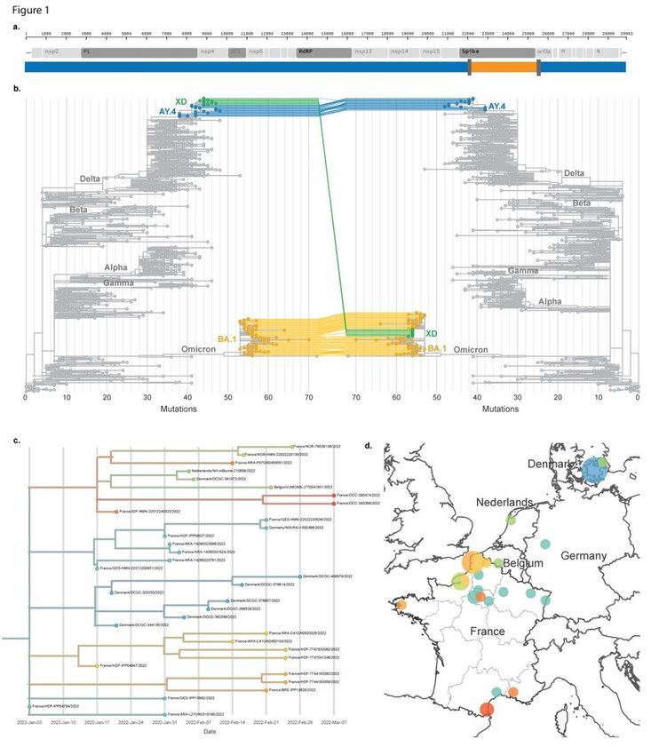
As described previously, we observed only a small reduction in the neutralizing titers against Delta
compared to the ancestral virus. In contrast, both the XD recombinant and BA.1 were barely neutralized
by sera sampled 1 or 6 months after 2 doses. The titers were all much higher 1 month after a third
immunization, although they displayed an 8-fold to 10-fold reduction in neutralization efficacy against
both BA.1 and XD compared to the original virus. Likewise, in individuals infected and vaccinated with
one dose, neutralization titers towards BA.1 and XD were reduced 3.4- and 3.7-fold (but not significant
differences), respectively. These results were obtained with a pseudovirus assay, which has been shown
Page 7/26to be strongly correlated with infectious virus experiments, but it cannot be excluded that differences in
susceptibility or escape to neutralization do exist for this chimeric virus.
Taken together, these results indicate that XD displays immune escape properties similar to those of BA.1,
and further reinforce the major role of the spike RBD epitopes in the vaccine induced neutralizing
response in our serum panel.
Neutralization of Omicron by monoclonal antibodies
We next assessed the sensitivity of the XD recombinant to a panel of therapeutic COVID-19 human
monoclonal antibodies using infectious viruses. We selected monoclonal antibodies that are currently in
clinical use and have been associated with varying reduction of activity against Omicron BA.1 24,25. We
included putative parental viruses (AY.4 and BA.1) and a B.1 isolate (carrying the spike D614G
substitution) for comparison.
Consistently with the binding targets of our antibodies, which correspond to sequences that are identical
between the parental BA.1 and recombinant virus, we observed highly similar reductions in neutralizing
activity for these viruses, and distinct from the AY.4 parental virus. As against BA.1, Tixagevimab lost all
detectable neutralizing potential against the XD virus, and Cilgavimab conserved a neutralizing activity
with a shift in EC50 from 29.7 ng/mL against B.1 to 402.8 ng/mL (fold change reduction of 13.5 for XD vs
16.9 for BA.1) (Table 3). The combination of these antibodies, proposed as the Evusheld cocktail,
accordingly, presented strongly reduced activity (fold change reduction of 37.9 for XD vs 36.7 for BA.1),
while Delta AY.4 was neutralized as efficiently as the B.1 virus. As reported previously, Sotrovimab
showed a small reduction (2.6-fold) in activity against BA.1 24,25, which was somewhat more pronounced
against the XD virus (5.1-fold reduction).
These results indicate that the XD recombinant, which possesses the RBD and S2 of Omicron BA.1,
shares the escape properties of the latter, and does not complexify the landscape of vaccines and
therapeutic efficacy.
XD recombinant is not attenuated in a mouse model of infection
Although it is challenging to infer the intrinsic severity of novel SARS-CoV-2 variants as they circulate in
populations with significant proportion of immunity with varying levels, Omicron BA.1 has been
associated with lower risks of severe disease compared to Delta 26,27. Consistently, BA.1 has been shown
to cause attenuated disease in rodent models, including in the highly susceptible model of human ACE2
transgenic (K18-hACE2) mice 28,29.
We infected K18-hACE2 mice with 2x10⁴ PFU of either the recombinant or each parental lineage virus. We
first compared the viral load in the lungs and nasal turbinates at 3 days post-infection (dpi). The levels of
viral RNA in the lungs of mice infected with BA.1 or XD viruses were comparable, and 3.7-fold lower than
those of AY.4-infected animals (Fig. 3a). In contrast, the viral load in nasal turbinates of the XD-infected
Page 8/26mice was 8.5-fold higher than that in BA.1-infected animals, and only slightly lower than that of AY.4
infected mice. Similar differences were noted when measuring infectious titers (Fig. 3b).
We also monitored mice for body weight loss and disease over 11 days (Fig. 3c,d, Extended Data Fig. 5).
Like the main Delta lineage, the parental AY.4 isolate resulted in disease with weight loss that accelerated
at 3 dpi, and 6/7 mice reached humane endpoints between dpi 7 and 9. As described, BA.1 infection was
not associated with weight loss nor lethality. With the XD virus, we did not observe weight loss during the
first 5 dpi, but all mice deteriorated rapidly between day 5 and 8 and died or had to be euthanized by day
9. Clinical scores followed the same progression and delayed onset in XD- compared with AY.4-infected
mice.
Together, these results indicate that the Delta-Omicron XD recombinant is associated with clinical
features distinct from those of Omicron BA.1 in K18-hACE2 mice. It further suggests that the attenuated
phenotype of BA.1 in this model cannot be only linked to the variations in the spike such as in the RBD or
near the furin cleavage site. Further experiments will be needed to determine the basis of the XD
recombinant intermediate phenotype in mice and to determine how this relates to XD infection in
humans.
In vivo replication
To further investigate potential differences in the replicative fitness of viruses of the XD lineage, we
measured its growth in comparison to Omicron BA.1, which was the dominant lineage in France and
most of Europe at the time of initial detection of the recombinant. As competition experiments may
expose differences in replication efficacy that are not detected in individual growth kinetic assays, we
inoculated groups of K18-hACE2 mice with a mixture of BA.1 and XD viruses at 1:1, 1:9 or 9:1 ratio and
used metagenomic sequencing to estimate the ratio of the viral genomes at 3dpi in both the lungs and
the nasal turbinates (Fig.3e). While BA.1 consistently outcompeted the XD recombinant in the lungs, the
reverse was observed in the nasal turbinates, both results being in accordance with what was observed
with each virus separately.
Taken together, these results indicate that XD and BA.1 each present a replicative advantage in different
environments. The apparent superior fitness of XD compared to Omicron BA.1 in the upper airways of
K18-hACE2 mice suggests that this trait is not tied to the spike RBD nor its furin cleavage efficacy.
Variations present in the first part of the spike N-terminal domain (from AY.4 in XD) might contribute to
these differences if related to the entry process itself. However, other specificities present in the genome
and in both structural and non-structural proteins might be involved in the XD phenotype (Extended Data
Fig. 6).
Potential limitations of our work include the low number of confirmed cases of XD virus infections, in a
context of overall high incidence and evolving testing strategies in Europe, potentially limiting the ability
to detect variants in asymptomatic cases. The pathogenicity and transmission potential in humans thus
remain unclear. The K18-hACE2 mouse model used here is highly susceptible to SARS-CoV-2 infection,
Page 9/26and importantly does not include any pre-existing immunity to the virus, which has been shown to
considerably reduce the risk of severe COVID-19. In addition, considering that the XD lineage has been
circulating at least since the beginning of January 2022, and although this lineage has been captured by
random genomic surveillance in multiple European countries, the limited detection might reflect the
inability of this lineage to outcompete Omicron BA.1 and BA.2 in human populations. Further work is
needed to dissect the epidemiological and virological characteristics of the XD recombinant.
The continued diversification observed during the COVID-19 pandemic, with the extreme case of the
emergence of VOCs and their intense circulation, is likely to provide further opportunities for
recombination, and continued worldwide monitoring and rapid risk assessment will be crucial to our long-
term response to SARS-CoV-2 circulation and understanding of its biology.
Methods
Virus sequencing
The consensus genome sequences of SARS-CoV-2 lineage XD / clade GKA described here have all been
deposited on the GISAID EpiCoV database (EPI_SET_20220325yn), and have been generated by different
PCR amplification strategies and on either Illumina or Oxford Nanopore technologies platforms, as
described in their sequences entries. We also deposited raw reads data (GISAID ID: EPI_ISL_9879437 and
EPI_ISL_9879436, with human host reads removed) for two primary samples generated by the National
Reference Center for Viruses of respiratory infection hosted at Institut Pasteur and part of the Emergen
consortium. These data were generated using a highly multiplexed short PCR amplicon approach (panel
ARTIC V4.1) 30, followed by library preparation with the Nextera XT kit. Viral stocks were verified by
untargeted metagenomic sequencing. RNA was extracted from culture supernatant, converted into double
stranded cDNA and libraries were prepared using the Nextera XT kit. Both genomic surveillance and virus
stocks libraries were sequenced on an Illumina NextSeq500 (2x150 cycles) at the Mutualized Platform for
Microbiology.
For the competition assays readout, we used an untargeted metagenomic sequencing approach with
ribosomal RNA (rRNA) depletion, which has been shown to capture the diversity of RNA species present
within a sample 31. RNA was extracted with the QIAamp Viral RNA extraction kit (Qiagen), with the poly-A
RNA carrier provided. Prior to library construction, carrier RNA and host rRNA were depleted using oligo
(dT) and custom probes respectively. The RNA resulting from selective depletion was used for random-
primed cDNA synthesis using the SuperScript IV RT (Invitrogen). Second-strand cDNA was generated
using Escherichia coli DNA ligase, RNAse H and DNA polymerase (New England Biolabs) and purified
using Agencourt AMPure XP beads (Beckman Coulter). Libraries were then prepared using the Nextera XT
kit and sequenced on an Illumina NextSeq500 platform (2x75 cycles).
Investigation of sequencing reads data
Page 10/26To rule out the possibility that the consensus sequences of XD lineage viruses correspond to coinfection
or contamination, we examined the raw read data generated by multiple platforms and approaches, to
estimate the frequency of intrahost single nucleotide variants (iSNVs) at all positions along the genome,
with a focus on positions that differs between the Delta and Omicron variants. We used iVAR 32 and thus
SAMtools 33 to compute the nucleotides frequencies. For comparison, we computed the same statistics
for samples that were rejected as possible contamination or coinfection by our sequencing curation
pipeline. The raw reads of samples hCoV-19/France/HDF-IPP04947/2022, hCoV-19/France/HDF-
IPP08027/2022 (amplicons), as well as direct metagenomic sequencing of the XD isolate viral stock
(hCoV-19/France/HDF-IPP08027i/2022) have been deposited on GISAID (EPI_SET_20220320tx).
Representative examples are shown in Extended Data Fig. 7.
Epidemiological investigations
Confirmed cases were defined as patients with a positive SARS-CoV-2 test and a sequencing result
demonstrating infection by recombinant XD. Suspected cases were defined as patients with a positive
SARS-CoV-2 test and an epidemiological link with a confirmed case. Cases were interviewed by the
Regional offices of Santé publique France (SpF, the French national Public Health Agency) using a
standard questionnaire. Collected data included demographic information (sex, age, place of residence),
travel history, contact persons, vaccination status, symptoms and outcome. The earliest XD case was a
family contact of a case with recent travel to Niger, and one case reported recent travel to Italy. A similar
investigation was performed during the emergence of Omicron (mostly BA.1) in France 18 and was used
for comparison.
Identification of putative parental sequences
To identify putative parental genome sequences, we retrieved all SARS-CoV-2 sequences available on the
GISAID EpiCoV database as of March 9th, 2022. We did not restrict our search to a window of time
corresponding to the earliest detection of BA.1 to the days prior to the earliest available sequence of
lineage XD, to be able to capture all possible related genomes of the parental lineages. We reduced this
dataset to sequences longer than 28000 nt and including less than 5% of Ns, on which we performed a
similarity search using gofasta (https://github.com/cov-ert/gofasta). As a query, we used XD genomes
after masking either the region corresponding to the AY.4-like or to the BA.1-like part of the genome.
Specifically, we set the boundary coordinates to the ends of sequential tracts of mutations specific to the
putative parental sequences. This is a conservative approach to assigning parental lineages and
consequently no parental lineage is assigned to those genome regions that do not contain unambiguous
lineage-defining mutations or deletions.
We conservatively masked the entirety of the breakpoint windows in each case, and the genome
extremities (keeping nt 101- 22034 and 25584-29802 for the AY.4 search, and 22193-25469 for the BA.1
search). All numberings are based on the Wuhan-Hu-1 reference genome (GenBank ID: NC_045512). The
AY.4 search resulted in 69 sequences post quality filtering. For BA.1, we restricted our selection to
Page 11/26sequences 100% identical to our recombinant query, resulting in 39557 sequences. We also selected all
sequences belonging to AY.4 lineage or one of its sub-lineages (995977 sequences), after annotating the
sequences with Pangolin (v3.1.20, pangolearn 2022-02-28, scorpio v0.3.16, constellation, v0.1.3,
pangodesign v1.2.32) with Usher placement.
We compiled statistics for each position of the sequences sets. We also inferred maximum likelihood
phylogenies for each putative parental sequences dataset (randomly subsampled for the related BA.1
genomes) (Extended Data Fig. 3).
Single nucleotide differences between the XD recombinant sequences and closely related putative
parental sequences were visualized using snipit (https://github.com/aineniamh/snipit), Extended
Data Fig. 3. Analysis of the variations within the putative parental AY.4 sublineage suggests that the
breakpoint window at the beginning of the spike could be further reduced to nt 22035 to 22096, as the
closest AY.4 sequences all carry an A at position 22097 (C in the recombinant sequences).
Phylogenetic analysis
To analyze the recombinant genomes, we generated a contextual dataset representative of the global
circulation of SARS-CoV-2 since its emergence using the subsampling strategy implemented in Nextstrain
(v.3.0.5) 34. We added high quality XD genomes (n = 16) reported here, as well as subsets of the AY.4 (n =
29) and BA.1 (n= 30) genomes identified by similarity search (EPISET EPI_SET_20220324xe).
As in the similarity search, we also used versions of the recombinant genomes where either one of the
regions with distinct ancestry was masked. To characterize the genetic diversity of lineage XD, the
complete genome was used, and the reference genome added to root the phylogeny.
Maximum-likelihood phylogenies were inferred using IQ-TREE v2.0.6 and branch support was calculated
using ultrafast bootstrap approximation with 1000 replicates 35,36. Prior to the tree reconstruction, the
ModelFinder application 37, as implemented in IQ-TREE, was used to select the best-fitting nucleotide
substitution model. The resulting phylogenies were visualized using Auspice v.2.29.1 and Figtree v.1.4.4
(http://tree.bio.ed.ac.uk/software/figtree/).
We separately downsampled this dataset to perform recombination analyses using methods
implemented in RDP5 38, including 3SEQ 39, confirming the clear signal for the two recombination
breakpoints.
Cell lines
VeroE6 cells (African green monkey kidney cells from ATCC (CCL-81)), were maintained in Dulbecco’s
Modified Eagle’s Medium (DMEM, Life Technologies) supplemented with 5% fetal bovine serum (FBS)
and penicillin/streptomycin. VeroE6/TMPRSS2 cells (ID 100978) were obtained from CFAR and were
maintained in minimal essential medium (Life Technologies) with 7.5% heat-inactivated fetal calf serum,
1% penicillin/streptomycin and G-418 (Life Technologies). Human embryonic kidney HEK-293T (ATCC
Page 12/26CRL-3216) and HEK-293T cells stably expressing human ACE2 (293T-ACE2) were maintained in DMEM
supplemented with 10% FBS and penicillin/streptomycin.
Viral Isolates
We isolated the XD virus and the putative parental AY.4 virus from nasopharyngeal swabs received in the
context of our random genomic surveillance. The sequences of the swabs and the outgrown viruses (XD:
EPI_ISL_10819657 and AY.4: EPI_ISL_11373018) were identical. All SARS-CoV-2 isolates were supplied by
the National Reference Centre for Respiratory Viruses hosted at Institut Pasteur (Paris, France). The
human sample from which XD strain hCoV-19/France/HDF-IPP08027i/2022 was isolated has been
provided by Dr. Jean-Marc Corcy Laboratoire Corcy et Associés, Soissons, France (hCoV-19/France/HDF-
IPP08027/2022 GISAID ID: EPI_ISL_9879437). The human sample from which the AY.4 virus hCoV-
19/France/IDF-IPP52092i/2021 was isolated has been provided by Dr. Stéphanie Marque-Juillet, CH de
Versailles, France (hCoV-19/France/IDF-IPP52092/2021 GISAID ID: EPI_ISL_8546521). The human
sample from which the BA.1 virus hCoV-19/France/PDL-IPP46934/2021 was isolated has been provided
by Dr. Axelle Paquin, CH de Laval, France (GISAID ID: EPI_ISL_8588473).
Virus isolation was performed on VeroE6 cells in the presence of 1 µg/mL TPCK trypsin as previously
described 40, followed by 2 passages on VeroE6 cells at a MOI of 10-4 to obtain a viral stock. We
performed metagenomic sequencing on the sample to check the presence of potential contaminants and
verify if the virus had acquired genomic changes during isolation, and each isolate sequence was
identical to the primary isolate sequence. All work with infectious virus was performed in biosafety level 3
containment laboratories.
SARS-CoV-2 isolate B.1 (BavPat1 EPI_ISL_406862) was obtained from Pr. C. Drosten through EVA
GLOBAL (https://www.european-virus-archive.com/). The Omicron BA.1 virus used in the monoclonal
antibodies assay was isolated from a nasopharyngeal swab of the 1st of December in Marseille, France.
The full genome sequence has been deposited on GISAID: EPI_ISL_7899754. The strain, called
2021/FR/1514, is available through EVA GLOBAL (www.european-virus-archive.com, ref: 001V-04436)
Antibodies and Sera
Post-vaccination sera: sera were collected in the context of the CORSER-4 cohort and provided by ICaReb.
Sera were from individuals with previous RT-PCR confirmed COVID-19 infection and sampled one month
after vaccination with one dose of Pfizer vaccine (BNT162b2, Tozinameran); individuals fully vaccinated
(2 doses) with the Pfizer or AstraZeneca (ChAdOx1-S) or combination thereof and sampled at one or 6
months after the second dose; individuals fully vaccinated having received a booster dose of the
Moderna vaccine (Elasomeran) and sampled at one month after the boost. Dates of vaccination and
dates of sampling are provided in Table 2.
Page 13/26Sotrovimab/ Vir-7831 was provided by GSK (GlaxoSmithKline). Cilgavimab and Tixagevimab
(AstraZeneca) were obtained from hospital pharmacy of the University hospital of La Timone (Marseille,
France).
Neutralization assay with pseudotypes
The XD recombinant spike-encoding plasmid was obtained by an In-Fusion reaction (Takara) using Delta
and BA.1 human codon-optimized synthetic genes as templates into the pVAX1 vector. Lentiviral particles
expressing a luciferase reporter gene and pseudotyped with the ancestral (Wuhan), Delta (B.1.617.2),
Omicron (BA.1) and recombinant XD spikes were produced by calcium phosphate transfection of 293T
cells with plasmids provided by the Bloom laboratory 41. The following reagent was obtained through BEI
Resources, NIAID, NIH: SARS-Related Coronavirus 2, Wuhan-Hu-1 Spike-Pseudotyped Lentiviral Kit, NR-
52948.
Four-fold serial dilutions of sera starting at 1:10 were mixed with 0,5 µl of pseudovirus in a final volume
of 50 µl of DMEM-10% FCS during 30 min at room temperature. After incubation, 293T-ACE2 cells were
added at 20,000 cells/well of a 96-well white culture plate. Luminescence was measured after 72 h using
the Bright-Glo luciferase substrate and a Berthold Centro XS luminometer. Neutralization was calculated
using the following formula: 1 – (RLU in presence of serum / (mean of RLU in absence of serum
determined in 48 wells – 3 x STD). The effective dose 50% (ED 50) was determined as the serum dilution
giving 50% of neutralization.
Neutralization assays with infectious virus
One day prior to infection, 5×10⁴ VeroE6/TMPRSS2 cells were seeded in 100μL assay medium
(containing 2.5% FCS) in 96 well culture plates. The next day, antibodies were diluted in PBS with ½
dilutions from 1000 to 0.97 ng/ml for Sotrovimab and from 5000 to 4.8 ng/ml for Cilgavimab,
Tixagevimab and its combination Evusheld. Eleven 2-fold serial dilutions of antibodies in triplicate were
added to the cells (25µL/well, in assay medium). Then, 25µL of a virus mix diluted in medium was added
to the wells. The amount of virus working stock used was calibrated prior to the assay, based on
replication kinetics, so that the viral replication was still in the exponential growth phase for the readout.
Plates were first incubated 15 min at room temperature and then 2 days at 37°C prior to quantification of
the viral genome by real-time RT-PCR. Synthetic RNA standards were used to quantify viral RNA
abundance in copies per reaction. Viral inhibition was calculated as follows: 100* (quantity mean VC-
sample quantity)/ quantity mean VC). The 50% effective concentrations (EC50 compound concentration
required to inhibit viral RNA replication by 50%) were determined using a nonlinear regression
(log(agonist) vs. response -- Variable slope (four parameters)) as described 25.
In vivo studies
Page 14/26Infection studies were performed in a biosafety level 3 (BSL-3) animal facility at the Institut Pasteur, in
Paris. All animal work was approved by the Institut Pasteur Ethics Committee (project dap 21050) and
authorized by the French Ministry of Research under project 31816 in compliance with the European and
French regulations. B6.Cg-Tg(K18-Ace2)2Prlmn/J mice were purchased from The Jackson Laboratory
(Maine, USA) and are bred at the Institut Pasteur under SOPF environment.
Seven- to eight-week-old mice were anesthetized by ketamine/xylazine and infected intranasally with 2
x104 PFU of SARS-CoV-2 isolates in 40 to 60 µl. In the first group of mice, clinical signs of disease and
weight loss were monitored daily for 11 days. Four criteria (ruffled fur, hunched back posture, reduced
mobility and difficulty to breath) were scored on a 0-2 scale and added up into a global clinical score.
Mice were euthanized when they reached predefined endpoints (clinical score of 6 or body weight loss >
25%). A second group of infected mice was euthanized 3 days post infection (dpi) for measurement of
viral load and viral titer. Right lung lobe and nasal turbinates were dissected and frozen at -80°C. Samples
were homogenized in 400 µl of cold PBS using lysing matrix M (MP Biomedical) and a MP Biomedical
FastPrep 24 Tissue Homogenizer. Viral RNA was extracted using an extraction robot IDEAL-32
(IDsolutions) and the NucleoMag Pathogen extraction kit (Macherey Nagel). Viral RNA quantification was
performed by quantitative reverse transcription PCR (RT-qPCR) using the IP4 set of primers and probe
(nCoV_IP4-14059Fw GGTAACTGGTATGATTTCG and nCoV_IP4-14146Rv CTGGTCAAGGTTAATATAGG
giving a 107bp product, and nCoV_IP4-14084Probe(+) TCATACAAACCACGCCAGG [5']Hex [3']BHQ-1) and
the Luna Universal Probe One-Step RT-qPCR Kit (NEB). Serial dilutions of a titrated viral stock were
analysed simultaneously to express viral loads as PFU equivalents (eqPFU) per gram of tissue.
For plaque assays, 10-fold serial dilutions of samples in DMEM were added onto VeroE6 monolayers in
24 well plates. After one-hour incubation at 37°C, the inoculum was replaced with an equivalent volume
of 5% FBS DMEM and 2% carboxymethylcellulose. Three days later, cells were fixed with 4%
formaldehyde, followed by staining with 1% crystal violet to visualize the plaques.
Analysis of the competition assays
For each sample, we trimmed the reads using trimgalore
(https://github.com/FelixKrueger/TrimGalore/tree/0.6.7). We mapped the reads on the reference
sequence Wuhan-Hu-1/2019 using bwa mem (v0.7.17) 42. We then counted the coverage of each
nucleotide at all positions of the genome using bam-readcount (v1.0.0) as performed in ViralFlow 43. We
selected the SARS-CoV-2 genomic positions that differ between the sequence of the recombinant isolate
(IPP08027i) and the BA.1 isolate (IPP46934i) using goalign (v0.3.5) 44, and counted the relative amounts
of each nucleotide corresponding either to the recombinant XD or to the BA.1 at each of these positions.
Statistical analysis
Statistical analysis was performed using R v4.1.2 and GraphPad Prism v9. Survival curves were
compared by logrank test. Viral loads and viral titers were compared by a Student's t-test. Epidemiological
data of XD and Omicron cases were compared using Chi2 tests and odds ratio.
Page 15/26Ethical statement
As per approval by the National Ethical Committee, SPF investigation teams have continual access to
personal data in order to investigate and control identified public health threats. No additional ethical
clearance was needed or sought. Personal information was anonymised in this publication.
Sera from human subjects who participated in the “Sero-epidemiological study of the SARS-CoV-2 virus
in France” were provided by ICAReB platform (Clinical Investigation & Access to Research Bioresources)
from the center for translational science, Institut Pasteur. All participants gave written informed consent
in the frame of CORSER-4 cohort after approval of the CPP Ile-de-France I Ethics Committee (2021, May
21st) and registered by the French national security agency for medicines and health products (IDRCB
2020-A00406-33) and Clinical trials (NCT 04325646).
Tables
Page 16/26Characteristics N %
Region (n=38) Auvergne-Rhône-Alpes 4 10.50%
Bretagne 1 2.60%
Grand Est 4 10.50%
Hauts-de-France 13 34.20%
île-de-France 4 10.50%
Normandie 3 7.90%
Occitanie 8 21.10%
Provence-Alpes-Côte d’Azur 1 2.60%
Sex (n=38) Females 15 39.50%
Males 23 60.50%
Travel history (n=34) Yes 2 5.90%
No 32 94.10%
Cluster (n=36) Yes 23 63.90%
No 13 36.10%
Previous SARS-CoV-2 infection (n=36) Yes 2 5.60%
No 34 94.40%
Vaccination status (n=34) Unvaccinated 2 5.90%
One dose 2 6%
Two doses 21 62%
Three doses 9 26.50%
Symptomatic infection (n=36) Yes 36 100%
No 0 0%
Risk factors (n=36) Yes 12 33.30%
No 24 66.70%
Hospitalization (n=36) Yes 2 5.60%
No 34 94.40%
Intensive care (n=36) Yes 0 0%
No 36 100%
Page 17/26Table 1: Characteristics of investigated cases infected by XD (N = 38). For each category, percentages
were calculated over the number of cases with available data, and respective denominators are indicated
in parenthesis.
Table 2: Characteristics of the serum donors.
Page 18/26virus
Antibody B.1 AY.4 BA.1 XD
GSK/ Vir Sotrovimab (vir-7831) EC50 62,7 218,1 163,7 317,5
fold-change - 3,5 2,6 5,1
AstraZeneca Cilgavimab (AZD1061) EC50 29,7 47,7 501,1 402,8
fold-change - 1,6 16,9 13,5
Tixagevimab (AZD8895) EC50 62,7 218,1 n.n n.n
fold-change - 3,5 - -
Evusheld (AZD7442) EC50 16,8 11,9 617,4 637,8
fold-change - 0,7 36,7 37,9
Table 3: Interpolated EC50 values of therapeutic antibodies against SARS-CoV-2 BavPat1, Delta (AY.4),
Omicron (BA.1) and the recombinant (XD) virus. EC50 values are expressed in ng/mL.
Declarations
Data availability
All data supporting the findings of this study are available in the manuscript. The SARS-CoV-2 genomic
data analysed here are available on GISAID (https://www.gisaid.org/).
Acknowledgments
We would like to thank all the healthcare workers, public health employees, and scientists involved in the
COVID-19 response. We acknowledge the authors, originating and submitting laboratories of the
sequences from GISAID and GenBank (Supplementary Table 1). We thank the Institut Pasteur C2RA
Animal Facility staff for their dedication to breeding and care of mice, the team of the Mutualized
Platform for Microbiology at Institut Pasteur, Dr Justine Schaeffer for scientific discussions, and Dr Cécile
Baronti and Dr Boris Pastorino for virus production and neutralizing activity experiments. We
acknowledge our colleagues of the Danish Covid-19 Genomics Consortium
(www.covid19genomics.dk/about), and our colleagues from the Covid19seq group of the Wisplinghoff
Laboratories. We are grateful to Anaïs Perilhou, Sandrine Fernandes Pellerin, Yanis Dahoumane and Tan-
Phuc Buivan from the center for translational science, Institut Pasteur, for the preparation and the
Page 19/26management of the cohort, to Laurence Arowas, Ayla Zayoud, Emmanuel Roux, Alain Li, Sophie Vacant,
Sophie Chaouche and Remy Yim of ICAReB platform from the Center for Translational Science, at the
Institut Pasteur for the management of the biobank for providing sera samples from donors. We
acknowledge the European virus archive-Marseille (EVAM, technological platforms of Aix-Marseille
university) for providing a BA.1 Omicron isolate. This work used the computational and storage services
provided by the IT department at Institut Pasteur, Paris.
Funding
This study has received funding from the EMERGEN programme (projects BIOVAR and MODVAR), from
Institut Pasteur, Santé publique France (the French national public health agency), the French
Government's Investissement d'Avenir programme, Laboratoire d'Excellence “Integrative Biology of
Emerging Infectious Diseases” (grant n°ANR-10-LABX-62-IBEID), REACTing (Research & Action Emerging
Infectious Diseases), the “Enhancing Whole Genome Sequencing (WGS) and/or Reverse Transcription
Polymerase Chain Reaction (RT-PCR) national infrastructures and capacities to respond to the COVID-19
pandemic in the European Union and European Economic Area” Grant Agreement ECDC/HERA/2021/007
ECD. 12221, the RECOVER project funded by the European Union’s Horizon 2020 research and innovation
programme under grant agreement No. 101003589 and the Caisse nationale d’assurance maladie
(Cnam), the national health insurance funds. ESL acknowledges funding from the INCEPTION
programme (Investissements d’Avenir grant ANR-16-CONV-0005).
Competing interests
AZ, TD and SK are employed by Inovie GEN-BIO. CS is employed by Dr. Wisplinghoff laboratories. NC and
HL are employed by BioPath. MPB, and AEB are employed by inBiome B.V.
Author contribution
Experimental strategy design, experiments: X.M., XdL, S.W. and E.S.-L. Data acquisition and curation: F.D.,
A.B., O.D., V.G., S.N., M.Be., J.Fo., M.R., S.K., A.Z., T.D., S.B., V.E., C.R., S.F., J-M.P., N.C., H.L., E.A-G., F.J., L.G.,
V.B., H.M., M.P.B., M.W., C.S., S.P., L.J., S.E-K., J.Fu., Y.Y. Vital materials: C.C., B.Cou., S.M-J., A.P., M.-C.H.-B.
Initial identification of the recombinant: S.N. Phylogenetic analysis: F.L., J.B., and E.S.-L. Virus isolation,
production, titration: F.D., A.B., O.D. Structural modelling: F.A.R. Infectious virus neutralization assays: F.T,
X.dL. Pseudovirus neutralization assays: S.M., M.A., F.A. S.vdW. Infection experiments in mice and
analysis: M.P., L.C., L.L., D., F.L., E.S.-L. and X.M. The Emergen consortium provided the framework for the
SARS-CoV-2 genomic surveillance in France. Writing - Original draft: E.S.-L. Writing – review and editing:
X.M., S.vdW., F.L., J.B.,V.G., F.T., X.dL., B.Coi., F.A.R., A.M. with inputs from all authors.
References
1 Worobey, M. & Holmes, E. C. Evolutionary aspects of recombination in RNA viruses. J Gen Virol
80 ( Pt 10), 2535-2543, doi:10.1099/0022-1317-80-10-2535 (1999).
Page 20/262 Simon-Loriere, E. & Holmes, E. C. Why do RNA viruses recombine? Nat Rev Microbiol 9, 617-626,
doi:10.1038/nrmicro2614 (2011).
3 WHO. Tracking SARS-CoV-2 variants, (2022).
4 Viana, R. et al. Rapid epidemic expansion of the SARS-CoV-2 Omicron variant in southern Africa.
Nature, doi:10.1038/s41586-022-04411-y (2022).
5 Plante, J. A. et al. Spike mutation D614G alters SARS-CoV-2 fitness. Nature 592, 116-121,
doi:10.1038/s41586-020-2895-3 (2021).
6 Saito, A. et al. Enhanced fusogenicity and pathogenicity of SARS-CoV-2 Delta P681R mutation.
Nature 602, 300-306, doi:10.1038/s41586-021-04266-9 (2022).
7 Syed, A. M. et al. Rapid assessment of SARS-CoV-2-evolved variants using virus-like particles.
Science 374, 1626-1632, doi:10.1126/science.abl6184 (2021).
8 Boni, M. F. et al. Evolutionary origins of the SARS-CoV-2 sarbecovirus lineage responsible for the
COVID-19 pandemic. Nat Microbiol 5, 1408-1417, doi:10.1038/s41564-020-0771-4 (2020).
9 Lytras, S. et al. Exploring the Natural Origins of SARS-CoV-2 in the Light of Recombination.
Genome Biol Evol 14, doi:10.1093/gbe/evac018 (2022).
10 Gutierrez, B. et al. Emergence and widespread circulation of a recombinant SARS-CoV-2 lineage
in North America. medRxiv, 2021.2011.2019.21266601, doi:10.1101/2021.11.19.21266601 (2021).
11 Jackson, B. et al. Generation and transmission of interlineage recombinants in the SARS-CoV-2
pandemic. Cell 184, 5179-5188 e5178, doi:10.1016/j.cell.2021.08.014 (2021).
12 VanInsberghe, D., Neish, A. S., Lowen, A. C. & Koelle, K. Recombinant SARS-CoV-2 genomes
circulated at low levels over the first year of the pandemic. Virus Evolution 7, veab059,
doi:10.1093/ve/veab059 (2021).
13 Wertheim, J. O. et al. Capturing intrahost recombination of SARS-CoV-2 during superinfection
with Alpha and Epsilon variants in New York City. medRxiv, 2022.2001.2018.22269300,
doi:10.1101/2022.01.18.22269300 (2022).
14 O'Toole, A. et al. Assignment of epidemiological lineages in an emerging pandemic using the
pangolin tool. Virus Evol 7, veab064, doi:10.1093/ve/veab064 (2021).
15 Aksamentov, I., Roemer, C., Hodcroft, E. B. & Neher, R. A. Nextclade: clade assignment, mutation
calling and quality control for viral genomes. Journal of Open Source Software 6, 3773,
doi:10.21105/joss.03773 (2021).
Page 21/2616 Nguyen, S. V. (2022).
17 ECDC. SARS-CoV-2 variants of concern as of 24 March 2022,
(2022).
18 Maisa, A. et al. First cases of Omicron in France are exhibiting mild symptoms, November 2021-
January 2022. Infect Dis Now, doi:10.1016/j.idnow.2022.02.003 (2022).
19 Elbe, S. & Buckland-Merrett, G. Data, disease and diplomacy: GISAID's innovative contribution to
global health. Glob Chall 1, 33-46, doi:10.1002/gch2.1018 (2017).
20 Simon-Loriere, E., Rossolillo, P. & Negroni, M. RNA structures, genomic organization and selection
of recombinant HIV. RNA Biol 8, 280-286, doi:10.4161/rna.8.2.15193 (2011).
21 Muslin, C., Mac Kain, A., Bessaud, M., Blondel, B. & Delpeyroux, F. Recombination in
Enteroviruses, a Multi-Step Modular Evolutionary Process. Viruses 11, doi:10.3390/v11090859 (2019).
22 Simon-Loriere, E. & Schwartz, O. Towards SARS-CoV-2 serotypes? Nat Rev Microbiol 20, 187-188,
doi:10.1038/s41579-022-00708-x (2022).
23 Cao, Y. et al. Omicron escapes the majority of existing SARS-CoV-2 neutralizing antibodies.
Nature, doi:10.1038/s41586-021-04385-3 (2021).
24 Planas, D. et al. Considerable escape of SARS-CoV-2 Omicron to antibody neutralization. Nature,
doi:https://doi.org/10.1038/d41586-021-03827-2 (2021).
25 Touret, F., Baronti, C., Bouzidi, H. S. & de Lamballerie, X. In vitro evaluation of therapeutic
antibodies against a SARS-CoV-2 Omicron B.1.1.529 isolate. Sci Rep 12, 4683, doi:10.1038/s41598-022-
08559-5 (2022).
26 Ulloa, A. C., Buchan, S. A., Daneman, N. & Brown, K. A. Estimates of SARS-CoV-2 Omicron Variant
Severity in Ontario, Canada. JAMA, doi:10.1001/jama.2022.2274 (2022).
27 Wolter, N. et al. Early assessment of the clinical severity of the SARS-CoV-2 omicron variant in
South Africa: a data linkage study. Lancet 399, 437-446, doi:10.1016/S0140-6736(22)00017-4 (2022).
28 Shuai, H. et al. Attenuated replication and pathogenicity of SARS-CoV-2 B.1.1.529 Omicron.
Nature, doi:10.1038/s41586-022-04442-5 (2022).
29 Halfmann, P. J. et al. SARS-CoV-2 Omicron virus causes attenuated disease in mice and
hamsters. Nature, doi:10.1038/s41586-022-04441-6 (2022).
30 Tyson, J. R. et al. Improvements to the ARTIC multiplex PCR method for SARS-CoV-2 genome
sequencing using nanopore. bioRxiv, 2020.2009.2004.283077, doi:10.1101/2020.09.04.283077 (2020).
Page 22/2631 Matranga, C. B. et al. Enhanced methods for unbiased deep sequencing of Lassa and Ebola RNA
viruses from clinical and biological samples. Genome Biol 15, 519, doi:10.1186/PREACCEPT-
1698056557139770 (2014).
32 Grubaugh, N. D. et al. An amplicon-based sequencing framework for accurately measuring
intrahost virus diversity using PrimalSeq and iVar. Genome Biol 20, 8, doi:10.1186/s13059-018-1618-7
(2019).
33 Li, H. et al. The Sequence Alignment/Map format and SAMtools. Bioinformatics 25, 2078-2079,
doi:10.1093/bioinformatics/btp352 (2009).
34 Hadfield, J. et al. Nextstrain: real-time tracking of pathogen evolution. Bioinformatics 34, 4121-
4123, doi:10.1093/bioinformatics/bty407 (2018).
35 Hoang, D. T., Chernomor, O., von Haeseler, A., Minh, B. Q. & Vinh, L. S. UFBoot2: Improving the
Ultrafast Bootstrap Approximation. Mol Biol Evol 35, 518-522, doi:10.1093/molbev/msx281 (2018).
36 Minh, B. Q. et al. IQ-TREE 2: New Models and Efficient Methods for Phylogenetic Inference in the
Genomic Era. Mol Biol Evol 37, 1530-1534, doi:10.1093/molbev/msaa015 (2020).
37 Kalyaanamoorthy, S., Minh, B. Q., Wong, T. K. F., von Haeseler, A. & Jermiin, L. S. ModelFinder:
fast model selection for accurate phylogenetic estimates. Nat Methods 14, 587-589,
doi:10.1038/nmeth.4285 (2017).
38 Martin, D. P. et al. RDP5: A computer program for analysing recombination in, and removing
signals of recombination from, nucleotide sequence datasets. Virus Evol, veaa087,
doi:10.1093/ve/veaa087 (2020).
39 Lam, H. M., Ratmann, O. & Boni, M. F. Improved Algorithmic Complexity for the 3SEQ
Recombination Detection Algorithm. Mol Biol Evol 35, 247-251, doi:10.1093/molbev/msx263 (2018).
40 Lescure, F. X. et al. Clinical and virological data of the first cases of COVID-19 in Europe: a case
series. Lancet Infect Dis 20, 697-706, doi:10.1016/S1473-3099(20)30200-0 (2020).
41 Crawford, K. H. D. et al. Protocol and Reagents for Pseudotyping Lentiviral Particles with SARS-
CoV-2 Spike Protein for Neutralization Assays. Viruses 12, doi:10.3390/v12050513 (2020).
42 Li, H. & Durbin, R. Fast and accurate short read alignment with Burrows-Wheeler transform.
Bioinformatics 25, 1754-1760, doi:10.1093/bioinformatics/btp324 (2009).
43 Dezordi, F. Z. et al. ViralFlow: A Versatile Automated Workflow for SARS-CoV-2 Genome
Assembly, Lineage Assignment, Mutations and Intrahost Variant Detection. Viruses 14,
doi:10.3390/v14020217 (2022).
Page 23/2644 Lemoine, F. & Gascuel, O. Gotree/Goalign: toolkit and Go API to facilitate the development of
phylogenetic workflows. NAR Genom Bioinform 3, lqab075, doi:10.1093/nargab/lqab075 (2021).
Figures
Figure 1
Page 24/26Mosaic genome and initial detection of the XD lineage. a. Scheme of the XD virus genome. The Delta-like
regions are shown in blue, and the BA.1 region in orange. The windows corresponding to the
recombination breakpoint are shown in dark grey. b. Maximum likelihood phylogenies of regions of the
recombinant genome corresponding to either Delta (left) or Omicron BA.1 (right). c. Time resolved
phylogeny of the XD lineage and d. geographic distribution.
Figure 2
XD recombinant and immune escape. a. Pseudovirus neutralization titers of sera from vaccine recipients
or individuals infected and vaccinated. b. Dose response curves showing the susceptibility of the XD virus
to a panel of therapeutic monoclonal antibodies. Data presented here correspond to 3 technical replicates
in VeroE6-TMPRSS2 cells, and error bars show mean ± s.d. A schematic of the XD spike mutations in
comparison to the parental sequences is shown in Extended Data Fig. 4.
Page 25/26Figure 3
Phenotypic differences of XD and parental viruses in K18-hACE2 mice. a. Viral RNA level and b. infectious
virus titer, in the lungs and nasal turbinates of K18-hACE2 mice inoculated with Delta AY.4 (n= 3, blue), XD
recombinant (n= 4, green) and Omicron BA.1 (n= 4, yellow) at 3 dpi. c. Weight change (mean ± sem) and
d. survival of K18-hACE2 mice inoculated with Delta AY.4 (n= 7, blue), XD recombinant (n= 8, green) and
Omicron BA.1 (n= 6, yellow). e. Proportion of XD (green) and BA.1 (yellow) inoculated (center) and
measured in the lungs (left) and nasal turbinates (right) of K18-ACE2 mice (each bar of the panels is an
independent mouse).
Supplementary Files
This is a list of supplementary files associated with this preprint. Click to download.
SupplTable1GISAIDacknowledgements.pdf
extendeddata.docx
Page 26/26You can also read